Iran University of Medical Sciences
_______________________________________________________________________________________________________________ 1. MD, Tracheal Diseases Research Center, National Research Institute of Tuberculosis and Lung Diseases (NRITLD), Masih Daneshvari Hospi-tal, Shahid Beheshti University of Medical Sciences, Tehran, Iran. [email protected]
2. MD, Clinical Tuberculosis and Epidemiology Research Center, National Research Institute of Tuberculosis and Lung Diseases (NRITLD), Masih Daneshvari Hospital, Shahid Beheshti University of Medical Sciences, Tehran, Iran. [email protected]
3. MD, Mycobacteriology Research Center, National Research Institute of Tuberculosis and Lung Diseases (NRITLD), Masih Daneshvari Hospi-tal, Shahid Beheshti University of Medical Sciences, Tehran, Iran. [email protected]
4. Msc, Mycobacteriology Research Center, National Research Institute of Tuberculosis and Lung Diseases (NRITLD), Masih Daneshvari Hos-pital, Shahid Beheshti University of Medical Sciences, Tehran, Iran. [email protected]
5. Msc, Pediatric Respiratory Diseases Research Center, National Research Institute of Tuberculosis and Lung Diseases (NRITLD), Masih Daneshvari Hospital, Shahid Beheshti University of Medical Sciences, Tehran, Iran. [email protected]
6. (Corresponding author) MD, Pediatric Respiratory Diseases Research Center, National Research Institute of Tuberculosis and Lung Diseases
(NRITLD), Masih Daneshvari Hospital, Shahid Beheshti University of Medical Sciences, Tehran, Iran. [email protected]
Evaluation of in-house polymerase chain reaction assay
sensitivity, can it be utilized in limited-resources settings?
Atosa Dorudinia1, Masoud Shamaei2, Shirin Karimi3, Alireza Javadi4
Leila Mohammadi Ziazi5, Mihan Pourabdollah6
Received:18 December 2013 Accepted:13 April 2014 Published:8 November 2014
Abstract
Background: Polymerase chain reaction (PCR) assay has widely used for the detection of tuberculosis (TB). This study tried to compare in-house PCR with some well-known commercial PCR kits for detection of TB agent.
Methods: Clinical samples obtained from 620 TB suspected patients were analyzed for the diagnosis of Myco-bacterium tuberculosis complex (MTC) by in-house PCR. All samples were obtained through pulmonary speci-mens consisted of 384 sputum, 148 bronchial aspirates and 88 pleural effusions.
Results: Considering culture as a golden criterion, in which its diagnostic sensitivity and specificity of PCR assay were 87.7% and 85.6%, respectively. The findings of this study also indicate 22.1% (137/620) of the spec-imens were detected as MTC by PCR. Both PCR and culture confirmed presence of MTC in 57 of the samples. In comparison to culture, the diagnostic sensitivity of PCR for sputum was 87.5% (42/48), bronchial aspirates 100% (12/12), and 60% (3/5) for pleural effusions. The sensitivity of in-house PCR method is comparable with the sensitivity of Amplicor and Cobas TaqMan for MTC.
Conclusion: The study illustrates the in-house PCR assay for detection of MTC has high sensitivity and speci-ficity versus approved commercial kits. This could be reliable test in the diagnosis of MTC in resource-limited countries.
Keywords: Mycobacterium tuberculosis, Polymerase chain reaction, Culture.
Cite this article as:Dorudinia A, Shamaei M, Karimi Sh, Javadi A, Mohammadi Ziazi L, Pourabdollah M. Evaluation of in-house poly-merase chain reaction assay sensitivity, can it be utilized in limited-resources settings?Med J Islam Repub Iran2014 (8 November). Vol. 28:126.
Introduction
The world today is facing with tuberculo-sis (TB) as one of the biggest health issues specifically since there are 8.8 million TB incidents worldwide and an estimated 1.7 million deaths in 2010 (1). According to WHO's report in 2011, TB incidence among Iranian populations is 16000 TB cases, approximately 21 per 100000 popu-lations (2). Thus, early diagnosis of TB is
required to begin desirable anti-tuberculosis therapies. Conventional methods with rapid and accurate diagnosis of TB can take a very long time from a few hours to several weeks, as a result of these long delays, TB infection can continue to spread (3). Among TB diagnostic techniques, acid-fast bacillus (AFB) smear can show result with-in 24 h after admission of TB suspects. However, smear is fairly insensitive for
tection of Mycobacterium tuberculosis complex (MTC) since it requires 103 to 104 organisms per ml of sputum and also needs expert specialists to report accurate diagno-sis of TB in suspects. Furthermore, this technique is unable to distinguish
Myco-bacterium tuberculosis (Mtb) from
non-tuberculosis mycobacterium (NTM) strains
and cannot differentiate drug-susceptible from resistant species (4, 5).
Bacterial culture accepted as a golden standard is a diagnostic technique for detec-tion of MTC and is much more sensitive and specific than direct smear. However, slow mycobacterial growth can cause a time delay for conclusive diagnosis of tu-berculosis in TB suspects and is much more complex test than microscopic examina-tions, and also being an expensive diagnos-tic technique that requires appropriate bi-osafety conditions (6, 7). Detection of M. tuberculosis nucleic acid by amplification tests potentially helps us for early diagnosis of TB patients. A number of PCR-based-methods, which are reliant on amplification of diverse target genes with pair probes, demonstrated high sensitivity and specifici-ty for the early detection of M. tuberculo-sis, differentiate tuberculosis from NTM strains, and reduce the time for diagnosis of TB (6). However, these methods also need expert technicians and high technology process. Today, real-time PCR tools using commercial diagnostic kits and high-purity DNA extraction kits have great advantage over PCR methods. Studies showed that IS6110/1S986 insertion elements from M. tuberculosis-specific gene sequences are two main target genes in many PCR diag-nostic protocols (8). In this case, MTC group including M. tuberculosis, M. bovis, M. africanum, M. microti, and M. bovis BCG contain copies of IS6110 gene se-quence (9). The current study was aimed to assess sensitivity of the in-house PCR tech-nique for detection of pulmonary TB sam-ples in resources-limited countries like Iran.
Methods
The current study was conducted at
Masih Daneshvari Hospital, a referral TB center in Tehran, Iran. From 2011 to 2012. 620 pulmonary specimens including 384 sputum, 148 bronchial aspirates and 88 pleural effusions were examined through in-house PCR and TB culture.
DNA preparation
Genomic DNA was extracted from the clinical samples by either, salting out method or classic phenol-chloroform pro-cedure.
The salting out protocol developed by Miller and collaborator in 1988 (10). For salting out, 500 µl of a decontaminated specimen was gently transferred to a 1.5 ml eppendorf tube and boiled at 84oC for 20 minutes. Sucrose with the final concentra-tion of 50% was added and the tube was spin down at 14000 rpm for 15 min at 4oC. The supernatant was discarded and the sed-iment was suspended with 100 µl of phos-phate buffer saline (PBS) and centrifuged at 8000 rpm for 1 min at 18oC. The obtained pellet was mixed with 50 µl of deionised water (dH20).
Phenol-chloroform procedure was done according to Barker et al in 1998 (11). The sample was first poured to a 1.5 ml eppen-dorf tube and centrifuged at 12000 rpm for 10 min. The sediment was mixed with 150 µl of lysing buffer and incubated at 80oC for 20 min. The sample was then treated with the aliquot of proteinase K (Fermentas Company, Germany) and incubated at 56oC for 30 min. To optimize pH, 50 µl of neu-tralizing buffer was added. Next, an aliquot of equilibrium phenol was transferred to the tube and centrifuged at 5000 rpm for 10 min. The upper aqueous layer containing the DNA of interest was then mixed with 200 µl chloroform followed by centrifuga-tion at 12000 rpm for 10 min. The upper aqueous layer was suspended with 120 µl of isopropanol and 20 µl of sodium acetate and finally incubated overnight at -20oC. The mixture was first spun down at 12000 rpm for 10 min at 4oC. The supernatant was discarded and 200 µl of 70% alcohol poured to the tube and centrifuged at 12000
rpm for 4 min at 4oC. The supernatant was removed and 50 µl of distilled water added to the tube.
PCR
The protocol: (MTC) was rapidly detect-ed by designdetect-ed pair primers, which targetdetect-ed a segment of 190 bp from IS6110 gene se-quence: TB-sense 5´ ATCCTGCGAGCG-TAGGCGTCGG 3´ and TB anti-sense 5´ CAGGACCACGATCGCTGATCCGG 3´. In each experiment, genomic DNA was qualified by amplification of a 330 bp seg-ment using designed pair primers: β-actin-sense 5'-TCCTGTGGCATCCACCAAAC T-3' and β-actin anti-sense 5'-GAAGCATT TGCGGTGGACCAT-3'. The amplification of all PCR reactions was performed in 200 µl micro-tubes and cycled in the program temp control system PC-320 thermo cyclers (ASTEC). The micro-tubes contained 25 µl PCR reaction consisted of 5 µl of isolated DNA, 1 X Buffer (Containing 20mM NH4SO4, 7 Mm Tris-HCl pH=8, and 0.1% Tween 20), 2mM of MgCl2, 0.2mM of each dNTP, 2 Unit/µl of Taq DNA polymerase (Fermentas Company, Germany), and 10 pmol/µl of each forward and reverse pri-mers of MTC and β-actin. Thermo cycler condition was as follows: denaturation for 2 min at 96°С and 35 cycles with 30 second at 96°С, 1 min at annealing temperature, and 72oC for 30 seconds followed by the extension step at 72oC for 5 min. Only 10
µl of the amplified products were loaded on a 2% agarose gel. The DNA band corre-sponding to 190bp was visualized by a Gel Doc Viber Trans illuminator.
The PCR inhibitor reported, in such that genomic DNA was undetectable by β-actin primers.
Statistical analysis
The sensitivity and specificity, positive and negative predictive values of in-house PCR versus bacterial culture was analyzed by Microsoft office excel 2010 with 95% confidence interval (CI).
Results
Of 620 suspected TB samples, 361 (58%) and 259 (42%) were male and female, re-spectively. The mean age (SD) of TB pa-tients was 22 which ranged from 1 to 96 years old.
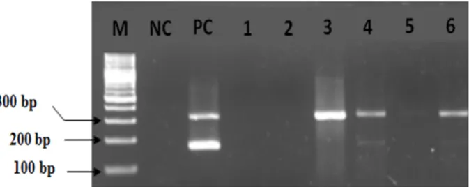
The PCR assay detected MTC DNA in 137 (22.1%) of the clinical samples. The figure 1 shows MTC bands corresponded to 190 bp.
To express the sensitivity, specificity and predictive values, the results of PCR were compared with those of TB culture as a golden standard method. The PCR sensitiv-ity was 87.7% (95% CI: 79-95) for both PCR and culture techniques which were positive for 57 clinical samples. The speci-ficity of PCR against culture as a gold standard was 85.6 %( 95% CI: 82-88)
(Ta-Fig.1. Analysis of 2% agarose gel. From left to right, lanes contain marker, negative control, positive control and follow-ing six clinical samples suspected of havfollow-ing pulmonary tuberculosis. The first sharp band in positive control shows Β-actin corresponding to 330 bp and it was found positive in clinical specimen 3, 4, 5 and 6. The second down clear band in posi-tive control lane presents the genomes of MTC identified at 190 bp, confirming that patients 4 and 6 are infected with TB. Both lanes number 1 and 2 have PCR inhibitors.
ble 1). The sensitivity and specificity of TB PCR were computed for clinical samples as: sputum (87.5%, 78.6%), bronchial aspi-rates (100%, 95.6%), and plural effusion (60%, 97.6%) (Table 1).
Discussion
MTC can be accurately detected through the PCR technique (12, 13). These molecu-lar techniques have been most often evalu-ated due to their variable rates of sensitivity and specificity (14). One approach might compare sensitivity and specificity of the in-house PCR assay with some commercial PCR systems like Amplicor and Cobas TaqMan for the detection of M. tuberculo-sis.
This study was performed to show the specificity and sensitivity of the PCR tech-nique for detection of MTC. The results of PCR were compared with those of TB cul-ture technique as the golden standard. Ob-viously, the sensitivity and specificity will increase when an optimal condition for DNA isolation and PCR processing consid-ered. IS6110 target gene is an insertion se-quence repeated in the genome of M. tuber-culosis. The sequence is not found in NTM such as M. avium complex, M. gastric etc. Various types of natural and extraneous impurities that may result during DNA ly-sate preparation make inhibitory impacts on PCR reaction (14, 18).
The findings of this study also indicate that the sensitivity of PCR for detecting the MTC was 87.7% and its specificity 85.6%. This rate of PCR sensitivity and specificity of PCR is comparable with the common ranges of 42% to 90.9% as clarified in most studies (19). Querol et al examined PCR
sensitivity for 314 respiratory specimens using IS6110 pair primers and showed PCR sensitivity about 97% (20). Another study by Thoe et al in Singapore used IS6110 primer and reported PCR sensitivity and specificity of 86.5% and 83.6%, respective-ly (21).
Our study indicate that 8 (12.3%) of total samples with PCR negative and culture positive results were expressed as PCR false-negative results due to, either, pres-ence of NTM strains in the samples, or presence of PCR inhibitors, especially in sputum. In the current study, the PCR in-hibitor was reported when genomic DNA from the samples was not detected using B-actin pair primers. A few review studies on diagnostic sensitivity of PCR have reported 5%-13% of PCR inhibitors in sputum sam-ples (22, 23). Of 384 sputum samsam-ples, the inhibitor was detected in 12 (3.1%) of the samples. This rate of PCR false-negative can be also a reason for low copy numbers of MTC DNA in the samples.
The occurrence of PCR false-positive in our study was a matter of concern. In this case, 80 (14.4%) Of PCR-positive were negatively determined by culture. A few studies on diagnostic evaluation of PCR have reported up to 6% of PCR false-positive results (24, 25). This might involve the facts that, either, cross-over contamina-tion in PCR processing, or some non-cultivable TB strains probably existed, or more importantly growth failure may be observed during TB culture (14). This issue can be a cause of low positive predictive value of PCR against MTC culture (totally 41/6 %( 95% CI: 33-49)) (Table 1).
Diagnostic sensitivity and specificity of Table1. Sensitivity, specificity and predictive value for in-house PCR
Variables PCR-positiveNo. (%) of culture-positiveNo. (%) of Sensitivity(95%CI) Specificity(95%CI) Positive Predic-tive value (95%CI)
Negative Pre-dictive Value (95%CI)
Sputum 114
(29.7%) (12.5%)48 (78-96)87.5% (74-82)78.6% (27.9-45.7)36.8% (96-99.5)97.7% Bronchial
aspirates (12.2%)18 (8.1%)12 (100-100)100% (92-99)95.6% (44.8-88.4)66.6% (100-100)100% Pleural
effusions (5.7%)5 (5.6%)5 (17-100)60% (94-100)97.6% (17-100)60% (94.2-100)97.5%
Total 137 65 87.7%
( 79-95) (82-88)85.6% (33-49)41.6% (97-99)98%
in-house PCR would be reliable indicator and can be compared with those found in other experiments. Gomez-Pastrana et al in 2000 used 235 gastric aspirates and 16 bronchoalveolar lavage specimens to detect TB in children (26). They reported 60% and 96.8% for the in-house PCR sensitivity and specificity, respectively. In 2011, Hyun Kim et al evaluated Cobas TaqMan MTC PCR and Cobas Amplicor MTC PCR for detection of Mycobacterium tuberculosis. They collected 406 samples from 247 pa-tients. Of these, 96 respiratory specimens were processed. The respiratory specimens were comprised of sputum, pleural and bronchoalveolar lavage (BAL) fluid. For respiratory specimens, they have also re-ported sensitivity and specificity of Cobas TaqMan for about 79.1%, 95.8%, and for Amplicor 58.3%, 98.6%, respectively (27).
Furthermore, Schirm et al assessed and compared Amplicor, in-house PCR, and bacterial culture for detection of Mycobac-terium tuberculosis in five hundred four clinical specimens (337sputum and 167 bronchial samples) from 340 patients. In-house PCR with the rate of 92.6% was rela-tively sensitive diagnostic technique com-pared to Amplicor M. tuberculosis test, cul-ture, and microscopy 70.4%, 88.9%, 52.4%, respectively. Obtained specificities for all of those four tests were more than 98% (28). It can be concluded that accurate performance of the in-house PCR using de-signed pair primers for diagnosis of M. tu-berculosis is a useful technique for diagnos-tic purposes. In the present study, sensitivi-ty and specificisensitivi-ty of in-house PCR were expressed in a valuable range correspond-ing to 87.7% and 85.6%, respectively with appropriate negative predictive val-ue(98%(95% CI: 97-99)). Thus, it can be highlighted that in-house PCR assay pro-vides valuable rate for sensitivity and speci-ficity for diagnosis of TB in developing countries such as Iran where preparation of TB diagnostic kits and high technology PCR tools are economically limited.
Conclusion
The study recommends that developing countries should apply the in-house PCR techniques for detection of TB. Proper per-formance of this technique needs to be con-sidered in clinical laboratories. At the end, it seems that in-house PCR can be used as a rapid assay for detection of M. tuberculosis and should be required in conjunction with other routine TB detection techniques such as direct smear and TB culture in the refer-ral health centers.
References
1. Bullo Saifullah, Mohd Zobir B Hussein, Samer Hasan Hussein Al Ali. Controlled-release approach-es towards the chemotherapy of tuberculosis. Int J Nanomedicine 2012; 7: 5451–5463.
2. World Health Organization (WHO) estimates of tuberculosis incidence by rate, 2011 (sorted by rate), public health England, 2011. Available at: http://www.hpa.org.uk
3. Pai M, Minion J, Sohn H, Zwerling A, Perkins MD. Novel and improved technologies for ztubercu-losis diagnosis: progress and challenges. Clin Chest Med 2009;30(4):701-16.
4. World Health Organization. Laboratory services in TB control, Part II: Microscopy. Geneva, WHO, 1998 (WHO/TB/98.258). Available at: http://www.who.int/tb/dots/laboratory/resourcs).
5. Morre DF, Curry JI. Detection and identifica-tion of Mycobacterium tuberculosis directly from sputum sediments by Amplicor PCR. J Clin Micro-biol 1995; 33: 2686-91.
6. Parvez MA, Hasan KN, Rumi MA, Ahmed S, Salimullah M, Tahera Y, Gomes DJ, Huq F, Hassan MS. PCR can help early diagnosis of pulmonary tuberculosis. Southest Asian j trop med public health 2003; 34(1):147-53.
7. World Health Organization. Laboratory ser-vices in TB control. Part III: Culture. Geneva, WHO, 1998 (WHO/TB/98.258. Available at: http://www.who.int/tb/dots/laboratory/resources).
8. Thierry D, Brisson-Noel A, Levy-Frebault V, Nguyen S, Guesdon L, Gicquen B. Characterization of a M. tuberculosis insertion sequence IS6110 and its application in diagnosis. J Clin Microbiol 1990; 28: 2668-73.
9. Cegielski JP, Devlin BH, Morris AJ, Kitinya JN, Pulipaka UP, Lema LE, et al. Comparison of PCR, culture, and histopathology for diagnosis of tuberculous pericarditis. J Clin Microbiol 1997; 35(12):3254-7.
10. Miller SA, Dykes DD, Polesky HF. A simple salting out procedure for extracting DNA from
man nucleated cells. Nucleic Acids Res 1988; 16:1215.
11. Phenol-Chloroform Isoamyl Alcohol (PCI) DNA Extraction [online]. [Modified from protocols by Barker et al (1998)] Available at: http://ccoon.myweb.usf.edu/ecoimmunology.org/Ab out_Home.html
12. Brisson-Noel A, Aznar C, Chureau C, et al. Diagnosis of tuberculosis by DNA amplification in clinical practice evaluation. Lancet 1991; 338: 364– 6.
13. Eisenach KD, Sifford MD, Cave MD, et al. Detection of Mycobacterium tuberculosis in sputum samples using a polymerase chain reaction. Am Rev Respir Dis 1991; 144:1160–3.
14. Sheikholslami MF, Ziaee AA, Khoshreza M, Masjedi MR, Mohammadi F, Farnia P, and Velayati AA. Detection of Pulmonary Tuberculosis by PCR Assay. Tanaffos 2005; 4(13), 63- 70.
15. Altwegg M. General problems associated with diagnostic applications of amplification methods. J Microbiol Methods 1995; 23: 21- 30.
16. Grosset J, Mouton Y. Is PCR a useful tool for the diagnosis of tuberculosis in 1995? Tuber Lung Dis 1995; 76 (3): 183-4.
17. Schirm J, Oostendorp LA, Mulder JG. Com-parison of Amplicor, in-house PCR, and conven-tional culture for detection of Mycobacterium tuber-culosis in clinical samples. J Clin Microbiol 1995; 33 (12): 3221- 4.
18. Weekes KM, Pearse MJ, Sievers A, Ross BC, d'Apice AJ. The diagnostic use of the polymerase chain reaction for the detection of Mycobacterium tuberculosis. Pathology 1994;
26 (4): 482- 6.
19. Lima SS, Clemente WT, Palaci M, Rosa RV, Antunes CM, Serufo JC. Conventional and molecu-lar techniques in the diagnosis of pulmonary tuber-culosis: a comparative study. J Bras Pneumol 2008; 34(12):1056-62.
20. Querol JM, Farga MA, Granda D, Gimeno C, Garcia-de-Lomas J. The utility of polymerase chain reaction in the diagnosis of pulmonary tuberculosis.
Chest. 1995;4:1631-35.
21. Thoe SY, Tay L, Sng EH. Evaluation of am-plicor and IS6110 PCR for direct detection of My-cobacterium tuberculosis complex in Singapore. Trop Med Int Health. 1997;2(11):1095-101.
22. Nolte FS, Metchock B, McGewan JE, et al. Direct detection of polymease chain reaction and DNA hybridazation. J Clin Microbiol 1993;3: 1771-82.
23. Wobeser WL, Krajden M, Colony J, et al. Evaluation of Roche Amolicor PCR assay for My-cobacterium tuberculosis. J Clin Microbiol 1996;34: 134-9.
24. Chakravorty S, Kamal Sen M, Sivaswami Tyagi J. Diagnosis of Extrapulmonary Tuberculosis by Smear, Culture, and PCR Using Universal Sam-ple Processing Technology. J Clin Microbiol 2005; 34 (9): 4357-4362.
25. Diagnostic Standards and Classification of Tu-berculosis in Adults and Children. This official statement of the American Thoracic Society and the Centers for Disease Control and Prevention was adopted by the ATS Board of Directors, July 1999. This statement was endorsed by the Council of the Infectious Disease Society of America, September 1999. Am J Respir Crit Care Med 2000;161(4 Pt 1):1376-95.
26. Gomez-Pastrana D, Torronteras R, Caro P, Anguita ML, López-Barrio AM, Andres A, Navarro J. Comparison of amplicor, in-house polymerase chain reaction, and conventional culture for the di-agnosis of tuberculosis in children. Clin Infect Dis 2001;32(1):17-22.
27. Kim JH, Kim YJ, Ki CS, Kim JY, Lee NY. Evaluation of Cobas TaqMan MTB PCR for detec-tion of Mycobacterium tuberculosis. J Clin Microbi-ol 2011;49(1):173-6.
28. Schirm J, Oostendorp LA, Mulder JG. Com-parison of Amplicor, in-house PCR, and conven-tional culture for detection of Mycobacterium tuber-culosis in clinical samples. J Clin Microbiol 1995;33(12):3221-4.