Published online 2016 January 3. Brief Report
The Mid-Term Changes of Pulmonary Function Tests After Phrenic Nerve
Transfer
Masoud Yavari,
1Seyed Esmail Hassanpour,
2and Mohammad Khodayari
1,* 1Department of Reconstructive Surgery, Shahid Behesti University of Medical Sciences, Tehran, IR Iran2Department of Plastic and Reconstructive Surgery, Shahid Behesti University of Medical Sciences, Tehran, IR Iran
*Corresponding author: Mohammad Khodayari, Department of Reconstructive Surgery, Shahid Behesti University of Medical Sciences, Tehran, IR Iran. Tel: +98-2188901108, Fax: +98-2188909193, E-mail: [email protected]
Received 2015 June 10; Accepted 2015 November 18.
Abstract
Background: In the restoration of elbow flexion, the phrenic nerve has proven to be a good donor, but considering the role of the phrenic nerve in respiratory function, we cannot disregard the potential dangers of this method.
Objectives: In the current study, we reviewed the results of pulmonary function tests (PFT) in four patients who underwent phrenic nerve transfer.
Patients and Methods: We reviewed the results of serial spirometry tests, which were performed before and after phrenic nerve transfer surgery.
Results: All patients regained Biceps power to M3 strength or above. None of our patients experienced pulmonary problems or respiratory complaints, but a significant reduction of spirometric parameters occurred after surgery.
Conclusions: This study highlights the close link between the role of the phrenic nerve and pulmonary function, such that the use of this nerve as a transfer donor leads to spirometric impairments.
Keywords: Phrenic Nerve Transfer, Pulmonary Function Tests, Elbow
Copyright © 2016, Kashan University of Medical Sciences. This is an open-access article distributed under the terms of the Creative Commons Attribution-Non-Commercial 4.0 International License (http://creativecommons.org/licenses/by-nc/4.0/) which permits copy and redistribute the material just in noncommercial usages, provided the original work is properly cited.
1. Background
A traumatic injury of the brachial plexus, the web of large nerves that conduct signals to the shoulder, arm and hand, can lead to partial or total denervation of the muscles of the upper extremities. Without proper and timely diagnosis and treatment, these are devastating injuries causing lifelong immobility (1). Although recov-ery takes place spontaneously in most patients, surgical intervention is required when clinical or electrical re-serve does not start within the three to six months after the injury (2).
Developments in microsurgery now have good success in brachial plexus restoration, and through treatments, such as neurolysis, nerve grafting, or nerve transfer (neu-rotization), patients can achieve rational mobility. Nerve transfer is a treatment option when root injuries involve the avulsion of the spinal nerve and proximal stumps are not accessible. During this procedure, a functional but less important nerve is transferred to the denervated nerve, which is functionally more important. Various nerves, such as the phrenic nerve (3), intercostal nerves (4), the medial pectoral nerve, (5) and the spinal acces-sory nerve (6) can be used as the source of transfer. The restoration of elbow flexion is the most important aim of any surgical treatment for severe brachial plexus in-jury, and the phrenic nerve alone or in combination with
multiple intercostal nerves has proved to be a good do-nor. However, considering the role of the phrenic nerve in respiratory function, we cannot disregard the poten-tial dangers. Some studies have revealed no significant reduction in pulmonary function subsequent to phrenic nerve transfer, but there are some reports of an effect on respiratory function in the long term.
2. Objectives
In the current study, we reviewed the results of pulmo-nary function tests (PFT) in four patients who underwent phrenic nerve transfer.
3. Patients and Methods
The injury was detected by physical examination and confirmed by preoperative and intraoperative electro-myography (EMG) and intraoperative exploration. We re-viewed the results of serial spirometries, which were per-formed before and after phrenic nerve transfer surgery.
The spirometry was performed with a computer-assist-ed spirometer (Pulmolab 435-spiro 235, Morgan, Eng-land) and according to international guidelines (7). The spirometric measurements were related to the predicted values for each patient.
3.1. Statistical Analysis
Descriptive statistical analyses were performed and
quantitative spirometric parameters were reported as the best measure for each value.
4. Results
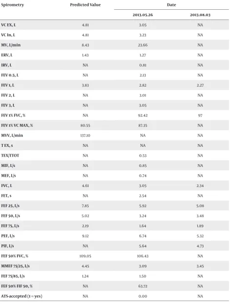
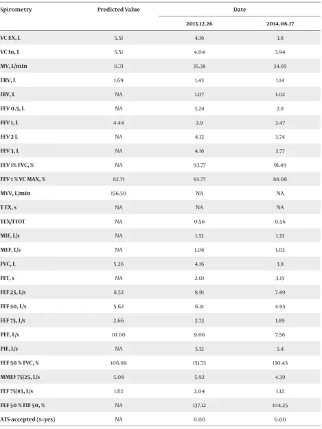
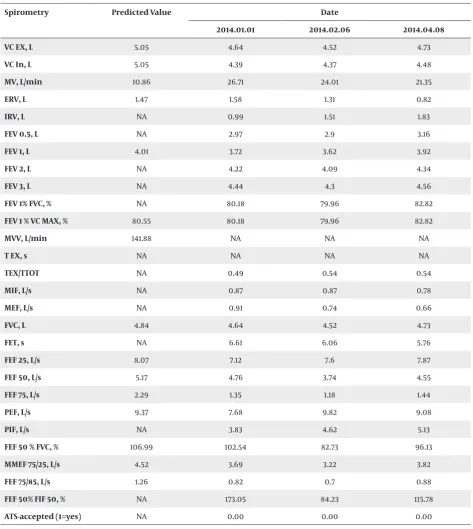
There were five patients who underwent phrenic nerve transfer for brachial plexus injury. All patients were male. The time interval between the injury and the nerve trans-fer was 3 - 6 months. All patients regained Biceps power to M3 strength or above. The lung function measurements are shown in detail in Tables 1 - 4. One patient did not re-turn for pulmonary function tests after surgery. None of our patients experienced pulmonary problems or respi-ratory complaints, but a significant reduction of spiro-metric parameters occurred after surgery.
Table 1. Patient 1, Date of Trauma :2013.05.18 , Date of Surgery :2013.08.17
Spirometry Predicted Value Date
2013.05.16 2013.10.15 2013.11.12 2014.04.06
VC EX, L 5.08 4.89 3.76 3.62 3.65
VC In, L 5.08 4.63 3.71 3.47 3.42
MV, L/min 11.00 35.01 30.23 28.94 30.87
ERV, L 1.64 1.53 0.68 0.75 0.79
IRV, L NA NA NA 0.69 NA
FEV 0.5, L NA 3.62 2.6 2.35 2.67
FEV 1, L 4.14 4.46 3.19 2.99 3.18
FEV 2, L NA 4.89 3.56 3.37 3.52
FEV 3, L NA 4.89 3.63 3.55 3.58
FEV 1% FVC, % NA 91.26 89.02 82.61 87.08
FEV 1% VC MAX, % 82.71 91.26 89.02 82.61 87.08
MVV, L/min 149.78 NA NA NA NA
T EX, s NA 1.86 2.44 NA 2.1
TEX/TTOT NA 0.53 0.56 0.59 0.58
MIF, L/s NA 1.24 1.15 1.17 1.22
MEF, L/s NA NA NA 0.82 NA
FVC, L 4.86 4.89 3.63 3.62 3.65
FET, s NA NA NA 4.62 NA
FEF 25, L/s 8.14 9.94 7.78 6.11 7.06
FEF 50, L/s 5.36 6.8 4.53 3.12 4.94
FEF 75, L/s 2.47 2.6 1.65 1.11 1.6
PEF, L/s 9.57 9.94 9.91 8.11 7.65
PIF, L/s NA NA NA 4.5 NA
FEF 50% FVC, % 110.45 139.04 132.62 86.26 135.24
MMEF 75/25, L/s 4.94 5.9 3.74 2.74 4.0
FEF 75/85, L/s 1.63 2.07 1.21 0.79 0.95
FEF 50 FIF 50, % NA 109.63 104.89 72.68 103.5
ATS-accepted (1 = yes) NA NA NA 0.00 NA
Table 2. Patient 2, Date of Trauma :2013.03.24 , Date of Surgery: 2013.05.29
Spirometry Predicted Value Date
2013.05.26 2013.08.03
VC EX, L 4.81 3.05 NA
VC In, L 4.81 3.23 NA
MV, L/min 8.43 23.66 NA
ERV, L 1.43 1.27 NA
IRV, L NA 0.81 NA
FEV 0.5, L NA 2.13 NA
FEV 1, L 3.83 2.82 2.27
FEV 2, L NA 3.01 NA
FEV 3, L NA 3.05 NA
FEV 1% FVC, % NA 92.42 97
FEV 1% VC MAX, % 80.55 87.35 NA
MVV, L/min 137.10 NA NA
T EX, s NA NA NA
TEX/TTOT NA 0.53 NA
MIF, L/s NA 0.85 NA
MEF, L/s NA 0.74 NA
FVC, L 4.61 3.05 2.34
FET, s NA 2.54 NA
FEF 25, L/s 7.85 5.92 5.08
FEF 50, L/s 5.02 3.24 3.48
FEF 75, L/s 2.19 1.64 1.89
PEF, L/s 9.12 6.74 5.32
PIF, L/s NA 5.64 4.73
FEF 50% FVC, % 109.05 106.43 NA
MMEF 75/25, L/s 4.45 3.09 3.45
FEF 75/85, L/s 1.24 1.50 NA
FEF 50% FIF 50, % NA 63.72 NA
ATS-accepted (1 = yes) NA 0.00 NA
Table 3. Patient 3, Date of Trauma :2013.09.16, Date of Sutgery :2014.02.01
Spirometry Predicted Value Date
2013.12.26 2014.06.17
VC EX, L 5.51 4.16 3.8
VC In, L 5.51 4.04 3.94
MV, L/min 11.71 35.38 34.95
ERV, L 1.69 1.43 1.14
IRV, L NA 1.07 1.02
FEV 0.5, L NA 3.24 2.8
FEV 1, L 4.44 3.9 3.47
FEV 2 L NA 4.12 3.74
FEV 3, L NA 4.16 3.77
FEV 1% FVC, % NA 93.77 91.49
FEV 1 % VC MAX, % 82.71 93.77 88.06
MVV, L/min 156.50 NA NA
T EX, s NA NA NA
TEX/TTOT NA 0.56 0.56
MIF, L/s NA 1.33 1.33
MEF, L/s NA 1.06 1.03
FVC, L 5.26 4.16 3.8
FET, s NA 2.01 3.15
FEF 25, L/s 8.52 8.91 7.49
FEF 50, L/s 5.62 6.31 4.95
FEF 75, L/s 2.66 2.72 1.89
PEF, L/s 10.00 9.06 7.56
PIF, L/s NA 5.12 5.4
FEF 50 % FVC, % 106.99 151.73 130.43
MMEF 75/25, L/s 5.08 5.92 4.39
FEF 75/85, L/s 1.62 2.04 1.12
FEF 50 % FIF 50, % NA 137.12 104.25
ATS-accepted (1=yes) NA 0.00 0.00
Table 4. Patient 4, Date of Trauma : 2013.08.16 , Date of Surgery :2014.01.03
Spirometry Predicted Value Date
2014.01.01 2014.02.06 2014.04.08
VC EX, L 5.05 4.64 4.52 4.73
VC In, L 5.05 4.39 4.37 4.48
MV, L/min 10.86 26.71 24.01 21.35
ERV, L 1.47 1.58 1.31 0.82
IRV, L NA 0.99 1.51 1.83
FEV 0.5, L NA 2.97 2.9 3.16
FEV 1, L 4.01 3.72 3.62 3.92
FEV 2, L NA 4.22 4.09 4.34
FEV 3, L NA 4.44 4.3 4.56
FEV 1% FVC, % NA 80.18 79.96 82.82
FEV 1 % VC MAX, % 80.55 80.18 79.96 82.82
MVV, L/min 141.88 NA NA NA
T EX, s NA NA NA NA
TEX/TTOT NA 0.49 0.54 0.54
MIF, L/s NA 0.87 0.87 0.78
MEF, L/s NA 0.91 0.74 0.66
FVC, L 4.84 4.64 4.52 4.73
FET, s NA 6.61 6.06 5.76
FEF 25, L/s 8.07 7.12 7.6 7.87
FEF 50, L/s 5.17 4.76 3.74 4.55
FEF 75, L/s 2.29 1.35 1.18 1.44
PEF, L/s 9.37 7.68 9.82 9.08
PIF, L/s NA 3.83 4.62 5.13
FEF 50 % FVC, % 106.99 102.54 82.73 96.13
MMEF 75/25, L/s 4.52 3.69 3.22 3.82
FEF 75/85, L/s 1.26 0.82 0.7 0.88
FEF 50% FIF 50, % NA 173.05 84.23 115.78
ATS-accepted (1=yes) NA 0.00 0.00 0.00
Abbreviation: NA, not available.
5. Discussion
Reviewing our experience with patients who under-went phrenic nerve transfer, we confirmed previous re-ports of spirometric impairments. The most common parameters used to interpret lung function in spirom-etry are VC, FEV1, FEV1/VC ratio and TLC. Although FVC is
frequently used instead of VC, the measured value of VC on inspiration (IVC), slow expiration (SVC) or forced expi-ration are more exact. The first finding of our study was the reduction of VC on both inspiration and expiration.
FEV1 decreased to < 80% of predicted values, and con-comitantly, a decrease occurred in FVC, while the FEV1/ FVC ratio was normal or even increased. This pattern most often happens when the patient cannot inhale or exhale successfully. A study by Chaliadapong et al. also showed a significant reduction of pulmonary function one year after phrenic nerve transfer surgery (8). Simi-lar to our findings, Beraldo et al. reported a significant reduction of FVC and FEV1 to 69% and 68% of predicted values, respectively. But in contrast, they also found a re-duction of the FEV1/FVC ratio to 81% of predicted value, which was increased in our patients (9). The reduced VC in combination with the increased FEV1/VC (> 85 - 90%) and the convex pattern of the flow-volume curve pro-poses a submaximal inspiratory or expiratory effort (7). It also results in an increase in the RV, because the pa-tients could not exhale long enough to empty the lungs from RV. These changes look to be the result of the in-ability of weakened muscles to force thoracic volume. Peak expiratory flow (PEF) was the other parameter that decreased after the surgery. It can be affected prior to FEV1 and FVC and represent the poor initial effort. Since MVV (maximal voluntary ventilation) correlates well with FEV1, it is not usually reported in PFT results. How-ever, a disproportionate decline relative to FEV1 can indi-cate neuromuscular disorders (10).
The results show that our patients did not improve with time after surgery. In contrast to our study, Beraldo et al. found an improvement of spirometric parameters with time after surgery (9).
5.1. Conclusions
This study highlights the close link between the role of the phrenic nerve and pulmonary function, such that the use of this nerve as a transfer donor leads to spirometric impairments.
Footnote
Authors’ Contribution:Masoud Yavari (study concept, design and supervision), Seyed Esmail Hassanpour (study concept and design), Mohammad Khodayari (Data collec-tion and analysis and manuscript writing ).
References
1. Venkatramani H, Bhardwaj P, Faruquee SR, Sabapathy SR. Func-tional outcome of nerve transfer for restoration of shoulder and elbow function in upper brachial plexus injury. J Brachial Plex Peripher Nerve Inj. 2008;3:15. doi: 10.1186/1749-7221-3-15. [PubMed: 18505571]
2. Monreal R. Restoration of Elbow Flexion by Transfer of the Phrenic Nerve to Musculocutaneous Nerve after Brachial Plexus Injuries. HAND. 2007;2(4):206–11. doi: 10.1007/s11552-007-9050-6. [PubMed: 18780054]
3. Gu YD, Wu MM, Zhen YL, Zhao JA, Zhang GM, Chen DS, et al. Phren-ic nerve transfer for brachial plexus motor neurotization. Micro-surgery. 1989;10(4):287–9. [PubMed: 2593799]
4. Krakauer JD, Wood MB. Intercostal nerve transfer for brachial plexopathy. J Hand Surg Am. 1994;19(5):829–35. doi: 10.1016/0363-5023(94)90196-1. [PubMed: 7806813]
5. Samardzic M, Grujicic D, Rasulic L, Bacetic D. Transfer of the medi-al pectormedi-al nerve: myth or remedi-ality? Neurosurgery. 2002;50(6):1277– 82. [PubMed: 12015846]
6. Songcharoen P, Mahaisavariya B, Chotigavanich C. Spinal acces-sory neurotization for restoration of elbow flexion in avulsion injuries of the brachial plexus. J Hand Surg Am. 1996;21(3):387–90. doi: 10.1016/s0363-5023(96)80349-2. [PubMed: 8724466] 7. Pellegrino R, Viegi G, Brusasco V, Crapo RO, Burgos F, Casaburi R,
et al. Interpretative strategies for lung function tests. Eur Respir J.
2005;26(5):948–68. doi: 10.1183/09031936.05.00035205. [PubMed: 16264058]
8. Chalidapong P, Sananpanich K, Kraisarin J, Bumroongkit C. Pul-monary and biceps function after intercostal and phrenic nerve transfer for brachial plexus injuries. J Hand Surg Br. 2004;29(1):8– 11. [PubMed: 14734060]
9. Beraldo P, Gepp R, Penna L, Ramalho S, Silva R, Neri E. Pulmonary function after phrenic nerve transfer for brachial plexus avul-sion injury. European Respir J. 2011;38(Suppl 55):p2398. 10. Serisier DE, Mastaglia FL, Gibson GJ. Respiratory muscle
func-tion and ventilatory control. I in patients with motor neu-rone disease. II in patients with myotonic dystrophy. Q J Med.