Iran University of Medical Sciences
_______________________________________________________________________________________________________________ 1. (Corresponding author) PhD, PT, Assistant Professor, Department of Sports Medicine, Hazrat Rasool-e-Akram Hospital, Iran University of Medical Sciences, Tehran, Iran. [email protected], [email protected]
2. MD, Sports Medicine Assistant, Hazrat Rasool-e-Akram Hospital, Iran University of Medical Sciences, Tehran, Iran. [email protected] 3. PhD. Assistant Professor, Epidemiology Department, Iran University of Medical Sciences, Tehran, Iran. [email protected]
The effects of scapular stabilization based exercise therapy on
pain, posture, flexibility and shoulder mobility in patients with
shoulder impingement syndrome: a controlled randomized
clini-cal trial
Azar Moezy1, Saeed Sepehrifar2, Masoud Solaymani Dodaran3
Received:25 September 2014 Accepted:26 April 2014 Published:27 August 2014
Abstract
Background: Dysfunction in the kinetic chain caused by poor scapula stabilization can contribute to shoulder injuries and Shoulder Impingement Syndrome (SIS). The purpose of this study was to compare the effectiveness of two treatment approaches scapular stabilization based exercise therapy and physical therapy in patients with SIS.
Methods: The study is a randomized clinical trial in which 68 patients with SIS were randomly assigned in two groups of exercise therapy (ET) and physical therapy (PT) and received 18 sessions of treatment. Pain, shoulders' range of abduction and external rotation, shoulder protraction, scapular rotation and symmetry as well as postural assessment and Pectoralis minor length were evaluated pre and post intervention. The paired-sample t test and the independent sample t test were applied respectively to determine the differences in each group and between two groups.
Results: Our findings indicated significant differences in abduction and external rotation range, improvement of forward shoulder translation and increase in the flexibility of the involved shoulder between the two groups (respectively ; p=0.024, p=0.001, p<0/0001, p<0/0001). No significant difference was detected in pain reduction between the groups (p=0.576). Protraction of the shoulder (p<0.0001), forward head posture (p<0/0001) and mid thoracic curvature (p<0.0001) revealed a significant improvement in the ET group. Apparent changes oc-curred in scapular rotation and symmetry in both groups but no significant differences were observed between the two groups (respectively; p=0.183, p=0.578).
Conclusion: The scapular stabilization based exercise intervention was successful in increasing shoulder range, decreasing forward head and shoulder postures and Pectoralis minor flexibility.
Keywords: Shoulder Impingement Syndromes, Posture, Exercise Therapy, Physical Therapy.
Cite this article as:Moezy A, Sepehrifar S, Solaymani Dodaran M. The Effects of Scapular Stabilization Based Exercise Therapy on Pain, Posture, Flexibility and Shoulder Mobility in Patients with Shoulder Impingement Syndrome: A Controlled Randomized Clinical Trial.Med J Islam Repub Iran2014 (27 August). Vol. 28:87.
Introduction
Shoulder impingement syndrome (SIS) is a common complaint for patients of all ages and different activity levels. It has been de-fined as compression and mechanical abra-sion of the rotator cuff structures as they pass beneath the coracoacromial arch dur-ing elevation of the arm (1). A wide range of different factors is involved in causing
SIS: including anatomic abnormalities of the coracoacromial arch or humeral head (2), tension overload ischemia (3), repeti-tive eccentric overload (2-3), aberrant kin-ematic patterns due to poor rotator cuff or scapular muscles function (2,4-5), and poor posture and scapular kinematic abnormali-ties (2) . People who are constantly doing repetitive overhead motions relate to their
occupations or athletic activities are also at risk of SIS (1, 6). Pain and dysfunction of patient with SIS occur when the shoulder is placed in positions of elevation, an activity that is commonplace during many sporting and daily living activities. Patients usually complain a general loss of strength in shoulder girdle muscles during working. Thus, the patients are at risk of losing their physical independence and jobs which have important socioeconomic implications.
The scapular position and motions on the thorax is a critical component of the normal glenohumeral function and plays great roles in facilitating optimal shoulder movements (6-8). In normal upper-quarter function, the scapula provides a stable base from which glenohumeral mobility occurs. Stability of the scapula depends on the surrounding musculature. These muscles must dynami-cally position the glenoid so that efficient glenohumeral movement can occur. During all movements of the glenohumeral joint especially overhead elevation of the arm, it is of great importance that the scapular-stabilizing musculature should be strong enough to properly position the scapula. The main scapula stabilizers are the Leva-tor Scapulae, Rhomboids major and minor, Serratus anterior, and Trapezii. These mus-cle groups function through synergistic co-contraction with rotator cuff to control the scapular movement (9). When weakness or dysfunction is present in the scapular mus-culature, normal scapular positioning and mechanics may become altered which re-sult in abnormal stresses to the capsular structures, rotator cuff compression and reduced performance (7).
There are some evidences suggesting that kinematic of scapulothoracic motions were impaired in SIS (2, 4,9-10).
The scapulothoracic kinematics can be al-tered in response to inappropriate or incor-rect movement pattern, macro and micro traumatic injuries, abnormal scapulo-humeral rhythm and other shoulder pathol-ogies (8,10). Altered muscle activity in the scapular muscles is commonly believed to be a crucial factor contributing to SIS (2,
11-12). The scapulothoracic kinematics are also affected by abnormal posture of tho-racic and cervical spine (13). Warner et al. demonstrated a pattern of increased scapu-lar winging with glenohumeral elevation. This winging pattern appears to represent scapular internal rotation and anterior tilt-ing (14). Recently, 3-dimensional kinemat-ic analysis has demonstrated decreased scapular posterior tilt, decreased upward rotation, and decreased scapular external rotation during glenohumeral elevation in SIS patients (2,5).
The treatment of SIS is % 90 -95 con-servative and often includes rotator cuff strengthening exercises (6,15), stretching exercises (16), immobilization, passive, active and active assisted range of motion exercises (ROM), various mobilization techniques, home exercise programs(17) and various physical therapy methods such as heat, transcutaneous electrical nerve stimulation (TENS) and ultrasound (US) and etc (18-19). In a conservative approach, exercise therapy is often being used and has an important role in shoulder rehabilitation. New insights in shoulder rehabilitation em-phasize the dynamic stabilization of the scapula as an essential part of the manage-ment because the ability to position and control movements of the scapula is very important for optimal upper limb function. When the scapula fails to perform its stabi-lization role, shoulder function is ineffi-cient, which can result not only in de-creased neuromuscular performance , but also may predispose the individual to shoulder injuries (20).
Unfortunately, the scapular musculature is often neglected in the treatment of SIS. This lack of attention may often lead into the incomplete treatment (7), therefore, reestablishment of normal shoulder func-tion and restoring normal scapular muscle activation patterns by scapular stabilization based exercises, in our view, are the keys to a successful rehabilitation program.
The purpose of our investigation was to compare the effectiveness of two treatment approaches for impingement syndrome of
the shoulder: (1) a 6-week scapular stabili-zation based exercises, and (2) a ROM ex-ercise program combined with physical modalities on pain, posture, flexibility and mobility of SIS patients.
Methods
The study design was a randomized clini-cal trial. Ethiclini-cal approval for this study was granted by the Research Ethics Committee of Tehran University of Medical Sciences (grant number 90-9-28-1992). Subjects were provided with information booklets explaining the purpose of the study and signed informed consent documents prior to participation. Subjects were free to with-draw from the study at any time. This study was conducted in Hazrat Rasool-e-Akram Hospital located in Tehran (Iran) between 2011and 2012. All measurements were tak-en before and after a 6-week intervtak-ention period.
Subjects: A total of 98 subjects was ini-tially recruited from Hazrat Rasool-e-Akram hospital sports medicine and ortho-pedic clinics and judged to meet the criteria for the study. Twenty six subjects did not fulfill the inclusion criteria (Fig 1). Inclu-sion and excluInclu-sion criteria were assessed for each subject based on a clinical exami-nation performed by the first author. The inclusion criteria were as follows: (1) Male and female mentally fitted between the ages of 18 to 75 years; (2) Unilateral shoulder pain of more than one month localized (an-terior and/or anterolateral) to the acromion; (3) Tenderness to palpation of the rotator cuff tendons; (4) Positive impingement tests, or a painful arc of movement (60°– 120°) ;(5) Pain produced or increased dur-ing flexion and/or abduction of the symp-tomatic shoulder.
All subjects tested positive for impinge-ment tests (which included the Hawkins, Neer, and Empty can tests) and underwent a full screening of cervical and shoulder ROM, resisted motions, and special tests. No single impingement test has 100% sen-sitivity or 100% specificity. Therefore, to correctly identify patients with shoulder
impingement, a combination of clinical tests is recommended. According to Ure et al. findings , multiple tests were able to cor-rectly distinguish SIS from other shoulder pathologies in 86% of cases(21-22).
Exclusion criteria were as follows: (1) cervical or shoulder symptoms reproduced by a cervical screening exam; (2) abnormal results with reflex or thoracic outlet tests; (3) symptoms of numbness or tingling in the upper extremity; (4) pregnancy, or (5) a history of the followings: onset of symp-toms due to traumatic injury, glenohumeral joint dislocation, acromioclavicular joint separation, shoulder fracture, surgery on the shoulder, fibromyalgia, use of any treatment within three months. Main partic-ipants were 72 SIS patients who were ran-domly allocated into two groups: (1) Scapular Stabilization based Exercise Ther-apy group (ET) and (2) Physical therTher-apy group (PT).
Random allocation of the subjects was done by using a random number table and block random sampling; A: ET; B: PT (a block size of 4).
Block Size:
Block 1: AABB Block 2: ABAB Block 3: BBAA Block 4: BABA Block 5: ABBA Block 6: BAAB
The subjects who were included in the study signed university-approved informed consent forms and completed demographic data sheets. Four subjects were excluded at the intervention stage and three subjects in ET group due to irregular attendance at therapy sessions, travel and an accident and one subject in PT group due to irregular attendance at therapy sessions (Flowchart. 1).
In addition to inclusion-exclusion crite-ria, the clinical evaluation included assess-ment of pain, active shoulder external rota-tion and abducrota-tion range, forward head posture, mid-thoracic curve, forward shoul-der translation, scapular protraction &
tion and Pectoralis minor length were done pre and post intervention.
It must be noted that the participants were blinded in each block. The patients were treated on different days and unaware of the other group. Also, the examiner who assessed was blinded to group allocation and clinical data. The interventions for both groups were done by two clinicians that were unaware of the treatments groups. One supervised exercise therapy for ET group on even days and the other did phys-ical therapy for PT group on odd days.
The power analysis of the study was per-formed to detect a 10% differences in pain and shoulder abduction with α=0/05 and a power of 80%, a sample size of 36 per
group was required.
Pain: Subjects were asked to record their maximal pain during the movements based on visual analogue scale (VAS) for pain. The VAS used in the study was a 10-cm line where the 0 was marked as no pain and the 10 as the worst pain imaginable (23).
Shoulder Range of Motion (ROM): The ranges of active external rotation and ab-duction were measured by a standard goni-ometer in both symptomatic and asympto-matic shoulders as follows: Shoulder ab-duction was measured in the seated chair position, as in flexion, with the trunk up-right. The arm was actively elevated in the strict coronal plane with the thumb pointed up toward the ceiling to allow the required Flowchart 1. Study profile for participants in ET and PT groups.
external rotation necessary to avoid im-pingement of the greater tuberosity on the acromion process. Once active end-range was achieved the measurements were doc-umented. Shoulder external rotation was measured in supine with the hips and knees flexed to approximately 45 degrees. The tested arm was supported on the table in 90 degrees of abduction, elbow flexed to 90 degrees, the forearm in midway between pronation/supination and the wrist in neu-tral. A towel roll was placed under the hu-merus to ensure neutral horizontal position-ing; which required the humerus to be level to the acromion process based on visual inspection. Once positioned, the participant was asked to rotate the arm into external rotation to the end available range without discomfort. The participant was instructed not to lift the lower back during this meas-urement. Once active end-range was achieved the measurement was recorded (24).
Forward Head Posture (FHP): To meas-ure FHP, a lateral photograph was then tak-en of the cervicothoracic region, using a Canon Camera (Model: IXY digital 3000 IS).The camera was placed 2 meters from the subject and mounted on a tripod, lev-eled with a bubble spirit level to control frontal and sagittal angles. This procedure has been used in previous published studies (25-27) . The method chosen to measure the FHP for the current investigation was direct measurement from lateral view pho-tographs of head and shoulder posture. To measure the angles, an A4-sized sheet of graph paper was photocopied onto trans-parency film for photocopiers. The graph paper had vertical and horizontal lines spaced at 1-mm intervals. The transparency film was then placed over the photograph and aligned so that one of the vertical lines was placed over the plumb line and the in-tersection of two vertical and horizontal lines coincided with the point the C7 mark-er came in contact with the skin. To calcu-late the position of the head in relation to C7 (C7-tragus angle), the angle between the horizontal line and the line connecting
tragus of the ear to spinous process of C7 , was measured with a goniometer and doc-umented in degrees.
Mid-Thoracic Curve: This curvature was measured by palpating and marking the spinous processes of the second thoracic vertebra (T2) and the twelfth thoracic ver-tebra (T12) by counting spinous processes, beginning with C7. The researcher placed the tip of the flexi ruler on T2 and conform it to the subject's spine, marking the flexi ruler at T12. The flexi ruler was transferred to a sheet of white paper, and the curve of the flexi ruler was traced. A metric ruler was used to measure the length and the flexi ruler was used to measure the height of the curve in centimeters. The following formula was used to determine mid-thoracic curvature:
Mid - thoracic curvature = θ = 4 × [arc tan (2 × H/L)]
where H = the height of the curve and L = the length of the curve (Fig 3) (25). Mid-thoracic curvature was measured and doc-umented in degrees for each subject.
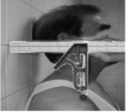
Forward Shoulder Translation(FST):
FST was measured using a combination square ruler (CL-01 model, E-Base measur-ing Tools Co, Taiwan) consists of a 40-cm to measure the distance from the wall to the anterior tip of the subject's acromion pro-cess. The patient stood in a relaxed position with their heels against a wall. The anterior tip of acromion process was marked and the distance of the point and wall was measured and documented in millimeter to determine the amount of FST. FST was as-sessed for both shoulders. This measure-ment was done three times for each
shoul-Fig. 1. Forward shoulder translation measurement
der and the average was recorded as FST (28) (Fig.1).
Scapular Protraction & Rotation: Scapu-lar protraction and rotation were measured with the subject standing, subjects nodded the head and neck forward and backward five times, then inhaled and exhaled deeply to produce a natural, reproducible standing posture and head and neck position (25). For determining scapular protraction, sub-jects were asked to adopt a comfortable and natural standing position. After palpation, non-allergenic adhesive markers 6 mm in diameter were attached to the following points (Fig. 2):
A= the root of the scapular spine
B= A mark on the thoracic spine corre-sponding to the root of the scapular spine
C= A mark on the thoracic spine corre-sponding to the inferior angle of the scapu-la
D= the inferior angle of the scapula E= the tip of the acromion of the scapula. The following formula was used to de-termine scapular protraction:
Scapular Protraction =BAEAE
Line BAE = the distance from the mark on the thoracic spine corresponding to the root of the scapular spine to the tip of the acromion.
Line AE= the distance from the root of the scapular spine to the tip of the acromi-on.
Scapular rotation was measured by palpating and marking the inferior angle of the scapula (D) and the corresponding mark on the thoracic spine (C).
The following formula was used to
determine scapular rotation: Scapular rotation= tan θ=CDBC
Line CD= the distance between the inferior angle of the scapula and the corresponding mark on the thoracic spine.
Line BC= the distance between the marks on the thoracic spine corresponding to the root of the scapular spine and the inferior angle of the scapula (Fig. 2).
Scapular rotation was measured and documented in degrees.Scapular protraction and scapular rotation were measured bilat-erally on each subject. The symmetry of the scapular was determined for each subject using the following formula:
Symmetry=LR
Where L= the ratio of left scapular pro-traction to left scapular rotation, And R= the ratio of right scapular protraction to right scapular rotation.
Anthropometric Measurement of Pecto-ralis Minor (PM) Length: The anthropo-metric measurement of the PM length was performed according to the protocol, de-scribed by Borstad (29). Before the PM length measurement, two anatomical land-marks were determined first the medial-inferior angle of the coracoid process and a second landmark just lateral to the sterno-costal junction of the inferior aspect of the fourth rib. A caliper was used to measure the distance between both bony reference points (Fig. 3). The PM length measure-ment was done three times for each method and the average was recorded.
Intervention protocols: Following the evaluation, Participants of ET and PT Fig. 2. Scapular protraction measurement Fig 3: Anthropometric Measurement of Pectoralis
Minor Length
groups began a six-week program (three times per week).
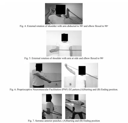
Exercise therapy intervention-After a brief explanation about the exercises proto-col, The ET subjects participated in three supervised exercise sessions per week over a 6-week period (16, 20, 30-32). This exer-cises protocol consisted of a 10 minute walking warm up on a treadmill (DK city-DX3-B1), flexibility, strengthening, scapu-lar stabilization and postural exercises. The patients were asked to avoid any other ex-ercises and severe daily activities during their treatment. At the first session, the sub-jects were introduced to the different levels of tubing exercises with Theraband. Thera-bands (The Hygienic Corp, Akron, Ohio) are color-coded, with each color
represent-ing a different resistance. Strength Trainrepresent-ing with Theraband consisted of exercises for rotator cuffs, scapular retractors, shoulder external rotators (Figs. 4, 5), D2-PNF pat-tern (Fig. 6) and Serratus anterior punches (Fig. 7). The level of difficulty of exercises was increased based on quality of shoulder motion and perceived intensity of pain. The level of tubing resistance was adjusted ac-cordingly for all subjects throughout the treatment process. At the first session, the ET subjects were asked to do five repeti-tions of each of the tubing exercises to see if they were too hard or too easy; Then the appropriate Theraband was chosen based on feedback from the subject, observation and palpation of the target muscles by the investigator. Five subjects of the ET group Fig. 4. External rotation of shoulder with arm abducted to 90ᵒ and elbow flexed to 90ᵒ
Fig. 5. External rotation of shoulder with arm at side and elbow flexed to 90ᵒ
Fig. 6. Proprioceptive Neuromuscular Facilitation (PNF) D2 pattern.(A)Starting and (B) Ending position.
Fig. 7. Serratus anterior punches. (A)Starting and (B) Ending position
used Tan (Ultrathin), sixteen used yellow (thin), and the remaining subjects used red (medium) Theraband. Each tubing exercise was performed as 3 sets of 10 repetitions with a 60-second rest period between each set. At the end of every week, the subjects were evaluated and progressed to the next higher level of resistance using yellow (thin), red (medium), green (heavy), blue (extra-heavy) and Black (Special Heavy) Therabands according to each subject.
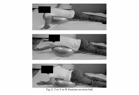
Another exercises program was applied in this study (Table 1) targeted the periscapu-lar muscle to improve stabilization of the scapula (Fig. 8). These exercises were done on a medium sized swiss ball.
Scapular-clock exercise was the other training we used in this study to facilitate the scapular motions of elevation, depres-sion, protraction, and retraction as well as joint kinesthesia and range of motion. The subject stood beside a plinth and put his hand on a ball and moved it to show 3, 6, 9 or 12 o'clock based on an imaginary clock he had on his mind (Fig. 9). This exercise also was done by pressing a ball and replac-ing it on a wall (Fig. 10).
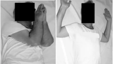
The flexibility exercises of our program consisted of sleeper stretch (Fig. 11), crossed arm stretch (Fig. 12), corner stretch (Fig. 13) and pectorals stretches (Fig. 14) which aimed to elongate Pectoralis major/ Table 1. Description of scapular stabilization exercises
Exercises Description
T to Y Patient was positioned in a prone on swiss ball with arms abducted to 90ᵒ (the letter T); Then flexed the elbows to 90ᵒ, retracted his scapulas and externally rotated his arms while keeping his arm in 90ᵒ abduction. Maintaining retraction of scapula, the patient raised his arms above the head and extended the elbow while arm flexed and abducted to 120ᵒ (the letter Y).
T to Y to W Patient was positioned in a prone on swiss ball and formed the letter T (as previous description) Then he changed his position to letter Y with his thumbs up. He retracted and depressed scapulas while he raised his arms 10-15 cm. Maintaining retraction of scapula, he flexed his elbows and extended his shoulders to form the letter W.(Fig 11) Scapular
Protrac-tion Patient was positioned in a prone with toes and forearms supported the body, Then hepushed up 1-2cm and protracted his scapulas.
Fig. 8. T to Y to W Exercise on swiss ball
minor muscles and stretch of shoulder cap-sule. Two sets of stretching exercises were performed with a minimum of 10 repeti-tions for 30 seconds.
Physical therapy intervention: A combi-nation of physical modalities and range of motion exercises was chosen as a conven-tional physical therapy in this study for PT group. Their protocol included pendulum and ROM exercises (Scaption, Abduction, Flexion, Extension, Horizontal Abduction, Horizontal Adduction and Rotations), infra-red therapy (33) (500 Watt IR lamp, Philips Co, Nederland); ultrasound therapy and TENS (Combined BTL-4825 S Topline, UK) (32) which performed three times per week for 6 weeks. Infrared lamp was in dis-tance of 45-50 cm from pain area according to the patient tolerance. Ultrasound therapy was applied on the subacromial region (US frequency: 1 MHz; US mode: continuous; time: 5 minutes; probe size: 5 cm2; and an
intensity of 1 W/cm2).We also used two-electrode TENS (pulse width: 50-250 ms, pulse rate: 90 – 130 Hz) over painful area according to the patient with the most com-fortable intensity level. Obviously, these modalities were chosen to reduce pain, im-prove tissue extensibility and increase range of shoulder motions.
Data Analysis
The Data were analyzed using the Statis-tical Package for the Social Sciences (SPSS, version 19, SPSS Inc, and Chicago, IL). Descriptive statistics (mean, SD, range) were computed for each study varia-ble. Normal distribution of data was deter-mined by observing histograms and One-Sample Kolmogorov-Smirnov test and par-Fig. 9. Scapular-clock exercise on the table
Fig. 10. Scapular-clock exercises on a wall
Fig. 11. Sleeper Stretch
Fig. 12. Crossed Arm Stretch (Posterior Capsule Stretch)
Fig. 13. Corner Stretch (Posterior Capsule Stretch)
Fig. 14. Pectorals Stretches. (Left)Starting and (Right) Ending position
ametric tests were used to analyze the data. A paired-sample t test was applied to de-termine the differences in each group. An independent sample t test was used to com-pare the baseline measurements between the groups at the beginning and at the end of training and also to analysis change scores of both groups after the test. The change scores of a group were defined as the increase or decrease of each variable from pre-test to post-test. The level of sig-nificance was set at p<0.05. In order to study the improvement process of partici-pants, the Change Score of all data were also calculated using the following equa-tion:
Change Score= Post Test Average - Pre Test Average
The change scores will reveal the increase
or decrease of each variable from pre-test to post-test.
To assess the Intra-tester reliability of ob-jective tests, 10 SIS subjects, who complet-ed informcomplet-ed consent documentation, had repeated measurements a week apart in a pilot study. Test-retest reliability of varia-bles was assessed using Paired Match T-Test (p≤0.05) and the correlation between the first and second assessing was also ob-tained (Table 2).
Results
Demographic data of subjects in both groups has been shown in Table 3. There were no significant differences between PT and ET groups for the demographic varia-bles (Table 3), indicating that the groups were well matched. There were 55 females Table 2. The Correlation between first and second measurements
Measurement Correlation Rate (p<0.05)
Right Abduction Range 100% <0/0001*
Left Abduction Range 100% <0/0001*
Right External Rotation Range 100% <0/0001*
left External Rotation Range 100% <0/0001*
Right Protraction 100% <0/0001*
Left Protraction 100% <0/0001*
Right Rotation 100% <0/0001*
Left Rotation 100% <0/0001*
Symmetry of Scapula 100% <0/0001*
Right Forward Shoulder Translation 99% <0/0001*
Left Forward Shoulder Translation 99% <0/0001*
Forward Head Posture 97% <0/0001*
Mid Thoracic Curve 98% <0/0001*
Right PML 100% <0/0001*
Left PML 100% <0/0001*
*= Significant
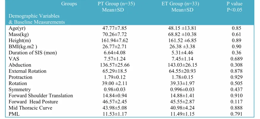
Table 3. Basic characteristics and the baseline measurements of the PT and ET groups Groups
Demographic Variables & Baseline Measurements
PT Group (n=35)
Mean±SD ET Group (n=33)Mean±SD P valueP<0.05
Age(yr) 47.77±7.85 48.15 ±13.81 0.85
Mass(kg) 70.26±7.72 68.82 ±10.38 0.61
Height(m) 161.94±7.62 161.52 ±6.85 0.89
BMI(kg.m2 ) 26.77±2.71 26.38 ±3.38 0.90
Duration of SIS (mon) 6.64±4.08 5.31±4.46 0.36
VAS 7.57±1.24 7.45±1.14 0.689
Abduction 136.57±25.66 143.03±26.15 0.308
External Rotation 65.29±18.5 64.55±20.93 0.878
Protraction 1.79±0.12 1.78±0.15 0.929
Rotation 39.00 ±2.11 39.33±1.97 0.505
Symmetry 0.98±0.03 0.996±0.03 0.437
Forward Shoulder Translation 14.84±0.94 14.88±1.41 0.910
Forward Head Posture 46.57±2.45 45.55±2.87 0.117
Mid Thoracic Curve 43.98±5.08 40.98±4.24 0.888
PML 11.53±1.17 11.49±1.15 0.791
and 13 males, with ages ranging from 21 to 78 years, 75.8% of subjects in ET group and 87.7% of PT group were female.
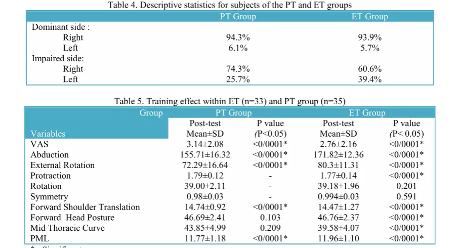
The majority of subjects in both groups were right dominant and right shoulder was affected in them (Table 4).
Pre Test Results: Independent- sample t test revealed no significant differences (p<0.05) between two groups at the begin-ning of the study (Table 3).
Post Test Results: There were significant differences in VAS, abduction, external rotation, forward shoulder translation,
Pec-toralis minor length in the PT group (Table 5) between pre and posttests (p<0.05) in PT Group. All variables showed significant differences in ET group except scapular rotation and symmetry (Table 5) between pre and posttests (p<0.05).
Comparison between Change scores in Training Groups: The improvement of shoulder abduction and external rotation ranges, postural parameters (forward shoulder translation, forward head posture, mid-thoracic curve and Pectoralis minor length) in ET group was significantly greater than that the PT group. VAS did not have significant difference between both Table 4. Descriptive statistics for subjects of the PT and ET groups
PT Group ET Group
Dominant side : Right
Left 94.3%6.1% 93.9%5.7%
Impaired side: Right
Left 74.3%25.7% 60.6%39.4%
Table 5. Training effect within ET (n=33) and PT group (n=35) Group
Variables
PT Group ET Group
Post-test
Mean±SD (P valueP<0.05) Mean±SDPost-test (P< 0.05)P value
VAS 3.14±2.08 <0/0001* 2.76±2.16 <0/0001*
Abduction 155.71±16.32 <0/0001* 171.82±12.36 <0/0001*
External Rotation 72.29±16.64 <0/0001* 80.3±11.31 <0/0001*
Protraction 1.79±0.12 - 1.77±0.14 <0/0001*
Rotation 39.00±2.11 - 39.18±1.96 0.201
Symmetry 0.98±0.03 - 0.994±0.03 0.591
Forward Shoulder Translation 14.74±0.92 <0/0001* 14.47±1.27 <0/0001*
Forward Head Posture 46.69±2.41 0.103 46.76±2.37 <0/0001*
Mid Thoracic Curve 43.85±4.99 0.209 39.58±4.07 <0/0001*
PML 11.77±1.18 <0/0001* 11.96±1.10 <0/0001*
*= Significant
Table 6. Comparison of change scores between ET (n=33) and PT group (n=35) Group
Variables
Group PT Change Score
Mean±SD
Group ET Change Score
Mean±SD
P value
(P< 0.05)
VAS -4.42 ± 1.86 -4.69 ± 2.06 0.576
Abduction 19.14± 14.42 28.78 ± 19.8 0.024*
External Rotation 7.00 ± 8.06 15.75 ± 12.99 0.001*
Protraction 0.0000 -0.016 ± 0.01 <0/001*
Rotation 0.0000 -0.15 ± 0.66 0.183
Symmetry 0.0000 -0.001 ± 0.01 0.578
Forward Shoulder Translation -0.10 ± 0.09 -0.40 ± 0.24 <0/001*
Forward Head Posture 0.11 ± 0.40 1.21 ± 0.92 <0/001*
Mid Thoracic Curve -0.13 ± 0.61 -1.40 ± 1.33 <0/001*
PML 0.23 ± 0.08 0.46 ± 0.20 <0/001*
*= Significant
groups. Moreover, scapular rotation and symmetry had no or very little changes be-tween PT and ET groups (Table 6).
Discussion
The purpose of the current study was to investigate the effect of a 6-week super-vised scapular stabilization exercise therapy on pain, scapular position, head and back posture, and shoulder mobility in compari-son with physical therapy in patients with SIS. Our findings indicate that the exercise protocol suggested in this study significant-ly decreased pain, improved scapular pro-traction, head and back posture and in-creased shoulder mobility. However, we did not observe between-group differences for scapular rotation and symmetry follow-ing the exercise therapy.
Pain: An equal effectiveness of physio-therapy and exercise physio-therapy in decreasing pain was obtained in this study and no sig-nificant difference was found in the VAS score and the shoulder pain was decreased in subjects of both groups. The effect of exercise therapy in reducing pain alone or in combination with other treatments has been shown in the previous studies (15, 34-36).
Rotator cuff muscles stabilize the humeral head in the glenoid, causing humerus to rotate outside while protecting the distance between large tubercle and acromion and preventing compression. It was why that the resistance training (Theraband) used in this study, was effective in reducing pain in ET group subjects. Furthermore, the stretching exercises of our program im-proved the flexibility of shoulder tight tis-sue that could be effective in pain decrease. It seems that our exercise therapy protocol is effective as a treatment for the reduction of pain in SIS patients.
In the present study, ultrasound and TENS was used for the treatment of PT group subjects. Despite the fact that ultra-sound has been used in the treatment of shoulder pain for decades, There are evi-dences showed that US alone has no benefit in decreasing SIS pain (18, 34, 37-39) and
also no available evidence for efficacy of TENS alone for patients with SIS pain (38). Başkurt et al. did not find any statistically significant difference in studying the im-mediate effects of TENS and heat on the pain related to stage one of SIS. (40) Herre-ra-Lasso et al. found positive effects of continuous ultrasound with high frequency TENS in decreasing pain and improving range of movement. (38) Therefore, in this study, a therapeutic package of continuous ultrasound with high frequency TENS and superficial heat (IR) was used which was effective in reducing pain between pre and post intervention in PT group due to the thermal effect of US and IR in combination with analgesic effect of TENS.
Shoulder Range of Motion (ROM): The findings of this study demonstrated the sig-nificant differences of affected shoulder abduction and external rotation between pre and post intervention in both group. Also there were significant differences in shoul-der ROM and its progression rate between PT and ET groups; which imply that ET group had remarkable improvement in shoulder ROM due to stretching exercises used in this study for decreasing tight shoulder capsule and shortened muscles especially pectoral muscles. Also increas-ing shoulder ROM may be due to decreas-ing patients' pain. Our finddecreas-ings agree with those of other researchers (37, 41-42) who have documented improvements in ROM following an exercise program in patients with SIS.
Posture: The effect of a six-week exer-cise intervention was evaluated in changing head, shoulder and back posture in compar-ison with physical modalities in this study. A decrease was observed in FHP, FST and mid-thoracic curve in both groups pre and post intervention but there was a significant difference between ET and PT groups. The results revealed that the exercise protocol used in current study significantly de-creased FHP, FST and mid-thoracic curve, suggesting that our exercises have im-proved patients' posture. The flexibility ex-ercises of this study were chosen based on
the results that indicate tightness of levator scapulae , pectorals in SIS (28) and also a previous study findings which has showed selective activation of deep cervical flexors, Middle and Lower Trapezes and also Serra-tus Anterior in SIS patient (43). It seems that the supervised stretching of the tight anterior shoulder muscles with strengthen-ing of the relatively weaker muscles in pre-sent protocol could have a significant syn-ergistic effect on patients' posture. Our findings are similar to Kluemper et al. and Lynch et al. findings who have studied the effect exercise intervention on FHP and FSP in swimmers (16, 28).
Scapular Protraction & Rotation: Anoth-er purpose of present study was to examine the effect of our exercise program on the position of scapula. The abnormal position of scapula in SIS patients may lead to in-stability of shoulder joint. The results of our study revealed significant differences in the scapular protraction of the ET group compared to the PT group within six-week exercise therapy, indeed there was no change in scapular protraction in PT group. But scapular rotation and symmetry did not indicate any significant difference between pre and post intervention in both group and also between groups. We expected to find more substantial changes in scapular rota-tion and symmetry after exercise based on Alizadeh et al. study (30). A significant changes in scapular symmetry and rotation might require a long term practice, perhaps due to short term duration of our exercise protocol or low accuracy of the measure-ment methods used in this study, scapular symmetry and rotation showed no signifi-cant changes.
Pectoralis Minor Length (PML): Abnor-mal kinematic of scapula in SIS might be lead to Pectoralis minor shortening and tightness, therefore in this study PML in patient involved shoulder was measured with anthropometric method .The results of this assessment indicate a significant dif-ference in PML between pre and post inter-vention in both group and also between groups. PML was increased more in ET
group compared to PT group because of applying stretching exercises. Our findings are similar to those of other researchers (44-46).
This study had several limitations. A ma-jor limitation of our work is that we have no follow-up period. May be if the subjects were followed-up, a significant difference in pain might be found after several weeks. Therefore, more high-quality trials with longer follow-ups are recommended. Also scapular and shoulder muscles strength was not assessed in this study and further re-search is needed, however, to evaluate shoulder girdle muscles strength.
Conclusion
The scapula plays a vital role in shoulder function, thus this study highlights exercise prescription to enhance scapular stabiliza-tion during the SIS rehabilitastabiliza-tion. The scapular stabilization based exercise inter-vention was successful at increasing shoul-der range of abduction and external rota-tion, decreasing forward head and shoulder postures and Pectoralis minor flexibility. This study supports basis for scapular stabi-lization based exercise therapy in the reha-bilitation of SIS. Also be noted that exer-cise therapy is effective as a treatment for the reduction of pain in these patients.
Acknowledgements
This project was supported by a grant from Research Deputy of Tehran Universi-ty of Medical Sciences with ID number for IRCT201111025486N2. The authors thank the participants for their excellent coopera-tion. The authors would like to acknowledge the generous assistance of the academic members and the staff of the Sports Medicine Clinic of Hazrat Rasool-e-Akram hospital, Tehran, Iran.
References
1. Koester MC, George MS, Kuhn JE. Shoulder impingement syndrome. The American journal of medicine. 2005;118(5):452-5.
2. Ludewig PM, Cook TM. Alterations in shoulder
kinematics and associated muscle activity in people with symptoms of shoulder impingement. Physical therapy. 2000;80(3):276-91.
3. Cook TM, Ludewig P. Translations of the humerus in persons with shoulder impingement symptoms. Journal of Orthopaedic & Sports Physical Therapy. 2002; 32(6): 248.
4. Graichen H, Stammberger T, Bonel H, Englmeier K-H, Reiser M, Eckstein F. Glenohumeral translation during active and passive elevation of the shoulder—a 3D open-MRI study. Journal of biomechanics. 2000;33(5):609-13.
5. Lukasiewicz A, McClure P, Michener L, Pratt N, Sennett B. Comparison of 3-dimensional scapular position and orientation between subjects with and without shoulder impingement. The Journal of orthopaedic and sports physical therapy. 1999;29(10):574.
6. Brumitt J. Scapular-stabilization exercises: early-intervention prescription. Athletic Therapy Today. 2006;11(5):15-8.
7. Voight ML, Thomson BC. The role of the scapula in the rehabilitation of shoulder injuries. Journal of Athletic training. 2000;35(3):364.
8. Ludewig PM, Reynolds JF. The association of scapular kinematics and glenohumeral joint pathologies. The Journal of orthopaedic and sports physical therapy. 2009;39(2):90.
9. Yano Y, Hamada J, Tamai K, Yoshizaki K, Sahara R, Fujiwara T, et al. Different scapular kinematics in healthy subjects during arm elevation and lowering: glenohumeral and scapulothoracic patterns. Journal of shoulder and elbow surgery. 2010;19(2):209-15.
10. Han KJ, Cho JH, Han SH, Hyun HS, Lee DH. Subacromial impingement syndrome secondary to scapulothoracic dyskinesia. Knee Surg Sports Traumatol Arthrosc. 2012 Oct;20(10):1958-60.
11. McQuade K, Dawson J, Smidt G. Scapulothoracic muscle fatigue associated with alterations in scapulohumeral rhythm kinematics during maximum resistive shoulder elevation. The Journal of orthopaedic and sports physical therapy. 1998;28(2):74.
12. Cools AM, Witvrouw EE, Declercq GA, Danneels LA, Cambier DC. Scapular Muscle Recruitment Patterns: Trapezius Muscle Latency with and without Impingement Symptoms. The American journal of sports medicine. 2003 July 1, 2003;31(4):542-9.
13. Michener LA, McClure PW, Karduna AR. Anatomical and biomechanical mechanisms of subacromial impingement syndrome. Clinical Biomechanics (Bristol, Avon). 2003;18(5):369-79.
14. Warner JJ, Micheli LJ, Arslanian LE, Kennedy J, Kennedy R. Scapulothoracic motion in normal shoulders and shoulders with glenohumeral instability and impingement syndrome. A study using Moire topographic analysis. Clin Orthop Relat Res. 1992;285:191-9.
15. Celik D, Akyuz G, Yeldan I. Comparison of the effects of two different exercise programs on pain in subacromial impingement syndrome. Acta Orthop Traumatol Turc. 2004;43(6):504-9.
16. Kluemper M, Uhl T, Hazelrigg H. Effect of stretching and strengthening shoulder muscles on forward shoulder posture in competitive swimmers. Journal of Sport Rehabilitation. 2006;15(1):58.
17. Faber E, Kuiper JI, Burdorf A, Miedema HS, Verhaar JA. Treatment of impingement syndrome: a systematic review of the effects on functional limitations and return to work. Journal of occupational rehabilitation. 2006;16(1):6-24.
18. Van der Heijden G, Van der Windt D, de Winter AF. Physiotherapy for patients with soft tissue shoulder disorders: a systematic review of randomised clinical trials. BMJ: British Medical Journal. 1997;315(7099):25.
19. Soibam I. Comparative study on the effectiveness of ultrasound (us) to transcutaneous electrical stimulation (tens) in the treatment of periarticular shoulder pain (psp). 2005.
20. Roy J-S, Moffet H, Hébert LJ, Lirette R. Effect of motor control and strengthening exercises on shoulder function in persons with impingement syndrome: A single-subject study design. Manual therapy. 2009;14(2):180-8.
21. Ure B, Tiling T, Kirchner R, Rixen D. [Reliability of clinical examination of the shoulder in comparison with arthroscopy. A prospective study]. Der Unfallchirurg. 1993;96(7):382.
22. Leroux J, Thomas E, Bonnel F, Blotman F. Diagnostic value of clinical tests for shoulder impingement syndrome. Revue du rhumatisme (English ed). 1995; 62(6):423.
23. Ferreira-Valente MA, Pais-Ribeiro JL, Jensen MP. Validity of four pain intensity rating scales. Pain. 2011;152(10):2399-404.
24. Kolber MJ, Hanney WJ. The reliability and concurrent validity of shoulder mobility measurements using a digital inclinometer and goniometer: A technical report. International journal of sports physical therapy. 2012;7(3):306.
25. Greenfield B, Catlin P, Coats P, Green E, McDonald J, North C. Posture in patients with shoulder overuse injuries and healthy individuals. The Journal of orthopaedic and sports physical therapy. 1995; 21(5):287.
26. Raine S, Twomey LT. Head and shoulder posture variations in 160 asymptomatic women and men. Archives of physical medicine and rehabilitation. 1997;78(11):1215-23.
27. Lewis JS, Wright C, Green A. Subacromial impingement syndrome: the effect of changing posture on shoulder range of movement. J Orthop Sports Phys Ther. 2005;35(2):72-87.
28. Lynch SS, Thigpen CA, Mihalik JP, Prentice WE, Padua D. The effects of an exercise intervention on forward head and rounded shoulder postures in elite swimmers. British journal of sports
medicine. 2010;44(5):376-81.
29. Borstad JD. Measurement of Pectoralis minor muscle length: validation and clinical application. The Journal of orthopaedic and sports physical therapy. 2008;38(4):169.
30. Alizadeh M, Daneshmandi H, Shademan B, Ahmadizad S. The Effects of Exercise Training on Scapula Position of Muscle Activity Measured by EMG. World. 2009;2(1):48-52.
31. De Mey K, Danneels L, Cagnie B, Cools AM. Scapular Muscle Rehabilitation Exercises in Overhead Athletes With Impingement Symptoms: Effect of a 6-Week Training Program on Muscle Recruitment and Functional Outcome. The American journal of sports medicine. 2012 August 1, 2012;40(8):1906-15.
32. Ginn K, Cohen M. Exercise therapy for shoulder pain aimed at restoring neuromuscular control: a randomized comparative clinical trial. Journal of Rehabilitation Medicine. 2005;37(2):115-22.
33. Leung MS, Cheing GL. Effects of deep and superficial heating in the management of frozen shoulder. Journal of Rehabilitation Medicine. 2008;40(2):145-50.
34. Kromer TO, Tautenhahn UG, de Bie RA, Staal JB, Bastiaenen CH. Effects of physiotherapy in patients with shoulder impingement syndrome: a systematic review of the literature. Journal of Rehabilitation Medicine. 2009;41(11):870-80.
35. Ginn KA, Herbert RD, Khouw W, Lee R. A randomized, controlled clinical trial of a treatment for shoulder pain. Physical therapy. 1997;77(8):802-9.
36. Bang MD.Comparison of Supervised Exercise With and Without~ anual Physical Therapy for Patients With Shoulder Impingement Syndrome. Journal of Orthopaedic & Sports Physical Therapy. 2000;30(3):126-37.
37. Michener LA, Walsworth MK, Burnet EN. Effectiveness of rehabilitation for patients with subacromial impingement syndrome: a systematic
review. Journal of hand therapy. 2004;17(2):152-64. 38. Johansson K, Oberg B, Adolfsson L, Foldevi M. A combination of systematic review and clinicians' beliefs in interventions for subacromial pain. The British Journal of General Practice. 2002; 52(475):145.
39. Green S, Buchbinder R, Hetrick S. Physiotherapy interventions for shoulder pain (Review). 2010.
40. Başkurt Z, Başkurt F, Özcan A, Yilmaz Ö. The immediate effects of heat and TENS on pressure pain threshold and pain intensity in patients with Stage I shoulder impingement syndrome. The Pain Clinic. 2006;18(1):81-5.
41. McClure PW, Bialker J, Neff N, Williams G, Karduna A. Shoulder function and 3-dimensional kinematics in people with shoulder impingement syndrome before and after a 6-week exercise program. Physical therapy. 2004;84(9):832-48.
42. Kuhn JE. Exercise in the treatment of rotator cuff impingement: a systematic review and a synthesized evidence-based rehabilitation protocol. Journal of shoulder and elbow surgery. 2009; 18(1):138-60.
43. Moseley J JF, Pink M, Perry J, Tibone J. EMG analysis of the scapular muscles during a shoulder rehabilitation program. Am J Sports Med. 1992; 20:128-34.
44. Borstad JD, Ludewig PM. Comparison of three stretches for the Pectoralis minor muscle. Journal of shoulder and elbow surgery. 2006; 15(3):324-30.
45. Cools AM, Johansson FR, Cambier DC, Velde AV, Palmans T, Witvrouw EE. Descriptive profile of scapulothoracic position, strength and flexibility variables in adolescent elite tennis players. British journal of sports medicine. 2010;44(9):678-84.
46. Muraki T, Aoki M, Izumi T, Fujii M, Hidaka E, Miyamoto S. Lengthening of the Pectoralis minor muscle during passive shoulder motions and stretching techniques: a cadaveric biomechanical study. Physical therapy. 2009;89(4):333-41.