Cite as
Koźluk E, Zyśko D, Piątkowska A, et al. Effectiveness compari-son of various atrial fibrillation ablation methods in patients with common venous trunk. Adv Clin Exp Med. 2019; 28(4):461–467. doi:10.17219/acem/78157
DOI
10.17219/acem/78157
Copyright
© 2019 by Wroclaw Medical University This is an article distributed under the terms of the Creative Commons Attribution Non-Commercial License (http://creativecommons.org/licenses/by-nc-nd/4.0/)
Address for correspondence
Dorota Zyśko E-mail: [email protected]
Funding sources
None declared
Conflict of interest
Edward Koźluk – proctor for ablation procedures (Medtronic and Johnson & Johnson) Piotr Lodziński – proctor for ablation procedures
(Johnson & Johnson) Paweł Balsam – proctor for ablation procedures
(St. Jude Medical)
Marek Kiliszek – proctor for ablation procedures (St. Jude Medical)
Other authors declare no conflict of interest
Received on March 22, 2017 Reviewed on June 25, 2017 Accepted on October 2, 2017
Published online on August 8, 2018
Abstract
Background. Atrial fibrillation (AF) is a common clinical problem. The left atrium anatomy makes up a factor that may significantly affect the effectiveness of the AF ablation.
Objectives. The aim of the study was to evaluate a long-term effectiveness ablation in patients with com-mon pulcom-monary vein trunk (CPVT) and AF.
Material and methods. The outcomes of 129 procedures in 95 patients with CPVT out of 1,475 proce-dures carried out in 1,150 patients with AF treated with ablation, were analyzed. Ablation with CARTO 3 system (Johnson & Johnson, New Brunswick, USA), cryoballoon, and the circular multipolar duty-cycled radiofrequency-based pulmonary vein ablation producer with catheter (PVAC) were considered as advanced methods. The following data was recorded for every patient: age, gender, AF duration and type, previous antiarrhythmic drugs, weight, height, any prior cardioversion, and comorbidities, including hypertension, diabetes, hypothyreosis, thyrotoxicosis, heart failure, and stroke/transient ischemic attack. The following anatomical factors were assessed: the presence of patent foramen ovale (PFO) and localization of the CPVT on the basis of venography or computed tomography (CT). In the 1st year after ablation, 24-hour Holter monitoring was performed 3–5 times, and the patients were encouraged to visit their doctor or an emergency department if a cardiac arrhythmia occurred. Long-term ablation effectiveness was assessed based on a tele-phone interview and patients’ answers to the questionnaires including 12-lead electrocardiography (ECG). Results. Sinus rhythm was maintained in 44 patients (43.6%) after a median of 42 months (range: 12–120). A lower number of clinical factors (odds ratio (OR) = 0.09; 95% confidence interval (95% CI) = 0.02–0.56; p < 0.01), and advanced ablation methods (OR = 3.1; 95% CI = 1.4–7.1; p < 0.01) were related to a better long-term effectiveness.
Conclusions. The long-term effectiveness of pulmonary vein (PV) isolation in patients with AF and CPVT is higher when advanced ablation techniques are used. Accumulation of clinical factors was found to be the most tremendous predictor of AF recurrence.
Key words: long-term outcome, atrial fibrillation, ablation, common trunk
Effectiveness comparison of various atrial fibrillation ablation methods
in patients with common venous trunk
Edward Koźluk
1,A–F, Dorota Zyśko
2,A,C–F, Agnieszka Piątkowska
1,2,A,B,E,F, Marek Kiliszek
3,A,B,E,F, Piotr Lodziński
1,A,B,E,F,
Sylwia Małkowska
1,A,B,E,F, Paweł Balsam
1,A,B,E,F, Dariusz Rodkiewicz
1,A,B,E,F, Małgorzata Żukowska
4,A,B,E,F, Grzegorz Opolski
1,A,E,F1 1st Chair and Department of Cardiology, Medical University of Warsaw, Poland
2 Chair of Emergency Medicine, Wroclaw Medical University, Poland
3 Department of Cardiology and Internal Diseases, Military Institute of Medicine, Warszawa, Poland
4 2nd Faculty of Radiology, Medical University of Warsaw, Poland
A – research concept and design; B – collection and/or assembly of data; C – data analysis and interpretation; D – writing the article; E – critical revision of the article; F – final approval of the article
Introduction
Atrial fibrillation (AF) is a common clinical problem associated with the occurrence of sudden and non-sud-den cardiac death, heart failure, stroke, an increased risk of hospitalizations, and decreased quality of life.1–3
In the treatment of patients with symptomatic AF, isola-tion of pulmonary vein (PV) is recommended by the cre-ation of circumferential lesions around the right and the left PV ostia.2–4 However, invasive procedures may
lead to complications.4–7 Looking for predictors of atrial
fibrillation relapse facilitates the process of qualifying the patient for the procedure, as well as the selection of the most effective methods to perform ablation.8–11
The left atrium anatomy makes up a factor that may significantly affect the effectiveness of the procedure. An unusual anatomy of PV, usually in the form of a com-mon pulof a com-monary vein trunk (CPVT), occurs in 13–29% of patients.12,13 The vascular anomaly, which usually refers
to single-sided PVs, is more common on the left side.4,13
The presence of the CPVT hampers PV isolation, the main reason for that possibly being a fact that systems for isolat-ing the PVs have been developed for a typical PV anatomy, and their effectiveness might alter in the case of an unusual anatomy. The aim of the study was to evaluate the long-term effectiveness of different methods used to ablate/ isolate PV in patients to treat AF.
Material and methods
The study was designed as the retrospective analysis of the long-term results of ablation procedures performed in patients with AF and CPVT between 2003 and 2012.
Out of the group of 1,150 patients (1,475 procedures) with AF, 107 (9.3%) had the CPVT recognized. Twelve patients did not appear at the follow-up after the 1st
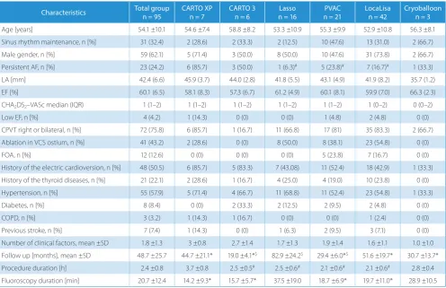
procedure. Finally, the effectiveness of 129 procedures performed in 95 patients with the CPVT was subjected to analyses in the study. Table 1 shows the patients’ char-acterization by the technique used for ablation. Among the studied population, 72 patients had 1 procedure only, 15 had 2 procedures, 6 had 3 procedures, 1 patient had 4 procedures, and 1 had 5 procedures. The mean number of ablation procedures amounted to 1.3 times per patient.
Data collection
The following data was recorded for every patient: age, gender, AF duration and type, previous antiarrhythmic drugs, weight, height, any prior cardioversion, and comor-bidities including hypertension, diabetes, hypothyreosis, thyrotoxicosis, heart failure, as well as stroke/transient ischemic attack. The following echocardiographic data was recorded: left ventricle ejection fraction (EF) and left atri-um diameter. Ejection fraction less than 50% was regarded
as low EF. For every ablation an average fluoroscopy and procedure time were recorded.
During ablation, the following anatomical factors were assessed: the presence of patent foramen ovale (PFO; if it could be used to introduce the catheter into the left atrium), and left-side or right-side localization of the CPVT recognized on the basis of venography or computed to-mography (CT). Furthermore, the method of ablation was recorded in each case and the nature of the procedure was referred to as the 1st or the repeated procedure.
Procedure details
The objective of the procedure was to isolate all PVs. Other ablation regions were performed if the surgeon de-cided so. In some patients with high far-field potentials from the superior vena cava (VCS) or the inferior vena cava (VCI) (n = 50 and n = 4, respectively), the vein was isolated, too. In patients with persistent AF, with a short distance between PV lines or with transformation of AF into an atypical atrial flutter, additional lines were per-formed. All lines were carried out using CARTO (John-son & Johnson, New Brunswick, USA) system, including left atrial roof (n = 10), posterior wall (n = 10) or the mitral isthmus (n = 8), right atrial isthmus (n = 13), coronary sinus (n = 8), complex fractionated atrial electrogram (CFAE) (n = 3), VCS VCI line (n = 1), and septum (n = 6).
In the presented study, fluoroscopy was used as the only imaging method in the subgroup of patients in whom Lasso electrode and ablation with 4-mm tip catheter were used for segmental PV isolation, using the method described by Haïssaguerre et al.14 The majority of the procedures
performed by the use of Haïssaguerre’s method were done with the LocaLisa system used to reduce fluoros-copy. Details of this technique were previously presented.14
The LocaLisa navigation system (Medtronic EP Systems, Minneapolis, USA) uses 3 low-amplitude, high-frequen-cy current fields. The fields are generated in 3 axes over the patient’s thorax and are dedicated to compute the po-sition of an electrode in the thorax relative to a reference electrode. The procedure of segmental PV isolation using the LocaLisa navigation system was precisely described in the previous publication.15 Briefly, the Lasso catheter
(Johnson & Johnson, New Brunswick, USA) was located in every PV under the fluoroscopic guidance. The ablation procedure was performed by the use of the ablation cath-eter under the LocaLisa system and fluoroscopy. The PV isolation was proved by disappearance or dissociation of potential recorded from the Lasso catheter.15
CARTO XP (Biosense Webster, Johnson & Johnson, New Brunswick, USA) visualized only the ablation catheter. This catheter was used to create the left atrial map. The next atrial PV linear isolation was performed based on the method presented by Pappone et al.16 Verification of the lines was
CARTO 3 (Biosense Webster, Johnson & Johnson, New Brunswick, USA) system is an upgraded version, al-lowing a more accurate visualization of the cardiac anato-my (e.g., to perform a quick anatomical map) and enabling the visualization of all catheters used. This PV isolation verification technology was used for the Lasso catheter.
Ablation with PV ablation producer with catheter (PVAC) (Medtronic, Carlsbad, USA) was described in most detail in the previous publication.17 Shortly after a PV selective
angiography a circular decapolar catheter is advanced over wire positioned selectively in every PV. After sev-eral radio frequency (RF) applications over all electrode pairs, the PVAC was rotated around the PV ostium to en-sure a complete isolation of the vein. During the ablation of large common ostia, electrode pairs with suboptimal contact to the atrial tissue were deactivated, preventing ineffective energy delivery.17
Cryoballoon ablation methods were described in the pre-vious publications.18 Shortly after an initial mapping of PV
using the Lasso catheter, the cryoballoon (Arctic Front – Cryocath, Medtronic, Mineapolis, USA) was introduced into the left atrium and positioned at PV ostia. Contact of the balloon with the ostial tissue was checked by contrast administration and temperature inside the balloon during cryoapplication. At every PV ostium 2 cryoapplications
with good contact were performed. Subsequently, remap-ping of cryoapplications in all veins was performed with Lasso catheters. In the absence of PV potentials, the pro-cedure was finished; otherwise, next cryoapplications were performed.18
Ablations with CARTO 3 system, cryoballoon, and PVAC catheter were considered as advanced methods, whereas those with CARTO XP, Lasso guided alone or with LocaLisa system were regarded as reference methods, potentially less effective because of hindered adaptation to the changed anatomy.
Follow-up
The details of follow-up procedures were presented else-where.19 Briefly, in the 1st year after ablation, 24-h Holter
monitoring was performed 3–5 times, and the patients were encouraged to visit their doctor or an emergency department if a cardiac arrhythmia occurred. The procedure was as-sessed as ineffective when an event of AF lasting at least 30 s was documented later than after 3 months of a blanking period following the procedure.8 Long-term ablation
effec-tiveness was assessed based on the telephone interview and patients’ answers to the questionnaires, including 12-lead electrocardiography (ECG). The gathered data was further
Table 1. Baseline demographics and clinical characteristics
Characteristics Total groupn = 95 CARTO XPn = 7 CARTO 3n = 6 n = 16Lasso n = 21PVAC LocaLisan = 42 Cryoballoonn = 3
Age [years] 54.1 ±10.1 54.6 ±7.4 58.8 ±8.2 53.3 ±10.9 55.3 ±9.9 52.9 ±10.8 56.3 ±8.1
Sinus rhythm maintenance, n [%] 31 (32.4) 2 (28.6) 2 (33.3) 2 (12.5) 10 (47.6) 13 (31.0) 2 (66.7)
Male gender, n [%] 59 (62.1) 5 (71.4) 3 (50.0) 8 (50.0) 10 (47.6) 31 (73.8) 2 (66.7)
Persistent AF, n [%] 23 (24.2) 6 (85.7) 3 (50.0) 1 (6.3)# 5 (23.8)# 7 (16.7)# 1 (33.3)
LA [mm] 42.4 (6.6) 45.9 (3.7) 44.0 (2.8) 41.8 (5.5) 43.1 (4.9) 41.9 (8.2) 35.7 (1.2)
EF [%] 60.1 (6.5) 58.1 (8.3) 57.3 (6.7) 61.2 (4.9) 60.1 (8.1) 59.9 (7.0) 66.3 (2.3)
CHA2DS2–VASc median (IQR) 1 (1–2) 1 (1–2) 1 (1–2) 1 (1–2) 1 (1–2) 1 (0–2) 0 (0–2)
Low EF, n [%] 4 (4.2) 1 (14.3) 0 (0) 0 (0) 1 (4.8) 2 (4.8) 0 (0)
CPVT right or bilateral, n [%] 72 (75.8) 6 (85.7) 1 (16.7) 11 (66.8) 17 (81) 35 (83.3) 2 (66.7)
Ablation in VCS ostium, n [%] 41 (43.2) 2 (28.6) 0 (0) 8 (50.0) 8 (38.1) 23 (54.8) 0 (0)
FOA, n [%] 12 (12.6) 0 (0) 0 (0) 0 (0) 5 (23.8) 7 (16.7) 0 (0)
History of the electric cardioversion, n [%] 48 (50.5) 6 (85.7) 5 (83.3) 7 (43.08) 11 (52.4) 18 (42.9) 1 (33.3) History of the thyroid diseases, n [%] 21 (22.1) 2 (28.6) 1 (16.7) 4 (25.0) 4 (19.0) 10 (23.8) 0 (0)
Hypertension, n [%] 55 (57.9) 5 (71.4) 4 (66.7) 11 (68.8) 11 (52.4) 23 (54.8) 1 (33.3)
Diabetes, n [%] 8 (8.4) 0 (0) 2 (33.3) 2 (12.5) 2 (9.5) 2 (4.8) 0 (0)
COPD, n [%] 3 (3.2) 1 (14.3) 1 (16.7) 0 (0) 0 (0) 1 (2.4) 0 (0)
Previous stroke, n [%] 7 (7.4) 1 (14.3) 0 (0) 1 (6.3) 2 (9.5) 3 (7.1) 0 (0)
Number of clinical factors, mean ±SD 1.8 ±1.3 3 ±0.8 2.7 ±1.4 1.7 ±1.3 1.9 ±1.4 1.6 ±1.1 1.0 ±1.0 Follow up [months], mean ±SD 48.7 ±25.7 44.7 ±21.1* 19.0 ±4.1*$ 82.9 ±24.2$ 29.4 ±6.0*$ 51.6 ±19.7* 30.7 ±13.7*
Procedure duration [h] 2.4 ±0.8 3.7 ±0.8 2.5 ±0.5# 2.5 ±0.6# 2.1 ±0.6# 2.1 ±0.6# 2.8 ±0.4
Fluoroscopy duration [min] 20.7 ±12.4 14.2 ±9.3* 15.7 ±5.7* 37.5 ±19.0 18.7 ±6.9* 19.7 ±11.0* 28.9 ±10.5
supplemented with the data from cardiology outpatient clinics and the results of the Holter monitoring. Had there been more than 1 ablation, all treatments preceding the last treatment (also in patients with whom the contact was lost) would have been considered ineffective.
Informed consent was obtained from all patients. The study was approved by the Bioethical Commission (Wroclaw Medical University, Poland).
Continuous variables were expressed as mean and stan-dard deviation (SD) and compared with analysis of vari-ance (ANOVA) test with Bonferroni correction for a mul-tiple comparison. Categorical variables were presented as numbers and percentages, and χ2 test together with
Fisher’s exact test were both used to assess the differences. The CHA2DS2–VASc score was calculated for each
patient. The number of clinical factors was calculated for each patient as a sum of points. One point was given for the presence of each diagnosis in the patients’ medi-cal history: age above 65 years, age above 75 years, hy-pertension, diabetes mellitus, previous stroke, previous myocardial infarction, congestive heart failure, the pres-ence of the permanent character of AF, previous electric cardioversion, thyroid diseases, and chronic obstructive pulmonary disease (COPD).
The logistic regression analysis was performed to find association between the AF recurrence, the advanced method of ablation and the number of clinical factors which may predict AF relapse.
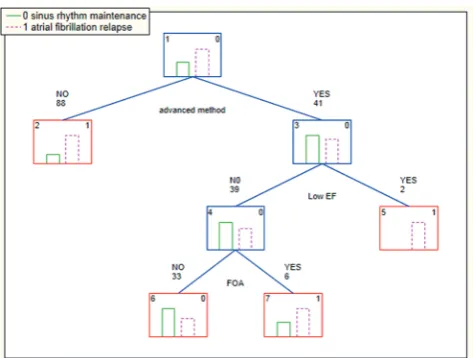
The classification and regression tree analysis were used to find associations among the long-term out-come, the method of ablation and the clinical predictors of the AF recurrence. Specificity, sensitivity, and positive and negative predictive values for each model were calcu-lated. The classification and regression tree analysis con-stitute non-parametric methods to establish predictors of the studied end-point. These methods were described by Breiman et al. and they consist in recursive partitioning of the study group for each possible point to find those dividing the entire group into subgroups which are dif-ferentiated the most regarding the evaluated events.19,20
The classification tree is a graphical method used for pre-senting the classification rules. The number in the up-per right corner of the rectangle is an information about which subgroup it was included to, based on the classifi-cation rules. In the case of the present study “0” means maintaining sinus rhythm and “1” is the AF recurrence. The number of factors associated with the AF recurrence was assessed for every patient.
Results
Demographics, clinical characteristics within the study group and subgroups selected on the basis of the 1st
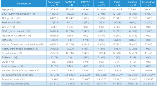
pro-cedure performed were presented in Table 1. Table 2 pictures the data regarding demographics and clinical
Table 2. Demographics and clinical characteristics in groups selected on the basis of the procedure performer
Characteristics Total group n = 129
CARTO XP n = 17
CARTO 3 n = 11
Lasso n = 19
PVAC n = 24
LocaLisa n = 52
Cryoballoon n = 6
Age [years] 54.7 ±10.2 57.5 ±6.8 59.9 ±6.6 54.2 ±10.4 54.4 ±10.8 52.5 ±11.1 59.8 ±7.1
Sinus rhythm maintenance, n [%] 44 (34.1) 4 (23.5) 5 (45.5) 2 (10.5) 12 (50.0) 16 (30.8) 5 (83.3)
Male gender, n [%] 84 (65.1) 11 (64.7) 7 (63.6) 9 (47.4) 13 (54.2) 39 (75.0) 5 (83.3)
Persistent AF, n [%] 27 (20.9) 8 (47.1) 3 (27.3) 1 (5.3) 5 (20.8) 9 (17.3) 1 (16.7)
Low EF, n [%] 6 (4.7) 1 (5.9) 0 (0) 0 (0) 2 (8.3) 3 (5.8) 0 (0)
CPVT right or bilateral, n [%] 96 (74.4) 12 (70.6) 5 (45.5) 14 (73.7) 19 (79.2) 41 (78.8) 5 (83.3)
Ablation in VCS ostium, n [%] 50 (38.8) 2 (11.8) 0 (0) 9 (47.4) 10 (41.7) 29 (55.8) 0 (0)
FOA, n [%] 14 (10.9) 0 (0) 1 (9.1) 0 (0) 5 (20.8) 8 (15.4) 0 (0)
History of the electric cardioversion, n [%] 66 (51.2) 12 (70.6) 6 (54.5) 9 (47.4) 13 (54.2) 23 (44.2) 3 (50.0) History of the thyroid diseases, n [%] 30 (23.3) 5 (29.4) 5 (45.5) 4 (21.1) 4 (16.7) 12 (23.1) 0 (0)
Hypertension, n [%] 75 (58.2) 12 (70.6) 5 (45.5) 13 (68.4) 12 (50.0) 32 (61.5) 1 (16.7)
Diabetes, n [%] 10 (7.8) 0 (0) 3 (27.3) 2 (10.5) 2 (8.3) 3 (5.8) 0 (0)
COPD, n [%] 4 (3.1) 2 (11.8) 1 (9.1) 0 (0) 0 (0) 1 (1.9) 0 (0)
Previous stroke, n [%] 10 (7.8) 3 (17.6) 0 (0) 1 (5.3) 2 (8.3) 4 (7.7) 0 (0)
Number of clinical factors, mean ±SD 1.8 ±1.2 2.5 ±1.2 2.2 ±1.7 1.6 ±1.2 1.8 ±1.3 1.7 ±1.1 1.0 ±0.9 Follow up [months], mean ±SD 48.7 ±10.1 51.5 ±26.3* 21.0 ±9.0*#$ 84.3 ±24.2 29.5 ±5.7*#$ 52.2 ±18.9* 25.5 ±12.8#$*
Procedure duration [h] 2.4 ±0.8 3.6 ±1.0 2.5 ±0.5# 2.4 ±0.6# 2.2 ±1.1# 2.1 ±0.6# 2.8 ±0.4#
Fluoroscopy duration [min] 21.4 ±13.2 14.6 ±9.5* 15.7 ±9.5* 35.5 ±12.4 18.7 ±6.9* 19.0 ±11.1* 28.9 ±10.5#
characteristics in groups selected on the basis of the pro-cedure performed. The subgroups did not differ in terms of age and gender distribution. The number of clinical factors associated with AF relapse was significantly greater in the group treated with CARTO XP system than with LocaLisa. Procedure times were the longest during proce-dures with CARTO XP.
Procedure safety
Two events of transient phrenic nerve palsy during cryoablation were observed, while in 1 patient a mild stenosis of PVs occurred during the long-term follow-up (from 16 × 14 mm before the procedure to 9 × 6 mm thereafter). Patient with the PV stenosis presented with hemoptysis and tussis. These symptoms subsided after a few months. This patient had the ablation performed with LocaLisa.
No significant differences were found with reference to the overall complication rate.
Follow-up
During the follow-up of a median of 42 months (range 12–120 months), the sinus rhythm was maintained in 44 patients (43.6%). Among 129 procedures, 85 proce-dures (65.9%) were assessed as ineffective.
Clinical variables associated
with atrial fibrillation recurrence
Thyroid diseases history was more prevalent in the group with AF recurrence than in the group without AF recur-rence (29.7% vs 6.5%; p < 0.02, respectively). No differ-ences in the occurrence of other clinical variables related to the AF recurrence were observed. However, it is worth-while to mention that the number of clinical factors associ-ated with recurring AF was significantly lower in patients without AF recurrence than in those with AF recurrence (1.4 ±1.2 vs 2.0 ±1.2; p < 0.01, respectively).
Multivariate analysis
The logistic regression analysis revealed that a lower number of clinical factors (odds ratio (OR) = 0.09, 95% confidence interval (95% CI) = 0.02–0.56; p < 0.01) and advanced ablation methods (OR = 3.1, 95% CI = 1.4–7.1; p < 0.01) were related to better long-term effectiveness of the given procedure.
The classification and regression analysis confirmed the significance of the advanced methods for good out-come, whereas the low EF and the presence of PFO were related to the AF recurrence (Fig. 1), with the specific-ity of that model being 0.85, sensitivthe specific-ity 0.90, and posi-tive and negaposi-tive predicposi-tive values being 0.87 and 0.69, respectively.
Discussion
The procedures of the PVs isolation are currently among the most effective therapeutic options for the AF treat-ment. The ablation effect on AF might be attributed to var-ious mechanisms, including the elimination of the trigger, modification of the arrhythmogenic substrate, interrup-tion of crucial pathways of conducinterrup-tion or rotors, or atrial denervation.
The PV isolation techniques differ with regards to the en-ergy source (radiofrequency or cryoento the en-ergy), mapping system (electrophysiological or electroanatomical) and electrode shape (focal, ring or balloon). The effectiveness of these techniques may vary in the settings of the CPVT and normal anatomy.
Difficult anatomy of venoatrial junction may impede the effective implementation of the ablation procedure. Common pulmonary vein trunk is a quite frequent-ly encountered variant of the PV anatomy. The size and shape of the veins ostia may be of particular im- portance for some of the techniques, whilst others may be more resistant to the associated difficulties.
Recent randomized studies comparing various meth-ods of ablation techniques were performed in the setting of 4 independent PVs and demonstrated that the simpli-fied strategy for PV cryoablation is inferior to PV isola-tion using open-irrigated radiofrequency catheters with electrophysiological and electroanatomical guidance.21
It was also observed that a complete PV conduction block is critical to the success of AF ablation. Malmborg et al. reported that either cryoballoon and/or the circular mul-tipolar duty-cycled radiofrequency-based PVAC proved to be comparably effective and safe in achieving acute PV isolation.22Other researchers have determined that among
different procedures of catheter ablation there are no sig-nificant differences in the success rate between the 2 single procedures. Notwithstanding, the success rates are higher for combined methods than for single methods.23
The accuracy of the spatial localization by different ad-vanced electroanatomical mapping systems seems to be comparable; however, in the case of the CPVT other factors may appear to be more important.
Furthermore, there are no established strategies for redo procedures after PV isolation. In the presented study, it was found that the advanced techniques are more effective than the older ones, regardless of whether used during the 1st
or the repeated procedure. Contrary to the presented find-ings, Pokushalov et al. reported that when patients require a redo PV isolation ablation procedure for the recurrent paroxysmal AF, RF appears to be the preferred method relative to cryoablation.24 However, the number
of cryobal-loon isolations in the presented study was too low to obtain conclusive results.
The clinical factors analyzed in the presented study overlap, but are not limited to, the parameters used in the CHA2DS2–VASc scale. Other factors assigned
to the clinical picture include the following: the permanent character of AF, previous electric cardioversion, thyroid diseases, and COPD.9
The importance of clinical factors in predicting the long-term outcome of ablation procedure was confirmed in the study. The increase of the number of clinical factors associated with the reduced effectiveness of long-term abla-tion was associated with an increased risk of AF recurrence by 30%. The classification and regression tree analysis re-vealed that thyroid disease constituted the most important clinical factor. It is well known that thyroid diseases often have the 1st clinical manifestation as cardiac
supraven-tricular arrhythmias. Thyroid hormones directly regulate the metabolism of myocardial cells, and thyroid diseases induce a diffuse atrial architecture disruption. This may predispose to ablation ineffectiveness.25–27 The PV
iso-lation procedure is targeted at removing the trigger off the PVs rather than treating the whole diseased atrial myo-cardium which constitutes the substrate for AF.
The presence of thyroid diseases may also represent the adverse effect of amiodarone therapy. Patients with a greater tendency to relapse could have been treated with amiodarone, which in turn could have induced thyroid diseases, further promoting AF.
The treatment with amiodarone may lead to thyroid dysfunction, which then reduces the effectiveness of ablation. Therefore, it should be considered whether in patients with paroxysmal AF, ablation should not take precedence over pharmacological treatment. A major problem in iatrogenic hyperthyroidism can particularly be the long time to get euthyroid.
Atrial fibrillation is a frequent rhythm in heart failure patients. It may exacerbate heart failure, which in turn may increase the susceptibility to AF occurrence. Heart failure is also a predictor of AF recurrence after ablation procedure, which was also determined in the presented study. An expla-nation for this observation may be the occurrence of chang-es in the myocardium of heart failure patients, which lead to the transformation of arrhythmia from trigger-driven
to substrate-mediated. The risk of AF recurrence after cath-eter ablation is also increased in another setting of the atrial myocardium damage as sick sinus syndrome.28
Such anatomical factors as the localization of the CPVT and the presence of PFO were also included in the anal-ysis because in the previous analin the anal-ysis we had found that these factors may have an impact on the effective-ness of the performed procedure. In the current analysis, contrary to the one performed earlier, only PFO presence was related to the lower effectiveness. The ablation meth-ods used may have various effectiveness levels in the case of the right-side CPVT, which made it difficult to show the adverse significance of the localization of the CPVT.
There are no established strategies for the redo proce-dures after the PV isolation. The presented study indicates the higher effectiveness of the advanced methods both in the 1st and in the redo procedures.
Limitations
This study is subjected to several limitations. Firstly, the follow-up duration varies among the patients’ groups based on methods that were used. However, even after tak-ing into account a 5% annual increase in the risk of recur-rence, the presented results indicate the higher effective-ness of newer ablation techniques.29 Secondly, the limited
number of patients and a single-center study design may produce a bias, thus these results may not be generalized.
The methods used for a long-term follow-up after AF catheter ablation are not as accurate as continuous ECG monitoring.30 However, these methods allow us to find
recurrences in the 2/3 of patients, although the immediate success rate was comparable with other studies.
Institutional experience over nearly 10 years may im-prove significantly; however, the total number of proce-dures was high, so the effect of a learning curve could affect only a small percentage of cases.
Conclusions
The long-term effectiveness of PV isolation in patients with AF and CPVT was higher when advanced ablation techniques were used.
The same group of ablation methods is more effective in treatment, regardless of whether it is applied during the 1st or the redo procedure.
Accumulation of the clinical risk factors was found to be the most tremendous predictor of AF recurrence, and this finding could be helpful in selecting patients with AF for an appropriate therapeutic strategy.
Thyroid disease seems to be the most significant clinical factor related to the AF recurrence after ablation.
References
1. Chen LY, Sotoodehnia N, Bůžková P, et al. Atrial fibrillation and the risk of sudden cardiac death: The atherosclerosis risk in communities study and cardiovascular health study. JAMA Intern Med. 2013;173(1):29–35. 2. Camm AJ, Lip GY, De Caterina R, et al. 2012 focused update of the ESC
Guidelines for the management of atrial fibrillation: An update of the 2010 ESC Guidelines for the management of atrial fibrilla-tion. Developed with the special contribution of the European Heart Rhythm Association. Eur Heart J. 2012;33(21):2719–2747.
3. European Heart Rhythm Association; European Association for Cardio-Thoracic Surgery; Camm AJ, Kirchhof P, Lip GY, et al. Committee for Prac-tice Guidelines. Guidelines for the management of atrial fibrillation: The Task Force for the Management of Atrial Fibrillation of the Europe-an Society of Cardiology (ESC). Europace. 2010;12:1360–1420. 4. Calkins H, Kuck KH, Cappato R, et al. 2012 HRS/EHRA/ECAS Expert
Consensus Statement on Catheter and Surgical Ablation of Atrial Fibrillation: Recommendations for patient selection, procedural techniques, patient management and follow-up, definitions, end-points, and research trial design. Europace. 2012;14:528–606. 5. Sotomi Y, Inoue K, Ito N, et al. Incidence and risk factors for very late
recurrence of atrial fibrillation after radiofrequency catheter abla-tion. Europace. 2013;15:1581–1586.
6. Cai L, Yin Y, Ling Z, et al. Predictors of late recurrence of atrial fibrilla-tion after catheter ablafibrilla-tion. Int J Cardiol. 2013;164(1):82–87. 7. Van Brabandt H, Neyt M, Devos C. Effectiveness of catheter ablation
of atrial fibrillation in Belgian practice: A cohort analysis on admin-istrative data. Europace. 2013;15:663–668.
8. Fein AS, Shvilkin A, Shah D, et al. Treatment of obstructive sleep apnea reduces the risk of atrial fibrillation recurrence following cath-eter ablation. J Am Coll Cardiol. 2013;62(4):300–305.
9. Chao TF, Cheng CC, Lin WS, et al. Associations among the CHADS(2) score, atrial substrate properties, and outcome of catheter abla-tion in patients with paroxysmal atrial fibrillaabla-tion. Heart Rhythm. 2011;8(8):1155–1159.
10. Beukema RP, Beukema WP, Smit JJ, et al. Efficacy of multi-electrode duty-cycled radiofrequency ablation for pulmonary vein discon-nection in patients with paroxysmal and persistent atrial fibrillation.
Europace. 2010;12:502–507.
11. Monteiro MM, Saraiva C, Castelo Branco J, Cavaco D, Adragão P. Char-acterization of pulmonary vein morphology using multi-detector row CT study prior to radiofrequency ablation for atrial fibrillation.
Rev Port Cardiol. 2009;28(5):545–559.
12. den Uijl DW, Tops LF, Delgado V, et al. Effect of pulmonary vein anatomy and left atrial dimensions on outcome of circumferential radiofrequency catheter ablation for atrial fibrillation. Am J Cardiol. 2011;107(2):243–249.
13. Sohns C, Sohns JM, Bergau L, et al. Pulmonary vein anatomy pre-dicts freedom from atrial fibrillation using remote magnetic nav-igation for circumferential pulmonary vein ablation. Europace. 2013;15:1136–1142.
14. Haïssaguerre M, Macle L, Jaïs P, et al. Electrophysiologically guided pulmonary vein isolation during sustained atrial fibrillation. J Car-diovasc Electrophysiol. 2003;14(3):255–260.
15. Koźluk E, Gawrysiak M, Lodziński P, et al. The LocaLisa system as the key to shortening the procedure duration and fluoroscopy time during ablation of atrial fibrillation. Kardiol Pol. 2008;66(6):624–629 (discus-sion 630–631).
16. Pappone C, Rosanio S, Oreto G, et al. Circumferential radiofrequency ablation of pulmonary vein ostia: A new anatomic approach for cur-ing atrial fibrillation. Circulation. 2000;102(21):2619–2628.
17. Koźluk E, Balsam P, Peller M, et al. Efficacy of multi-electrode duty-cycled radiofrequency ablation in patients with paroxysmal and per-sistent atrial fibrillation. Cardiol J. 2013;20:618–625.
18. Koźluk E, Gaj S, Piatkowska A, et al. Evaluation of safety and the suc-cess rate of cryoballoon ablation of the pulmonary vein ostia in patients with atrial fibrillation: A preliminary report. Kardiol Pol. 2010;68(2):175–180.
19. Koźluk E, Zyśko D, Piątkowska A, et al. Clinical and anatomical mod-els of atrial fibrillation ablation in patients with common pulmonary vein trunk: Preliminary report. Pol Prz Kardiol. 2013;15(4):241–249. 20. Breiman L, Friedman JH, Olshen A, Stone CJ. Classification and
Regres-sion Trees. Belmont, CA: Wadsworth; 1984.
21. Pérez-Castellano N, Fernández-Cavazos R, Moreno J, et al. The COR trial: A randomized study with continuous rhythm monitoring to compare the efficacy of cryoenergy and radiofrequency for pul-monary vein isolation. Heart Rhythm. 2014;11(1):8–14.
22. Malmborg H, Lönnerholm S, Blomström P, Blomström-Lundqvist C. Ablation of atrial fibrillation with cryoballoon or duty-cycled radiofrequency pulmonary vein ablation catheter: A randomized controlled study comparing the clinical outcome and safety. The AF-COR study. Europace. 2013;15:1567–1573.
23. Lin G, Lu HH, Shen Y, Huang JF, Shi LS, Guo YN. Meta-analysis of the therapeutic effects of various methods for the treatment of chronic atrial fibrillation. Exp Ther Med. 2013;6(2):489–496. 24. Pokushalov E, Romanov A, Artyomenko S, et al. Cryoballoon versus
radiofrequency for pulmonary vein re-isolation after a failed initial ablation procedure in patients with paroxysmal atrial fibrillation.
J Cardiovasc Electrophysiol. 2013;24(3):274–279.
25. Tang RB, Liu DL, Dong JZ, et al. High-normal thyroid function and risk of recurrence of atrial fibrillation after catheter ablation. Circ J. 2010;74:1316–1321.
26. Mikhaylov EN, Orshanskaya VS, Lebedev AD, Szili-Torok T, Lebe-dev DS. Catheter ablation of paroxysmal atrial fibrillation in patients with previous amiodarone-induced hyperthyroidism: A case-control study. J Cardiovasc Electrophysiol. 2013;24(8):888–893.
27. Zimetbaum P. Amiodarone for atrial fibrillation. N Engl J Med. 2007;356(9):935–941.
28. Wu JT, Dong JZ, Sang CH, Tang RB, Li XH, Ma CS. Efficacy of cathe-ter ablation for atrial fibrillation in patients with a permanent pace-maker for sick sinus syndrome. Intern Med. 2013;52(20):2305–2310. 29. Weerasooriya R, Khairy P, Litalien J, et al. Catheter ablation for atrial
fibrillation: Are results maintained at 5 years of follow-up? J Am Coll Cardiol. 2011;57(2):160–166.