This is an open access journal, and articles are distributed under the terms of the Creative Commons Attribution-Non Commercial-ShareAlike 4.0 License, which allows others to remix, tweak, and build upon the work non-commercially, as long as appropriate credit is given and the new creations are licensed under the identical terms.
© 2018 Journal of Advanced Pharmacy Education & Research | Published by SPER Publication 75
Effect of experimentally induced diabetes mellitus on the
exocrine part of pancreas of adult male albino rat and the
possible protective role of Silymarin: light and electron
microscopic study
Ibrahim Labib Abd Allah
1, Medhat Mohamed Morsi
1, Maha Khaled Abd-El wahed
2, Waleed
Mahmoud Ragab
2*, Layla Ahmed Rashed
31Anatomy and Embryology Department, Faculty of Medicine, Cairo University, Egypt,2Anatomy and Embryology Department, Faculty of Medicine, Fayoum University,
Egypt, 3Biochemistry Department, Faculty of Medicine, Cairo University, Egypt.
Correspondence: Waleed Mahmoud Ragab, Anatomy and Embryology Department, Faculty of Medicine, Fayoum University, Egypt. E_mail:[email protected]
ABSTRACT
Diabetes is a chronic metabolic disorder that remains a major worldwide health problem. The present study aimed to demonstrate the effect of experimentally induced diabetes on the exocrine part of pancreas and the possible protective effect of Silymarin. Forty adult male albino rats were randomly distributed into four groups (10 rats each). Group I and II (control groups), Group III (diabetic group): the rats received Streptozotocinintraperitoneally once in a dose of 55 mg/kg, and group IV (diabetic group were given Silymarin in a dose of 200mg /kg by oral gavage daily for four weeks). At the end of experimental time, the rats were sacrificed. The pancreas was excised and processed for histological (light and ultrastructural studies) and biochemical examination. Light microscopic examination of pancreatic sections of diabetic rats displayed loss of architecture of pancreatic acini, widening of spaces between acini, dilated interlobular duct, and congestion of blood vessels, excessive collagen fibers deposition around blood vessels and around interlobular ducts. Ultra structurally, the pancreatic sections of diabetic rats showed little secretory granules, widely separated RER, irregular nuclear membrane and clumping of chromatin, fragmented mitochondria, rarefaction, and vacuolation of cytoplasm. Silymarin induction to diabetic rats led to normal architecture of some pancreatic acini but there are wide spaces between them, minimal collagen fibers deposition around acini and around blood vessels. Ultra structurally there were euchromatic nuclei, many secretory granules. Few of the rough endoplasmic reticulum were widely separated. Biochemically GPx and SOD levels in the pancreatic tissues of diabetic rats were significantly lower than the other groups. Treatment with Silymarin for four weeks led to restoration of GPx and SOD to normal level in the pancreatic tissues. The present study demonstrated the pathological effects of induced diabetes on the exocrine part of pancreas and that the use of Silymarin could ameliorate these effects.
Keywords:Diabetes, pancreas, rat, Silymarin.
Introduction
Diabetes is a chronic metabolic disorder that remains major
worldwide health problem. Although diabetes has no known cause; complex interplay of several factors including genetic, social, and environmental factors were involved in its etiology [1].
Hyperglycemia, the primary clinical manifestation of diabetes, is the main factor for the development of numerous chronic
diabetic complications [2]. hyperglycemia injuries cells by many
mechanisms leading to functional changes which are multifunctional and include oxidative stress, non-enzymatic glycation of proteins, increased metabolism of glucose via the sorbitol pathway, greater cholesterol levels and changes in the production of vasoactive substances such as, prostanoids and nitric oxide (NO) and endothelin [3].
Access this article online
Website:www.japer.in E-ISSN: 2249-3379
How to cite this article: IbrahimLabibAbd Allah, Medhat Mohamed Morsi, MahaKhaledAbd-El wahed, Waleed Mahmoud Ragab, Layla Ahmed Rashed.Effect of experimentally induced diabetes mellitus on the exocrine part of pancreas of adult male albino rat and the possible protective role of Silymarin: light and electron microscopic study. J Adv Pharm Edu Res 2018;8(1):75-81.
Bajaj and Khan[4] reported that there are several sources of reactive oxygen species (ROS) production in diabetes including those of mitochondrial and non-mitochondrial origins; ROS hastens the four important molecular mechanisms tangled in hyperglycemia-induced oxidative tissue damage. These four pathways are activation of protein kinase C (PKC), increased hexosamine pathway flux, increased advanced glycation end-product (AGE), and augmented polyol pathway flux.
Streptozotocin (STZ) is an alkylating agent that has been used frequently to induce diabetes mellitus in animals. STZ causes pancreatic ß-cell death by inducing poly-ADP-ribose synthetase activation, followed by fatal nicotinamide adenine dinucleotide (NAD) exhaustion. STZ moreover impairs the anti-oxidative
defense system and increases free radical production [5].
As concerning the effect of diabetes on the pancreatic
antioxidant enzymes Gawlik et al, [6] reported that diabetes led
to significantly lower levels of glutathione peroxidase and higher levels of glutathione reductase both in plasma and
hemolysate. Varsha et al. [7] revealed that diabetes led to
significant decrease in the level of superoxide dismutase (SOD), glutathione perioxidase (GPx) and catalase (CAT). In addition,
Nurdiana et al. [8] reported that both GSH and SOD activities
decreased in the pancreas of diabetic rats, suggesting that pancreatic oxidative stress was increased.
Silymarin is a flavonoid obtained from the milk thistle Silybummarianum.Its protective effects against the oxidative peroxidation in several experimental models and in human
hepatic and pancreatic injury has been formerly proved [9,
10]Silymarin working as a free radical scavenger, increasing
reduced glutathione (GSH) which functions as a detoxificant of intermediary oxygen reactive products of
lipoperoxidation[11].Moreover, Karimi et al. [12]reported that
Silymarin increases serum insulin; reduces serum glucose and raises antioxidant enzymes and glutathione. As well as recovers endocrine function and pancreatic morphology in diabetic
models. In addition, Kumas et al. [13] reported that Silymarin
hinders the entrance of noxious agents into cells by increasing cell membrane resistance.
Several studies pointed out that diabetes led to pathological changes of the exocrine part of the pancreas. Several exocrine acini displayed focal acinar destruction in the form of pyknotic nuclei, cytoplasmic vacuolation, increased collagen fibers deposition around the acini and swelling of the intimal cells of the congested stromal blood vessels [14, 15].
Campbell et al. [16] revealed an unpredicted gathering of
neutrophils in the exocrine part of pancreas. These infiltrating neutrophils mostly localized at the level of small blood vessels and to a minor extent nearby to acinar cells. Similarly, the study
of Sheweita et al. [17] revealedinflammatory cells infiltrate
around the pancreatic duct, disturbed acinar pattern and congested blood vessels.
The aim of current study is to demonstrate histopathological and biochemical changes of the exocrine part of pancreas in streptozotocin-induced diabetes in male albino rat model and to investigate the possible protective effect of Silymarin.
Materials and Methods
Ethical approval
All the ethical protocols for animal treatment were followed and supervised by the animal house, Faculty of Medicine, Cairo University. We followed the guidelines of the ethical standards of the National Institutes of Health guide for the care and use of Laboratory Animals (NIH Publications No. 8023, revised 1978).
This study was performed using 40 adult male albino rats weighing 200-250 gm. The rats were acclimatized in the laboratory for a period of two weeks before carrying out the experiment. They were housed in cages, five rats/cage, under standard laboratory and environmental conditions. The animals were given food and water ad libitum. The rats were divided into four experimental groups (10 rats each).
Group I (Normal control): Received no medications. Group II (Sham control): The ten rats were divided into 2 groups
• Group II A: Received 0.5 ml citrate buffer intraperitoneally once
• Group II B: Received 0.4 ml distilled water by gastric tube daily for four weeks.
Group III (diabetic group): Rats received
Streptozotocinintraperitoneally once in a dose of 55 mg/kg [18].
Group IV (diabetic group treated with Silymarin): Rats
received streptozotocinintraperitoneally once in a dose of 55mg
/ kg and siylmarin by gastric tube daily at 11 am for four weeks
in a dose of 200mg /kg starting 3 days after STZ injection
(when rats were confirmed to be diabetic) [9].
Three days after STZ treatment, development of diabetes was confirmed by measuring blood glucose levels in venous blood samples from the rat s tail. Diabetes was confirmed by Ames One Touch Glucometer. Rats with blood glucose levels of 250 mg/dl or higher was considered to be diabetic.
After four weeks the rats were sacrificed by cervical decapitation. The pancreas was excised and prepared for 1- Light microscopic examination:
Pancreatic specimens of each group were fixed in 10 % formalin. Sections of 7 microns’ thickness were made and stained with:
Hematoxylin and Eiosin [19] and Masson’s trichrome stain [20]
2- Electron microscopic examination: [21]
Electron micrographs from all groups were compared to establish the ultra-structural changes.
3- Biochemical analysis (antioxidant enzyme assay): Measuring of the level of antioxidant enzymes: Glutathione perioxidase (GPx) and Superoxide dismutase (SOD) in pancreatic tissue of all groups by the method of prasad el al, [22]. 4- Image analysis:
Journal of Advanced Pharmacy Education &Research |Jan-Mar 2018 | Vol 8 | Issue 1 77 micrometer units. Using the measuring field menu, the area,
area % and standard measuring frame of a standard area equal
to 118476.6 m2 were chosen from the parameters in ten fields.
In each chosen field of the slides stained by Masson's Trichrome, the pancreatic sections were enclosed inside the standard measuring frame and then the collagen fibres area was measured.
Area % of collagen fibres
=Area of standard measuring frame Area of collagen fibres × 100
5- Statistical analysis:
The obtained data from the image analyzer and the histobiochmeical study were recorded for descriptive statistics and tables. Values were presented as means ± standard error of mean (S. E. M.). One-way analysis of variance (ANOVA) was done for comparison between groups, the significance of the data was determined by P value (P ≤ 0.05 was considered significant).
Results
1-Light microscopic examination:
Group I and Group II (normal and sham
control groups)
Light microscope examination of both normal control and sham control groups revealed that both groups were indistinguishable from each other. Rat pancreas of control groups stained by haematoxylin& eosin showed pancreatic exocrine acini arranged in lobules separated by narrow septa. Acinar cells contain rounded vesicular basal nuclei and apical acidophilic cytoplasm. Endocrine islets of Langerhans appeared as lighter staining areas. The islet of Langerhans looked aswell circumscribed, pale stained, oval or rounded areas within the pancreatic lobules. They were formed of groups of cells arranged in irregular, branching, and anastomosing cords (Fig. 1a).
Sections of rat pancreas stained by Masson's trichrome stain demonstrated fine collagen fibers appear in-between the acini of pancreas, around islets cells, around interlobular duct and blood vessels (Fig.1b).
Group III (diabetic group)
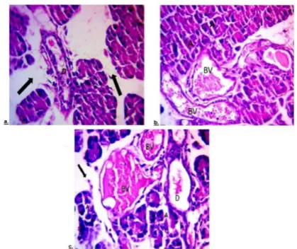
Pancreatic sections of diabetic rats stained by haematoxylin&eosin showed focal affection of the pancreas in the form of loss of architecture of pancreatic acini (Fig. 2c), widening of spaces between acini (Fig 2a&2c), dilated interlobular duct (Figs.2a&2c), and congestion of blood vessels (Figs.2b&2c). Masson’s trichrome stain demonstrated excessive collagen fibers deposition around blood vessels and around interlobular ducts (Figs. 3a&3b).
Group IV (diabetic group treated with
Silymarin)
Pancreatic sections stained by haematoxylin& eosin showed normal architecture of some acini, loss of architecture of other acini, but there are wide spaces between them and islet cells appeared to be normal (Fig.5a). Masson’s trichrome stain showed minimal collagen fibers deposition around acini and around blood vessels (Fig5b).
2-Electron microscopic examination:
Group I and Group II (normal and sham
control groups)
Ultra structural examination of acinar cells showed euchromatic nucleus, many electron dense secretory granules, well developed rough endoplasmic reticulum and mitochondria (Fig.1c).
Figure 2: a. photomicrograph of section of a rat pancreas from group III showing dilated interlobular duct (D) and wide spaces between acini (black arrows). b. photomicrograph of section of a rat pancreas from group III showing congested blood vessels (BV) and loss of architecture of acini (A) c. photomicrograph of section of a rat pancreas from group III showing loss of architecture of pancreatic acini (A), congested blood vessels (BV), dilated duct (D) and wide spaces between acini (black arrow) (Hx.&E.x400).
Group III (diabetic group)
Ultrastructural examination showed euchromatic nucleus, little secretory granules and widely separated RER (Fig.4a), irregular nuclear envelop and clumping of chromatin (Fig.4b, 4c & 4d), destructed mitochondria with loss of cristae (Fig.4c) rarefaction and vacuolation of cytoplasm (Figs. 4b&4d)
Group IV (diabetic group treated with
Silymarin)
Ultrastructural examination of acinar cells showed euchromatic nuclei, many secretory granules. Some of the rough endoplasmic reticulum were widely separated and others were normal (Fig.5c&5d).
3-Biochemical analysis (antioxidant
enzymes assay):
A)
Glutathione peroxidase (GPx) level in
the pancreas
GPx level in the pancreatic tissues of diabetic rats was significantly lower than the other groups. On the other hand, treatments with Silymarin for four weeks lead to an increase in GPx level to normal level in the pancreatic tissues. Statistical comparison of mean GPx level in different groups, there was statistically significant difference between normal control & diabetic groups, sham control & diabetic groups and between diabetic &diabetic treated with Silymarin groups while there were no significant differences between other groups. P value is significant when less than 0 .05 (table 1).
B)
Superoxide dismutase (SOD) level in
the pancreas
SOD level in the pancreatic tissues of diabetic rats was significantly lower than the other groups. On the other hand, treatment with Silymarin for four weeks led to an increase in
SOD level to normal level in the pancreatic tissues. Comparison of mean SOD level in different groups, there was statistically significant difference between normal control & diabetic groups, sham control & diabetic groups and between diabetic &diabetic treated with Silymarin groups while there were no significant differences between other groups (table 1)
4-Image analysis:
Mean of area % of collagen fibers in the
pancreas:
The mean of area % of collagen fibersin pancreatic sections of
diabetic rats was significantly higher than the other groups. On the other hand, treatment with Silymarin for four weeks led to decrease in the mean of area % of collagen fibers in pancreatic tissues to normal level (table 1). Statistical comparison of mean of area % of collagen in different groups, there was significant difference between normal control & diabetic groups, sham control & diabetic groups and between diabetic &diabetic treated with Silymarin groups while there were no significant differences between other groups (Table 1).
Journal of Advanced Pharmacy Education &Research |Jan-Mar 2018 | Vol 8 | Issue 1 79
Figure 4: a. An electron photomicrograph of a rat pancreas from group III showing acinar cell having aneuchromatic nucleus (N), widely separated RER (yellow arrow), little secretory granules (black arrow) and apparently normal mitochondria (M). b. An electron photomicrograph of a rat pancreas from group III showing acinar cell having a nucleus (N) with irregular nuclear envelope and clumping of chromatin (black arrows), vacuolation (V) and rarefaction of cytoplasm (R). c. An electron photomicrograph of a rat pancreas from group III showing acinar cell with indentation of nucleus (white arrow), clumping of chromatin (black arrows) also showing destruction of mitochondria with loss of cristae (M). d. An electron photomicrograph of a rat pancreas from group III showing acinar cell with vacuolation (V), indentation of nucleus (white arrow), clumping of chromatin (black arrow) and rarefaction of cytoplasm (R) (TEM x8000).
Figure 5: a. photomicrograph of a section of a rat pancreas of group IV showing normal architecture of some acini (A) and loss of architecture of other acini (blue arrows) and islets cells (I) appear to be normal (Hx. &E.x400). b. A photomicrograph of section of rat pancreas from group IV showing minimal collagen fibers (black arrow) around acini (A) and ducts (D) (Masson's trichrome X 400). c. An electron photomicrograph of rat pancreas from group IV showing acinar cell having an euchromatic nucleus (N), many secretory granules (black arrows), Some rough endoplasmic reticulum are widely separated (yellow arrows) and others are normal (white arrow) (TEM x 8000). d. An electron photomicrograph of rat pancreas from group IV showing acinar cell having a euchromatic nucleus (N). Manysecretory granules (black arrows), apparently normal mitochondria (M). Some rough endoplasmic reticulum is widely separated (blue arrow) and others are normal (white arrow) (TEM x 8000).
Table 1: showing the mean glutathione (GPx) and (SOD) level measured in umol/g tissue in the pancreas and the mean area % of collagen, in the different studied groups.
P-value Mean collagen %± SD P-value Mean SOD± SD P-value Mean GTH± SD Group 3.29 ±0.93 3.66 ±0.43 59.74 ±12.67 I(normal control) 0.693* <0.0001*** 0.075**** 2.92 ±0.87 0.916* <0.0001*** 0.121**** 3.62 ±0.42 0.756* <0.0001*** 0.101**** 58.07 ±10.25 II (sham control) <0.0001* <0.0001** <0.0001**** 10.24 ±2.56 <0.0001* <0.0001** 0.001**** 1.61 ±0.86 <0.0001* <0.0001** <0.0001**** 22.69 ±6.15 III (diabetic) 0.152* 0.075** <0.0001*** 4.67 ±0.71 0.099* 0.121** 0.001*** 3.01 ±0.72 0.055* 0.101** <0.0001*** 49.00 ±6.46 IV (diabetic treated with Silymarin)
*compared to group 1, ** compared to group 2, ***compared to group 3, ****compared to group 4
. P value ≤ 0 .05 was considered significant.
Discussion
The most common approaches to induce diabetes are established on streptozotocin (STZ) or alloxan (toxic glucose analogs) administration in rodents. They selectively accumulate
in the β-cells and result in a marked hypoinsulinemia and
subsequent hyperglycemia [23].
In the present study, manifestations of the pathological impact of diabetes mellitus on the exocrine part of pancreas were recorded. Pancreatic sections of rats of diabetic rats stained by Hematoxylin and Eosin showed focal affection of the exocrine part in the form of loss of architecture of pancreatic acini, widening of spaces between acini, dilated interlobular duct and cytoplasmic vacuolation. Other sections revealed congested blood vessels.
The present work supported the work of Attia [24] who reported
disturbance in the architecture of pancreatic acini, small vacuoles in the cytoplasm and thickening of the septa between
acini. Similar findings were reported by Bera et al. [18] who
found that diabetes led to degeneration of pancreatic acini.
The present work coincided with the work of Abdul-Hamid and
Moustafa [14] who reported that diabetes led to pathological changes of exocrine part of the pancreas in the form of acinar destruction presented by pyknotic nuclei and cytoplasmic vacuolation.
On the other hand, El-Desouk et al, [25] reported appearance of
leucocytic (mononuclear cells) infiltration between acini which was not present in the current work and that may be due to induction of diabetes by alloxan which caused insulinitis. The present work was in partial agreement with the work done
by Sheweita et al. [17] who reported that diabetes caused
histological changes in in the form of disturbance of the acini pattern structure, pyknotic nuclei of some acinar cells, vacuolated acini, dilatation, thickening, and congestion of the blood vessels, and inflammatory cells infiltrate around the pancreatic duct.
Nurdiana et al. [8] demonstrated that pancreatic sections of
diabetic rats showed swelling of the acinar cells and small vacuoles were observed in almost all acinar cells.
were in agreement with previous work done by El-Desouk et
al. [25] who demonstrated increased collagen fibers deposition
around blood vessels. Similar findings were reported by Abdul
-Hamid and Moustafa [14] who pointed out that diabetes led to formation of condensed collagen fibers around the acini. Hypertrophy and thickening were noticed in the media with swelling in the endothelium of the intima of the congested blood vessels.
The increase in collagen synthesis in diabetes resulted from activation of pancreatic stellate cells (which have fibroblast like action) in response to increased free fatty acids and lipid peroxidation [26]. Abunasef et al. [27] noticed condensed collagen fibers around some pancreatic ducts and blood vessels. In addition, the later observed that some islets which were completely fibrosed after 6 weeks from the onset of diabetes.
Ultra structural examination of pancreatic sections of diabetic
rats revealed acinar cells with rarefied cytoplasm, little
secretory granules, widely separated rough endoplasmic reticulum and irregular nuclear envelope with clumping of chromatin. Other sections showed destruction of mitochondria with loss of cristae and vacuolation of cytoplasm.
The present work was in agreement with work of Abdul-Hamid
and Moustafa [14] who noticed thatultrastructural examination of the diabetic rats revealed marked changes in pancreatic acini represented by little secretory granules, cytoplasmic vacuolation, destructed mitochondria, autophagic vacuoles and irregular shapes of nuclei.
As regarding GPx and SOD level in the pancreatic tissues of
diabetic rats was significantly decreased than other groups. On the other hand, treatment with Silymarin for four weeks leads to an increase in GPx level to normal level in the pancreatic tissues. The present study was in agreement with the work done by Gupta et al., [28]and Varsha et al [7] who revealed that STZ administration led to significant decrease in the level of SOD, GSH and CAT, however treatment with vitamin k1 significantly increase the level of these enzymes to normal level
Nurdiana et al. [8] reported that both GPx and SOD activities
decreased in the pancreas of diabetic rats, suggesting that pancreatic oxidative stress was increased. In conclusion, the present study demonstrated the pathological effects of induced diabetes on the exocrine part of pancreas and that the use of Silymarincould ameliorate these effects. Therefore, it is recommended to use Silymarin to prevent the side effects of diabetes.
References
1. Erejuwa, O. O. (2014): Effect of honey in diabetes
mellitus: matters arising. Journal of Diabetes & Metabolic Disorders, 13(1): 23-26.
2. Baynes, J.W. (1991): Role of oxidative stress in the
development of complications in diabetes, Diabetes, 40: 405–412.
3. Calles-Escandon, J. andCipolla, M. (2001): Diabetes and
endothelial dysfunction: aclinical perspective. Endocr. Rev., 22:36–52.
4. Bajaj, S. and Khan, A. (2012): Antioxidants and diabetes.
Indian journal of endocrinology and metabolism, 16(Suppl 2): S267-S271.
5. Yang, H. and Wright, JR. (2002): Human beta cells are
exceedingly resistant to streptozotocin in vivo. Endocrinology, 143: 2491-2495.
6. Gawlik, K.; Naskalski, J. W.; Fedak, D.;
Pawlica-Gosiewska, D.; Grudzień, U.; Dumnicka, P., and
Solnica, B. (2015): Markers of antioxidant defense in patients with type 2 diabetes. Oxidative medicine and cellular longevity: 1-6.
7. Varsha, M. S.; Thiagarajan, R.; Manikandan, R. and
Dhanasekaran, G. (2015): Vitamin K1 alleviates streptozotocin-induced type 1 diabetes by mitigating free
radical stress, as well as inhibiting NF-κB activation and
iNOS expression in rat pancreas. Nutrition, 31(1): 214-222.
8. Nurdiana, S.; Goh, Y. M.; Ahmad, H.; Dom, S. M.;
Azmi, N. S. A.; Zin, N. S. N. M. and Ebrahimi, M. (2017): Changes in pancreatic histology, insulin secretion and oxidative status in diabetic rats following treatment with Ficusdeltoidea and vitexin. BMC complementary and alternative medicine, 17(1): 290-306.
9. Soto, C., Recoba, R., Barrón, H., Alvarez, C., &Favari,
L. (2003): Silymarin increases antioxidant enzymes in alloxan-induced diabetes in rat pancreas. Comparative Biochemistry and Physiology Part C: Toxicology & Pharmacology, 136(3): 205-212.
10. El-Samaligy, M. S; Afifi, N. N. and Mahmoud, E. A.,
(2006): Evaluation of hybrid liposomes-encapsulated Silymarin regarding physical stability and in vivo performance. Int. J. Pharm., 319: 121–129.
11. Pradeep, K.; Mohan, C. V. R.; Gobianand, K. and
Karthikeyan, S. (2007): Silymarin modulates the oxidant– antioxidant imbalance during diethylnitrosamine induced oxidative stress in rats. European journal of pharmacology, 560(2): 110-116.
12. Karimi, G.; Vahabzadeh, M.; Lari, P.; Rashedinia, M. and
Moshiri, M. (2011): Silymarin”, a promising pharmacological agent for treatment of diseases. Iranian journal of basic medical sciences, 14(4): 308-317.
13. Kumas, M.; Esrefoglu, M. and Ozer, O. F. (2016):
Protective Effects of Silymarin against Cardiac Tissue Injury Caused by a High-dose Administration of Isotretinoin in Mice. Bezmialem Science, 4(2): 43-50.
14. Abdul-Hamid, M. andMoustafa, N. (2013): Protective
effect of curcumin on histopathology and ultrastructure of pancreas in the alloxan treated rats for induction of diabetes. The Journal of Basic & Applied Zoology, 66(4): 169-179.
15. Roy, V. K.; Chenkual, L. and Gurusubramanian, G.
Journal of Advanced Pharmacy Education &Research |Jan-Mar 2018 | Vol 8 | Issue 1 81 responses in pancreas of diabetic rats. Actahistochemica,
118(2): 152-163.
16. Campbell-Thompson, M.; Rodriguez-Calvo, T.
andBattaglia, M. (2015). Abnormalities of the exocrine pancreas in type 1 diabetes. Current diabetes reports, 15(10), 79.
17. Sheweita, S. A.; Mashaly, S.; Newairy, A. A.; Abdou, H.
M. and Eweda, S. M. (2016): Changes in oxidative stress and antioxidant enzyme activities in streptozotocin-induced Diabetes mellitus in rats: Role of AlhagiMaurorum extracts. Oxidative medicine and cellular longevity, 2016.
18. Bera, T. K.; Chatterjee, K.; Jana, K.; Ali, K. M.;
Debasis, D. A.; Maiti, S. and Ghosh, D. (2012): Antihyperglycemic and antioxidative effect of hydro - methanolic (2:3) extract of the seed of Swieteniamahagoni (L.) Jacq. In streptozotocin-induced diabetic male albino rat: An approach through pancreas. Genomic Medicine, Biomarkers, and Health Sciences, 4: 107-117.
19. Drury, R.A. and Wallington, E.B. (1980): Carltons
Histological techniques. Oxford University Press; 5:139-239.
20. Bancroft, J. and Gamble, M. (2008): Theory and Practice
of Histological Techniques, 6th edition, Churchill Livingstone, London, pp: 85-107.
21. Hayat, M. A. (2000): Principles and techniques of
electron microscopy: Biological applications. Cambridge Univ. Press: Cambridge, 4: 546-8.
22. Prasad K, Lee P, Mantha SV, Kalra J, Prasad M, Gupta JB:
Detection of ischemia-reperfusion cardiac injury by cardiac muscle chemi- luminescence. Mol Cell Biochem 115: 49-58, 1992
23. Lenzen, S. (2008): Oxidative stress: The vulnerable β
-cell. BiochemSoc Trans 36:343–347.
24. 3. Attia, A. A. (2009): Histological and electron
microscopic studies of the effect of ß-Carotene on the pancreas of streptozotocin (STZ)-Induced diabetic rats. Pakistan Journal of Biological Sciences, 12(4), 301-314.
25. El-Desouk, N.; Basyony, M.; El-Nenaey, M. and
Abdel-Magied, R. (2007): Histological and cytological studies on the effect of melatonin on experimentally induced pancreatic diabetes in rats. Egypt. J. Exp. Biol. (Zool.), 3: 69-82.
26. Zhang, X.; Cui, Y.; Fang, L. and Li, F. (2008): Chronic
high-fat diets induce oxide injuries and fibrogenesis of pancreatic cells in rats. Pancreas, 37(3): 31-38.
27. Abunasef, S.K.; Amin, H.A. and Abdel-Hamid, G.A.
(2014): A histological and immunohistochemical study of beta cells in streptozotocin diabetic rats treated with caffeine. Folia HistochemicaetBiologica, 52(1): 42–50.
28. Gupta, R.; Sharma, A. K.; Sharma, M. C.and Gupta, R.
S. (2012): Antioxidant activity and protection of
pancreatic β cells by embelin in streptozotocin induced