Ersel Dag
1, A–D,
Zeynep O. Dag
2, A–D, Giyasettin Baydas
3, A, C, Mehmet Tuzcu
4, A, C,
Tahir K. Yoldas
5, A, C, Bulent Mungen
6, A, C, Ramazan Bal
7, A, C, DEffects of Lamotrigine and Topiramate
on Brain Maturation and Cognitive Functions
in Offspring of Pregnant Rats – Preliminary Study
1 Department of Neurology, Faculty of Medicine, Kirikkale University, Kirikkale, Turkey
2 Department of Obstetrics and Gynecology, Faculty of Medicine, Kirikkale University, Kirikkale, Turkey 3 Bingol University Rectorate, Bingol University, Bingol, Turkey
4 Department of Biology, Faculty of Science, Firat University, Elazig, Turkey 5 Department of Neurology, Faculty of Medicine, Harran University, S.Urfa, Turkey 6 Department of Neurology, Faculty of Medicine, Firat University, Elazig, Turkey 7 Department of Physiology, Faculty of Medicine, Firat University, Elazig, Turkey
A – research concept and design; B – collection and/or assembly of data; C – data analysis and interpretation;
D – writing the article; E – critical revision of the article; F – final approval of article; G – other
Abstract
Background. Antiepileptic drugs (AED) which are used to treat seizures in pregnant women, infants, and young children may cause cognitive impairment or other uncertain injury. However, the precise mechanisms responsible for the negative effects of new AEDs like lamotrigine (LTG) and topiramate (TPM) in the developing brain are still unclear.
Objectives. To investigate the GFAP, NCAM and S100B levels in the whole brain of newborn rats on postnatal 1 day and in the hippocampus of adult rats to find out the effect of TPM and LTG on cognitive impairment and brain maturation.
Material and Methods. Twenty eight pregnant rats were randomly divided into 7 groups with 4 animals in each group. The first group, receiving no drugs, was assigned as the control group. The study groups received intra-peritoneal TPM or LTG injections in each trimester. Western blot analysis of the GFAP, NCAM and S100B was performed in the offspring. Behavioral tests were performed at postnatal day 75.
Results. The rats in the TPM-I and TPM-III groups had a significant impairment in escape latency on the 5th day
as compared to the control rats in a Morris water maze test. In addition, in the expression of astrocyte derived markers, GFAP was upregulated, whereas S100β and NCAM were downregulated in the whole brain on postnatal day 1, in offspring exposed to LTG and TPM in utero.
Conclusions. The detrimental effects of TPM and LTG appear to be confined particularly to the early stages of brain development. And TPM seems to have a partial role in the cognitive impairment (Adv Clin Exp Med 2014, 23, 5, 691–698).
Key words: topiramate, lamotrigine, brain development, cognitive functions.
Adv Clin Exp Med 2014, 23, 5, 691–698 ISSN 1899–5276
ORIGINAL PAPERS
© Copyright by Wroclaw Medical University
The new generation AEDs such as lamotrig-ine (LTG) and topiramate (TPM) represent a po-tential improvement for patients whose seizures are incompletely controlled or who experience signifi-cant adverse effects with older anticonvulsants [1]. These two drugs are teratogenic and prenatal ex-posure has been associated with an increased risk
of congenital abnormalities affecting a variety of organs, including the central nervous system [2]. High doses of TPM have been reported to be asso-ciated with cognitive impairments [3]. Impairments in cognitive functions such as learning and memory have usually been associated with congenitally oc-curring structural and molecular alterations in the
limbic system, particularly the hippocampus of off-spring [4]. These alterations are not clearly seenat birth but form the basis for various functional de-fects noticible gradually during further maturation.
Although a limited number of animal model studies show less teratogenic effects with the new AEDs compared to the known effects in animal models exposed to the older agents, it is still not known whether these new antiepileptic drugs have deleterious effects on brain maturation and cog-nitive functions of the fetusus of pregnant wom-an. A recent study has shown that prenatal expo-sure to lamotrigine during the neuronal migration stage causes neocortical and hippocampal altera-tions [5]. It is likely that these antiepileptic drugs might affect fetal brain maturation and cognitive functions.
There are a number of markers indicating brain injury affecting cognitive functions, such as neural cell adhesion molecules (NCAM), glial fi-brillary acidic protein (GFAP) and S100β. NCAM is known to participate in synaptogenesis in neu-ronal plasticity and particularly involved in syn-aptic changes underlying memory formation in adult individuals [6]. Glial fibrillary acidic pro-tein (GFAP) is one of the intermediate filament proteins of glial cells and is involved in neuronal differentiation and maturation, which is recog-nized as an astrocyte maturation marker and re-active astrocytes [7]. In response to neuronal dam-age such as brain injury [8], stroke [9], or induced seizures [10], reactive hypertrophy and prolifera-tion of astroglial cells can be promoted, a process known as astrogliosis. S100β is a Ca2+ binding
pro-tein expressed in Schwann cells and astroglia in the central nervous system, which is overexpressed in response to neuronal damage [11].
Therefore, we aimed to study the expression levels of NCAM, GFAP and S100β protein and performed Morris water maze tasks to search for possible mechanisms of brain injury and cognitive impairment in the offspring of pregnant rats ex-posed to intraperitoneal LTG or TPM injections.
Material and Methods
Animals and Experimental
Design
The experimental protocols were approved by the local Animal Use Committees. Twenty eight healthy adult female Wistar albino rats, aged 4–5 month and weighing in the range of 210–250 g, were used, and were housed in polycarbonate cag-es in a room with a 12 h day–night cycle, tempera-ture of 24 ± 3°C and humidity of 45 to 65%.
Pro-estrous animals were kept overnight with male rats and the next day was taken as gestation-al day 0 when spermatozoa were seen in a vagi-nal smear. The pregnant rats were randomly di-vided into 7 groups with 4 animals in each group in the following way: the first group of rats, re-ceiving no drugs, was assigned as the control group (n = 4). The animals receiving intraperito-neal topiramate injection at 25 mg/kg in the first trimester were assigned as (TPM I), the second trimester as (TPM II) and the third trimester as (TPM III), respectively. Similarly, the animals re-ceiving intraperitoneal lamotrigine injection at 25 mg/kg in the first trimester as (LTG I), the sec-ond trimester as (LTG II) and the third trimes-ter as (LTG III). The pregnant rats in group LTG I had 34 offspring, LTG II had 32, LTG III had 36, TPM I had 28, TPM II had 32, TPM III had 36 and the control animals had 42. After delivery on post-natal day (PND) 1, the litter size, body weight and gender of the offspring of each litter were record-ed. Half of the offspring per group were decapi-tated (total n = 120) and their brains were stored at –70 oC for the Western blot analysis of NCAM,
GFAP and S100β on PND1. The remaining off-spring from each experimental litter were weaned from their dams when they were 21 days old and they were separated into male and female cages. The Morris water maze test [12] was employed to study the cognitive status of the animals, regard-ing spatial learnregard-ing and memory, at postnatal day 75. After 24 h, young-adult offspring (n = 120) from all groups were decapitated. Brain tissues were removed and the hippocampi were dissected and stored at –70oC.
The detailed descriptions of the methods on imminoblotting for the measurements of NCAM, GFAP and S100β, and the Morris water maze test for studying the cognitive status of the animals re-garding spatial learning have been described in our earlier publication [13].
Statistical Analysis
The results were expressed as mean ± SD one- -way analysis of variance (ANOVA) and post hoc Tukey-HSD tests were used to determine differ-ences in all parameters between groups. Differdiffer-ences were considered statistically significant if the p-value was < 0.05.
Results
There were no still births in any of the groups except group topiramate I, in which there were 27% still birth. The duration of pregnancy and lit-ter size were not statistically different between the groups. After birth, all the living animals survived the procedure, with no evidence of neurological deficits.
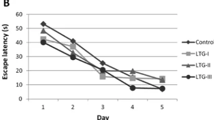
The effects of prenatal TPM and LTG exposure on learning and memory performance were exam-ined using the Morris water maze test of a hidden- -platform, a hippocampus-dependent task, which requires an animal to learn and remember the rela-tionships between the platform location and mul-tiple distal cues to escape the water [14], the mean escape latencies, which is the time for rats to reach the platform, in control, LTG-I, LTG-II, LTG-III, TPM-I, TPM-II and TPM-III groups were com-parable in the first trial, suggesting that their mo-tor performances (ability to swim) were unaffected by prenatal LTG and TPM exposures. All animals were able to swim in a normal way during all trials and control rats and rats whose mothers were ex-posed to LTG and TPM during pregnancy learned the task, as evidenced by a decrease in the es-cape latency to find the platform from the first to the last day of training (day 5) (p< 0.001). How-ever, the rats in the TPM-I and TPM-III groups had a significant impairment in escape latency on the 5th day as compared to the control rats, since
TPM-I and TPM-III groups tended to take longer to find the platform than the controls in the last trials (p < 0.05 vs p < 0.001) (Fig. 1 A). Whereas, there were no significant differences between the performances of LTG-I, LTG-II, LTG-III and con-trol groups (Fig. 1B).
Levels of GFAP, S100β and
NCAM in the Offspring
of Pregnant Rats Exposed
to LTG and TPM
In order to demonstrate if LTG and TPM ex-posure during the first, second or third trimester causes reactive gliosis in the offspring, expression levels of GFAP, NCAM and S100β were studied using semiquantitative Western blotting.
GFAP protein had main band of relative mo-lecular weights of 49 kDa and several bands for degradation products of smaller molecular weight at roughly 42–47 kDa (Fig. 2, shown by arrows). The main bands and degradation products of GFAP protein in the whole offspring brain was sig-nificantly elevated in all groups except the TPM-I group, compared to the control, indicating an in-duced glial hyperactivity (Fig. 2A2). There were also significant increases in GFAP content in the hippocampal tissues from adult rats in only LTG-I and TPM-III groups (Fig. 2B2).
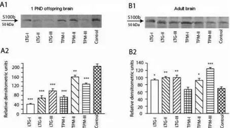
The major band representing the main S100β polypeptide migrates at a relative mobili-ty of about 50 kDa (Fig. 3, shown by arrows). The amount of S100β protein in the whole offspring brain of rats from all TPM and LTG groups were significantly lower than in the control group, sug-gesting a delayed maturation of astrocytes, which is linked to brain maturation (Fig. 3A2). Where-as, the amounts of S100β protein was higher in the hippocampus of adult rats from TPM and LTG groups except TPM-I compared to control (Fig. 3B2).
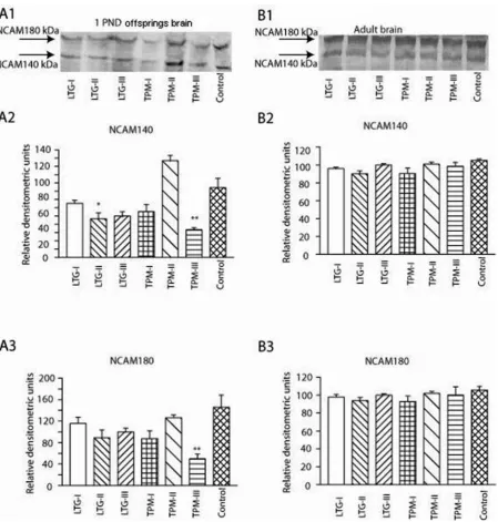
As shown in the figure (indicated by an ar-row), two bands in each line could be detected. The 140-kDa isoform of NCAM in LTG-II and TPM-III groups were found to be significantly lower in comparison to the control group in the whole offspring brain (Fig. 4A2). Similarly, the 180-kDa isoforms of NCAM were found to be sig-nificantly lower in the whole offspring brain of the TPM-III group in comparison to the control group (Fig. 4A3). No significant difference was discerned in the 140- and 180-kDa isoforms of NCAM in the hippocampus of adult LTG&TPM rats compared to control group (Fig. 4B2 and B3).
Fig. 1B. Morris water maze test results of rats in the LTG group and controls
Discussion
Recent studies have reported that new genera-tion AEDs are less teratogenic and have less cog-nitive impairment than traditional AEDs [15–18].
However, there are few studies about the long-term effects of AEDs such as cognitive impair-ment [19, 20]. The exact mechanisms responsible for the adverse effects of AEDs on both brain mat-uration and long term cognitive impairment are Fig. 2. Western blot analysis of GFAP. Representative bands in whole offspring brain (A1) and in the hippocampal tissues from adult rats (B1) and their respective densitometric analysis in whole offspring brain (A2) and in the hippo-campal homogenates from adult rats (B2). *P, 0:05, **P, 0:01 and ***P, 0:001 vs. control values
still not clear. Therefore, in the current study, we investigated both the brain maturation of the off-spring and the cognitive status of the animals re-garding spatial learning and memory.
In previous studies, high doses of TPM and LTG have been reported to be associated with brain maturation defects including cognitive impair-ments in some offspring of TPM- or LTG-treat-ed mothers [3]. Impairments in cognitive func-tions such as learning and memory have usually been associated with congenitally occurring struc-tural and molecular alterations in the limbic sys-tem, particularly the hippocampus of offspring [4].
However, to the best of our knowledge, there is on-ly one study evaluating the effects of maternal ex-posure to TPM and LTG on cognitive behavior in offspring [19].
took significantly longer to find the hidden plat-form than the controls in the last trials of the Mor-ris water maze test. Various mechanisms have been proposed for the teratogenicity of AEDs, in-cluding folate-related action, ischemia, neuronal suppression, reactive intermediates (free radicals) and AED-induced neuronal apoptosis. Anatomi-cal and behavioral or cognitive teratogenesis differ in mechanisms. Because of the fetal organ devel-opment in the first trimester, AED exposure may pose the highest risk for anatomical malforma-tions, and the above causes may have caused a dif-ference in still births or cognitive performance in group TPM I. In the third trimester of pregnan-cy, the human brain develops fast structural and functional changes. During this developing pro-cess, neuronal and glial cells differentiate, mature and migrate until the term infant brain architec-ture is fully developed [22]. With these structural changes functional changes are occurring also. For this reason, there is a difference between groups TPM I and TPM III in cognitive performance. This data suggests that rats exposed to TPM in utero had a long-lasting deficit in spatial learning and memory. The learning deficit in the rats exposed to TPM appears to result from impairments in spa-tial learning, not from swimming ability or visual acuity because there was no significant difference between the groups in latency to reach the plat-form in the first trial [23].
Impairment in spatial learning in the rats ex-posed to AEDs in utero might be owing to develop-mental delays in brain maturation and neurogen-esis during gestation, since memory impairments have been related to a decline in synaptic plastic-ity [4]. Therefore, the levels of glial intermediate filaments and NCAM expression were studied in the whole brain from PND1 offspring and in the hippocampus of 81 day-old young rats exposed to LTG and TPM in utero.
Expression levels of GFAP is commonly used as a marker for changes in astroglial cells during brain development and injury [24]. Injury of the brain resulting from trauma, disease, genetic dis-orders, or chemical insult causes astrocytes to be-come reactive, a condition characterized by an increase in GFAP [25]. In the current study, expo-sure to LTG and TPM in utero caused significant increases in GFAP in the whole brain from PND1 offspring as measured by Western blotting, indi-cating reactive gliosis. Although increased GFAP is commonly associated with astroglial activation, increased GFAP alone does not allow us to draw conclusions about the type of astroglial response, be it beneficial or deleterious [26].
S100β protein, a cytosolic constituent of the astrocytes, is a calcium binding protein
predominantly expressed and secreted by astro-cytes in the central nervous system [27]. As shown in Fig. 3A1 and A2, the amount of S100β protein in the whole offspring brain of rats from all TPM and LTG groups were significantly downregulat-ed compardownregulat-ed to the control. This suggest a delaydownregulat-ed maturation of astrocytes [13]. However, expression of brain tissue S100β has been upregulated in neu-rodegenerative disorders, such as Alzheimer’s dis-ease [28]. The incrdis-eased level of S100β in the hip-pocampus of 81 day-old young rats exposed to LTG and TPM in utero (Fig. 3B1 and B2) appearantly results from upregulation of S100β expression in astrocytes [28], which is likely to be due to neuro-degeneration induced by TPM and LTG in utero. It has been suggested that increments of S100β may improve neurogenesis, particularly in the hippo-campus [29] and also stimulate glial proliferation and neuronal survival and protect neurons against glutamate excitotoxicity [30]. NCAM is capable of incorporating long chains of α 2,8 polysialic ac-id (PSA) [31]. The polysialated NCAM seems to be involved in several developmental events such as neuronal migration [32], synaptogenesis [31], axonal outgrowth and fasciculation [33]. The ex-pression of PSA-NCAM is developmentally regu-lated [34]; PSA-NCAM is dramatically downregu-lated in the brain except in areas associated with neuroplastic events such as in the olfactory bulb and hippocampus [35]. The findings in our study that animals exposed to TPM in utero had im-paired spatial learning is meaningful, since NCAM expression in the whole brain of PND1 offspring exposed to TPM in the, particularly, third trimes-ter in utero was downregulated.
In conclusion, the lower levels GFAP, S100β, and NCAM, in particular GFAP and NCAM, in the hippocampus of young rats exposed to TPM and LTG suggest that the detrimental effects of TPM and LTG appeared to be confined particu-larly to the early stages of brain maturation. Addi-tionally, the early effects of TPM exposure in utero resulted in impairments of spatial learning. These results indicate that TPM, but not LTG, seem to have a partial role on the cognitive impairment.
Study Limitations
References
[1] Pippenger CE: Pharmacology of Neural Tube Defects. Epilepsia 2003, 44, 24–32.
[2] Ornoy A: The impact of intrauterine exposure versus postnatal environment in neurodevelopment toxicity: long term neurobehavioral studies in children at risk for developmental disorders. Toxicol Lett 2003, 140, 171–181.
[3] Park SP, Kwon SH: Cognitive effects of antiepileptic drugs. J Clin Neurol 2008, 4, 99–106.
[4] Spear LP: Age at the time of testing reconsidered in neurobehavioral teratological research. In: Neurobehavioral Teratology. Ed.: Yanai J. Elsevier, New York 1984, 315–328.
[5] Manent JB, Jorquera I, Franco V, Ben-Ari Y, Perucca E, Represa A: Antiepileptic drugs and brain maturation: fetal exposure to lamotrigine generates cortical malformations in rats. Epilepsy Res 2008, 78, 131–139.
[6] Tzeng SF, H Cheng, YS Lee, JP Wu, BJ Hoffer, JS Kuo: Expression of neural cell adhesion molecule in spinal cords following a complete transaction. Life Sci 2001, 68, 1005–1012.
[7] Cerutti SM, Chadi G: S100 immunoreactivity is increased in reactive astrocytes of the visual pathways following a mechanical lesion of the rat occipital cortex. Cell Biol Int 2000, 24, 35–49.
[8] Hausmann R, Rieß R, Fieguth A, Betz P: Immunohistochemical investigations on the course of astroglial GFAP expression following human brain injury. Int J Legal Med 2000, 113, 70–75.
[9] Herrmann M, Vos P, Wunderlich MT, Brujin CHMM, Lamers KJB: Release of glial tissue-specific proteins after acute stroke – A comparative analysis of serum concentrations of protein s-100 B and glial fibrillary acidic protein. Stroke 2000, 31, 2670–2677.
[10] Stringer GL: Repeated seizures increase GFAP and vimentin in the hippocampus. Brain Res 1996, 717, 147–153.
[11] Schäfer BW, CW Heizmann: The S100 family of EF-hand calcium-binding proteins: functions and pathology. Trends Biochem Sci 1996, 21, 134–140.
[12] Morris RG, Garrud P, Rawlins JN, O’Keefe J: Place navigation impaired in rats with hippocampal lesions. Nature 1982, 297, 681–683.
[13] Baydas G, Koz ST, Tuzcu M, Nedzvetsky VS: Melatonin prevents gestational hyperhomocysteinemia-associated alterations in neurobehavioral developments in rats. J Pineal Res 2008, 44, 181–188.
[14] Schenk F, Morris RGM: Dissociation between components of spatial memory in rats after recovery from the effects of retrohippocampal lesion. Exp Brain Res 1985, 58, 11–28.
[15] Vajda FJ, Graham J, Roten A, Lander CM, O’Brien TJ, Eadie M: Teratogenicity of the newer antiepileptic drugs – the Australian experience. J Clin Neurosci 2012, 19, 57–59.
[16] Holmes LB, Hernandez-Diaz S: Newer anticonvulsants: lamotrigine, topiramate and gabapentin. Birth Defects Res A Clin Mol Teratol 2012, 94, 599–606.
[17] Hernández-Díaz S, Smith CR, Shen A, Mittendorf R, Hauser WA, Yerby M, Holmes LB: North American AED Pregnancy Registry; North American AED Pregnancy Registry: Comparative safety of antiepileptic drugs during pregnancy. Neurology 2012, 22, 1692–1699.
[18] Ditte MN,Anders H: Newer-Generation Antiepileptic Drugs and the Risk of Major Birth Defects. JAMA2011, 305, 1996–2002.
[19] Shi XY, Wang JW, Cui H, Li BM, Lei GF, Sun RP: Effects of antiepileptic drugs on mRNA levels of BDNF and NT-3 and cell neogenesis in the developing rat brain. Brain Dev 2010, 32, 229–235.
[20] Chen J, Quan QY, Yang F, Wang Y, Wang JC, Zhao G, Jiang W: Effects of lamotrigine and topiramate on hip-pocampal neurogenesis in experimental temporal-lobe epilepsy. Brain Res 2010, 1313, 270–282.
[21] Hajikhani R, Ahmadi A, Naderi N, Yaghoobi K, Shirazizand Z, Rezaee NM, Niknafs BN: Effect of phencycli-dine derivatives on anxiety-like behavior using an elevated-plus maze test in mice. Adv Clin Exp Med 2012, 21, 3, 307–312.
[22] Volpe JJ: Neuronal proliferation migration organization and myelination. In: Neurology of the newborn. WB Saunders, Philadelphia 2008, 5th ed., 82–90.
[23] Schuck PF, Ferreira Gda C, Viegas CM, Tonin AM, Busanello EN, Pettenuzzo LF, Netto CA, Wajner M:
Chronic early postnatal administration of ethylmalonic acid to rats causes behavioral deficit. Behav Brain Res 2009, 11, 364–370.
[24] Eng LF, Ghirnikar RS, Lee YL: Glial fibrillary acidic protein: GFAP-thirty-one years (1969–2000). Neurochem Res 2000, 25, 1439–1451.
[25] O’Callaghan JP, Sriram K: Glial fibrillary acidic protein and related glial proteins as biomarkers of neurotoxicity. Expert Opin Drug Saf 2005, 4, 433–442.
[26] Liberto CM, Albrecht PJ, Herx LM, Yong VW, Levison SW: Pro-regenerative properties of cytokine-activated astrocytes. J Neurochem 2004, 89, 1092–1100.
[27] Marenholz I, Heizmann CW, Fritz G: S100 proteins in mouse and man: from evolution to function and pathology (including an update of the nomenclature). Biochem Biophys Res Commun 2004, 322, 1111–1122.
[28] Rothermundt M, Peters M, Prehn JH, Arolt V: S100B in brain damage and neurodegeneration. Microsc Res Tech 2003, 60, 614–632.
[29] Kleindienst A, Ross Bullock M: A critical analysis of the role of the neurotrophic protein S100B in acute brain injury. J Neurotrauma 2006, 23, 1185–1200.
[30] Van Eldik LJ, Wainwright MS: The Janus face of glial-derived S100B: beneficial and detrimental functions in the brain. Restorative Neurol Neurosci 2003, 21, 97–108.
[32] Ono K, Tomasiewicz H, Magnuson T, Rutishauser U: N-CAMmutation inhibits tangential neuronal migration and is phenocopied by enzymatic removal of polysialic acid. Neuron 1994, 13, 595–609.
[33] Zhang H, Miller RH, Rutishauser U: Polysialic acid is required for optimal growth of axons on a neuronal sub-strate. J Neurosci 1992, 12, 3107–3114.
[34] Edelman GM: Modulation of cell adhesion during induction, histogenesis, and perinatal development of the ner-vous system. Annu Rev Neurosci 1984, 7, 339–377.
[35] Chuong CM, Edelman GM: Alterations in neural cell adhesion molecules during development of different regions of the nervous system. J Neurosci 1984, 4, 2354–2368.
Address for correspondence:
Ersel Dag
Department of Neurology Faculty of Medicine Kirikkale University Kirikkale
Turkey
Tel: +90 318 225 24 85 22 81 E-mail: [email protected] Conflict of interest: None declared Received: 14.01.2013