Address for correspondence
Nuray Bayar Muluk
E-mail: [email protected]
Funding sources
None declared
Conflict of interest
None declared
Acknowledgements
With the exception of data collection, the preparation of this paper, including design and planning, was supported by the Continuous Education and Scientific Research Association. Only scientific support was provided; no grant or funding was received.
Received on April 3, 2015 Revised on November 10, 2015 Accepted on July 21, 2016
Abstract
Background. Oxidative stress has been associated with pathological processes involved in acoustic trauma. Objectives. In this prospective experimental study, we investigated the potential preventive effect of N-acetyl cysteine (NAC) in rats exposed to acoustic trauma (AT). Light microscopic and scanning electron microscopic (SEM) evaluations were performed.
Material and methods. Healthy Wistar albino rats (n = 18) were divided into 3 groups: group 1 (control group, n = 6), group 2 (acoustic trauma group, n = 6), and group 3 (AT+NAC group, n = 6). The rats in group 2 were exposed to AT. The rats in group 3 received NAC at a dose of 100 mg/kg/day by gavage for 7 days, and then 10 min after the 7th-day dose, they were exposed to AT.
Results. From light and scanning electron microscopy evaluations in the control group, the cochlear struc-ture and epithelium were normal. In group 2 (AT group), extensive hair cell loss was observed in the cochlea by light microscopy evaluation. In the SEM evaluation, various epithelial damage and loss of stereocilia were also observed. In group 3 (AT+NAC group), decreased damage with preserved cochlear structures was seen by light microscopy. In the SEM evaluation, although stereocilia loss was also seen, nearly normal cell structures and vertical and symmetrical alignment of stereocilia structures were observed compared to the AT group.
Conclusions. NAC reduced cochlear damage due to acoustic trauma. Because NAC has antioxidant capac-ity, AT mat have caused an increase in free radicals and death of outer hair cells. NAC is an antioxidant agent and it prevented cochlear damage due to AT in rats.
Key words: N-acetyl cysteine (NAC), acoustic trauma (AT), light microscopy, scanning electron microscopy (SEM) DOI
10.17219/acem/64332
Copyright
© 2017 by Wroclaw Medical University This is an article distributed under the terms of the Creative Commons Attribution Non-Commercial License (http://creativecommons.org/licenses/by-nc-nd/4.0/)
Potential protective effect of N-acetyl cysteine in acoustic trauma:
An experimental study using scanning electron microscopy
Servet Ada
1, A–C, F, Deniz Hanci
2, A, C, F, Seçkin Ulusoy
3, A, C, F, Djanan Vejselova
4, A–C, F,
Dilek Burukoglu
5, A–C, F, Nuray Bayar Muluk
6, A, C, D, F, Cemal Cingi
7, A, C, D, F1 Luleburgaz State Hospital, ENT Department, Kırklareli, Turkey 2 Okmeydanı Training and Research Hospital, ENT Clinics, Istanbul,Turkey 3 Şişli Kolan International Hospital, ENT Clinics, Istanbul, Turkey
4 Department of Biology, Faculty of Science, Anadolu University, Eskişehir, Turkey 5 Department of Histology, Medical Faculty, Eskişehir Osmangazi University, Turkey 6 Department of Otorhinolaryngology, Faculty of Medicine, Kirikkale University, Turkey 7 Department of Otorhinolaryngology, Medical Faculty, Eskişehir Osmangazi University, Turkey
A – research concept and design; B – collection and/or assembly of data; C – data analysis and interpretation; D – writing the article; E – critical revision of the article; F – final approval of article
Oxidative stress has been associated with pathologi-cal processes involved in auditory trauma including mitochondrial injury, activation of cell death pathways, activation of mediators of inflammation, glutamate
ex-citotoxicity, and increased levels of lipid peroxidase.1–3
These findings suggest that antioxidants have the po-tential to block molecular cascades that are triggered by auditory trauma, which induces oxidative stress and results in permanent threshold shifts (PTS) and hearing loss.4
In blast-exposed ears, cochlear pathology includes scar formation replacing dead hair cells, fused and damaged stereocilia, and in some extreme cases, separation of the organ of Corti from the basilar membrane. Most of the hair cell loss was in the outer hair cell (OHC) region and generally in the middle or basal turn. Although hair cell loss was found in the apical turn, the damage was less pronounced. N-acetyl cysteine (NAC), which was used after blast exposure, significantly reduced the permanent structural and functional damage resulting from expo-sure to blast overpresexpo-sure. Drug treatment suppressed
both functional and physical damage to the OHCs.4
In the present study, we investigated the potential pre-ventative effect of NAC in rats exposed to acoustic trau-ma (AT). In this experimental study, light microscopy and scanning electron microscopy (SEM) evaluations were performed.
Material and methods
The study occurred at Eskişehir Osmangazi University, Faculty of Medicine. The animals were maintained at the Experimental Animal Breeding and Experimental Stud-ies Centre of Eskişehir Osmangazi University. Adaptation
and care of the animals and experimental studies were performed at the same center in compliance with the
principles of the Declaration of Helsinki.5 Ethics
com-mittee approval was obtained from Eskişehir Osmangazi University.
Animal subjects
The study was performed in 18 healthy Wistar albino rats, weighing 190–220 g. Each group of rats was housed separately in a climate-controlled room at 20°C. The ani-mals were randomly divided into 3 groups: group 1 (con-trol; n = 6): rats in this group received no drug and were not subjected to AT. Group 2 (n = 6): rats in this group re-ceived no drugs and were exposed to AT. Group 3 (n = 6): rats in this group received NAC dissolved in distilled wa-ter at a dose of 100 mg/kg/day via gavage for 7 days. Then,
10 min after the 7th-day dose, they were exposed to AT
(NAC+AT).
Anesthesia procedure
The rats were anesthetized during experiments via in-tramuscular injection of 40 mg/kg ketamine hydrochlo-ride (Ketalar, Parke-Davis, USA), and 5 mg/kg xylazine hydrochloride (Rompun, Bayer, Germany). Eye-blink reflexes and respiratory rhythms were followed during the experiments, and deep anesthesia was achieved by repeated doses.
Acoustic trauma (AT)
To create AT in rats with 1–12 kHz band white noise, a MATLAB program that produced a single unit of vari-ance sound was used. Then, the sound was filtered with
a 1–12 kHz FIR type digital filter, and an additional filter at 200 Hz. Then, the filtered noise was recorded in a com-puter-based Wav file. Set with a decibel meter, the AT was 110 dB of noise for 6 h (continuous noise).
Electron microscopy evaluation
After the aforementioned procedures were performed, the rats were sacrificed using 80 mg/kg pentothal. The time of sacrifice was 30 min after AT. Immediately after death, the temporal bones were removed, and the
otic bullas were excised.6 Under a dissecting microscope,
the bony capsule of the cochlea was carefully removed, and then the lateral wall was cut away to reveal the organ of Corti. We analyzed hair cell damage in 2 different ar-eas of the middle and basal turns.
Scanning electron microscopy analysis
The cochlea and organ of Corti were dissected out and fixed for SEM preparation; all samples were fixed and post-fixed in the same way. Then, the cochlea was dehy-drated through a graded ethanol series, prior to desicca-tion, using a critical-point drying method described by
Lovell et al.7 Fully desiccated samples were mounted on
a specimen stub using a carbon tab. After critical-point
drying using CO2 and sputter-coating with gold
accord-ing to standard procedures, specimens were investigated with a Zeiss Ultra 50 SEM operating at 5 kV accelerating
voltage.7
Results
Light microscopy and SEM evaluations showed that there was no cochlear damage in group 1 (control group). In group 2 (AT-exposed group), cochlear dam-age was evident. Inner hair cell stereocilia were mis- sing in some regions, and OHC stereocilia were severe-ly damaged by the noise trauma, with a large number prostrate or missing. The damage was more evident in the OHCs in the basal turns of the cochlea. In group 3 (NAC+AT group), although there was cochlear damage, it was less than in group 2 (AT-exposed group without NAC). In group 3, there were fewer missing cells in the inner and OHC group. There was slight damage to the OHC stereocilia.
Light microscopic evaluation results
of groups 1–3 (Fig. 1)
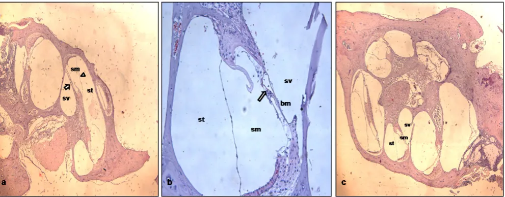
Control group: Normal-appearing cochlear structures were observed (Fig. 1a). Acoustic trauma group: Exten-sive hair cell loss was detected in the cochlea (Fig. 1b). Acoustic trauma+NAC group: Decreased damage with preserved cochlear structures was seen (Fig. 1c).
Scanning electron microscopy evaluation
results of Groups 1–3 (Fig. 2)
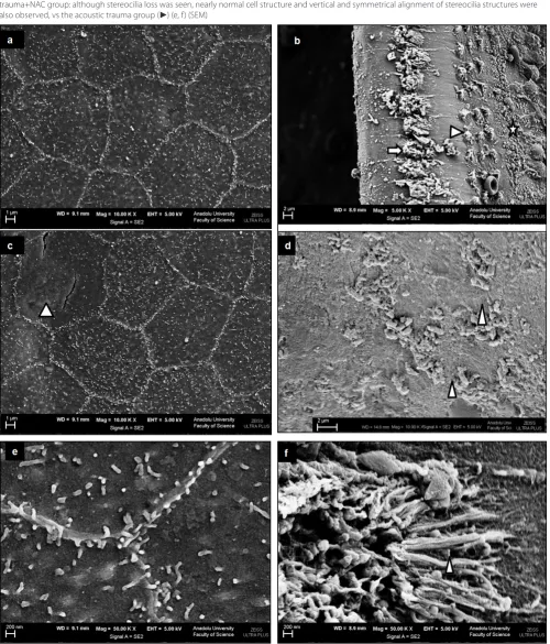
Control group: In the control group, SEM views of epithelium showed normal structures (Fig. 2a), outer hair cells, inner hair cells, and Hensen’s cells (Fig. 2b). Acoustic trauma group: In cross-sectional images of this group, various epithelial damage and loss of stereocilia were observed (Fig. 2c, d). Acoustic trauma+NAC group: Although stereocilia loss was also present in this group, nearly normal cell structures and vertical and symmetri-cal alignment of stereocilia structures were also observed versus the acoustic trauma group (Fig. 2e, f).
Discussion
Acoustic overstimulation of the cochlea generates free radicals and reactive oxygen species (ROS), which may
induce OHC death.8–10 Elevated levels of ROS activate the
upregulation of cochlear antioxidant enzyme activity and
modulate the key antioxidant compound, GSH.11–13
Vari-ous agents with antioxidant properties have been shown to attenuate threshold shifts and/or hair cell loss after the
exposure to damaging noise.14
Oxidative stress plays a crucial role in the pathogenesis of NIHL and NIT. During exposure to excessive noise, the OHCs suffer metabolic depletion, leading to the ac-cumulation of ROS and reactive nitrogen species (RNS),
which may ultimately lead to necrosis and/or apoptosis.15
While necrosis is a passive form of cell death, usually oc-curring after gross physical or chemical insult, associ-ated with cell swelling and eventually causing cell rup-ture and loss of function, apoptosis is an active process of cell death which also occurs under normal metabolic
circumstances.16 Oxidative stress from the production of
excessive amounts of free radicals and nitric oxide and
peroxynitrite is involved in noise-related injury.8,16,17
Oxidative damage is also associated with
medication-induced hearing impairment.18
In light microscopic and SEM evaluations of the con-trol group, the cochlear structure and epithelium were
normal. In group 2 (AT group), extensive hair cell loss
was observed in the cochlea by light microscopy. In the SEM evaluation, various epithelial damage and loss of stereocilia were also observed. In group 3 (AT+NAC group), light microscopy showed decreased damage with preserved cochlear structures. In the SEM evaluation,
although stereocilia loss occurred in this group, nearly
normal cell structure and vertical and symmetrical align-ment of stereocilia structures were also observed
com-pared to the AT group.
Single agents have demonstrated some preventative ben-efits against noise-induced damage and medication-relat-ed ototoxicity. Vitamin E rmedication-relat-educes cochlear damage from
these factors.19,20 NAC can also protect the inner ear
appa-ratus.21,22 Coenzyme Q10 and vitamin C both have shown
damage-preventing properties.23,24 High-dose NAC has
been combined with acetyl-L-carnitine before and after
noise exposure to attenuate hearing loss.25,26
The use of NAC has been validated in preventing
noise-induced permanent threshold shift (NIPTS).14
Ewert et al. reported that a combination of antioxidants, 2,4-disulfonyl α-phenyl tertiary butyl nitrone (HPN-07) and NAC could enhance temporary threshold shift (TTS) recovery and prevent permanent threshold shift (PTS) by reducing damage to the mechanical and neural compo-nents of the auditory system when administered shortly
after blast exposure.27
In the present study, NAC reduced the cochlear dam-age due to AT. Because NAC has antioxidant capacity, it is possible that AT caused increased free radicals and death of OHCs. NAC is an antioxidant agent and prevents co-chlear damage due to AT in rats. If it is used before AT, its preventative effects are more obvious. Thus, we recom-mend the use of NAC in places with high noise exposure.
References
1. Abi-Hacehm RN, Sine Z, van De Water TR. The injured cochlea as a target for inflammatory processes, initiation of cell death path-ways and application of related otoprotective strategies. Recent Patents CNS Drug Discov. 2010;5:147–163.
2. Clausen F, Marklund N, Lewe’n A, Hillered L. The tirone free radi-cal scavenger NXY-059 is neuroprotective when administered after traumatic brain injury in the rat. J Neurotrauma. 2008;25:1449–1457. 3. Haase GM, Prasad KN, Cole WC, Baggett-Strehlau JM, Wyatt SE.
Antioxidant micronutrient impact on hearing disorders: Concept, rationale, and evidence. Am J Otol. 2011;32:55–61.
4. Ewert DL, Lu J, Li W, Du X, Floyd R, Kopke R. Antioxidant treatment reduces blast-induced cochlear damage and hearing loss. Hear Res.
2012;285(1–2):29–39. DOI: 10.1016/j.heares.2012.01.013. Epub 2012 Feb 6.
5. 52nd WMA General Assembly. World Medical Association
Declara-tion of Helsinki: Ethical principles for medical research involving human subjects. JAMA. 2000;284:3043–3049.
6. Chole RA, Quick CA. Experimental temporal bone histopathology in rats deprived of dietary retinol and maintained with supplemen-tal retinoic acid. J Nutr. 1978;108:1008–1016.
7. Lovell BM, Harper GM. The morphology of the inner ear from the domestic pig (sus scrofa). J Microsc. 2007;228:345–357.
8. Ohlemiller KK, Wright JS, Dugan LL. Early elevation of cochlear reactive oxygen species following noise exposure. Audiol Neuroot-ol. 1999;4:229–236.
9. Yamane H, Nakai Y, Takayama M, et al. The emergence of free rad-icals after acoustic trauma and strial blood flow. Acta Otolaryngol Suppl. 1995;519:87–92.
10. Yang WP, Henderson D, Hu BH, Nicotera TM. Quantitative analysis of apoptotic and necrotic outer hair cells after exposure to differ-ent levels of continuous noise. Hear Res. 2004;196:69–76.
11. Jacono AA, Hu B, Kopke RD, Henderson D, van De Water TR, Stein-man HM. Changes in cochlear antioxidant enzyme activity after sound conditioning and noise exposure in the chinchilla. Hear Res.
1998;117:31–38.
12. Kopke R, Allen KA, Henderson D, Hoffer M, Frenz D, van de Water T. A radical demise: Toxins and trauma share common pathways in hair cell death. Ann N Y Acad Sci. 1999;884:171–191.
13. Yamasoba T, Nuttall AL, Harris C, Raphael Y, Miller JM. Role of gluta-thione in protection against noise-induced hearing loss. Brain Res. 1998;784:82–90.
14. Coleman J, Huang X, Liu J, Kopke R, Jackson R. Dosing study on the effectiveness of salicylate/N-acetylcysteine for prevention of noise-induced hearing loss. Noise Health. 2010;12:159–165. 15. Lynch ED, Kil J. Compounds for the prevention and treatment of
noise-induced hearing loss. Drug Discov Today. 2005;15:1291–1298. 16. Henderson D, Bielefeld EC, Harris KC, Hu BH. The role of oxidative
stress in noise-induced hearing loss. Ear Hear. 2006;15:1–19. 17. Yamashita D, Jiang HY, Schacht J, et al. Delayed production of free
radicals following noise exposure. Brain Res. 2004;1019:201–209. 18. Minami SB, Sha SH, Schacht J. Antioxidant protection in a new
ani-mal model of cisplatin-induced ototoxicity. Hear Res. 2004;198: 137–143.
19. Joachims HZ, Segal J, Golz A, et al. Antioxidants in treatment of idiopathic sudden hearing loss. Otol Neurotol. 2003;24:572–575. 20. Kalkanis JG, Whitworth C, Rybak LP. Vitamin E reduces cisplatin
ototoxicity. Laryngoscope. 2004;114:538–542.
21. Duan M, Qiu J, Laurell G, et al. Dose and time-dependent protec-tion of the antioxidant N-acetyl-L-cysteine against impulse noise trauma. Hear Res. 2004;192:1–9.
22. Tepel M. N-Acetylcysteine in the prevention of ototoxicity. Kidney Int. 2007;72:231–232.
23. McFadden SL, Woo JM, Michalak N, et al. Dietary vitamin C supple-mentation reduces noise-induced hearing loss in guinea pigs. Hear Res. 2005;202:200–208.
24. Sergi B, Fetoni AR, Paludetti G, et al. Protective properties of ide-benone in noise-induced hearing loss in the guinea pig. Neurore-port. 2006;17:857–861.
25. Kopke R, Bielefeld E, Liu J, et al. Prevention of impulse noise-induced hearing loss with antioxidants. Acta Otolaryngol. 2005;125: 235–243.
26. Coleman JK, Kopke RD, Liu J, et al. Pharmacological rescue of noise-induced hearing loss using N-acetylcysteine and acetyl-L carnitine.
Hear Res. 2007;226:104–113.
27. Ewert DL, Lu J, Li W, Du X, Floyd R, Kopke R. Antioxidant treatment reduces blast-induced cochlear damage and hearing loss. Hear Res.