Development of novel BMIP algorithms for human eyes
affected with glaucoma and hardware implementation
using VLSI based embedded systems
Pavithra G
1, *Manjunath TC
2and Anushree G
31,2,3Department of ECE, DSCE, Bangalore, India
Glaucoma is one of the second driving eye maladies on the planet, if not treated legitimately may prompt lasting visual impairment. There are no particular side effects when the glaucoma disease is considered, especially for this type of eye disease, the effect of which is the vision loss in the human eyes. Because of measuring, the container zone increments, which will result in the vision impairment in the human eyes. Normally exceptionally prepared opthalmogists physically review eye pictures as tedious way. In this unique circumstance, we are attempting to build up some novel calculations for programmed recognition of eyes influenced with glaucoma utilizing picture preparing separating and change strategies and actualize the same on equipment utilizing micro-controller framework. The product that will be created by us could be implanted on the equipment to test the sound and undesirable fundus pictures for the recognition of glaucoma. The calculations that could be created can be actualized wrt the eye pictures in HDL language utilizing Xilinx ISE, MATLAB and MODELSIM, TI based unit or NI based pack (any one) is the equipment apparatus that is considered for execution purposes.
Keywords: Glaucoma, Eye, Pressure, Hardware, Software, Disease.
INTRODUCTION
In this section, a brief review of the concepts relating to the glaucoma disease, its types, how it can be detected, etc., is being presented.
(a) (b)
Fig. 1: Enlarged view of normal & affected eye with glaucoma
A. Normal non-glaucoma eye (b) Neo-vascular glaucoma affected eye
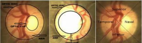
Fig. 2 : Normal Disc, Glaucomatic Disc, ISNT Quadrants
*Corresponding author: Manjunath TC, Department of
ECE, DSCE, Bangalore, India. Email:
Vol. 3(2), pp. 099-103, November, 2017. © www.premierpublishers.org, ISSN: 3254-1213
x
Fig. 3 :Medical image of normal and affected eye
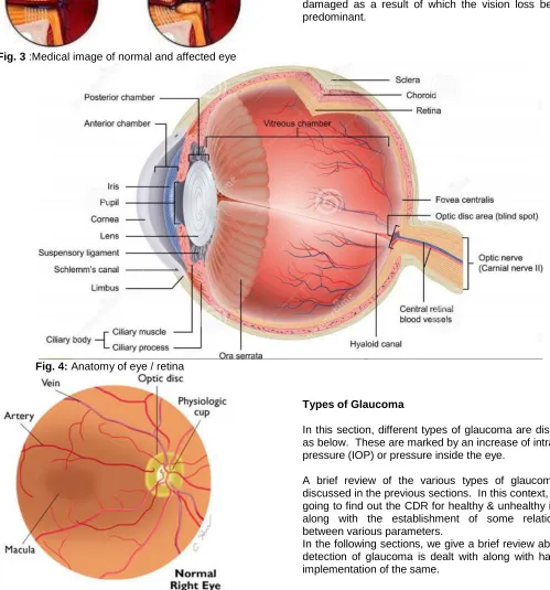
Anatomy of the normal eye
An anatomy of human eye is approximately a spherical organ & is shown in the Fig. 4. The container to-plate proportion is an estimation utilized as a part of ophthalmology and optometry to survey the movement of glaucoma. The optic plate is the anatomical area of the eye’s blind side. It is the territory where the optic nerve and veins enter the retina. The optic plate can be level or it can have a specific measure of ordinary measuring. If there is a loss of pressure, then definitely, the optical nerve gets damaged as a result of which the vision loss becomes predominant.
Fig. 4: Anatomy of eye / retina
Fig. 5: Enlarged view of the retina with optic disc
Types of Glaucoma
In this section, different types of glaucoma are discussed as below. These are marked by an increase of intraocular pressure (IOP) or pressure inside the eye.
A brief review of the various types of glaucoma was discussed in the previous sections. In this context, we are going to find out the CDR for healthy & unhealthy images along with the establishment of some relationships between various parameters.
LITERATURE SURVEY
Glaucoma disease in human beings is considered as one of the important diseases which affects the nervous systems & may lead to the loss of vision. Glaucoma damages the optic nerve which carries visual information to the brain. The brain can recognize the objects in the foreground and in the background or at a certain distance with the help of eyes. The damage to the optic nerve leads to permanent blindness or to loss of vision. So, detection of glaucoma plays an important role in order to prevent the loss of vision.
The various parameters for the pri-glauco could be thought of as below one after another as
• Measurement of the IP, • Determination of the ONH, • R N F L and
• Defect in the visual effects of the eyes.
Extensive research is being carried out on the secondary glaucoma issues in the world @ various research centers till date. A number of researchers have worked on the topic so far, some of them have advantages & some of them dis-advantages. A brief exhaustive review of the similar work done in the relevant chosen field by different authors w.r.t. glaucoma is summarized as follows.
G.C. Escher et al communicated that optical circle (OD) estimate in Ocular Hypertension (OHT) patients is littler contrasted with POAG patients and normal eye in the human beings. The even and the vertical distances across were measured. S.Sekhar et al. utilized Hough change to recognize optical disc parameters. To discover the shapes of disc O D, a locale of intrigue (ROI) is found from the twofold picture gotten after pre-preparing. Morphological calculations are utilized to compute the extent slope or the grads for edge location of the ODs.
Mahdad Esmaeili et al effective optical discs confinement and division (segmenting) are vital undertakings in robotized retinal screening of the human eyes which are affected with glaucoma. In this computerized curvelet change (DCUT) of the improved retinal picture is taken and its coefficients are altered in light of the sparsely of curvelet coefficients to get likely area of OD.
Bock Rudger et al. developed a novel computerized glaucoma identification framework in which, Glaucoma Risk Index figuring comprises of 3 stages: pre-handling/processing to kill the malady autonomous varieties from the information picture, extracting the features of the diseased eye by the P C A methodology to change the pre-prepared info information to trademark and minimal portrayal, and a 2-stage probabilistic SVM classifier to produce the Glaucoma indexed values, from which we can detect whether the human has been affected with the disease or not.
Chrastek et al in their research paper displayed a technique for optic nerve head division and its approval, i.e., they validated the detection of the ONH by conducting some real time works. The technique depends on morphological calculations, Hough trans, and a tied down dynamic form display. Gopal Dat Joshi et al in their developed a new methodology of the Glaucoma discovery by figuring glass to plate proportion (CDR), i.e., the ratio between the cup and the disc of the eye. Morphological calculations and Hough changes are connected w.r.t. each other in order to distinguish the O D.
Niemeijer Meindert et al, executed a quick strategy to distinguish the position of the optic plate and the fovea in retinal pictures of the human eyes which are affected with glaucoma. Jaeyoung Kim et al, actualized ongoing picture preparing program utilizing Open CV library by using different types of mobile phones such as apple, Samsung, android smart phones, etc.... Sopharak et al, executed location of OD in light of entropy channel.
Jalal Rashid Qureshi et al, actualized a mix of the distinctive calculations for the discovery of OD and Macula. Aby P.K et al, actualized picture handling calculations on DM3730 for face location applications using various types of sophisticated IP algorithms by considered the face of the human in which the eye is a part of the face.
Shifeng Hu, proposed a driver weariness eye highlights discovery calculation in light of Open CV, Hiroki Sugano in their research paper, proposed/developed a parallel execution of morphological handling advanced technology for Cell Broadband Defects in the diseased eye affected with glaucoma.
CuljakSIvan et al, in their research paper portrayed numerous PC vision calculations to make a peruser comfortable with Open CV and introduced numerous fundamental and mainstream Computer Vision calculations, alongside many key references for an intrigued peruser to seek after further subtle elements.
Objectives of the project work
The main objective of our M.Tech dissertation (project) work is to develop some algorithms for
• the diagnosis & detection of glaucoma by developing sophisticated algorithms using different types of transformation techniques &
• To compare them for their best performance for glaucoma detection by finding out the performance indices.
• Two cases may be considered in this project work, i.e., for healthy images & unhealthy images (affected with glaucoma& injury).
• Using MATLAB / LABVIEW as a tool to achieve this implementation process.
• The main objective of our proposed research work is to develop a simulation methodology for the identification of the glaucoma and try to implement it using real time hardware by using various types of hardware implementation kits such as the Spartan, HDL kit or the FPGA kit or the micro-controller kit or the D S P kit or in the Verilog kit.
The above-mentioned objective of our dissertation work may be achieved using the following steps:
1. Collecting images of human eyes (Both healthy and unhealthy) using appropriate image capturing devices… a large number of samples (data base of image collection) from various sources from hospitals & image databases.
2. Preparation of desired image data bases using state-of-art techniques.
3. Performing image pre-processing (segmentation, enhancement), processing, and analysis and application of mathematically developed equations in spatial & frequency domains.
4. Finding the ROI using different IP techniques.
5. Use of filtering & transformation techniques to get a fine image
6. Simulating the same using Matlab/LabVIEW 7. Implementation using hardware kits.
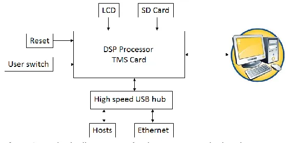
Fig. 6: Block-diagram of the proposed hardware implementation methodology
Hardware kits used
The hardware kits that are going to be used for the project work is DSP card with CCS-Code Composer Studio & the National Instruments Kits / FPGA Spartan kits for the experimentation purposes / Xilinx Spartan Kits.
Motivation / Problem statement definition
The motivation for carrying out the project work is depicted in this section along with the problem statement. Doctors are finding problems in the earlier detection of the infected region in case of eye as the glaucoma disease is the 2nd most affected disease in the world to which many people are falling victims.
At the same time, it is a very expensive process to detect the disease using the modern tools as a result of which we are developing a methodology for detection such that it is affordable by all the sections of the society, also it can be detected at the early stage & prevention can be taken.
Hence in continuation, with zeal of this work, we are proposing some novel methodologies for detection of glaucoma by developing some software algorithms using some types of transformation techniques& filtering techniques in Matlab/LabVIEW & finally implementing the same using hardware (VLSI techniques) in FPGA, the problem finally, being defined as “Simulation, development of bio-medical image processing algorithms of eyes affected with glaucoma & hardware implementation using VLSI techniques”.
Proposed Methodology
The proposed algorithm can be developed for the detection of glaucoma using the areas of cup and disc & can be applied for the glaucoma detection in the humans who are affected with the disease. Finally, the methodology could be experimentally verified using labview software. It can also be verified by converting the labview codes into the .c codes and finally implemented using the dsp kits.
At long last, these C projects could be supplanted with Open CV programs as the single board PC framework works on open source Linux stage & observing the experimental results. Finally, the result could be obtained and can be presented in the final stage, which would conclude the effectiveness of proposed methodology that is going to be developed by us.
Applications of our project work
The project work can be developed w.r.t. rural community with less experienced doctors even in the field of eye diagnosis affected with serious ailments. It can also be used in public places like in malls, so that the human being who is affected with glaucoma can be detected immediately, precaution could be given so that the proper diagnosis can be done at the earlier stage to avoid loss of vision.
CONCLUSION
A brief review of the work related to the project undertaken was depicted in the previous sections in the form of introduction, followed by literature survey. The objectives of the project work were also explored & arrived at the definition of the problem that had to be tackled with.
The methodology is proposed in the form of a block diagram to solve the above defined problem using Matlab/LabVIEW/Xilinx kits and implementation using hardware tools such as DSP / FPGA kits in order to arrive at the expected results.
Once, the hardware implementation is done, then the simulation results what we have got can be authenticated, proved in the real time environment of the hardware implementation using interfacing kits.
REFERENCES
DRIONS-DB: Digital Retinal Images for optic Nerve Segmentation Database.
High Resolution Fundus Image database
https://www5.cs.fau.de/research/data/fundus-images/
http://cvit.iiit.ac.in/projects/mip/drishti-gs/mip-dataset2/Home.php
http://dx.doi.org/10.14257/ijmue.2015.10.9.14
http://www.ia.uned.es/~ejcarmona/DRIONS-DB.html Kaur, Husandeep, and Amandeep Kaur (2014). Early
Stage Glaucoma Detection in Diabetic Patients A Review. International Journal of Advanced Research in Computer Science and Software Engineering, Volume 4, Issue 5. ISSN: 2277 128X , Page 271-274.
Khan, Fauzia, et al. (2013). Detection of glaucoma using retinal fundus images”, Biomedical Engineering International Conference (BMEiCON), 6th.
Manjula Sri, M. Raghupathi Reddy, K.M.M. Rao (2011). Hardware Implementation of Detection of Glaucoma from Color Fundus Images. pp. 340-345.
Opticdisc.org Database
http://www.optic-disc.org/library/normal-discs/page7.html
Praveen Vanaparthy, Sahitya G., Krishna Sree, Dr. C.D.Naidu (2013). FPGA implementation of image enhancement algorithms for biomedical image processing. International Journal of Advanced Research in Electrical, Electronics and Instrumentation Engineering, An ISO 3297: 2007 Certified Organization, ISSN (Print) : 2320 – 3765, ISSN (Online): 2278 – 8875, pp. 5747 – 5753, Vol. 2, Issue 11.
R. C. Gonzalez, R. E. Woods, and S. L. Eddins (2004). Digital Image Processing using MATLAB, New York: Pearson Prentice Hall, Prentice Hall, ISBN 0-13-094659.
Srinivasan Aruchamy, Partha Bhattacharjee and Goutam Sanyal (2015). Automated Glaucoma Screening in Retinal Fundus Images. International Journal of Multimedia and Ubiquitous Engineering, ISSN: 1975-0080 IJMUE, Copyright ⓒ 2015 SERSC, , Vol. 10, No. 9, pp.129 136.
Accepted 23 October, 2017
Citation: Pavithra G, Manjunath TC and Anushree G (2017). Development of novel BMIP algorithms for human eyes affected with glaucoma and hardware implementation using VLSI based embedded systems. International Research Journal of Power and Energy Engineering, 3(2): 099-103.