Proteomic progress in studying tuberculosis from 2010
to 2011
Lijun Zhang1,2*, Douglas Lowrie2, Honghao Zhou1
1
Department of Pharmacogenetics, Institute of Clinical Pharmacology, Central South University, Changsha, China; [email protected]
2
Shanghai Public Health Clinical Center, Fudan University, Shanghai, China. *Corresponding Author: [email protected]
Received 17 June 2011; revised 25 July 2011; accepted 3 August 2011.
ABSTRACT
It is well accepted that rapid and early detection of Mycobacterium tuberculosis infection and understanding the mechanism of microbiology- host interaction. Herein, we review the recently published papers related to TB proteomics from 2010 to 2011, including new technologies used in TB proteome research, diagnosis biomarkers of TB-associated diseases, disease pathogene- sis and antigens for drug development. Through this review, we wish to offer some help for TB diagnosis and treatment.
Keywords:Proteomics; Tuberculosis; Diagnosis; Treatment
1. INTRODUCTION
Mycobacterium tuberculosis (Mtb), the causative agent of tuberculosis (TB), kills nearly two million people an- nually and has been a major health threat for centuries, mostly in low and middle-income countries [1-3].
However, the study for anti-TB was very slowly. One is due to no reliable biomarker for diagnosis. The gold standard for TB diagnosis is strain culture that usually costs as long as 8 weeks and the sensitivity is as low as 50% or less [4,5]. The other is no new drug. Therefore, it is very important to find new biomarkers for TB diag- nosis and drug targets.
The development of proteomics has opened new ways for TB study due to its facilitation in investigating many complex issues of TB and TB-host interactions. Although proteomics has lagged behind genomics and transcrip- tomics due to instrumental and sensitivity problems, the application of proteomics to the study of infectious agents is beginning to emerge. Such applications include sear- ching potential biomarkers for diagnosis [6], identifying complete virulence factor inventories [7], studying the response of both host and pathogen to the infection
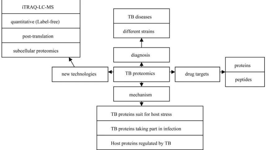
process (reviewed by Bhavsar AP [8] and Boshoff HI [9]), and elucidating mechanistic actions of virulence factors as they interface with host cells [10]. In this study, we reviewed proteomic progresses in TB studies from 2010 to 2011 as shown in Figure 1.
2. NEW TECHNOLOGIES DEVELOPED
IN TB STUDIES
2.1. Quantitative Proteomic Studies
There are two general technologies for TB proteomic studies including: 1) two-dimensional electrophoresis (2DE) combined with mass spectrometry (2DE-MS) [11] or western blot (named 2DE-western blot(2DE-WB)) [12, 13]; 2) isotope labeling followed with multiple-dimen- tional liquid chromatography separation combined with mass spectrometry analysis [11]. During the last two years, a label-free proteomic method based on 1DE se- paration, LC-MS identification, followed by emPAI quantification was reported to identify differentially abun- dant proteins in closely related hypo- and hyper-virulent clinical Mycobacterium tuberculosis Beijing isolates [14]. Another label-free proteomics based on SDS-PAGE se- paration followed by MaxQuant peak intensity calcula- tions was developed to analyze membrane proteins of Mycobacterium tuberculosis H37Rv and H37Ra strains [7].
2.2. Proteomics for Post-Translation Modification
Figure 1. Illustration of proteomic progresses in TB studies from 2010 to 2011.
sinized PUPylated peptides, a global pull-down of pro- tein targets for PUPylation in Mycobacterium smegmatis (Mycobacterium tuberculosis (Mtb)’s avirulent strain) revealed 103 candidate PUPylation targets and 52 con- firmed targets[17]. Phosphorylation is another important post-translation. Prisic et al. [18] reported a phospho- proteomic study, in which they studied Ser/Thr phos- phorylation in TB and found more than 500 phosphory- lation events in 301 proteins.
2.3. Subcellular Proteomics
It is very difficult to study whole cell or TB proteome due to the overlap of higher abundant proteins to the lowers. In order to find proteins with low abundance or difficult to solve, subcellular proteomics was developed and used for TB study.
A proteomic study was carried in cell wall of Myco- bacterium tuberculosis [19]. A total of 528 proteins were identified through a combination of detergent extraction, 2DGE, multidimensional liquid chromatography and mass spectrometry. These proteins play a role in host- pathogen interactions and define new potential drug tar- gets via discovery of unique biosynthetic or metabolic processes. Another proteomic study was carried out in culture filtratefrom M. bovis BCG Moreau, the Brazilian vaccine strain, comparing it to that of BCG Pasteur [20]. 101 differential proteins were identified, which were con- sidered of great importance given their dominant immu- nogenicity and role in pathogenesis, being available for interaction with host cells since early infection. Further-
more, a proteomic strategy coupling blue native PAGE to liquid chromatography tandem mass spectrometry (LC- MS/MS) was developed [21]. 40 proteins (including 12 integral membrane proteins) such as Pks7 and Pks8 were unambiguous identified. These Mycobacterial membrane and membrane-associated protein complexes play impor- tant roles in various cellular processes, and the protein- protein interactions.
3. DISCOVERING BIOMARKERS FOR TB
DIAGNOSIS
3.1. Potential Biomarkers Related to TB Infection
non-tuberculosis (non-TB) controls, respectively. Ka- shyap RS [22] found that the 65-kDa HSP, 71-kDa HSP, 14-kDa HSP and Ag 85 complex proteins were over- expressed in TB ascites compared with non-TB ascites patients through proteomic study. Takahiro Tanaka [24] found that RBP4 and fetuin-A were significantly lower expressed in samples from patients with active TB than in their controls (P < 0.0001) through analyzing the whole blood supernatant.
3.2. Potential Biomarkers to Distinguish Different Strains
Except diagnosing TB infection, the ability to readily and accurately distinguish among strains is important be- cause different strains have different virulence and drug- resistances characters that necessitate different treat- ments. A label-free quantitative proteomic approach [23] showed that 48 proteins were over-represented in the hypo-virulent isolate, while 53 were overrepresented in the hyper-virulent. These proteins such as ESAT-6, Esx- like proteins and fbpD (MPT51) reduced in the hyper- virulent strain might be used for distinguishing hypo and hyper-virulent TB. Similarly, a label-free quantitative proteomic approach was performed in the virulent H37- Rv strain and its attenuated counterpart H37Ra. The re- sults showed that 29 membrane-associated proteins with a 5 or more fold difference in their relative abundance in one strain compared to the other. Of which, 19 mem- brane- and lipo-proteins such as three ABC transporter proteins (Rv0933, Rv1273c and Rv1819c) had higher abundance in H37Rv, while another 10 proteins had a higher abundance in H37Ra [7]. Furthermore, Mehaffy et al. [11] studied the proteomes of secreted and cytosolic proteins of genetically closely related strains of M. tu-berculosis through 2DE and iTRAQ-LC-MS methods, and found that some enzymes such as GltA2, SucC, Gnd1 and Eno were expressed differently in different strains.
4. MECHANISM OF TB INFECTION
Although TB has been found for a long time, the know- ledge about TB infection is still limited. Proteomics as a new technology has shown great advance in the study of TB infection.
4.1. TB Proteins Suit for Host Stress
During the TB infection, the tubercle bacilli are likely to be exposed to stress that can result in the formation of aberrant proteins with altered structures. Bacteria have evolved accurate regulatory systems to control the ex- pression and function of potentially destructive proteases and chaperones. For example, the Clp Gene Regulator
(ClgR)of M.tuberculosis activates the transcription of at least ten genes including four of which encode protease systems (ClpP1/C, ClpP2/C, PrtB and an HtrA-like pro- tease-Rv1043c) and three of which encode chaperones (Acr2, ClpB and Rv3269) [25]. HtrA-like serine protease encoded by pepD, are responsible for degrading or re- folding protein substrates following exposure to stress. A proteomic study further showed that the HtrA-Like Ser- ine Protease PepD interacts with and modulates the My- cobacterium tuberculosis 35-kDa antigen outer envelope protein [26].
4.2. TB Proteins Taking Part in Infection
Considerable effort has been directed toward control- ling tuberculosis through understanding host-pathogen interactions leading to biomarker expression. During the early and chronic stages of disease, it is proteins from TB that regulates host cells. Through a guinea pig model of tuberculosis, 500 unique proteins were identified in the infected lung. Of which cell wall and cell wall processes, and intermediary metabolism and respiration were the two major functional classes, including Rv2209, Rv2315c, etc [27]. Kunnath VS [12] also found that sera from worldwide TB suspects recognized approximately 10% of the bacterial proteome and the M. tuberculosis immu- noproteome were rich in membrane-associated and ex- tracellular proteins through carrying a systems immu- nology approach.
4.3. Host Proteins Regulated by TB
TB can regulate host proteome to finish its infection. Proteins involved in membrane trafficking and signal transduction such as Ras GTPase-activating-like protein IQGAP1 were detected to be regulated through a proteo- mic study [28]. In this study, the phagosomes from BCG- infected human macrophages were purified, and 447 human host proteins were identified. Furthermore, My-cobacteria release active membrane vesicles (MVs) to deliver ligands that can be recognized by host cells. Ex- tensive proteomic analysis revealed that only MVs from the virulent strains contained TLR2 lipoprotein agonists. The interaction of MVs with macrophages isolated from mice stimulated the release of cytokines and chemokines in a TLR2-dependent fashion [29].
pathways that contribute to the arrest of phagosome ma- turation were found to be regulated [10].
5. PROTEOMICS IN DRUG
DEVELOPMENT
5.1. Proteins for Drug Development
The inadequate protection afforded by bacillus Cal- mette-Guérin (BCG) vaccination provides continued im- petus for the discovery of immunodominant Mycobacte- rium tuberculosis (MTB) antigens to develop improved vaccines. The 6-kDa early secretory antigenic target of Mycobacterium tuberculosis (ESAT-6) [30] and the 10- kDa culture filtrate antigen (CFP-10) [31] are the most immunodominant and highly M. tuberculosis (MTB)-spe-cific antigens. Ag85B-ESAT-6 adjuvanted with IC31 promotes strong and long-lived Mycobacterium tubercu- losis specific T cell responses in naive human volunteers [30]. Rv3615c [Esx-1 substrate protein C (EspC)], a pro-tein with similar size and sequence to CFP-10 and ESAT- 6, was found to be a highly immunodominant RD1 (Re-gion of Difference 1)-dependent secreted antigen specific for Mycobacterium tuberculosis infection through a quantitative proteomics and metabolically labeled mutant and genetically complemented MTB strains [32]. Giri et al. [33] used LC-MS/MS to identify 41 mycobacterial proteins present in exosomes released from M. tubercu-losis-infected J774 cells. Another quantitative proteomic analysis showed a higher expression of immunogenic pro- teins such as Rv1860 (BCG1896, Apa), Rv1926c (BCG- 1965c, Mpb63) and Rv1886c (BCG1923c, Ag85B) in BCG Moreau when compared to BCG Pasteur from their culture filtrate [20]. These antigens might offer new tar-gets for vaccine development. Nearly,an immunoproteo- me microarray study of M. tuberculosis-proteins with sera from patients was developed [34], in which, 3 novel antigens, namely, Rv1987, Rv3807c, and Rv3887c, pro-vided better sensitivity and accuracy for TB detection.
Sarah L. Kinnings built a TB-drugome(http://funsite.- sdsc.edu/drugome/TB) database. In this TB-drugome, 123 of the 274 drugs are connected to 447 of the 1730 proteins. These proteins are the potential targets for de-veloping some safe and efficient anti-tubercular drugs [35].
5.2. Peptides for Drug Development
It is proposed that low-similarity peptides be a poten- tial epitope against M. tuberculosis. The TB sequence were examined for similarity score to the proteins of the host in which the epitopes were defined [36]. Addition- ally, a peptide microarray consisting of 7466 unique pep-tides derived from 61 M. tuberculosis proteins for
poten-tial MHC class II-presented epitopes was constructed. Two hundred and twenty-two peptides that formed MHC class II-peptide complexes had previously been de-scribed as exclusively recognized by IgG in sera from patients with active pulmonary tuberculosis [37].
6. CONCLUSIONS
This review summarized the new proteomic technolo- gies used in anti-TB studies and provided a better under- standing for possible new biomarkers and infection me- chanism as well as drug development in TB study through proteomic technologies. Although the proteins provided by proteomic studies might not be used for biomarkers or drug targets at once, they will be helpful for anti-TB re- search and must promote TB treatment development through further studies. In all, proteomics may open up new horizons for understanding the pathogenesis of tu-berculosis and developing drugs.
7. ACKNOWLEDGEMENTS
This work was supported by “China Postdoctoral Science Founda-tion (20100471238)”, the NaFounda-tional Basic Research Program of China (also called 973 Program) (2011CB910700) and the Postdoctoral Sci-ence Foundation of Central South University.
REFERENCES
[1] Prasad, R. (2010) Multidrug and extensively drug-resis- tant TB (M/XDR-TB): Problems and solutions. Indian Journal of Tuberculosis, 57, 180-191.
[2] Ahmad, S. (2010) New approaches in the diagnosis and treatment of latent tuberculosis infection. Respiratory Research, 11, 169. doi:10.1186/1465-9921-11-169 [3] (2010) WHO global tuberculosis control report 2010.
Summary. Central European Journal of Public Health,
18, 237.
[4] Sendagire, I., Schim Van der Loeff, M., Mubiru, M., Konde-Lule, J. and Cobelens, F. (2010) Long delays and missed opportunities in diagnosing smear-positive pul- monary tuberculosis in Kampala, Uganda: A cross-sec- tional study. PLoS One, 5, e14459.
doi:10.1371/journal.pone.0014459
[5] Bailey, S.L., Roper, M.H., Huayta, M., Trejos, N., Lopez Alarcon, V. and Moore, D.A. (2010) Missed opportunities for tuberculosis diagnosis. The International Journal of Tuberculosis and Lung Disease, 15, 205-210.
[6] Liu, Q., Chen, X., Hu, C., Zhang, R., Yue, J., Wu, G., et al.
(2010) Serum protein profiling of smear-positive and smear-negative pulmonary tuberculosis using SELDI- TOF mass spectrometry. Lung, 188, 15-23.
doi:10.1007/s00408-009-9199-6
[7] Malen, H., De Souza, G.A., Pathak, S., Softeland, T. and Wiker, H.G. (2011) Comparison of membrane proteins of
Mycobacterium tuberculosis H37Rv and H37Ra strains.
BMC Microbiology, 11, 18. doi:10.1186/1471-2180-11-18 [8] Bhavsar, A.P., Auweter, S.D. and Finlay, B.B. (2010) Pro-
lecular boundaries. Future Microbiology, 5, 253-265. doi:10.2217/fmb.09.114
[9] Boshoff, H.I. and Lun, D.S. (2010) Systems biology ap- proaches to understanding mycobacterial survival mecha- nisms. Drug Discovery Today: Disease Mechanisms, 7, 75-82. doi:10.1016/j.ddmec.2010.09.008
[10] Shui, W., Petzold, C.J., Redding, A., Liu, J., Pitcher, A., Sheu, L., et al. (2011) Organelle membrane proteomics reveals differential influence of mycobacterial lipogly- cans on macrophage phagosome maturation and auto- phagosome accumulation.Journal of Proteome Research,
10, 339-348. doi:10.1021/pr100688h
[11] Mehaffy, C., Hess, A., Prenni, J.E., Mathema, B., Krei- swirth, B. and Dobos, K.M. (2010) Descriptive proteomic analysis shows protein variability between closely related clinical isolates of Mycobacterium tuberculosis. Pro- teomics, 10, 1966-1984. doi:10.1002/pmic.200900836 [12] Kunnath-Velayudhan, S., Salamon, H., Wang, H.Y., Davi-
dow, A.L., Molina, D.M., Huynh, V.T., et al. (2010) Dy- namic antibody responses to the Mycobacterium tuber- culosis proteome. Proceedings of the National Academy of Sciences of the United States of America, 107, 14703- 14708. doi:10.1073/pnas.1009080107
[13] Deenadayalan, A., Heaslip, D., Rajendiran, A.A., Velayu- dham, B.V., Frederick, S., Yang, H.L., et al. (2010) Im- munoproteomic identification of human T cell antigens of
Mycobacterium tuberculosis that differentiate healthy contacts from tuberculosis patients. Molecular & Cellular Proteomics, 9, 538-549.
doi:10.1074/mcp.M900299-MCP200
[14] de Souza, G.A., Fortuin, S., Aguilar, D., Pando, R.H., McEvoy, C.R., van Helden, P.D., et al. (2010) Using a label-free proteomics method to identify differentially abundant proteins in closely related hypo- and hyperviru- lent clinical Mycobacterium tuberculosis Beijing isolates.
Molecular & Cellular Proteomics, 9, 2414-2423. doi:10.1074/mcp.M900422-MCP200
[15] Festa, R.A., McAllister, F., Pearce, M.J., Mintseris, J., Burns, K.E., Gygi, S.P. and Darwin, K.H. (2010) Pro-karyotic ubiquitin-like protein (Pup) proteome of Myco-bacterium tuberculosis [corrected]. PLoS One, 5, 8589. doi:10.1371/journal.pone.0008589
[16] Poulsen, C., Akhter, Y., Jeon, A.H., Schmitt-Ulms, G., Meyer, H.E., Stefanski, A., et al. (2010) Proteome-wide identification of mycobacterial pupylation targets. Mo-lecular Systems Biology, 6, 386.
doi:10.1038/msb.2010.39
[17] Watrous, J., Burns, K., Liu, W.T., Patel, A., Hook, V., Bafna, V., et al. (2010) Expansion of the mycobacterial “PUPylome”. Molecular Biosystems, 6, 376-385.
doi:10.1039/b916104j
[18] Prisic, S., Dankwa, S., Schwartz, D., Chou, M.F., Lo- casale, J.W., Kang, C.M., et al. (2010) Extensive phos- phorylation with overlapping specificity by Mycobacte- rium tuberculosis serine/threonine protein kinases. Pro-ceedings of the National Academy of Sciences of the United States of America, 107, 7521-7526.
doi:10.1073/pnas.0913482107
[19] Wolfe, L.M., Mahaffey, S.B., Kruh, N.A. and Dobos K.M. (2010) Proteomic definition of the cell wall of Mycobac-terium tuberculosis. Journal of Proteome Research, 9, 5816-5826. doi:10.1021/pr1005873
[20] Berredo-Pinho, M., Kalume, D.E., Correa, P.R., Gomes, L.H., Pereira, M.P., Silva, R.F., et al. ( 2011) Proteomic profile of culture filtrate from the Brazilian vaccine strain Mycobacterium bovis BCG Moreau compared to M. bo- vis BCG Pasteur. BMC Microbiology, 11, 80.
doi:10.1186/1471-2180-11-80
[21] Zheng, J., Wei, C., Zhao, L., Liu, L., Leng, W., Li, W. and Jin, Q. (2011) Combining blue native polyacrylamide gel electrophoresis with liquid chromatography tandem mass spectrometry as an effective strategy for analyzing poten- tial membrane protein complexes of Mycobacterium bo- vis bacillus Calmette-Guerin. BMC Genomics, 12, 40. doi:10.1186/1471-2164-12-40
[22] Kashyap, R.S., Saha, S.M., Nagdev, K.J., Kelkar, S.S., Purohit, H.J., Taori, G.M. and Daginawala, H.F. (2010) Diagnostic markers for tuberculosis ascites: A prelimi- nary study. Biomark Insights, 5, 87-94.
[23] Desouza, G.A., Fortuin, S., Aguilar, D., Pando, R.H., Mc Evoy, C.R., van Helden, P.D., et al. (2010) Using a la-bel-free proteomic method to identify differentially abun- dant proteins in closely related hypo- and hyper-virulent clinical Mycobacterium tuberculosis Beijing isolates. Mo- lecular & Cellular Proteomics, 9, 2414-2423.
doi:10.1074/mcp.M900422-MCP200
[24] Tanaka, T., Sakurada, S., Kano, K., Takahashi, E., Yasuda, K., Hirano, H., et al. (2011) Identification of tuberculo- sis-associated proteins in whole blood supernatant. BMC Infectious Diseases, 11, 71. doi:10.1186/1471-2334-11-71 [25] Estorninho, M., Smith, H., Thole, J., Harders-Westerveen,
J., Kierzek, A., Butler, R.E., et al. (2010) ClgR regulation of chaperone and protease systems is essential for Myco- bacterium tuberculosis parasitism of the macrophage.
Microbiology, 156, 3445-3455. doi:10.1099/mic.0.042275-0
[26] White, M.J., Savaryn, J.P., Bretl, D.J., He, H., Penoske, R.M., Terhune, S.S. and Zahrt, T.C. (2011) The HtrA-like serine protease PepD interacts with and modulates the
mycobacterium tuberculosis 35-kDa antigen outer enve-lope protein. PLoS One, 6, 18175.
doi:10.1371/journal.pone.0018175
[27] Kruh, N.A., Troudt, J., Izzo, A., Prenni, J. and Dobos, K.M. (2010) Portrait of a pathogen: The Mycobacterium tuberculosis proteome in vivo. PLoS One, 5, 13938. doi:10.1371/journal.pone.0013938
[28] Lee, B.Y., Jethwaney, D., Schilling, B., Clemens, D.L., Gibson, B.W. and Horwitz, M.A. (2010) The mycobacte-rium bovis bacille calmette-guerin phagosome proteome.
Molecular & Cellular Proteomics, 9, 32-53. doi:10.1074/mcp.M900396-MCP200
[29] Prados-Rosales, R., Baena, A., Martinez, L.R., Luque- Garcia, J., Kalscheuer, R., Veeraraghavan, U., et al. (2011) Mycobacteria release active membrane vesicles that mo- dulate immune responses in a TLR2-dependent manner in mice. The Journal of Clinical Investigation, Epub ahead of print. doi:10.1172/JCI44261
[30] van Dissel, J.T., Arend, S.M., Prins, C., Bang, P., Ting- skov, P.N., Lingnau, K., et al. (2010) Ag85B-ESAT-6 ad- juvanted with IC31 promotes strong and long-lived My- cobacterium tuberculosis specific T cell responses in na- ive human volunteers. Vaccine, 28, 3571-3581.
doi:10.1016/j.vaccine.2010.02.094
preclinical testing of new tuberculosis vaccines. Clinical Microbiology Reviews, 23, 781-794.
doi:10.1128/CMR.00005-10
[32] Millington, K.A., Fortune, S.M., Low, J., Garces, A., Hin- gley-Wilson, S.M., Wickremasinghe, M., et al. (2010) Rv3615c is a highly immunodominant RD1 (Region of Difference 1)-dependent secreted antigen specific for My- cobacterium tuberculosis infection. Proceedings of the National Academy of Sciences of the United States of America, 108, 5730-5735. doi:10.1073/pnas.1015153108 [33] Giri, P.K., Kruh, N.A., Dobos, K.M. and Schorey, J.S.
(2010) Proteomic analysis identifies highly antigenic proteins in exosomes from M. tuberculosis-infected and culture filtrate protein-treated macrophages. Proteomics,
10, 3190-3202. doi:10.1002/pmic.200900840
[34] Li, Y., Zeng, J., Shi, J., Wang, M., Rao, M., Xue, C., et al.
(2010) A proteome-scale identification of novel anti- genic proteins in Mycobacterium tuberculosis toward
di-agnostic and vaccine development. Journal of Proteome Research, 9, 4812-4822. doi:10.1021/pr1005108
[35] Kinnings, S.L., Xie, L., Fung, K.H., Jackson, R.M., Xie, L. and Bourne, P.E. (2010) The Mycobacterium tubercu-losis drugome and its polypharmacological implications.
PLoS Computational Biology, 6, 1000976. doi:10.1371/journal.pcbi.1000976
[36] Lucchese, G., Stufano, A. and Kanduc, D. (2010) Propos-ing low-similarity peptide vaccines against Mycobacte-rium tuberculosis. Journal of Biomedicine and Biotech-nology, 832341.
[37] Gaseitsiwe, S., Valentini, D., Mahdavifar, S., Reilly, M., Ehrnst, A. and Maeurer, M. (2010) Peptide microar-ray-based identification of Mycobacterium tuberculosis