GREEN SYNTHESIS OF NANOSILVER AND ITS EFFECT ON PHASE
SOLUBILITY OF FEW BCS CLASS II DRUGS
1
Dr. Ajit Shankarrao Kulkarni*, 2Mr. Kailas Madhukar Karande, 3Dr. Nagesh Hanumantrao Aloorkar, 4Mr. Jayprakash Sitaram Suryavanshi,
1
Professor and Vice Principal, Satara College of Pharmacy, Plot No.1539, New Additional MIDC, Degaon, Satara. 415004
2
Assistant Professor, Department of Pharmaceutics, Satara College of Pharmacy, Satara
3
Professor and HOD, Department of Pharmaceutics, Satara College of Pharmacy.
4
Assistant Professor and HOD, Department of 4Pharmacognosy, Satara College of Pharmacy.
ABSTRACT
Nanometals are developing tools for drug delivery systems owing to their unique properties viz. surface plasmon resonance, high surface to volume ratio, ability to interact not only with biomolecules but also drug molecules, precise therapeutic targeting etc. These core properties pertaining to drug delivery make them potential tool in pharmaceutical research.In present investigation, silver nanoparticles were synthesized using bioexcreta, cow urine and gelatin as stabilizing agent. Synthesized nanosilvers were characterized by absorption spectroscopy, infrared spectroscopy, transmission electron spectroscopy and XRD. Absorption spectroscopy revealed successful synthesis of nanosilver. Fourier transform infrared spectroscopy showed contribution of functional groups stabilizing nanosilver. TEM depicted spherical uniform size and shape of nanosilver. XRD study revealed face centered cubic crystals. These nonfunctionalized nanosilvers were subsequently evaluated for acute toxicity study and found safe. Studies were also extended to confirm effect of synthesized NS on phase solubility of simvastatin and atorvastatin, and revealed appreciable increase in solubility of these drugs. Present investigations suggest that a green method can be used to synthesize NS which is ecological, economical, and time efficient. Further, NS could enhance solubility of simvastatin and atorvastatin.
Volume 3, Issue 6, 440-456. Research Article ISSN 2277 – 7105
Article Received on 13 June 2014,
Revised on 08 July 2014, Accepted on 03 August 2014
*Correspondence for
Author
Dr. Ajit Shankarrao Kulkarni
KEY WORDS: Silver Nanoparticles, Green synthesis, Simvastatin, Atorvastatin.
1. INTRODUCTION
Metal nanoparticles are gaining much more importance and still there has been a remarkable research interest in the area of nanotechnology to develop reliable processes for the synthesis of metal nanoparticles[1] and subsequently to functionalize them. Nanosilver (NS) is one of the examples of metal nanoparticle. Particularly for drug delivery, polymeric nanoparticles, dendrimers, liposomes and metal nanoparticles are being widely explored[9]. Research into the medical applications of NS has been extremely active, with a variety of commercially available products being used clinically[8]. Ample of methodologies have been reported in the past, to synthesize various metal nanoparticles like Ag, Au, Pt, Cu, Zn etc[26,27,28,29]. Out of those all, biosynthetic methods have received considerable attention due to the growing need to develop ecological and economical methodologies. Most of biosynthetic methods include green methods and are of natural origin. Natural environment is a reach source of crude untreated extract from tissue of diverse widespread flora and fauna like terrestrial and aquatic plants and marine organism, microorganism etc[2]. Animal waste also is the part of natural environment. Cow, Bos indicus is most valuable animal in all veda and it is called as the mother of all. The composition containing cow‟s excretions urine, dung, milk, curd, ghee five ingredients together known as “Pachgavya” which is the main ingredient of many of ayurvedic preparations [3]. According to ancient literatures distillate of cow urine was the one to be used mainly and the distillate was found to exhibit anti oxidant effect[4] which is preliminary requisite for the synthesis of metal nanoparticles. Fresh cow urine is more active than its distillate [3] hence fresh cow urine was considered in present investigation.
For drugs with poor aqueous solubility, the rate at which the drug dissolves is often the slowest step and exhibits a rate limiting effect on drug bioavailability. According to modified Noyes-Whitney analysis, drug solubility can be increased by increasing the surface area available for dissolution by decreasing the particle size of the solid compound and or by optimizing the wetting characterization of the compound to decrease the boundary layer thickness [6]. Unique nanoscale dimension of NS provides a large surface area for efficient conjugation and protection of drugs. The attachment of payload can be readily achieved by either noncovalent interactions or covalent chemical conjugation[7]. This interaction may detach drug molecule/s from the poorly soluble crystal/drug particles; concomitantly decreases the particle size and increases the solubility.In present investigation green method was reported for synthesis of nonfunctionalized NS which were further tested for its effect on phase solubility of few class II drugs like Ibuprofen, Simvastatin and Atorvastatin.
2. MATERIALS AND METHODS
2.1. Carboxy methyl cellulose, hydroxyl propyl methyl cellulose, agar, starch, sodium alginate, microcrystalline cellulose, methyl cellulose, gelatin and AgNO3 were procured from Sigma–Aldrich. Fresh cow urine was collected from local cow yard using sterile plastic container and stored at cool temperature. All aqueous solutions were prepared in double distilled, membrane filtered water.
2.2. Synthesis of NS
The onset of optimization was done using several polymers like carboxyl methyl cellulose, hydroxyl propyl methyl cellulose, agar, starch, sodium alginate, microcrystalline cellulose, methyl cellulose, gelatin etc. These were screened for suitable template for NS. 0.25, 0.5, 0.75, 1, 2 % w/v solutions (each four) of each of above polymer solutions were prepared. 0.5, 1, 1.5, 2 %V/V of cow urine were added in each solution. 1ml of 0.1M aqueous solution of silver nitrate was poured in each solution and autoclaved at 15 psi at 1210C for 5 min. Solutions of same concentrations were also kept at room temperature without autoclaving. Solution showing better result compared to other solutions was selected for further study
2.3. Characterization of Nanosilver 2.3.1. Absorption study[35,36]
2.3.2. FTIR study [37]
The prominent IR peaks of NS, silver nitrate and gelatin were recorded using Perkin-Elmer FT-IR spectrophotometer. Spray dried and freeze dried samples were used for IR studies.
2.3.3. Transmission electron microscopy [35, 36, 37]
The size and shape of nanoparticles were analyzed using transmission electron micrographs. Micrographs were obtained using a Philips EM 208 from Bombay IIT (TEM operating at 200 kV).
2.3.4. X- ray diffraction analysis [14]
The aqueous solution NS was spray dried in a Labultima Lab spray drier (Model – LU 222Advanced) at 1000C. Spray drying was performed at an inlet air temperature of 1350C , corresponding to an outlet temperature 900c. The powder was collected from I & II cyclones of the spray dryer. The powder X-ray diffraction was performed using a Philips, Holland – PW3710 diffractometer. The diffracted intensities were recorded from 100 to 650 2θ angles.
3. Phase solubility[10]
Phase solubility study was done according to Higuchi and Connors. An excess amount of drug (Ibuprofen, Simavastatin, and Atorvastatin) was placed into 50 ml volumetric flask containing different percent of NS. The flasks were kept in a Remi shaker and continued stirring for 48 hrs. After 48 hrs content of each flask was analyzed spectrophotometrically. All solubility measurements were performed in duplicate, in and off presence of NS
4. Acute Toxicity Study of NS
The acute oral toxicity was conducted using the recommended Organization for Economic Cooperation and Development (OECD) guidelines for the resulting of chemicals for safety evaluation. Furthermore, lethal dose 50 (LD50) in acute oral toxicity was evaluated24. Twenty albino male mice between weight ranges 18-22gm were selected and divided into 4 groups.
5. Antibacterial Activity of NS [25]
6. RESULTS AND DISCUSSION
containing 2% W/V of gelatin & 1.5 % V/V of cow urine during preliminary screening (results are shown in figure. 3, 4, 5, 6). Although studies were not extended to confirm formation of quantum dots, but TEM result, literature review[11] and visual experimental observations(appearance of fluorescence) may support formation of quantum dots. More is the symmetry of the peak less is the aggregation [18]. Moreover, absorbance shown by this solution was more intense compared to all other solution. If, NS appeared with less aggregation, more & more surface was available & hence more was the plasmon, which lead to resonate strongly with the resonating wavelength of incident light & hence intensity of incident light decreased appreciable with increased absorbance[19].
6.1. FTIR Spectra
FTIR measurement, were carried out to identify the stabilization mechanism or effect owing to capping agent. The FTIR spectrum of gelatin and NS is shown in figure 7 and 8 respectively.
The stabilization of NS could be owing to functional groups available on the molecular structure of gelatin. The roles of these functional groups in stabilization of NS were confirmed using FTIR measurements, the comparative spectrum of which is shown in figure 7 and 8. The band at 3743.32 cm-1 corresponds to O-H stretching due to H-bonding. The peak at 2924.96 cm-1 corresponds to O-H stretch in carboxylic acid (- COOH residue of amino acid). The assignment at 1645.05 cm-1 corresponds to C-N stretching of amine group (especially aromatic), 1095.71 cm-1 due to C-O stretching vibration. Therefore, the synthesized NS were surrounded by proteins & confirmed carbonyl group of the amino acid residues & hydroxyl groups were confirmed to show the stronger ability to bind metal indicating that gelatin could stabilize NS.
6.2. Transmission Electron Microscopy
6.3. X- Ray Diffraction Study
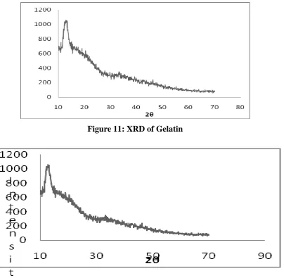
The X-ray diffractogram of gelatin showed number of diffused peak. The diffractogram of gelatin showed sharp peaks indicating crystalline nature, while the diffractogram of NS embedded in the gelatin matrix showed sharp peaks with high intensity. X-RD pattern of pure gelatin showed diffused peaks at 10.70,11.90, 12.80, 130, 140, 15.50, 16.30, 17.50, 18.50 and NS embedded in the gelatin matrix showed intense peaks at 10.30, 11.80, 12.90, 13.30, 140, 150, 160, 17.10, 18.80. Results are shown in figure 11 and 12. Reduction in crystallinity was determined by comparing some representative peak heights in the diffraction patterns of pure gelatin (reference) and gelatin impregnated with NS. Pure gelatin showed diffused peak at 12.80 (2θ) with lowest peak intensity 969. Gelatin impregnated with NS showed sharp peak at 13.30(2θ) with highest peak intensity 1059.The relative degree of crystallinity (RDC) was calculated according to the equation:
RDC= ISample/Ireference
Where, ISampleis the peak height of the sample and Ireference is the peak height at the same angle
for the reference with the highest intensity. The peak height at 13.30 (2θ) was used for calculating the RDC of NS and its RDC value was found to be 1.09 which indicate that crystallinity of sample was found to be increased as the value of RDC was less than 1.
6.4. Phase Solubility Study
6.5. Acute Toxicity Test
The mild irritation on eye, mouth, skin, paw region was observed after 24 hours of dosing when 5000mg/kg of NS was administered to mice. This irritation reaction may be caused due to accumulation of NS in oral, eye, dermal region. In acute oral toxicity test, the mice treated with the dose of 5000mg/kg body weight showed no significant changes in body weight. In addition, no death of animals was noted during the observation time.
The result of the acute oral toxicity study indicated that LD 50 of NS was greater than 5000mg/kg according to the OECD 423 guideline. The NS was found to be less toxic when oral toxicity test in mice were performed. Further long- term toxicity, mutagenicity, carcinogenicity are required to clarify any adverse effects.
6.6. Antibacterial Activity Of NS
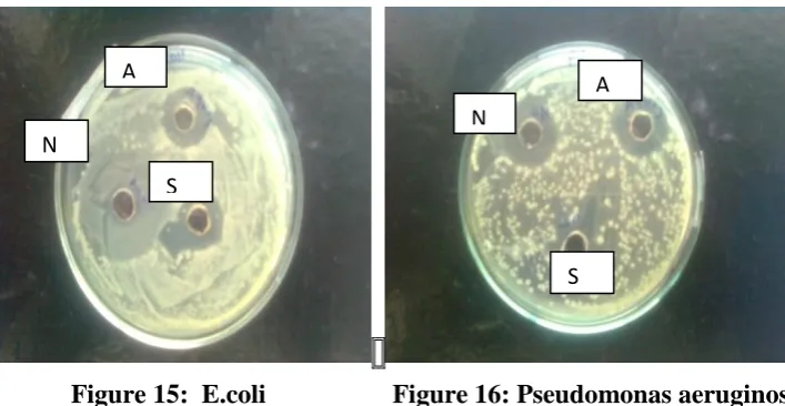
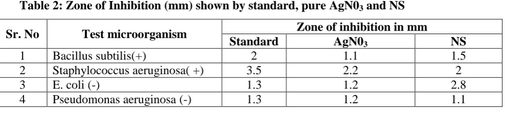
In present investigations, NS were reported to have more antibacterial activity against gram negative bacteria than gram positive bacteria. Antibacterial activity of NS essentially due to the electrostatic attraction between positively charged NS and negatively charged bacterial cells [25]. Owing to fast internalization of NS from the cell wall of bacteria that leads to fast inactivation of protein structure, causing structural changes to cause cell death, may be other reason. Antimicrobial activity may also depend upon the shape/morphology of nanoparticles. However, study was not extended to prove the same. Results are shown in figure 13,14,15,16 and table no.2.
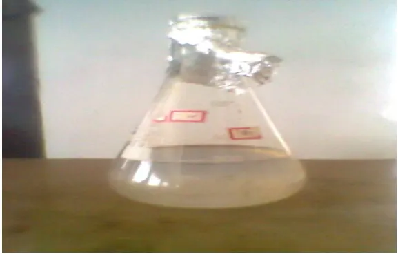
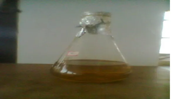
[image:8.595.153.442.501.685.2]Figure 2 (After autoclaving)
Figure 3: Absorption peak of HPMC and cow urine
[image:9.595.153.446.67.236.2]
[image:10.595.157.445.87.250.2]
Figure 5: Absorption peak of gelatin and cow urine
[image:10.595.151.446.523.697.2]Figure 6: Absorption peak of sodium alginate and urine
Figure 7: IR spectra of gelatin
Figure 8: IR spectra of NS
Figure 9: TEM image of NS (Scale-20nm)
[image:11.595.136.499.514.708.2]Figure 11: XRD of Gelatin
Figure12: XRD of Gelatin impregnated NS
Figure 13: Bacillus subtilis Figure 14: Staphylococcus aureus
A S
N
N S
[image:12.595.94.510.523.689.2]Figure 15: E.coli Figure 16: Pseudomonas aeruginosa (A: AgN03S: Benzyl penicillinN: Silver nanoparticles)
Table 1: Solutions used during preliminary screening of polymer. The data represented is for gelatin, similar concentrations were prepared for screening of other polymers
% w/v of gelatin Quantity of Cow Urine % V/V
Quantity of
Silver nitrate Absorbance
A) 0.25 0.5 1 2.35±0.45
0.5 0.5 1 2.20 ±0.32
0.75 0.5 1 2.60 ±0.30
1 0.5 1 2.07 ±0.03
2 0.5 1 2.01 ±0.02
B) 0.25 1 1 2.81 ±0.32
0.5 1 1 2.78 ±0.30
0.75 1 1 2.72 ±0.02
1 1 1 2.71 ±0.01
2 1 1 2.61 ±0.32
C) 0.25 1.5 1 3.99 ±0.33
0.5 1.5 1 3.71 ±0.32
0.75 1.5 1 3.46 ±0.02
1 1.5 1 3.69 ±0.15
2 1.5 1 3.61 ±0.01
D) 0.25 2 1 3.99 ±0.23
0.5 2 1 3.99 ±0.20
0.75 2 1 3.98 ±0.32
1 2 1 3.67 ±0.01
2 2 1 3.61 ±0.01
N
A
S
A N
[image:13.595.121.475.69.252.2]Table 2: Zone of Inhibition (mm) shown by standard, pure AgN03 and NS
Sr. No Test microorganism Zone of inhibition in mm
Standard AgN03 NS
1 Bacillus subtilis(+) 2 1.1 1.5 2 Staphylococcus aeruginosa( +) 3.5 2.2 2
3 E. coli (-) 1.3 1.2 2.8
4 Pseudomonas aeruginosa (-) 1.3 1.2 1.1
7. CONCLUSION
Applications of NS to pharmacy are still under budding phase & needed to be explored. NS can be synthesized by physical, chemical and green methods. Green methods are always better option for eco-continuation. Hence, present investigation had more emphasize on synthesis of NS by green method. NS were successfully prepared by using gelatin & cow urine. Method was found to be very simple, have least wastage, ecological & economical.
Characterization & other evaluation parameters viz. toxicity, antibacterial activity & phase solubility study were found to be satisfactory & being pertinent to drug delivery may give impetus to the pharmabiotech research. Moreover, the raw materials, used in both methods were widely available from renewable sources. These raw materials were simply processed to form NS. Characterization in present investigation confirmed successful formation of NS. Phase solubility study revealed increase in solubility. However, characterization of conjugation of NS with drug molecules is to be studied. Antibacterial activity was also appreciable when compared with standard. NS synthesized were found to be stable in aqueous solution for three months at room temperature. These findings may have great significance in preformulation study in the context to enhance solubility of poorly soluble drugs. Findings in this investigations may be amenable to pharma industry and potentially open several avenues in pharma research, however, it warrant further investigation.
8. ACKNOWLEDGEMENT
The authors are thankful to Prof. Madan Jagtap, Chairman, Gourishankar education Society, Satara for providing infrastructure for this work also without his motivation; help and support this work would have not been completed. We, further extend our deep gratitude to Bombay IIT, Shivaji University, Kolhapur and Government College of Pharmacy, Karad for providing all necessary facilities during work.
REFERENCES
kondagogu (Cochlospermum gossypium)”, Colloids and Surfaces B, Biointefaces, Vol. 83, pp.291-298, 2011.
2. Amarendra DD, Krishna G, “Biosynthesis of silver and gold nanoparticles using
Chenopodium album leaf extract”, Colloids and surfaces A, Physicochem. and
Engineering aspects, Vol. 369,pp.27-33, 2010.
3. Jarald E, Edwin S, Tiwari V, Garg R and Toppo E, “Antioxidant and Antimicrobial Activities of Cow Urine”, Global Journal of Pharmacology, Vol.2,nu.2, pp.20-22, 2008. 4. Krishnamurthi K, Dutta D, Devi SS, Chakrabarti T, “ Protective effect of diatillate and
redistillate of cow,s urine in human polymorphonuclear leukocytes challenged with established genotoxic chemicals” Biomed. Environ. Sci. Vol.17, pp.57-66, 2004.
5. Riddin T, Gericke M, Whiteley CG, “Biological synthesis of platinum nanoparticles: Effect of initial metal concentration”, Enz. Microbial. Technol., Vol.46, pp. 501-505, 2010.
6. Allamneni Y, Potturi P, Nulu R, Ramesh R, Chary Pd, Kalekar A, , “Enhancement of Dissolution of poorly water soluble drug using super disintegrants by kneading method”, Pharmatutor-ART-1273.
7. Han G, Ghosh P, Rotello VM, “Multi-Functional Gold Nanoparticles for Drug Delivery”, In: Warren CWC, editor. Bioapplications of nanoparticles, Landes Bioscience/ Springer science, ISBN. 978-0-387-76712-3, pp.48
8. Karla Chaloupka K, Malam Y, Seifalian AM, “Nanosilver as a new generation of nanoproduct in biomedical application”, Trends in Biotechnology, Vol. 28, nu.11, pp.580-588, 2010.
9. Mirza AZ, “.Prepartion and characterization of doxorubicin functionalized gold nanoparticles”, Europian Journal of Medicinal Chemistry, Vol.46, pp.1857-1860, 2011. 10.Higuchi T, Connors KA, “Phase solubility techniques” In: Reilly CN, editor. Advances in
Analytical Chemistry Instrumentation. Vol. 4, New York, NY: Interscience, pp.117–212, 1965.
11.Pompa PP, Martiradonna L, Dello A, Carbone L, Del LL, “Fluorescence enhancement in colloidal semiconductor nanocrystals by metallic nanopatterns”, Sensors and actuators B, Vol.126, pp.187-192, 2007.
13.Bajpai SK, Mohan YM, Bajpai M, Tankhiwale R, Thomas V, “Synthesis of polymer stabilized silver and gold nanostructures” J Nanosci Nanotechnol, Vol.7, nu.9, pp.2994-3010, 2007.
14.Darroudi M, Ahmad M, Abdullah A, Ibrahim N, “Green synthesis and characterization of gelatin – based and sugar – reduced silver nanoparticles”, International journal of nanomedicine, Vol. 6, pp.569-574, 2011.
15.Richard MC, Victor C, Buford I. Lemon I, Li S, “Synthesis, Characterization, and Applications of Dendrimer-Encapsulated Metal and Semiconductor Nanoparticles”, In:. Feldheim DL, Foss CA, editor, Metal Namoparticles, Synthesis, Characterization and applications, ISBN: 0-8247-0604-8, Marcel Dekker, Inc pp. 262, 2002.
16.Park Y, Hong Y, Weyers A, Kim Y, Linhard R, “Polysaccharide and phytochemicals: a natural reservoir for the green synthesis of gold and silver nanoparticles” ITE nanotechnology, Vol.5, nu.3, pp.69-78,2011.
17.Darroudi M, Mansor BA, Khorsand Z, Reza Z, Mohammad H, “Fabrication and characterization of gelatin stabilized silver nanoparticles under UV-Light”, Int. J. Mol. Sci, Vol.12, nu.9, pp.6346-6356, 2011.
18.Yusheng L, Shimou C, Lei Z, Guozhong W, “Preparation of high-stable silver nanoparticle dispersion by using sodium alginate as a stabilizer under gamma radiation”, Radiation Physics and Chemistry, Vol. 78, pp. 251–255, 2009.
19.Muhamad M, Darroudi M, Zak A, Huang N, Hakimi M, “Green synthesis of colloidal silver nanoparticles by sonochemical method”, Vol.66, pp.117-120, 2012.
20.Hermanson GT, “Preparation of colloidal-gold labelled proteins, In: Bioconjugate techniques, Academic Press, San Diego, pp.593-601, 1996.
21.Patra CR, Bhattacharya R, Mukhopadhyay D, Mukherjee P, “Fabrication of gold nanoparticles for targeted therapy in pancreatic cancer”, Advanced Drug Delivery Reviews, Vol.62, pp.346–361, 2010.
22.Hyatt AD, “Protein A-gold: nonspecific binding and cross-contamination” In: Hayat MA (ed), Colloidal gold: principles, methods and applications, Vol 2. Academic Press, San Diego, pp.19–31,1989.
23.Chaloupka K, Malam Y, Seifalian AM, “ Nanosilver as a new generation of nanoproduct in biomedical applications”, Trends in Biotechnology, Vol.28, No.11, pp.580-588,2010. 24.Bilberg K, Hovgaard M, Besenbacher F, Baatrup E, “In vivo toxicity of silver
25.Venkatpurwar V, Pokharkar V, “Green synthesis of silver nanoparticles using marine polysaccharides: study of in-vitro antibacterial activity”,Materials Letters,2011;65:999-1002.
26.Stani V, Dimitrijevi S, Anti JS, Miodrag M, Bojan J, Ilija B, Ple, Slavica R, “Synthesis, characterization and antimicrobial activity of copper and zinc-doped hydroxyapatite nanopowders”, Applied Surface Science, Vol. 256, pp.6083–6089, 2010.
27.Shiying H, Zhirui G, Yu Z, Song Z, Jing W, Ning G, “Biosynthesis of gold nanoparticles using the bacteria Rhodopseudomonas capsulate”, Materials Letters, 2007;61:3984–3987, 28.MubarakAli D, Thajuddin N, Jeganathan K, Gunasekaran M, “ Plant extract mediated synthesis of silver and gold nanoparticles and its antibacterial activity against clinically isolated pathogens”, Colloids and Surfaces B: Biointerfaces, Vol.85, pp.360–365, 2011. 29.Kathiresan K, Manivannan S, Nabeel MA, Dhivya B, “Studies on silver nanoparticles
synthesized by a marine fungus, Penicillium fellutanum isolated from coastal mangrove sediment”, Colloids and Surfaces B: Biointerfaces, Vol.71, pp.133–137, 2009.
30.Shahverdi AR, Minaeian S, Hamid RS, Hossein J, Ashraf AN, “ Rapid synthesis of silver nanoparticles using culture supernatants of Enterobacteria: A novel biological approach”, Process Biochemistry, Vol. 42, pp.919–923, 2007.
31.Harekrishna B, Dipak KB, Gobinda P. Sahoo, Priyanka S, Santanu P, Ajay M, “ Green synthesis of silver nanoparticles using seed extract of Jatropha curcas”, Colloids and Surfaces A: Physicochem. Eng. Aspects, Vol.348, pp.212–216, 2009.
32.Virender KS, Ria A, Yngard, Y, Lin, “Silver nanoparticles: Green synthesis and their antimicrobial activities” Advances in Colloid and Interface Science,145,83–96, 2009. 33.Feldheim DL, Colby AF, “Overview”, In. Metal Namoparticles, Synthesis,
Characterization and applications, ISBN: 0-8247-0604-8, Marcel Dekker, Inc, pp.1, 2002. 34.Mahendra R, Alka Y, Aniket G, “Silver nanoparticles as a new generation of
antimicrobials”, Biotechnology Advances, Vol. 27, pp.76–83, 2009.
35.Halim ESA, Salem S, Al-Deyaba, “Utilization of hydroxypropyl cellulose for green and efficient synthesis of silver Nanoparticles”, Carbohydrate Polymers,2011 :86; 1615–1622. 36.Vigneshwaran N, Nachane RP, Balasubramanya RH, Varadarajan PV, “ A novel one-pot „green‟ synthesis of stable silver nanoparticles using soluble starch”, Carbohydrate Research, Vol.341, pp.2012–2018, 2006.