Dependent Bidirectional Modulation of Motor Cortex
Output
Graphical Abstract
Highlights
d
During movement, reduced V
mvariance lowers spike
probability in L5B output neurons
d
Noradrenaline mediates a tonic depolarization in L5B
enhneurons during movement
d
Noradrenaline selectively enhances signal-to-baseline ratio
of L5B
enhneurons
d
Blocking noradrenaline receptors in M1 reduces contralateral
forepaw motor coordination
Authors
Julia Schiemann, Paolo Puggioni, ...,
Mark C.W. van Rossum, Ian Duguid
Correspondence
[email protected]
In Brief
Schiemann et al. show that, in mouse
motor cortex, layer 5B pyramidal neuron
firing rates are suppressed or enhanced
during movement due to a global
reduction in membrane potential
variability and coincident
noradrenaline-mediated depolarization in a
subpopulation of neurons, respectively.
Blocking noradrenergic input to M1
impairs motor coordination.
Schiemann et al., 2015, Cell Reports11, 1–12 May 26, 2015ª2015 The Authors
Cellular Mechanisms Underlying Behavioral
State-Dependent Bidirectional Modulation
of Motor Cortex Output
Julia Schiemann,1,4Paolo Puggioni,1,2,3,4Joshua Dacre,1Miha Pelko,2,3Aleksander Domanski,1Mark C.W. van Rossum,2 and Ian Duguid1,*
1Centre for Integrative Physiology and Patrick Wild Centre, University of Edinburgh, Hugh Robson Building, George Square,
Edinburgh EH8 9XD, UK
2Institute for Adaptive and Neural Computation, School of Informatics, University of Edinburgh, Edinburgh EH8 9AB, UK 3Neuroinformatics Doctoral Training Centre, School of Informatics, University of Edinburgh, Edinburgh EH8 9AB, UK 4Co-first author
*Correspondence:[email protected] http://dx.doi.org/10.1016/j.celrep.2015.04.042
This is an open access article under the CC BY-NC-ND license (http://creativecommons.org/licenses/by-nc-nd/4.0/).
SUMMARY
Neuronal activity in primary motor cortex (M1)
corre-lates with behavioral state, but the cellular
mecha-nisms underpinning behavioral state-dependent
modulation of M1 output remain largely unresolved.
Here, we performed in vivo patch-clamp recordings
from layer 5B (L5B) pyramidal neurons in awake
mice during quiet wakefulness and self-paced,
voluntary movement. We show that L5B output
neurons display bidirectional (i.e., enhanced or
sup-pressed) firing rate changes during movement,
medi-ated via two opposing subthreshold mechanisms: (1)
a global decrease in membrane potential variability
that reduced L5B firing rates (L5B
suppressedneurons),
and (2) a coincident noradrenaline-mediated
in-crease in excitatory drive to a subpopulation of L5B
neurons (L5B
enhancedneurons) that elevated firing
rates. Blocking noradrenergic receptors in forelimb
M1 abolished the bidirectional modulation of M1
output during movement and selectively impaired
contralateral forelimb motor coordination. Together,
our results provide a mechanism for how
noradren-ergic neuromodulation and network-driven input
changes bidirectionally modulate M1 output during
motor behavior.
INTRODUCTION
Neuronal activity in layer 5 (L5) of primary motor cortex (M1) cor-relates with rhythmic voluntary movements (Armstrong and Drew, 1984a, 1984b). During walking or running, pyramidal neu-rons display changes in firing rate that reflect periods of coordi-nated muscle activity (Armstrong and Drew, 1984a; Beloozerova et al., 2003). Although spontaneous locomotor activity can be controlled by central pattern generators (CPGs) in the spinal
cord (Forssberg et al., 1980; Grillner, 1981; Grillner and Zangger, 1979), descending motor commands from M1 are integrated with ongoing rhythmic spinal cord signals and sensory input from the periphery to initiate, adjust, and maintain locomotor function (Armstrong and Drew, 1984a; Beloozerova et al., 2003; Orlovsky, 1972; Ueno and Yamashita, 2011). In lower mammals, such as cats, rabbits, and mice, discrete subpopula-tions of L5 output neurons display enhanced or suppressed (i.e., bidirectional) firing rate changes during locomotion (Armstrong and Drew, 1984a; Beloozerova et al., 2003; Costa et al., 2004). In rodents, these changes can be either abrupt, sustained changes—so-called on-off responses—or gradual frequency changes linked to the velocity of running (Costa et al., 2004). Although we are now beginning to understand how patterns of motor cortex activity relate to changes in behavioral state in rodents (i.e., quiet wakefulness to movement), the cellular mech-anisms underpinning bidirectional modulation of M1 output dur-ing self-paced movement remain largely unresolved.
Several mechanisms could underlie the bidirectional modula-tion of M1 output, such as a change in cortical state-dependent network-driven input structure, intracortical or long-range excit-atory input, and/or neuromodulation. During quiet wakefulness or slow-wave sleep, cortical networks remain in a synchronized state that consists of slow, large-amplitude oscillations in neuronal population activity (Cowan and Wilson, 1994; Steriade et al., 1993c). During active behavior, cortical networks enter an activated state characterized by a reduction in slow oscillations and, in some cases, an increase in higher frequency activity (Steriade et al., 1993b; Timofeev et al., 2001). This change profoundly alters the subthreshold Vm dynamics and spike
A
C D E
B
F
I J K
G H
behavior. Anatomical and functional connectivity mapping have shown the presence of a strong top-down laminar organization of local excitatory microcircuits in M1, with feedforward projec-tions from layer 2/3 (L2/3) targeting multiple classes of projection neurons in L5 (Kaneko et al., 1994; Weiler et al., 2008). Given that L2/3 pyramidal neurons can display dense clustered activity dur-ing head-restrained locomotion in mice (Dombeck et al., 2009), changes in descending excitation from upper-layer pyramidal neurons could be a mechanism for generating bidirectional mod-ulation of M1 output. Alternatively, neuromodulators are impor-tant for cortical processing, with noradrenaline and acetylcholine release being associated with changes in arousal, vigilance, and behavioral state (Berridge and Waterhouse, 2003; Carter et al., 2010; Castro-Alamancos and Gulati, 2014; Constantinople and Bruno, 2011; Eggermann et al., 2014; Fu et al., 2014; Polack et al., 2013; Steriade et al., 1993a). Thus, how local, long-range, and neuromodulatory inputs regulate L5 pyramidal neuron Vm
dynamics during changes in behavioral state remains to be fully established.
Here we combined in vivo patch-clamp recordings in awake mice with selective pharmacology to investigate the cellular mechanisms underpinning behavioral state-dependent modula-tion of motor cortex output. We found that changing behavioral state, from quiet wakefulness to movement, bidirectionally modulated (i.e., enhanced or suppressed) M1 output via two opposing subthreshold mechanisms: (1) a global decrease in network-driven, slow, large-amplitude Vmfluctuations, which
reduced Vmvariability, spike probability, and firing rates in L5B
pyramidal neurons (L5Bsuppressedneurons); and (2) a coincident
increase in excitatory drive to a subpopulation of L5B neurons (L5Benhanced), which depolarized mean Vmand enhanced firing
rates. We found that the movement-related tonic depolarization in L5Benh neurons was dependent on the interplay between
ascending motor thalamic input, which maintained Vm near
threshold, and noradrenergic input from the locus coeruleus (LC). The behavioral state-dependent release of noradrenaline increased the signal-to-baseline ratio (SBR) for movement-evoked responses in L5Benh neurons. Selectively blocking
noradrenergic input in the forelimb region of M1 significantly reduced motor coordination in the contralateral forelimb during motor behavior. Thus, our findings provide a mechanism for
how noradrenergic neuromodulation and network-driven input changes bidirectionally modulate M1 output during self-paced voluntary movement.
RESULTS
Membrane Potential Dynamics of L5B Pyramidal Neurons during Self-Paced, Voluntary Movement To investigate the cellular mechanisms underpinning behav-ioral state-dependent modulation of M1 output, we obtained whole-cell patch-clamp recordings from L5B pyramidal neurons (forelimb motor cortex, 620–880mm from the pial surface; see
Experimental Procedures; n = 45 neurons) during quiet wake-fulness and self-paced, voluntary movements (i.e., walking, running, or grooming on a single-axis, cylindrical treadmill; Fig-ure 1A). During periods of quiet wakefulness, all L5B pyramidal neurons displayed large-amplitude Vm fluctuations (Vm SD =
3.8± 0.2 mV) and a relatively depolarized average Vm(Vm=
51.1±0.8 mV). The interplay among mean Vm, distance from
threshold, and Vmvariability resulted in moderate basal firing
rates (5.7±0.6 Hz, range: 0.0–15.9 Hz;Figures 1B–1K andS1). During switches in behavioral state (i.e., quiet wakefulness to movement), characterized by a low-amplitude, high-frequency local field potential signal in L5B (Figure 1A), the vast majority of L5B pyramidal neurons (90%) displayed significant modula-tion of their basal firing rates. To funcmodula-tionally classify individual neurons, we compared the variability in quiet wakefulness firing rate with the average firing rate during self-paced movement (seeExperimental Procedures). If the average movement-related firing rate was lower than the first percentile of the distribution of firing rate changes during quiet wakefulness, neurons were clas-sified as suppressed (L5Bsupp, n = 17;Figures 1C and 1F;Table S1), while neurons that displayed an average movement-related firing rate above the 99thpercentile were classified as enhanced (L5Benh, n = 24;Figures 1E and 1H;Table S1). A small proportion
of L5B neurons (n = 4/45) did not significantly change their firing rates during movement and were classified as non-re-sponding neurons (L5Bn-r;Figures 1D and 1G). The proportion
of L5 pyramidal neurons in which spike frequency decreased (38%), increased (53%), or did not change (9%) during move-ment was consistent with previous reports (Beloozerova et al.,
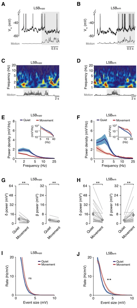
Figure 1. Membrane Potential Dynamics of L5B Pyramidal Neurons in M1 during Self-Paced, Voluntary Movement
(A) Patch-clamp recording configuration in head-fixed mice mounted on a single-axis, cylindrical treadmill. Local field potential (LFP) recordings (black traces, n = 2 mice) of L5B activity and moderate speed (60 frames/s) digital imaging were used to confirm changes in behavioral state (quiet wakefulness to movement) and to calculate motion index (gray traces).
(B) Representative voltage traces from two L5B pyramidal neurons that displayed either a decrease (top) or increase (bottom) in firing rate during movement (light gray shading). The motion index (dark gray) defines the magnitude and duration of each forelimb movement. In this figure and all subsequent figures, action potentials have been truncated to highlight subthreshold Vmchanges during movement.
(C–E) Representative change in firing rate probability distributions during quiet wakefulness (blue) and movement (gray) in L5Bsuppressed(C), L5Bnon-responding(D),
and L5Benhanced(E) neurons. Gray dotted lines represent the 1 st
(left) and 99th(right) percentiles. Solid colored lines represent the average firing rate change in L5Bsuppressed(yellow), L5Bnon-responding(black), and L5Benhanced(purple) neurons during movement.
(F–H) Average firing rate during quiet wakefulness and movement in L5Bsuppressed(F, n = 17), L5Bnon-responding(G, n = 4), and L5Benhanced(H, n = 24) neurons.
Filled circles represent data from individual neurons. Insets depict the average firing rate±SEM during quiet wakefulness (Q) and movement (M). **p < 0.01; ns, non-significant.
(I–K) Average Vm(left-hand plot) and VmSD (right-hand plot) in L5Bsuppressed(I, n = 17), L5Bnon-responding(J, n = 4), and L5Benhanced(K, n = 24) neurons during
quiet wakefulness and movement. Solid gray lines represent data from individual neurons while open symbols represent mean±SEM. *p < 0.05, **p < 0.01; ns, non-significant.
2003; Costa et al., 2004). Moreover, the functional classification of individual neurons remained consistent during repeated bouts of movement and was not dependent on the type of motor behavior being executed (Figure S1). To further demonstrate the coexistence of functionally distinct subpopulations of L5B pyramidal neurons in M1, we performed multiple recordings from the same mouse and identified L5Benh, L5Bsupp, and
L5Bn-rpyramidal neurons during the execution of similar forelimb
movements (i.e., repeated forepaw swing/stance cycles, n = 8 recordings from three mice; L5Bsupp/L5Benh/L5Bn-rratio: 4:3:1,
note similar ratio of functionally classified neurons when compared to the population data inFigure 1;Figure S1).
We next investigated the subthreshold mechanisms underpin-ning bidirectional modulation of M1 output. During movement, L5Bsuppneurons displayed1 mV hyperpolarization in mean Vm
(p = 2310 2) and reduced V
mvariability (VmSD quiet = 3.5±
0.2 mV, VmSD movement = 2.5±0.1 mV, p = 3310 4), which
lowered the probability of reaching threshold and reduced overall firing rates (quiet 6.4±1.0 Hz, movement 2.8±0.6 Hz, p = 3310 4;Figures 1F and 1I). In L5Benhneurons, movement
also reduced Vmvariability (VmSD quiet = 4.1±0.3 mV, VmSD
movement = 3.2±0.2 mV, p = 2310 3), but this was counteracted by a depolarization in average Vm(quiet 52.4±1.1 mV,
move-ment 47.9±1.0 mV, p = 2310 6), which significantly increased
spike probability and firing rates (quiet 5.7±0.8 Hz, movement 12.9±1.5 Hz, p = 2310 5;Figures 1H and 1K). Moreover,
move-ment-related firing rate changes strongly correlated with the level of Vmdepolarization in individual L5Benhneurons (Figure S2). By
contrast, Vmdynamics and firing rates of L5Bn-rneurons were
not affected by the transition from quiet wakefulness to movement (Figures 1G and 1J). Interestingly, the functional classification of L5B pyramidal neurons (L5Bsuppversus L5Benh) was not
depen-dent on their basic electrophysiological properties (Table S1) or the projection-class identity of individual neurons based on retro-grade tracing and selective expression of the transcription factors CTIP2 (thick-tufted pyramidal tract [PT]-type neurons) and SATB2 (thin-tufted intratelencephalic [IT]-type neurons; Leone et al., 2008;Figures S3andS4;Table S2). Together, our results suggest that movement-related modulation of L5Benhfiring rates is
primar-ily mediated by a tonic depolarization in Vm, while reduced firing
rates in L5Bsuppneurons result from a moderate hyperpolarization
and significant reduction in Vmvariance.
L5B Input-Output Transformations during Voluntary Movement
Behavioral state-dependent changes in Vm dynamics can
pro-foundly affect the integrative mode and output firing patterns of neocortical neurons. What effects do movement-related changes in Vmdynamics have on input-output transformations in M1 L5B
pyramidal neurons? In principle, both changes in Vm SD and
mean can influence the responsiveness and firing dynamics of a neuron (Chance et al., 2002; H^o and Destexhe, 2000). To test this, we performed current injection experiments (i.e., somatic in-jection of excitatory postsynaptic current [EPSC]-like waveforms) in a subset of L5Bsuppand L5Benhneurons in vivo (Figures 2A
and 2B;Supplemental Experimental Procedures) and measured the spike probability during quiet wakefulness and voluntary movement. Although current injection at the soma disregards dendritic non-linearities, synaptic properties, and locations, it provides a robust measure to assess the relationship between synaptic conductances arriving at the soma and spike output probability during behavior. During movement L5Bsuppneurons,
which experience a decrease in VmSD with relatively little change
in mean Vm(Figure 1), displayed a 2-fold reduction in spike
prob-ability (DSpike probability = 0.6±0.1, n = 5;Figures 2C and 2E). By contrast, L5Benhneurons, which experience a decrease in Vm
SD and an increase in mean Vm(Figure 1), displayed a 2-fold
increase in spike probability (DSpike probability = 1.7 ±0.4, n = 6;Figures 2D and 2F). Although both L5B subpopulations dis-played moderate changes in input resistance during movement, they did not significantly differ from quiet wakefulness (n = 5 and 5, respectively, p = 0.32;Figure S2).
Changes in L5B Input Structure during Movement To further investigate the mechanisms underpinning L5Bsuppand
L5Benhneuron Vmdynamics, we explored changes in Vmspectral
components before and after movement onset. During quiet wakefulness, we observed slow (1.5–4 Hz,dfrequency band), large-amplitude Vm fluctuations in all L5B pyramidal neurons
A
C
E F
[image:5.603.62.295.97.350.2]D B
Figure 2. Input-Output Transformations in L5Bsupp and L5Benh
Pyramidal Neurons during Movement
(A and B) Representative voltage traces (upper trace, black) during somatic EPSC-like current injections in vivo (lower trace, dark gray) in L5Bsupp(A) and
L5Benh(B) pyramidal neurons during quiet wakefulness and movement (light
gray shading).
(C and D) Input-output transformations in L5Bsupp(C, n = 5) and L5Benh
(D, n = 6) neurons recorded in vivo during quiet wakefulness (blue) and movement (red). Symbols represent mean±SD; solid lines are fits to a trun-cated error function.
(E and F) Mean change in spike probability for L5Bsupp(E, n = 5) and L5Benh
(F, n = 6) neurons. Filled symbols represent data from individual neurons and black open squares represent mean±SD.
(Figures 3A–3D), which were suppressed during movement (L5Bsuppquiet 7.8±1.3 mV2, movement 3.6±0.5 mV2, n = 17,
p = 2 310 3; L5B
enhquiet 16.4±3.1 mV2, movement 6.2±
1.2 mV2, n = 24, p = 1310 4;Figures 3A–3H). The reduction indpower led to reduced VmSD, which together with a moderate
hyperpolarization (1 mV) could account for the reduction in spike probability observed in L5Bsupppyramidal neurons during
movement (Figures 1andS2). In L5Benhneurons, the
suppres-sion of slow Vmfluctuations was counteracted by an increase
in power (12–30 Hz) in thebfrequency band (12–30 Hz; quiet 3.0±0.4 mV2, movement 7.4±1.4 mV2, n = 24, p = 3310 5;
Figures 3F and 3H). The magnitude of increasedbpower dis-played a strong positive correlation with the magnitude of Vm
depolarization in individual L5Benhneurons (Figure S2),
suggest-ing this could be the source of the increased excitatory drive. To examine this further, we developed an event detection algorithm to estimate the level of excitatory input during quiet wakefulness and movement. Due to the high frequency of afferent input (estimated range: 5–15 kHz, data not shown), we were unable to isolate single excitatory postsynaptic potentials (EPSPs). However, we could reliably detect compound synaptic inputs (R1 mV) occurring in a time window (5 ms) shorter than the average membrane time constant (8.2±0.7 ms, n = 10; Fig-ure S2). The detection threshold corresponded to twice the size of the average unitary synaptic response measured in L5 pyrami-dal neurons in vitro (Deuchars et al., 1994; Reyes and Sakmann, 1999). Events that occurred within ±10 ms of a spike were excluded from the analysis. During quiet wakefulness, we detected fast-rising compound EPSPs (range: 1–9.7 mV) with similar rates in both L5Bsuppand L5Benhpyramidal neurons ( Fig-ures 3I and 3J), indicating both subpopulations of neurons receive a comparable level of excitatory drive. During move-ment, the rate of compound events in L5Bsupp neurons was
not affected (n = 17; Figure 3I), whereas L5Benhneurons
[image:6.603.54.287.100.624.2]dis-played a significant increase in compound EPSP rate (n = 24;
Figure 3J). Remarkably, we did not detect any compound events with amplitudes greater than 9.4 mV, even though neurons spent approximately 50% of the time >10 mV from threshold. Thus, L5Benh neurons appear to preferentially receive a net
increase in excitatory drive during movement, which enhances the firing rate by depolarizing mean Vm and increasing spike
probability.
Effects of Local and Long-Range Input to L5B Pyramidal Neurons during Self-Paced Movement
To investigate the possible source(s) of the increased excitatory drive to L5Benhneurons, we examined the activity of local and
long-range inputs from L2/3 and motor thalamus, respectively.
A
C
E
G
I J
H F D B
Figure 3. Movement Reduces Slow, Large-Amplitude Vm
Fluctua-tions but Increases Excitatory Drive in L5BenhNeurons
(A and B) Representative high-time resolution voltage traces for L5Bsupp(A) and
L5Benh(B) neurons during quiet wakefulness and movement (gray shading).
(C and D) Low-time resolution wavelet spectrograms for L5Bsupp(C) and
L5Benh(D) neurons during quiet wakefulness and movement. Representative
examples correspond to neurons shown in (A and B).
(E and F) Average Vmpower density for L5Bsupp(E, n = 17) and L5Benh(F, n =
24) pyramidal neurons during quiet wakefulness (blue) and movement (red). Data represent mean±SD. Insets show average Vmpower density between 5
and 40 Hz.
(G and H) Average Vmpower ind(1.5–4 Hz) andb(12–30 Hz) frequency bands
in L5Bsupp(G, n = 17) and L5Benh(H, n = 24) pyramidal neurons during quiet
wakefulness and movement. Gray lines represent data from individual neurons and black symbols represent mean±SEM. **p < 0.01.
(I and J) Average rate density of compound synaptic events in L5Bsupp
(I, n = 17) and L5Benh(J, n = 24) pyramidal neurons during quiet wakefulness
(blue) and movement (red). Data represent mean ± SD. **p < 0.01; ns, non-significant.
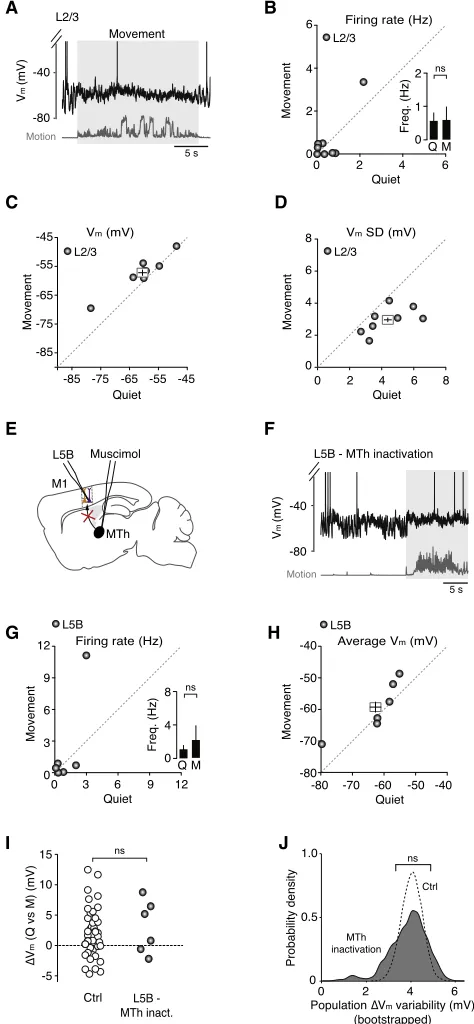
Previous studies have shown that M1 L2/3 neurons can be highly active during head-restrained locomotion on a spherical treadmill (Dombeck et al., 2009) and this descending excitation could potentially influence the activity of output neurons in L5 (Weiler et al., 2008). To test this possibility, we obtained whole-cell patch-clamp recordings from L2/3 pyramidal neurons (180–420 mm from the pial surface; Figure 4A). During quiet wakefulness, L2/3 neurons displayed relatively low firing rates, which were unaffected by the onset of movement (quiet 0.6±
0.3 Hz, movement 0.6±0.4 Hz, n = 8;Figure 4B). Although the average Vmof L2/3 neurons depolarized by4 mV (Figure 4C),
this was counteracted by a significant reduction in VmSD ( Fig-ure 4D), which maintained baseline spike probability and firing rates. Given that our sample of L2/3 neurons displayed low spike rates during both quiet wakefulness and movement, this sug-gests that descending input from L2/3 is unlikely to be the pri-mary source of the increased excitatory drive to L5Benhneurons
in our experimental paradigm (see alsoDombeck et al., 2009and
Discussion).
Given that thalamocortical neuron firing rates vary markedly depending on behavioral state and ventroanterior/ventrolateral (VA/VL) thalamic input to M1 displays bidirectional modulation during simple locomotion (Marlinski et al., 2012), we investigated the role of motor thalamus in regulating M1 output during move-ment. Blocking thalamic input by local infusion of the GABAA
re-ceptor agonist muscimol into the VA/VL complex (Experimental Procedures;Figure 4E) enhanced the amplitude of slow, large-amplitude Vmfluctuations (control VmSD = 3.8±0.2 mV versus
thalamic inactivation SD = 5.1±0.6 mV; n = 45 and n = 6, respec-tively; p = 3.4310 2, Mann-Whitney U test) and produced a
hy-perpolarizing shift in average Vm(control Vm= 51.1±0.8 mV
versus thalamic inactivation Vm= 62.5±3.6 mV; n = 45 and
n = 6, respectively; p = 8.0310 4), which significantly reduced the basal firing rate of L5B neurons compared to control condi-tions (control 5.7 ±0.6 Hz versus thalamic inactivation 1.1±
0.5 Hz; n = 45 and n = 6, respectively; p = 1.1310 3;Figures 4G and 4H; see alsoFigure 1). The hyperpolarization associated with thalamic inactivation increased the distance to threshold
A
C
E
G
I J
H F
[image:7.603.60.297.103.615.2]D B
Figure 4. Effect of Descending L2/3 and Ascending Motor Thalamic Input on L5B Pyramidal Neuron VmDynamics during Quiet
Wake-fulness and Movement
(A) Representative voltage trace shows an L2/3 pyramidal neuron during quiet wakefulness and movement (gray shading).
(B–D) Average firing rate (B), mean Vm(C), and VmSD (D) in L2/3
py-ramidal neurons (gray symbols, n = 8) before and after movement. Filled circles represent data from individual neurons while square symbols represent mean ± SEM. Inset in (B) depicts average L2/3 pyramidal neuron firing rate during quiet wakefulness (Q) and movement (M). ns, non-significant.
(E) Schematic representation shows an L5B pyramidal neuron recording after inactivation of ipsilateral motor thalamus (MTh) by local perfusion of muscimol. (F) Representative voltage trace showing an L5B pyramidal neuron after ipsi-lateral inactivation of motor thalamus.
(G and H) Average firing rate (G) and mean Vm(H) in L5B pyramidal neurons
after motor thalamic inactivation (n = 6). Filled circles represent data from in-dividual neurons while the square symbol in (H) represents the mean±SEM. Inset in (G) depicts the average firing rate of L5B neurons during quiet wake-fulness (Q) and movement (M). ns, non-significant.
(I) Change in average Vm(DVm) during movement in the presence (Ctrl, open
symbols, n = 41) and absence of motor thalamic input (gray symbols, n = 6). Control data (Ctrl) were taken fromFigure 1for comparison. Mann-Whitney U test; ns, non-significant.
(J) Probability density distributions ofDVmvariability across the L5B pyramidal
neuron population (Ctrl and MTh inact.), measured as the SD of theDVm
dis-tributions shown in (I) (PopulationDVmSD) using bootstrap analysis (10,000
bootstrap replicates). Black dashed line represents populationDVmvariability
distribution in control (Ctrl), and gray shading represents population DVm
(data not shown) such that movement-related firing rate changes were abolished (Figure 4G), precluding the functional classifica-tion of L5Bsuppand L5Benhneurons. However, during movement
50% of L5B neurons (n = 3/6) still experienced a 5–10 mV depo-larization in mean Vm(Figure 4I) and increased rate of compound
EPSPs (Figure S5), similar to that observed in L5Benhneurons
un-der control conditions (Figures 1and3). We analyzed this further by plotting theDVmvariability across the L5B pyramidal neuron
population, measured as the SD of theDVmdistributions shown
in Figure 4I (population DVm SD), using bootstrap analysis
(10,000 bootstrap replicates;Figure 4J). We found that motor thalamic inactivation did not affect the populationDVmvariability
in L5B pyramidal neurons compared to control (Figures 4I and 4J), suggesting input from the motor thalamus—either direct or indirect—is essential for maintaining L5B pyramidal neuron Vm
near threshold, but is unlikely to be the main source of the increased excitatory drive.
Noradrenergic Neuromodulation Selectively Enhances Excitatory Drive and Signal-to-Baseline Ratio in L5Benh Neurons
Given that the movement-related increase in excitatory drive and tonic depolarization in L5Benhneurons could not be directly
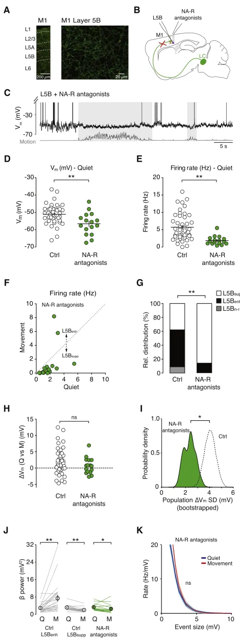
ex-plained by increased excitation from L2/3 or motor thalamus, we next explored the role of noradrenergic neuromodulation, which has been shown to be important during changes in arousal, attention, and behavioral state (Berridge and Water-house, 2003; Carter et al., 2010; Castro-Alamancos and Gulati, 2014; Constantinople and Bruno, 2011; Polack et al., 2013). Se-lective immunohistochemical staining for the noradrenaline transporter (NAT), expressed exclusively in noradrenergic axons (Lorang et al., 1994), revealed dense axonal innervation of all layers in forelimb M1 (Figure 5A). To test the importance of noradrenergic input in regulating L5B pyramidal neuron Vm
dy-namics during movement, we topically applieda1,a2, and b noradrenergic receptor antagonists (1 mM prazosin, 1 mM yohimbine, and 1 mM propranolol, respectively) to the forelimb region of M1 (Figures 5B and 5C). The local infusion of noradren-ergic receptor antagonists via the craniotomy selectively disrup-ted noradrenergic signaling in forelimb M1 (Figure S6), whereas direct manipulation of LC activity, via electrical stimulation or op-togenetics, would have widespread effects across many brain areas and spinal cord circuits. Moreover, topical application was preferred due to the technical limitations of simultaneously pressure ejecting drugs at multiple sites along the entire somato-dendritic length of L5B pyramidal neurons during intracellular recordings.
Blocking noradrenergic receptors reduced the mean Vm
(con-trol 51.1 ±0.8 mV versus noradrenergic receptor blockade 56.6±1.6 mV; p = 4.0310 3) and quiet wakefulness firing rate of L5B pyramidal neurons (control 5.7±0.6 Hz versus norad-renergic receptor blockade 1.9±0.3 Hz; n = 45 and n = 16, respectively; p < 1310 4;Figures 5D and 5E), and significantly
reduced the proportion of L5B neurons that displayed enhanced firing rates during movement (control L5Benh 24/45 neurons
[53.3%] versus noradrenergic receptor blockade L5Benh2/16
neurons [12.5%]; p < 1310 2;Figures 5F and 5G). The change in relative distribution of L5Bsupp/L5Benhneurons could be
ex-plained in part by the moderate hyperpolarization in Vm and
increased distance to threshold during movement (Figure S6). Although noradrenergic receptor blockade did not affect the mean population DVmcompared to control conditions, due to
both distributions being centered around 0 mV (Figure 5H), we did observe a significant decrease inDVmvariability across the
L5B pyramidal neuron population, measured as the SD of the DVmdistributions shown inFigure 5H (populationDVmSD) using
bootstrap analysis (10,000 bootstrap replicates; Figure 5I). Consistent with the idea that noradrenergic signaling underpins a large proportion of the increased excitatory drive to L5Benh
neurons during movement, blocking noradrenergic receptors also abolished the increase in Vm b-band power (Figure 5J)
and rate of compound synaptic events associated with move-ment (Figures 5K andS5).
Given that pre-application of noradrenergic receptor antago-nists precludes the prior identification of L5Benhneurons prior
to receptor blockade, we also performed long-term (40- to 80-min) recordings from identified L5Benhneurons before (Figure 6A)
and after (Figure 6B) receptor block. If noradrenergic neuromo-dulation underpins the Vmdepolarization in L5Benhneurons
dur-ing movement, then blockdur-ing noradrenergic receptors should have a disproportionately larger effect on movement-related firing rates compared to quiet firing rates. Accordingly, we found that receptor blockade resulted in a modest, time-dependent reduction in L5Benhbasal firing rates and a strong suppression
of movement-related firing (Figure 6C). The drug diffusion and time dependency of the antagonist effects in L5B were consis-tent with our dye diffusion mapping results (Figure S6). To assess the extent to which noradrenaline facilitates L5Benhoutput during
movement, we examined the Signal-to-Baseline Ratio (SBR), defined as the ratio of the movement-related spike rate to the spike rate during quiet wakefulness. Blocking noradrenergic neurotransmission significantly reduced the SBR compared to control conditions (sham control SBR: 1.1±0.1, noradrenergic receptor antagonist SBR: 0.3±0.1; p = 6310 3; n = 3 and 3, respectively;Figure 6D).
(data not shown). Together, our results demonstrate that norad-renergic input from the LC is necessary for controlling M1 output and motor coordination during self-paced voluntary movement.
DISCUSSION
In this paper we present three main findings. First, we show that behavioral state-dependent bidirectional modulation of M1 output is governed by two opposing subthreshold mechanisms (1) a global decrease in network-driven, slow, large-amplitude Vmfluctuations, which reduced Vmvariability, spike probability,
and firing rates in L5Bsuppneurons; and (2) a coincident increase
in excitatory drive in a subpopulation of L5B neurons (L5Benh),
which increased spike probability and firing rates. Second, we demonstrate that the movement-related tonic depolarization in L5Benhneurons requires the interplay between ascending input
from the motor thalamus, which maintained Vmnear threshold,
and noradrenergic input from the LC, which enhanced the SBR for movement-evoked responses. Finally, we show that selective A
C
D
F
H
J K
I G E
B Figure 5. Blocking Input from the LC Reduces Behavioral State-Dependent Increase in Excitatory Drive in L5BenhNeurons (A) Noradrenergic axons in the forelimb region of M1 were labeled using an anti-noradrenaline transporter antibody and secondary antibody conjugated to AlexaFluor 488.
(B) Schematic representation of an L5B pyramidal neuron recording after blocking noradrenergic input from the LC.
(C) Representative voltage trace from a L5B pyramidal neuron in the absence of noradrenergic input.
(D and E) Average Vm(D) and firing rate (E) of L5B pyramidal neurons during
quiet wakefulness in the absence (open symbols, n = 45) and presence (green symbols, n = 16) of noradrenergic receptor (NA-R) antagonists. Filled circles represent data from individual neurons, black bars represent mean±SEM. Control data (Ctrl) were taken from the dataset presented in Figure 1for comparison. Mann-Whitney U test, *p < 0.017, **p < 0.003.
(F) Average firing rate of L5B pyramidal neurons in the presence of norad-renergic receptor antagonists (n = 16) during quiet wakefulness and move-ment. Filled circles represent data from individual neurons.
(G) Relative distributions of L5Bsupp, L5Benh, and L5Bn-r neurons in the
absence (Ctrl) and presence (NA-R antagonists, n = 16) of noradrenergic re-ceptor antagonists. Control data (Ctrl) were taken from the dataset presented inFigure 1for comparison. Chi-square test, **p < 0.01.
(H) Change in average Vm(DVm) during movement in the absence (Ctrl, n = 41)
and presence of noradrenergic receptor antagonists (green symbols, n = 16). Control data (Ctrl) were taken fromFigure 1for comparison. Mann-Whitney U test; ns, non-significant.
(I) Probability density distributions ofDVmvariability across the L5B pyramidal
neuron population (Ctrl and NA-R antagonists), measured as the SD of theDVm
distributions shown in (H) (Population DVmSD) using bootstrap analysis
(10,000 bootstrap replicates). Black dashed line represents populationDVm
variability distribution in control (Ctrl), and green shading represents popula-tionDVmvariability distribution following noradrenergic receptor blockade.
Control data (Ctrl) were taken fromFigure 1for comparison. F test, *p < 0.025. (J) Average L5B pyramidal neuron Vmpower in thebfrequency band (12–
30 Hz) during quiet wakefulness and movement in the presence (Ctrl: L5Bsupp,
n = 17 and L5Benh, n = 24) and absence of noradrenergic input (NA-R
antag-onists, n = 16). Solid lines represent data from individual neurons, symbols represent mean± SEM. Control data (Ctrl) were taken fromFigure 3for comparison. *p < 0.05.
(K) Average rate density of compound synaptic events in L5B pyramidal neurons during quiet wakefulness (blue) and movement (red) in the absence of noradrenergic input (n = 16). Compare withFigures 3I and 3J.
[image:9.603.59.294.97.723.2]blockade of noradrenaline signaling in forelimb M1 reduces motor coordination in the contralateral forelimb, resulting in a significant decrease in precise forepaw placements. Together, our findings reveal the subthreshold and circuit mechanisms that regulate behavioral state-dependent bidirec-tional modulation of M1 output during self-paced, voluntary movement.
Behavioral State-Dependent Modulation of Input-Output Transformations in L5
Physiologically relevant changes in Vmvariance or mean have
been shown to profoundly influence neuronal input-output transformations (Chance et al., 2002; H^o and Destexhe, 2000). But, this has never been explored in L5 pyramidal neurons in the awake cortex. Our current injection experiments in vivo demonstrate that changes in VmSD (L5Bsupp) or Vm SD and
mean (L5Benh) have quantitatively similar—but functionally
opposing—effects on spike probability when examined over a behaviorally relevant input amplitude range (1–10 mV). This sim-ilarity arises due to the non-linear relationship between Vmand
firing probability, such that moderate depolarization can pro-duce a non-linear additive increase in the sensitivity of a neuron to small-amplitude inputs, while decreased VmSD produces a
divisive reduction in input sensitivity (Brozovic et al., 2008; Mur-phy and Miller, 2003). The behavioral state-dependent bidirec-tional modulation of neuronal responsiveness in L5B pyramidal neurons (i.e., increased or decreased spike probability) could facilitate the routing of sensorimotor information through specific M1 neuronal assemblies during movement.
Local and Long-Range Inputs to M1 during Self-Paced Voluntary Movement
M1 receives input from a variety of brain areas (e.g., ipsilateral primary and secondary somatosensory cortices, secondary mo-tor cortex, and orbitofrontal cortex), with ascending input from motor thalamus and descending input from L2/3 providing strong feedforward excitation directly to L5B neurons ( Castro-Alamancos and Connors, 1996; Hooks et al., 2013; Weiler et al., 2008). We found that movement did not affect firing rates in our sample of L2/3 pyramidal neurons, suggesting that de-scending excitation from L2/3 may not be the primary source of the tonic depolarization in L5Benhneurons during simple
loco-motion on a cylindrical treadmill. These findings are in direct contrast to a previous study by Dombeck and colleagues, where locomotion on a spherical treadmill resulted in large-scale, clus-tered activity of L2/3 neurons in mouse forelimb M1 (Dombeck et al., 2009). The reason for this discrepancy is unclear. One pos-sibility is that our recordings undersampled L2/3 population ac-tivity; however, if locomotion induced dense activity similar to that observed in Dombeck et al., (2009), we would have ex-pected to observe movement-related firing rate changes in a sig-nificant proportion of our intracellular recordings. Moreover, Dombeck and colleagues did not identify individual neuronal subtypes, so the large-scale activity could be due, in part, to elevated L2/3 interneuron activity. Alternatively, dense L2/3 ac-tivity could result from mice having to balance and oppose the inertial forces of a rotating air-cushioned ball when changing direction. In principle, this could generate a sensorimotor A
C
E F
[image:10.603.53.293.97.510.2]D B
Figure 6. Blocking Noradrenergic Input Reduces the SBR in L5Benh
Neurons and Impairs Contralateral Forepaw Motor Coordination
(A and B) Schematic representation of the experimental design and repre-sentative voltage traces from an L5Benhpyramidal neuron prior to (A) and after
(B, >30 min) topical application of noradrenergic receptor antagonists. Gray shading depicts movement.
(C) Time course shows quiet (blue) and movement-related (red) firing rates in an L5Benhpyramidal neuron before and after noradrenergic receptor blockade
(green bar).
(D) Movement-induced SBR in L5Benhpyramidal neurons before and >30 min
after application of noradrenergic receptor antagonists (green circles, n = 3) or saline (gray circles, n = 3). Square symbols represent mean±SEM. Unpaired t test, **p < 0.01.
(E) Behavioral assessment of forepaw placement precision in head-fixed mice mounted on a single-axis, cylindrical runged treadmill. Video sequences were used to score contralateral and ipsilateral forepaw placements. (F) Percentage of precise contralateral forepaw placements before and 60 min after application of noradrenergic receptor antagonists (green circles, n = 5) or saline (gray circles, n = 3). Square symbols represent mean±SEM. Unpaired t test, **p < 0.01.
mismatch between the rotational direction of the ball and the intended movement trajectory of the mouse, leading to contin-uous sensory feedback to M1. Thus, it will be important for future studies to investigate the extent to which descending L2/3 input contributes to L5B frequency modulation during simple versus complex motor behaviors.
Direct thalamic input to cortical pyramidal neurons can drive output by reducing slow Vm fluctuations, depolarizing mean
Vm, and reducing the distance to threshold (Castro-Alamancos and Connors, 1996; Constantinople and Bruno, 2013; Hirata and Castro-Alamancos, 2010; Poulet et al., 2012). Consistent with previous findings in sensory cortex, we found that inactiva-tion of thalamus (VA/VL region) increased slow, large-amplitude Vm fluctuations, but did not abolish the activated state during
behavior (Hirata and Castro-Alamancos, 2010; Poulet et al., 2012), suggesting thalamic input to M1 is sufficient but not necessary for generating the activated cortical state. However, ascending motor thalamic input—direct or indirect—appears to be necessary for maintaining the average Vmrelatively close
to threshold, providing a mechanism whereby subtle changes in input structure can generate positive or negative changes in M1 output during movement.
Noradrenergic Neuromodulation
We have shown that noradrenaline release during different behavioral states (i.e., quiet wakefulness versus movement) has profound effects on M1 cortical dynamics. Similar to thalamic inactivation, blocking noradrenergic input from the LC reduced basal firing rates by hyperpolarizing mean Vm and
increasing distance to threshold, suggesting tonic input from both the LC and motor thalamus are necessary to generate moderate firing rates in L5B pyramidal neurons during quiet wakefulness. Our finding that noradrenaline generated a tonic depolarization in a selected subpopulation of L5B pyramidal neurons differs from results obtained in superficial layers of sen-sory cortex (Polack et al., 2013), highlighting the importance of understanding the sublayer-specific effects of noradrenaline in the awake cortex. In primary visual cortex (V1), locomotion-dependent noradrenaline release generates a global depolariza-tion of L2/3 pyramidal neurons, which may enhance visual atten-tion by increasing the gain and signal-to-noise ratio of visually evoked responses (Bennett et al., 2013; Polack et al., 2013).
The fact that we also observed a movement-related tonic de-polarization in the majority of M1 L2/3 pyramidal neurons, which was abolished by noradrenergic receptor blockade (Figure S6), suggests that noradrenaline may differentially affect cortical pro-cessing in superficial versus deep-layer pyramidal neurons dur-ing active behavior. Topical application of high concentrations of noradrenergic receptor antagonists could potentially produce off-target effects. However, given that low doses of antagonists affect L2/3 Vmdynamics in the same way as high concentrations,
albeit smaller in magnitude, suggests relatively selective antag-onist effects (Polack et al., 2013). Although noradrenaline ap-pears to underpin the majority of the locomotion-dependent Vm depolarization in V1, cholinergic disinhibition of
somato-statin-containing interneurons is likely to further enhance behav-ioral state-dependent gain modulation (Fu et al., 2014). We did not directly test the role of acetylcholine in our study, but given
its importance in regulating Vmdynamics in other cortical areas
(Constantinople and Bruno, 2011; Eggermann et al., 2014; Fa-vero et al., 2012; Fu et al., 2014; Polack et al., 2013), it will be important for future studies to investigate its role in M1 during motor behavior.
How does noradrenaline generate the tonic depolarization in L5Benh neurons during movement? Previous studies have
shown that noradrenaline modulates voltage-dependent and voltage-independent potassium conductances and hyperpolar-ization-activated cyclic nucleotide-gated (HCN) channels, thus generating a tonic depolarization by reducing the spike after-hy-perpolarization and prolonging the depolarizing effect of excit-atory synaptic inputs (Favero et al., 2012; Sheets et al., 2011; Wang et al., 2007; Wang and McCormick, 1993). This combined with modulation of basal firing rates is thought to alter the signal-to-noise ratio of neuronal responses to synaptic input (Berridge and Waterhouse, 2003). Alternatively, we cannot rule out the possibility that noradrenaline selectively reduces the activity of local GABAergic interneurons, thus releasing L5Benhneurons
from inhibition and generating a depolarization in Vm. Therefore,
identifying the specific expression patterns and subcellular localization ofaand badrenergic receptors in excitatory and inhibitory neurons in M1 will be an important next step in under-standing how noradrenaline exerts its sublayer- and cell-type specific effects.
Functional Implications
What function does behavioral state-dependent bidirectional modulation of L5 output serve? The flexible modulation of L5B output channels (PT type and IT type) provides an important control mechanism to modulate and update activity patterns in downstream cortical and subcortical areas during changes in behavioral state. PT output provides online information about the state of cortical activation to downstream areas involved in motor control. This continuously updating flow of information generates a basic pattern of input to brainstem and spinal cord circuits in order to generate appropriate behavioral responses in accordance with changes in behavioral state. We demonstrate that blocking noradrenergic receptors in forelimb M1 selectively disrupts motor coordination in the contralateral forepaw, thus confirming the importance of noradrenergic neuromodulation and descending M1 output for motor control. Given that output from sensory and non-sensory cortices have overlapping down-stream targets (Hattox and Nelson, 2007; Kita and Kita, 2012), we speculate that our findings might generalize to other cortical output layers and that noradrenergic neuromodulation and network-driven input changes are common mechanisms to bidi-rectionally modulate cortical output during active behavior.
EXPERIMENTAL PROCEDURES
Animals and Surgery
procedures were performed under 1.5% isoflurane anesthesia. After 24-to 48-hr recovery, a craniotomy (3003300mm) was performed and the dura removed above the right forelimb region of M1. Using intracortical microstimu-lation (seeSupplemental Experimental Procedures), the center of M1FLwas
located 0.7 mm rostral and 1.5 mm lateral to bregma. The craniotomy was sealed with (1.5%) agar and Kwik-Cast sealant (WPI) and mice recovered for 2 hr before recording commenced.
Motion Index and Motor Pattern Discrimination
An optical encoder was used to capture movement of the treadmill and loco-motion was defined as periods with speed > 0.01 m/s for more than 2 s. Changes in behavioral state (quiet wakefulness to movement [grooming or locomotion]) were captured using an elevated, front-mounted, moderate-speed (60 frames/s) digital video camera synchronized with each electrophys-iological recording.
In Vivo Electrophysiology and Pharmacology
Mice were habituated to the head restraint and experimental setup for 45–60 min before each recording session. Whole-cell patch-clamp record-ings were obtained from awake head-restrained mice at a depth of 180– 420mm (layer 2/3) or 620–880mm (layer 5B) from the pial surface, using a Multiclamp 700B amplifier (Molecular Devices). The signal was filtered at 10 kHz and acquired at 20 kHz using PClamp 10 software in conjunction with a DigiData 1440 DAC interface (Molecular Devices). No bias current was injected during recordings and the membrane potential was not cor-rected for junction potential. Resting membrane potentials were recorded immediately after attaining the whole-cell configuration (break-in). Series resistances (Rs) ranged from 15 to 40 MUand experiments were terminated
if Rsexceeded 60 MU. Current injection was performed only if Rs< 35 MU.
Patch pipettes (5–7 MU) were filled with internal solution (285–295 mOsm) containing: 135 mM K-gluconate, 4 mM KCl, 10 mM HEPES, 10 mM sodium phosphocreatine, 2 mM MgATP, 2 mM Na2ATP, 0.5 mM Na2GTP, and
2 mg/ml biocytin (pH adjusted to 7.2 with KOH). External solution con-tained: 150 mM NaCl, 2.5 mM KCl, 10 mM HEPES, 1 mM CaCl2, and
1 mM MgCl2(adjusted to pH 7.3 with NaOH).
For inactivation of the motor thalamus, the GABAAreceptor agonist
musci-mol (1 mM muscimusci-mol hydrobromide, Sigma-Aldrich) was dissolved in external solution, and 100 nl was stereotaxically injected into the right VA/VL complex ( 1 mm caudal, 1.1 mm lateral to bregma, and 3.2 mm below the pial surface). Whole-cell patch-clamp recordings of L5B pyramidal neurons were carried out approximately 2 hr after muscimol injection.
To block noradrenergic receptors, a mixture ofa1,a2, andbnoradrenergic receptor antagonists (1 mM prazosin, yohimbine, and propranolol; Sigma-Al-drich) in external solution (adjusted to pH 7.3) was applied topically to the craniotomy and recordings were performed >40 min after antagonist application.
Functional Classification of Recorded Neurons
For each L5B cell, we (1) divided quiet periods into 1-s epochs; (2) randomly assigned each epoch into two groups, quiet 1 (q1) and quiet 2 (q2); and (3) calculated the firing rate difference between q1 and q2. We repeated steps (1) to (3) 10,000 times for each cell to obtain the distribution probability of the difference of firing rate in q1 and q2 (seeFigures 1C–1E). If during move-ment the firing rate change was higher than the 99th
percentile or lower than the 1st
percentile, we classified the neuron as enhanced or suppressed, respectively. If the firing rate change fell within the first and 99thpercentiles, the cell was classified as non-responding.
Statistical Analyses
Summary data are expressed as mean±SEM unless otherwise stated. Statis-tical significance was determined using Wilcoxon signed-rank tests (paired data) and rank-sum tests (unpaired data) unless otherwise stated. Wilcoxon signed-rank tests on the areas underlying the rate-density curves were used
in Figures 3I and 3J. The relative distribution of functional phenotypes
(L5Bsupp, L5Benh, and L5Bn-r) was analyzed using Pearson chi-square test
sta-tistics (based on 106
permutations). Statistical significance in populationDVm
variability (Figures 4J and5I) was determined using two-sample F tests. To
de-pict the variance of the underlying populations, 10,000 bootstrap samples (random samples with replacement) of each population were taken, and a probability density function of the variances of the bootstrap samples was plotted. For statistical tests, p < 0.05 was considered significant (*p < 0.05 and **p < 0.01). For repeated statistical comparisons with the control dataset, resulting p values were compared to Bonferroni-corrected alpha levels and stated accordingly.
SUPPLEMENTAL INFORMATION
Supplemental Information includes Supplemental Experimental Procedures, six figures, and two tables and can be found with this article online athttp:// dx.doi.org/10.1016/j.celrep.2015.04.042.
AUTHOR CONTRIBUTIONS
J.S., P.P., M.C.W.v.R., and I.D. designed the experiments. All experiments were carried out by J.S., P.P., and J.D. with help from I.D. Analysis was per-formed by P.P., J.S., M.P., and J.D. A.D. wrote MATLAB scripts to extract fore-paw motion statistics. Neuronal reconstructions were carried out by J.S., P.P., J.D., and I.D. I.D., P.P., and J.S. wrote the manuscript, and all authors contrib-uted to discussion and interpretation of the results.
ACKNOWLEDGMENTS
We are grateful to M. Nolan, M. London, P. Magill, K. Harris, T. Branco, L. Acerbi, M. Ludwig, N. Rochefort, and members of the I.D. lab for helpful discussions and for comments on the manuscript; M. Cowper-Coles for neuronal reconstructions; and T. Theil for advice on CTIP2/SATB2 immunohis-tochemistry. Confocal microscopy was performed in the IMPACT Imaging Facility at the University of Edinburgh. This work was supported by grants from the Engineering and Physical Sciences Research Council, Eurospin Erasmus Mundus Program, Leopoldina fellowship program (LPDS 2012-11 to J.S.), the Shirley Foundation, Medical Research Council (MR/K014137/1, awarded to P. Kind), and a Wellcome Trust Career Development fellowship (WT086602MF) to I.D.
Received: June 11, 2014 Revised: March 16, 2015 Accepted: April 20, 2015 Published: May 14, 2015
REFERENCES
Armstrong, D.M., and Drew, T. (1984a). Discharges of pyramidal tract and other motor cortical neurones during locomotion in the cat. J. Physiol.346, 471–495.
Armstrong, D.M., and Drew, T. (1984b). Locomotor-related neuronal dis-charges in cat motor cortex compared with peripheral receptive fields and
evoked movements. J. Physiol.346, 497–517.
Beloozerova, I.N., Sirota, M.G., and Swadlow, H.A. (2003). Activity of different
classes of neurons of the motor cortex during locomotion. J. Neurosci.23,
1087–1097.
Beloozerova, I.N., Farrell, B.J., Sirota, M.G., and Prilutsky, B.I. (2010). Differ-ences in movement mechanics, electromyographic, and motor cortex activity
between accurate and nonaccurate stepping. J. Neurophysiol.103, 2285–
2300.
Bennett, C., Arroyo, S., and Hestrin, S. (2013). Subthreshold mechanisms
underlying state-dependent modulation of visual responses. Neuron 80,
350–357.
Berridge, C.W., and Waterhouse, B.D. (2003). The locus coeruleus-noradren-ergic system: modulation of behavioral state and state-dependent cognitive
processes. Brain Res. Brain Res. Rev.42, 33–84.
Brozovic, M., Abbott, L.F., and Andersen, R.A. (2008). Mechanism of gain
modulation at single neuron and network levels. J. Comput. Neurosci.25,
Carter, M.E., Yizhar, O., Chikahisa, S., Nguyen, H., Adamantidis, A., Nishino, S., Deisseroth, K., and de Lecea, L. (2010). Tuning arousal with optogenetic
modulation of locus coeruleus neurons. Nat. Neurosci.13, 1526–1533.
Castro-Alamancos, M.A. (2004). Absence of rapid sensory adaptation in
neocortex during information processing states. Neuron41, 455–464.
Castro-Alamancos, M.A., and Connors, B.W. (1996). Short-term plasticity of a thalamocortical pathway dynamically modulated by behavioral state. Science 272, 274–277.
Castro-Alamancos, M.A., and Gulati, T. (2014). Neuromodulators produce distinct activated states in neocortex. J. Neurosci.34, 12353–12367.
Castro-Alamancos, M.A., and Oldford, E. (2002). Cortical sensory suppression during arousal is due to the activity-dependent depression of thalamocortical synapses. J. Physiol.541, 319–331.
Chance, F.S., Abbott, L.F., and Reyes, A.D. (2002). Gain modulation from
background synaptic input. Neuron35, 773–782.
Constantinople, C.M., and Bruno, R.M. (2011). Effects and mechanisms of
wakefulness on local cortical networks. Neuron69, 1061–1068.
Constantinople, C.M., and Bruno, R.M. (2013). Deep cortical layers are
acti-vated directly by thalamus. Science340, 1591–1594.
Costa, R.M., Cohen, D., and Nicolelis, M.A. (2004). Differential corticostriatal
plasticity during fast and slow motor skill learning in mice. Curr. Biol.14,
1124–1134.
Cowan, R.L., and Wilson, C.J. (1994). Spontaneous firing patterns and axonal projections of single corticostriatal neurons in the rat medial agranular cortex. J. Neurophysiol.71, 17–32.
Crochet, S., and Petersen, C.C. (2006). Correlating whisker behavior with membrane potential in barrel cortex of awake mice. Nat. Neurosci.9, 608–610.
Deuchars, J., West, D.C., and Thomson, A.M. (1994). Relationships between morphology and physiology of pyramid-pyramid single axon connections in rat neocortexin vitro. J. Physiol.478, 423–435.
Dombeck, D.A., Graziano, M.S., and Tank, D.W. (2009). Functional clustering of neurons in motor cortex determined by cellular resolution imaging in awake
behaving mice. J. Neurosci.29, 13751–13760.
Eggermann, E., Kremer, Y., Crochet, S., and Petersen, C.C. (2014). Cholinergic
signals in mouse barrel cortex during active whisker sensing. Cell Rep.9,
1654–1660.
Favero, M., Varghese, G., and Castro-Alamancos, M.A. (2012). The state of
somatosensory cortex during neuromodulation. J. Neurophysiol.108, 1010–
1024.
Forssberg, H., Grillner, S., Halbertsma, J., and Rossignol, S. (1980). The loco-motion of the low spinal cat. II. Interlimb coordination. Acta Physiol. Scand. 108, 283–295.
Fu, Y., Tucciarone, J.M., Espinosa, J.S., Sheng, N., Darcy, D.P., Nicoll, R.A., Huang, Z.J., and Stryker, M.P. (2014). A cortical circuit for gain control by behavioral state. Cell156, 1139–1152.
Grillner, S. (1981). Control of locomotion in bipeds, tetrapods and fish. In
Hand-book of Physiology (Bethesda: American Physiology Society), pp. 1179–1236.
Grillner, S., and Zangger, P. (1979). On the central generation of locomotion in the low spinal cat. Exp. Brain Res.34, 241–261.
Hattox, A.M., and Nelson, S.B. (2007). Layer V neurons in mouse cortex projecting to different targets have distinct physiological properties.
J. Neurophysiol.98, 3330–3340.
Hirata, A., and Castro-Alamancos, M.A. (2010). Neocortex network activation
and deactivation states controlled by the thalamus. J. Neurophysiol.103,
1147–1157.
Ho, N., and Destexhe, A. (2000). Synaptic background activity enhances^
the responsiveness of neocortical pyramidal neurons. J. Neurophysiol.84,
1488–1496.
Hooks, B.M., Mao, T., Gutnisky, D.A., Yamawaki, N., Svoboda, K., and Shep-herd, G.M. (2013). Organization of cortical and thalamic input to pyramidal
neurons in mouse motor cortex. J. Neurosci.33, 748–760.
Kaneko, T., Caria, M.A., and Asanuma, H. (1994). Information processing within the motor cortex. II. Intracortical connections between neurons receiving somatosensory cortical input and motor output neurons of the
cor-tex. J. Comp. Neurol.345, 172–184.
Kita, T., and Kita, H. (2012). The subthalamic nucleus is one of multiple inner-vation sites for long-range corticofugal axons: a single-axon tracing study in the rat. J. Neurosci.32, 5990–5999.
Leone, D.P., Srinivasan, K., Chen, B., Alcamo, E., and McConnell, S.K. (2008). The determination of projection neuron identity in the developing cerebral cor-tex. Curr. Opin. Neurobiol.18, 28–35.
Lorang, D., Amara, S.G., and Simerly, R.B. (1994). Cell-type-specific expres-sion of catecholamine transporters in the rat brain. J. Neurosci.14, 4903–4914.
Marlinski, V., Nilaweera, W.U., Zelenin, P.V., Sirota, M.G., and Beloozerova, I.N. (2012). Signals from the ventrolateral thalamus to the motor cortex during
locomotion. J. Neurophysiol.107, 455–472.
Murphy, B.K., and Miller, K.D. (2003). Multiplicative gain changes are induced by excitation or inhibition alone. J. Neurosci.23, 10040–10051.
Orlovsky, G.N. (1972). The effect of different descending systems on flexor and
extensor activity during locomotion. Brain Res.40, 359–371.
Polack, P.O., Friedman, J., and Golshani, P. (2013). Cellular mechanisms of
brain state-dependent gain modulation in visual cortex. Nat. Neurosci.16,
1331–1339.
Poulet, J.F., Fernandez, L.M., Crochet, S., and Petersen, C.C. (2012). Thalamic control of cortical states. Nat. Neurosci.15, 370–372.
Reyes, A., and Sakmann, B. (1999). Developmental switch in the short-term modification of unitary EPSPs evoked in layer 2/3 and layer 5 pyramidal neu-rons of rat neocortex. J. Neurosci.19, 3827–3835.
Sheets, P.L., Suter, B.A., Kiritani, T., Chan, C.S., Surmeier, D.J., and Shep-herd, G.M.G. (2011). Corticospinal-specific HCN expression in mouse motor cortex: I(h)-dependent synaptic integration as a candidate microcircuit
mech-anism involved in motor control. J. Neurophysiol.106, 2216–2231.
Steriade, M., Amzica, F., and Nun˜ez, A. (1993a). Cholinergic and noradrenergic modulation of the slow (approximately 0.3 Hz) oscillation in neocortical cells.
J. Neurophysiol.70, 1385–1400.
Steriade, M., McCormick, D.A., and Sejnowski, T.J. (1993b). Thalamocortical
oscillations in the sleeping and aroused brain. Science262, 679–685.
Steriade, M., Nun˜ez, A., and Amzica, F. (1993c). A novel slow (< 1 Hz) oscilla-tion of neocortical neurons in vivo: depolarizing and hyperpolarizing
compo-nents. J. Neurosci.13, 3252–3265.
Timofeev, I., Grenier, F., and Steriade, M. (2001). Disfacilitation and active in-hibition in the neocortex during the natural sleep-wake cycle: an intracellular
study. Proc. Natl. Acad. Sci. USA98, 1924–1929.
Ueno, M., and Yamashita, T. (2011). Kinematic analyses reveal impaired loco-motion following injury of the motor cortex in mice. Exp. Neurol.230, 280–290.
Wang, Z., and McCormick, D.A. (1993). Control of firing mode of corticotectal and corticopontine layer V burst-generating neurons by norepinephrine,
acetylcholine, and 1S,3R-ACPD. J. Neurosci.13, 2199–2216.
Wang, M., Ramos, B.P., Paspalas, C.D., Shu, Y., Simen, A., Duque, A., Vijayr-aghavan, S., Brennan, A., Dudley, A., Nou, E., et al. (2007). Alpha2A-adreno-ceptors strengthen working memory networks by inhibiting cAMP-HCN channel signaling in prefrontal cortex. Cell129, 397–410.