Article No. BB970565
The Cytotoxicity of Nitroxyl: Possible Implications
for the Pathophysiological Role of NO
David A. Wink,*
,1Martin Feelisch,† Jon Fukuto,‡ Danae Chistodoulou,§ David Jourd’heuil,
ØMatthew B. Grisham,
ØYoram Vodovotz,* John A. Cook,* Murali Krishna,*
William G. DeGraff,* SungMee Kim,* Janet Gamson,* and James B. Mitchell*
*Tumor Biology Section, Radiation Biology Branch, National Cancer Institute, Bethesda, Maryland 20892, †Department of
Medicine, Division of Cardiology, Pulmonary Diseases, Heinrich Heine University, 4100 Reustrasse 5, D-40225 Dusseldorf, Federal Republic of Germany, ‡Department of Molecular Pharmacology, University of California, Los Angeles California 90095, §Laboratory of Comparative Carcinogenesis, National Cancer Institute, FRDC, Frederick, Maryland 21702; and
Ø
Department of Molecular and Cellular Physiology, Louisiana State University Medical Center, Shreveport, Louisiana 71130
Received October 22, 1997, and in revised form December 15, 1997
completely prevented by coincubation of cells with fer-ricyanide or Tempol. The data in this study suggest In addition to the broad repertoire of regulatory
that nitroxyl may contribute to the cytotoxicity associ-functions nitric oxide (NO) serves in mammalian
phys-ated with an enhanced expression of theL-arginine:NO
iology, theL-arginine:NO pathway is also involved in
pathway under different biological conditions. q1998
numerous pathophysiological mechanisms. While NO
Academic Press
itself may actually protect cells from the toxicity of reactive oxygen radicals in some cases, it has been sug-gested that reactive nitrogen oxide species formed from nitric oxide synthase (NOS) can be cytotoxic. In
Nitric oxide (NO) has been postulated as a causative
addition to NO, the one electron reduction product
agent in a variety of pathophysiological states.
How-NO0 has been proposed to be formed from NOS. We
ever, effects ascribed to NO are often mutually
contra-investigated the potential cytotoxic role of nitroxyl
dictory and have therefore been a source of some
confu-(NO0), using the nitroxyl donor Angelis’s salt, (AS;
so-sion (1). For instance, NO, in the presence of oxygen
dium trioxodinitrate, Na2N2O3) as the source of NO0.
or superoxide, can generate reactive nitrogen oxide
spe-AS was found to be cytotoxic to Chinese hamster V79
cies (RNOS)2
, which can oxidize nitrate, or nitrosate,
lung fibroblast cells over a concentration range of 2 –
4 mM. The presence of equimolar ferricyanide (Fe(III)- other biomolecules (2, 3). RNOS are proposed to
medi-(CN6)30), which converts NO0to NO, afforded dramatic ate various toxic and cytotoxic mechanisms. In
con-protection against AS-mediated cytotoxicity. Treat- trast, NO can protect cells from damage caused by
reac-ment of V79 cells with L-buthionine sulfoximine to re- tive oxygen species (ROS) (4 – 6), as a result of different
duce intracellular glutathione markedly enhanced AS chemical reactions than those involving RNOS (7 – 10). cytotoxicity, which suggests that GSH is critical for NO participates in chemical reactions associated cellular protection against the toxicity of NO0.
Fur-with both oxidative and nitrosative stress which can
ther experiments showed that low molecular weight have deleterious consequences in vivo. Studies suggest transition metal complexes associated with the forma- that there exists a balance between NO and two of tion of reactive oxygen species are not involved in AS- the major reactive nitrogen oxide species, dinitrogen mediated cytotoxicity since metal chelators had no ef- trioxide (N
2O3) and peroxynitrite (ONOO0) (11 – 13)
fect. However, under aerobic conditions, AS was more
that can determine the toxicological outcome under
toxic than under hypoxic conditions, suggesting that oxygen dramatically enhanced AS-mediated
cytotoxic-ity. At a molecular level, AS exposure resulted in DNA 2Abbreviations used: RNOS, reactive nitrogen oxide species; AS,
double strand breaks in whole cells, and this effect was Angelis’s salt; DETAPAC, diethylenetriaminepentaacetic acid; DF, desferrioxime; TPH, tempol-H; NADPH,b-nicotinamide adenine di-nucleotide phosphate; PBS, phosphate-buffered saline; GSH,
intra-1To whom correspondence and reprint requests should be ad- cellular glutathione; ER, enhancement ratio; ESR, electron spin
ture by the reaction of AgNO3(50 g 294 mmol) in 100 mL of CH3CN
conditions involving both or either NO and ROS.
How-added to 33.3 mL (294 mmol) of n-BuI in five portions with
acetoni-ever, as the NO story unfolds, it becomes clear that
trile (CH3CN). The reaction was stirred for 72 h until a yellow
precipi-other species may be involved in the modulation of tate formed. The solution was filtered though celite (80 mg celite in physiologic responses. For instance, another often over- 110 mL CH3CN) with CH3CN (100 mL). A small fraction of the
solu-tion was tested for excess silver by adding tetrabutlyammonium
chlo-looked nitrogen oxide species, nitroxyl (NO0
), has been
ride (NBu4Cl). Further Ag/was removed by stirring in the presence
thought to participate in the biology of nitric oxide.
of a saturated NaCl solution. The precipated AgCl (white solid) was
NO0is the one electron reduction product of NO. Two
removed by filtration and the organic layer retested for Ag/until no
reports suggest that NO0
is formed directly by nitric further precipitate was formed. The organic layer was dried over oxide synthase (NOS) (14, 15), the source of NO in anhydrous Na2SO4. The filtration of Na2SO4 gave a clear organic
solution. The organic layer was distilled under nitrogen to remove
mammals. These two reports postulate that the
prod-solvent. The remaining residue was pale yellow with a yield of 13.7
uct of NOS is NO0
which was then converted to NO by
g. Next, NaOEt was freshly made by dissolving small pieces of
ether-SOD and other suitable electron acceptors. However, rinsed Na metal (4.03 g) into ethanol. At the same time NH
2OHrHCl
NOS also can produce NG
-hydroxy-L-arginine (HO-Arg) (4.05 g, 58 mmol) was stirred in heated EtOH (35 mL). The ethanolic
(16), which can be oxidized by catalase and hydrogen solution of NH2OHrHCl was added to the solution of NaOEt and the
white solid (NaCl) removed by filtration. To the stirring filtrate was
peroxide to give NO0
(17). This would suggest that NOS
added n-BuONO2(7 g, 58 mmol). The solution turned light brown,
can produce NO0
directly or from precursors such as
followed then by formation of a white precipitate. The solution was
HO-Arg under oxidative stress. Finally, NO0 can be
capped and stored at0207C, where a solid formed overnight. The
formed under nitrosative stress (18, 19). The formation solution was taken in a dry-box, filtered, and washed with diethyl
ether (20 mL) to yield 4.01 g of crude solid. The resulting filtrate was
of S-nitrosothiols in the presence of nitrosating agents
treated with 100 mL of diethyl ether and stored at0307C overnight to
formed either from the NO/O2 reaction or the NO/O20
obtain additional precipitate. The product was dissolved in 25 mL
reaction can decompose to yield NO0
in the presence
50 mM NaOH and filtered through celite. 150 mL EtOH was added
of excess thiol by reactions described for S-nitrosothiols and the combined solutions stored at0207C. In 3 days, needle-shaped under acidic conditions (20). Therefore, in biological crystals were formed, which were washed with acetone and dried in an inert (nitrogen) atmosphere at ambient pressure. Purity was
systems, the formation of NO0
can result either directly
determined by the extinction coefficient at 250 nm in 20 mM NaOH
from the enzymatic activity of NOS, metabolism of the
solution. Stock solutions were made by dissolving AS in 20 mM
decoupled NOS product HO-Arg under oxidative stress,
NaOH solution as previously described (28).
or under nitrosative stress through the interaction with
NADPH measurements. The oxidation of NADPH was assayed
thiols. Given these diverse pathways to form NO0 in
by examining the loss of the characteristic absorbance at 340 nm. A vivo, we investigated the effect of NO0
on cell survival phosphate-buffered saline (PBS) solution of NADPH (100mM) con-using Angelis’s salt as a spontaneous NO0
donor com- taining 0.05 mM DETAPAC and 10 mM Hepes was treated with AS at 377C for 30 min. Peroxynitrite (ONOO0) was synthesized as
pound.
previously described (29) and added to NADPH-containing buffer at
In our previous reports, NONOate complexes have
room temperature. Anaerobic experiments were carried out in
been used to explore the toxicology of NO (5, 21 – 25). sealed-vials with rubber septa which were degassed with nitrogen. These amine derivatives release NO in a time-depen- DEA/NO ((C2H5)2N[N(O)NO]0Na/) was supplied by Dr. J. Saavedra
dent fashion when dissolved at neutral pH. Likewise, and was prepared as previously described (28).
AS possesses a similar functional group (X-N(O)NO) Cell culture and incubations. Chinese hamster V79 lung fibro-blasts were cultured in F12 medium supplemented with 10% fetal
and also gradually decomposes at neutral pH to yield
calf serum and antibiotics. Cell survival was assessed by clonogenic
NO0instead of NO (26). AS is therefore a useful tool
assay; plating efficiencies for V79 cells were 0.74{0.1. Stock cultures
for the investigation of the potential cytotoxic role of
of exponentially growing cells were trypsinized, rinsed, and plated
NO0
. We report here that NO0
derived from AS is more (71 105 cells/dish) into a number of 100 cm2culture dishes and
toxic than NO under similar conditions. Such findings incubated 16 h at 377C prior to use in experimental protocols. Cells were exposed to varying concentrations of AS (0 – 5 mM) for 1 h.
may provide useful insights which may better define
Modulation of AS cytotoxicity was studied by various drug additions
the ambiguities and consequences of NO metabolism
immediately prior to addition of AS, including equimolar final
con-and the function of NOS-derived products.
centrations of potassium ferricyanide (Fig. 1A), potassium ferricya-nide (final concentration of 1 mM, Fig. 2A), or Tempol (TP) (final concentration, 1 mM) or Tempol-H (TPH) (final concentration, 1 MATERIALS AND METHODS
mM). Some studies involved pretreatment of cells for 2 h with desfer-rioxamine (DF) or DETAPAC at final concentrations of 0.5 or 0.1
Chemicals. Desferrioxamine (DF),
diethylenetriaminepentaace-tic acid (DETAPAC), NH2OHrHCl potassium ferricyanide, silver ni- mM, respectively. DF and DETAPAC were left on the cells during
the 1-h treatment with AS. For studies involving GSH depletion prior trate (AgNO3), butyl iodine (n-BuI), tetrabutylammoninium chloride
(NBu4Cl), sodium sulfate (Na2SO4), and Tempol (4-hydroxy-2,2,6,6- to AS treatment, cells were pretreated with 0.5 mM BSO for 19 h
prior to experimental procedures as described above. In control stud-tetramethyl-1-piperinyloxyl) were purchased from Aldrich
(Milwau-kee, WI). Tempol-H (the hydroxylamine form of Tempol; TPH) was ies, AS (0 – 5 mM) was allowed to decompose (denoted as ‘‘Decom-posed AS’’ on survival curves) in full medium overnight at 377C prior purchased from Molecular Probes (Eugene, OR).b-Nicotinamide
ade-nine dinucleotide phosphate (reduced form, NADPH) was purchased to addition to cells. For all treatment conditions, 10 mM Hepes was added to the medium to maintain pH at 7.2. Following treatment, from Sigma (St. Louis, MO). AS was prepared by a modification of
a previously reported method that used n-Butylnitrate (n-BuONO2) the cells were washed twice with phosphate-buffered saline (PBS),
tempera-For each dose determination, cells were plated into triplicate dishes scintillation counter. The data is expressed as ‘‘percentage of DNA remaining in the well’’ and was calculated as follows:
and each experiment was repeated a minimum of two times. Plates
were incubated for 7 days, after which colonies were fixed with meth- At least two independent experiments were run for each treatment, with duplicate plugs run at each dose level or control.
anol/acetic acid (3:1), stained with crystal violet, and counted.
Colo-nies containingú50 cells were scored. Error bars represent S.D. of Electrochemical determination of NO. The determination of NO the mean and are shown when larger than the symbol. Survival produced under different experimental conditions was performed as curves were corrected for plating efficiency and cytotoxicity of the previously described (34, 35). NO determination was evaluated by various drugs used where appropriate. observing current changes utilizing a Nickel porphyrin/Nafion elec-For exposure to AS under hypoxic conditions, cells from exponen- trode at 377C. However, unlike previous determination of NO re-tially growing stock cultures were plated into specially designed 25 leased from NONOate (5) and other NO donor compounds (6), AS cm3glass flasks (2.51105cells in 2 mL of medium flask) equipped
itself produced significant current under these conditions; 10 nA/mM with ground glass side arms which, when inverted, could deliver 0.2 AS. To determine whether NO was produced, oxyhemoglobin (HbO2) mL of solution of AS (in 20 mM NaOH) or decomposed AS to the was used to scavenge NO. Addition of oxyhemoglobin did not alter culture media and cell monolayer (30). Stoppered flasks connected the observed current with only AS present. Tempol (1 mM) also in series were gassed with a humidified gas mixture of 95% nitrogen/ showed an increase in current (10 nA/mM). This background was 5% CO2(Matheson Gas Products) for 1 h at 377C. The gassing proce- subtracted from the observed current to reflect the changes in current dure resulted in an equilibrium between the gas and liquid phase resulting from the interference by AS. Experiments (time zero) were and yielded oxygen concentrations in the effluent gas phase ofõ10 initiated by delivery of AS. Ferricyanide or Tempol were added to ppm as measured by a Thermox probe (30). Following the gassing buffer prior to AS. HbO
2was prepared as previously described by
period, the hypoxic AS (or decomposed AS) solution in the side arms reduction of hemoglobin (Sigma, St. Louis, MO) with dithionite and was added to the cell monolayer and the cells were exposed to varying desalted by passage through a Sephadex P-25M column (Pharmacia, concentrations of AS or decomposed AS (1.0 – 4.0 mM) for 1 h while Sweden).
maintaining hypoxic conditions. Following treatment, cell survival was assayed as described above. Cells were exposed to the specified
RESULTS agents for 60 min as described for the cytotoxicity assay, rinsed twice,
then evaluated for intracellular GSH. Intracellular glutathione was
When Chinese hamster V79 lung fibroblast cells
measured as previously described (31).
were exposed to increasing concentrations of AS for 1
DNA double strand breaks. Cells for electrophoresis were plated
h, there was a marked decrease in cell survival (Fig.
as described above, and the DNA was labeled by incubating the cells
1A). If AS was first allowed to decompose (half-life is
with 0.02mCi/ml14C-thymidine for 24 h prior to exposure to test
compounds. DNA was prepared for electrophoresis as follows (32, 2.3 min (28)) in media before addition to cells, no
appre-33). After exposure to AS, as well as other combinations of ferricya- ciable toxicity was observed, which indicates that a nide and Tempol, the cells were trypsinized, rinsed, and resuspended transient toxic intermediate derived from AS mediated in PBS at 107cells/ml. An equal volume of 1% low-gelling temperature
the observed cytotoxicity (Fig. 1A). Ferricyanide ([Fe(III)
agarose was added, and the cell suspension was drawn into 3/32 inch
(CN)6]30) converts AS from a nitrous oxide (N2O)
gener-(i.d.) silicone tubing with a syringe. Both ends of the tubing were
clamped, and the tubing was immersed in an ice bath to rapidly ating compound to an NO generating compound (36).
solidify the agarose. The agarose was then extruded from the tubing, Ferricyanide treatment alone resulted in no cytotoxic-cut into 5-mm lengths, and these ‘‘plugs’’ were placed into 1.5-ml ity; however, addition of equimolar AS and ferricyanide centrifuge tubes. The procedure results in approximately 105 cells
resulted in marked protection against AS-induced
tox-per 5-mm plug. Cellular DNA with the plugs was prepared for
elec-icity, suggesting that the electron acceptor [Fe(III)
trophoresis by incubating at 557C in ESP buffer (0.5 M EDTA, 1%
Sarkosyl, and 1mg/mL proteinase K) for 24 h. The plugs were then (CN)6]30 neutralizes the toxic intermediate derived
rinsed in TE buffer (10 mM Tris, 1 mM EDTA) for 24 h with a total from AS (Fig. 1A). To further determine the influence of three buffer changes. RNA was digested by incubation with 0.1 of other electron acceptor compounds on AS-mediated mg/ml boiled RNAse A in TE buffer for 2 h at 377C.
toxicity, cells were treated with and without addition
Agarose gels (0.8%) were cast in TBE (TBE contained 45 mM Tris,
of the superoxide dismutase-mimic, Tempol (37).
Si-45 mM boric acid, 1.25 mM EDTA), loaded into 21615-mm wells,
and the wells were sealed with melted agarose. Electrophoresis was multaneous treatment of cells with AS and Tempol
(fi-carried out for 24 h at 56 volts (4 volts/cm), with a 3:1 ratio of forward nal concentration 1.0 mM) resulted in significant pro-to reverse pulse time. The initial forward pulse time was 7.5 s (re- tection against AS-induced cytotoxicity (Fig. 1B). When verse pulse 2.5 s), increasing to a final forward pulse time of 90
the same experiment was repeated with the
one-elec-s (final reverone-elec-se pulone-elec-se 30 one-elec-s). The running buffer (0.51 TBE) was
tron reduction product of Tempol, TPH (final
concen-recirculated and cooled to maintain a temperature of 12 – 157C. These
electrophoresis conditions were chosen based on methods of Stamato tration 1.0 mM), no protection was observed (Fig. 1B).
and Denko (33), and the desire to keep the released DNA concen- The marked protection by the one electron oxidants, trated in a narrow band to facilitate quantitation (see below). ferricyanide and Tempol, can be explained by the
con-After electrophoresis, the gels were soaked in 0.5mg/mL ethidium
version of AS from a nitroxyl donor to an NO donor.
bromide for 30 min, destained with distilled water for 30 min, and
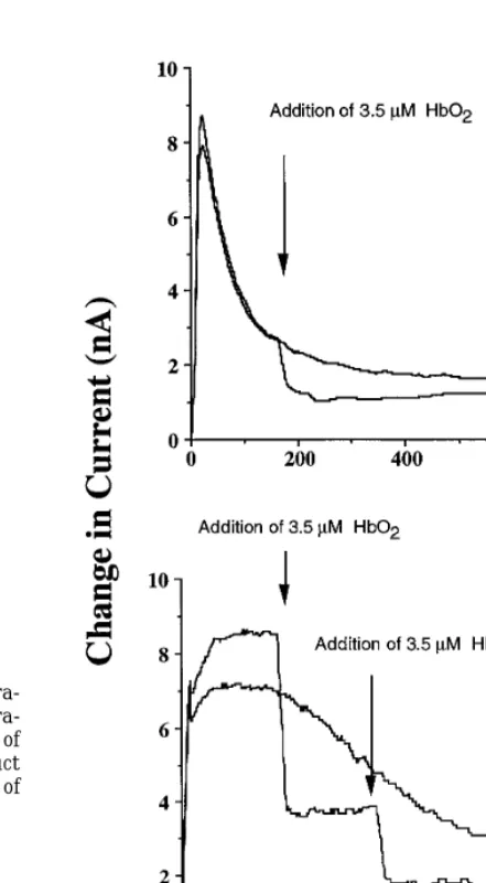
To verify this, the presence of NO was evaluated using
photographed on a UV light box. The lanes were separated from one
another, and the well containing the plug was separated from the a Ni porphyrin/Nafion-coated electrode. When 0.1 mM
portion of the lane containing the released DNA. These small pieces AS was allowed to decompose in PBS buffer at 377C, of agarose were put into separate scintillation vials, and the agarose there was an immediate increase in current that de-was melted by placing each vial on a hot plate, after adding 50mL
creased in a fashion consistent with the decomposition
of concentrated HCl to prevent resolidification of the agarose. Fifteen
kinetics expected for AS under these conditions. To
de-milliliters of Euoquint (National Diagnostics) was added to each vial,
NO (Fig. 2B). Control experiments showed that both Tempol and its one electron reduced product TPH pro-duce current under the above experimental conditions (10 nA/mM); however, exposure of HbO2 did not alter
the current. This control experiment eliminated the possibility of the reduction in current being due to a reaction between Tempol or TPH and HbO2. Taken
together, the results clearly show that there was in-creased NO production from AS in the presence of ei-ther ferricyanide or Tempol.
The next series of experiments involved exploring factors that might influence AS cytotoxicity.
Intracellu-FIG. 1. Cell survival of V79 cells exposed to varying tions of AS for 1 h in the presence or absence of equimolar concentra-tions of ferricyanide (Fe(III)(CN)603) (A); or a final concentration of
1.0 mM Tempol or the corresponding one electron reduction product TPH (B). AS or decomposed AS was exposed to cells for a period of 1 h at 377C.
of 3.5 mM HbO2 had no effect on the electrochemical
signal, which suggested that no detectable NO was pro-duced. The same experiment was then conducted in the presence of 1 mM ferricyanide. As shown in Fig. 2A, there was a dramatic increase in signal which could be attributed to the presence of NO. To verify this, 3.5
mM HbO2 was added to the solution after which the FIG. 2. Electrochemical detection of NO in the presence of AS with
electrochemical signal decreased (Fig. 2A). Such obser- ferricyanide or Tempol. AS (0.1 mM) was added to PBS buffer at 377C containing either 1 mM ferricyanide (A) or 1 mM Tempol (B).
vations confirmed the formation of NO under these
con-3.5mM HbO2was added to verify NO was present. Addition of HbO2
ditions. When an analogous experiment was performed
did not alter the observed changes in current with AS present only.
with 1 mM Tempol, there again was an increase in Therefore, the baseline due to AS decay was subtracted from the current (Fig. 2B). Addition of 3.5mM HbO2resulted in current changes observed in the presence of ferricyanide or Tempol.
The NOmM per current changes was 1.7mM/nA.
[image:4.612.31.257.33.434.2] [image:4.612.292.513.190.591.2]lar glutathione (GSH) is a critical part of the cellular detoxification network against a variety of chemical insults mediated by reactive chemical species. To deter-mine if GSH affected the cytotoxicity of AS, V79 cells were treated with BSO for 19 h to deplete intracellular GSH levels prior to treatment with AS. Using these conditions, BSO treatment alone resulted in no cytotox-icity and reduced intracellular GSH levels to õ5% of control values (data not shown). When the BSO-treated cells were exposed to increasing concentrations of AS, there was a marked enhancement in cytotoxicity over that observed for AS treatment alone, with an enhance-ment ratio (ER) of 2.2 at the 1% survival level (Fig. 3A). As demonstrated in Fig. 1A, [Fe(III)(CN)6]30treatment
alone was not cytotoxic; however, cotreatment of BSO-treated cells with AS and [Fe(III)(CN)6]
30
resulted in marked protection against cytotoxicity induced by AS (Fig. 3A).
Since oxygen could react with NO0
[image:5.612.282.520.29.425.2]to potentially form a variety of RNOS that might cause cellular dam-age, the influence of oxygen on AS cytotoxicity was evaluated. Cells were therefore maintained under hyp-oxic conditions and exposed to varying concentrations of AS for 1 h and the cytotoxicity was evaluated (Fig. 3B). AS treatment under hypoxic conditions exhibited modest cytotoxicity; however, the cytotoxicity was much less when compared to aerobic AS treatment. The products from decomposed AS resulted in no cyto-toxicity if treatment was conducted under hypoxic con-ditions. The results show that oxygen enhanced the cytotoxicity of AS to V79 cells. One possible explanation for these results could involve ROS derived from metal-based Fenton chemistry. We therefore evaluated cyto-toxicity in V79 fibroblast cells induced by AS in the presence of the metal chelators, desferrioxamine and
FIG. 3. (A) Cell survival of V79 cells exposed to varying
concentra-DETAPAC. Under aerobic conditions, neither of these
tions of AS for 1 h in the presence or absence of BSO pretreatment
agents afforded significant protection against AS
cyto-or with and without ferricyanide (Fe(III)(CN)603). (B) Cell survival
toxicity (Fig. 4). The concentrations of metal chelators of V79 cells exposed to varying concentrations of AS for 1 h under used were sufficient to prevent cellular damage and aerobic or anoxic conditions.
death mediated by Fenton-derived ROS (37). There-fore, these findings suggest a limited role for metals in
the cytotoxicity of AS. cellular GSH; however, such reduction in GSH levels can be prevented by one electron acceptors.
Since depletion of GSH was associated with an
in-creased toxicity of AS, changes in the intracellular GSH Treatment of V79 cells with AS resulted in DNA dou-ble strand breaks as shown in Figs. 5A and 5B. Either levels were examined after exposure to AS. Exposure
of 1 mM AS to V79 cells for 60 min resulted in a de- Tempol or [Fe(III)(CN)6]30added at equimolar
concen-trations completely abolished the double strand breaks crease in intracellular GSH levels by 75% (Table I).
In the presence of 3 mM AS, no detectable GSH was caused by AS; whereas, Tempol-H did not. Our findings would indicate that DNA double strand breaks observed. In contrast, treatment with DEA/NO
re-sulted in a decrease of only 25% in intracellular GSH sulting from AS exposure may be the direct result of nitroxyl toxicity. Likewise, the reversal of AS-mediated (25). Treatment of V79 cells with solution of
decom-posed AS, which contains primarily nitrite and N2O, DNA double strand breaks by [Fe(III)(CN)6] 30
and Tempol correlates with their ability to provide protec-reduced intracellular levels of GSH by 25% (Table I).
However, when cells were treated with AS in the pres- tion against the cytotoxicity of AS (Figs. 1A and 1B). We next examined the AS-mediated oxidative chemis-ence of ferricyanide or Tempol, depletion of GSH was
FIG. 4. Cell survival of V79 cells exposed to varying concentrations of AS for 1 h in the absence of presence of pretreatment with 0.1 mM DF (final concentration, 0.5 mM) or DETAPAC (final concentra-tion, 0.1 mM).
FIG. 5. (A) Field inversion electrophoresis gel of DNA extracted from V79 cells treated with 4 mM AS (or decomposed AS) for 1 h in the absence or presence of 4.0 mM ferricyanide, 1.0 mM Tempol, or 1.0 mM Tempol-H. Column numbers correspond to: 1, control; 2, AS
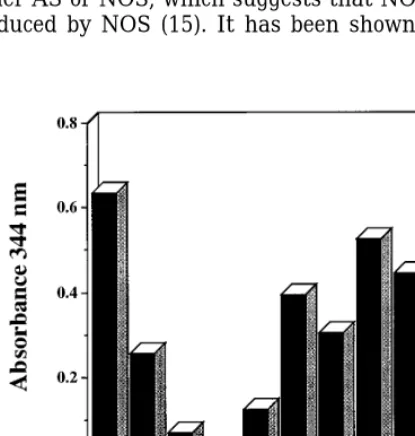
NADPH can undergo two-electron oxidation to form
alone; 3, decomposed AS alone; 4, ferricyanide alone; 5, ferricyanide
NADP/
. This oxidation can be measured by observing
/AS; 6, Tempol/AS; 7, Tempol-H/AS. Treatment of cells with
the loss of characteristic absorbance peak of NADPH at Tempol or Tempol-H alone resulted in no DNA damage, data not 340 nm. When solutions of NADPH were exposed to shown. (B) Quantitation of DNA damage (expressed as percentage increasing amounts of AS, NADPH oxidation was ob- of DNA released) of conditions shown in A. Column numbers on the
x-axis correspond to column numbers in A.
served (Fig. 6). Experiments using 0.2 mM AS under anaerobic conditions resulted in slightly less oxidation of NADPH, which indicates that oxidation may occur through either NO0 or some other RNOS (Fig. 6).
assay because it oxidized NADPH. One possible chemi-NADPH oxidation was also prevented by 0.5 mM
Tem-cal intermediate in the decomposition of AS is peroxyni-pol (Fig. 6), again consistent with the data shown in Fig. trite (ONOO0) derived from the reaction between NO0
1B. Ferricyanide could not be used in this particular
and oxygen. Peroxynitrite at 0.2 mM also oxidized NADPH (Fig. 6); however, Tempol did not prevent the peroxynitrite-mediated oxidation of NADPH, suggesting
TABLE I that Tempol acts by scavenging NO0
, not ONOO0
.
Hy-Effect of AS on Intracellular GSH Levelsa
droxylamine (NH2OH), which has been shown to
scav-enge NO0
, also prevented NADPH oxidation mediated
Conditions Percentage of GSHb
by AS. However, hydroxylamine did not prevent NADPH oxidation meditated by ONOO0
(Fig. 6).
Controlc 100/3
AS 25/4
Ferricyanide 82/6 DISCUSSION
AS/Ferricyanide 66/6
The pathophysiological role of RNOS has received
Rel AS 75/6
Tempol 118/4 much attention since the discovery of the endogenous
AS Tempol 60/6 formation of NO (38,39). Exposure of cells to NO
deliv-AS (3 mM) N.D.d
ered either as a bolus solution saturated with gaseous
AS (3 mM)/Tempol 7/2
NO or from the decomposition of NO donor compounds
TPH 101/9
AS/TPH 21/0.3 results in little toxicity (23), unless the cells are ex-posed chronically to very high amounts of NO (ú 5
aAS, ferricyanide, tempol, and TPH were done at 1 mM unless
mM total) (40). However, in the presence of oxygen or
otherwise indicated.
superoxide, RNOS such as N2O3 and ONOO0 are
bExperiments were conducted in triplicate.
formed. These reactive molecules can modify numerous
cTotal GSH was 0.7mg per 106cells.
[image:6.612.18.268.483.618.2](41). The chemistry involving these intermediates is can undergo one electron reduction to convert NO0to
NO (Eq. [3]) (44, 45). often invoked as the causative agent in numerous
dele-terious biological effects of NO.
NO0
/SOD(Cu2/
)jNO/SOD (Cu/
) [ 3 ] Recent reports suggest that another RNOS, NO0
,
may be of critical importance in the biology of NO (14, The similarity between the SOD concentration re-15, 42). NO0
is the one-electron reduction species of quired to form NO from NOS and AS, as well as our NO. However, its formation in vivo through reduction observation that NO is often cytoprotective (4), is unlikely because oxygen, which is generally in 100 prompted us to examine whether NO0
would prove to times excess over NO, is more easily reduced than NO be cytotoxic. Our results indeed demonstrate that NO0
(18). However, NO0
may be formed (1) directly, through delivered by AS is orders of magnitude more toxic than the enzymatic activity of NOS (14,15); (2) indirectly, equal concentrations of NO delivered by a similar NO via of HO-Arg mediated by hydrogen peroxide/hemo- donor DEA/NO (Fig. 1) (5, 6).
protein (17); or (3) through the heterolytic decomposi- Addition of an electron acceptor such as [Fe(III) tion of S-nitrosothiols (20). (CN)6]30
or Tempol markedly protected V79 cells from Angelis’s salt yields N2O by the formation of NO
0
the toxic effects of NO0. Initial experiments using an
(Eqs. [1] and [2]), unlike related amine NONOate com- NO0sensitive electrode showed that the presence of
ei-pounds such as DEA/NO which yield exclusively NO. ther electron acceptor, [Fe(III)(CN)6]30
or Tempol, re-sulted in the release of NO from AS (data not shown). N2O
02 3 /H
/
jHNO/NO0
2 [ 1 ]
This can readily be explained by the oxidation of NO0
by either oxidizing agents (Eqs. [4] and [5]): 2HNO jN2O/H2O [ 2 ]
NO0
/[Fe(III)(CN)30
6 jNO/Fe(II)(CN)460 [ 4 ]
It has been proposed that the primary decomposition product of AS at neutral pH is singlet NO0(26), though
NO0
/TempoljNO/Tempol0H(TPH) [ 5 ] other intermediates have been proposed (43). Two
re-cent reports have suggested that NO0
is a product of Tempol is a stable free radical which loses its charac-NOS (14, 15). One report showed that similar concen- teristic electron spin resonance (ESR) signal upon re-trations of SOD were required for NO production from duction. Preliminary ESR studies revealed that the either AS or NOS, which suggests that NO0is in fact
conversion of Tempol to the ESR silent TPH follows produced by NOS (15). It has been shown that SOD the decomposition rate of AS (data not shown). Fur-thermore, there was no significant difference of reduc-tion rate at different concentrareduc-tions of Tempol. This would indicate there is no direct oxidation of AS by Tempol and suggests that the interception of NO0by
Tempol generates NO. Comparison of Fig. 2A with 2B suggests that part of the conversion reaction of NO0
to NO by ferricyanide may occur through the direct oxidation of AS. Direct oxidation of AS also leads to NO formation (18). The importance of the interconver-sion of NO0to NO is that the cytotoxicity of NO0can
be compared to that of NO by exposing cells to AS in the presence and absence of either [Fe(III)(CN)6]30 or
Tempol.
Further comparison with NO0
and other reactive in-termediates can be made. The production of simultane-ous NO/O20produces ONOO0and other RNOS. It has
been proposed that the NO/O20reaction is one of the
major causative agents in numerous pathophysiologi-cal conditions. The hypothesis is based on bolus doses of ONOO0. However, several studies have shown that
cogeneration of NO/O20under biological conditions is
[image:7.612.37.245.388.606.2]not appreciably toxic (4). The NO donor SIN-1, which
FIG. 6. Oxidation of NADPH as measured by loss of absorbance at produces both O20
and NO, added at concentrations up
340 nm with increasing amounts of AS (0, 0.1, 0.2, 0.5 mM), 0.2 mM to 5 mM, produces little or no toxicity mammalian cells AS/Tempol, 0.2 mM AS/NH2OH. These values were compared to 0.2 (46). A comparison of similar conditions showed that
mM ONOO0, 0.2 mM PN/0.5 mM Tempol and 0.5 mM PN/0.5 mM
exposure of V79 cells to the superoxide generating
sys-NH2OH. Samples were done in triplicate and incubated for 30 min.
DEA/NO (1 mM DEA/NO gives equimolar NO as 2 mM role in the toxicity of alkylhydroperoxides (data not shown). Trolox, a water soluble antioxidant that may AS gives NO0) does not result in appreciable toxicity
(4), while addition of hypoxanthine/xanthine oxidase abate lipid peroxidation (48) afforded no protection against AS-toxicity (data not shown). Another possibil-alone was toxic. Therefore, simultaneous generation of
NO and O2 0
is considerably less toxic than NO0
, despite ity is that NO0
can react with oxygen to form ONOO0
. However, NO0
is released from AS in the singlet state the presence of the toxic RNOS, peroxynitrite. In fact,
under these conditions NO is protective against toxicity (26), which may not have a sufficient rate of reaction with triplet oxygen to form ONOO0
(49). We found in mediated by ROS.
We examined the cellular factors which may govern preliminary experiments that the oxidation chemistry of dihydrorhodamine with ONOO0
or the intermediate the extent of NO0
toxicity. Reduced glutathione (GSH)
is crucial for the detoxification of the RNOS, N2O3, and produced by AS are similar. However, we also found
that AS hydroxylates benzoate 10 times more effi-ONOO0(47), as well as for H
2O2. GSH appears to play
a similar role in the case of NO0. Depletion of intracel- ciently than ONOO0 and, unlike the latter, does not
appear to nitrate phenols (data not shown). These re-lular GSH resulted in a considerable increase in the
toxicity of AS. It has been shown that NO0
derived from sults may suggest that either the formation of peroxy-nitrite is modified or there exists another intermediate. AS readily oxidizes thiols (43). In fact, in preliminary
competitive studies, we have found that NO0 reacts We are currently examining the chemical mechanisms
of these reactions. with thiols 100 times more rapidly than other RNOS
such as ONOO0
(data not shown). The chemical and The finding that AS induces double strand breaks in cells is surprising, especially since neither ONOO0
nor cellular results support the notion that GSH is critical
in the initial detoxification of NO0
. Examination of H2O2induce this type of cellular damage. These strand
breaks occur from the chemistry associated with NO0
GSH levels in Table 1 shows that while exposure of
cells to 1 mM AS was not toxic, this concentration of and are not mediated by peroxide/metal-mediated oxi-dation. This activity of AS suggests that neither AS reduced GSH as much as 80%. It may be surmised
that NO0
first depletes intracellular GSH, which then ONOO0
nor H2O2 is the primary chemical species
re-sponsible for DNA damage. Similar to the effects of renders other cellular targets susceptible to the
chem-istry of NO0
. ionizing radiation and some chemotherapeutic drugs such Adriamycin, NO0appears to be effective in
caus-Another important aspect of our observations is their
implication for the chemistry of the L-Arginine:NO ing in this type of DNA damage.
Our findings may explain some of the discrepancies pathway under different pathophysiological conditions.
The depletion of GSH by endogenously produced NO0
in the literature as to the protective or deleterious ef-fects of NO in biological systems. While NO can abate may render cells susceptible to other cytotoxic agents.
For instance, intracellular GSH abates the toxicity of the toxicity of peroxides via reactions with radicals and other components of the Fenton reaction, NO0actually
peroxide and RNOS under conditions of increased oxi-dative stress. The concomitant formation of NO0
in the enhances this toxicity. In vivo nitroxyl may by formed in one of three ways: (1) enzymatically, through the presence of oxidative stress may enhance levels of
per-oxide toxicity since GSH would not be available to de- direct production of NO0
from NOS; (2) through the incomplete oxidation of L-arginine to form HO-Arg
toxify H2O2. In a previous report, AS markedly
en-hanced the cytotoxicity of hydrogen peroxide (6). There- which is further oxidized to form NO0, perhaps at a
distal site from its source, or (3) via degradation of fore, even at subtoxic doses of NO0
, a cell can be
rendered susceptible to damage by oxidative stress. S-nitrosothiols. We hypothesize that NOS inhibitors
would prevent the formation of NO0
by any of these Studies on oxidative stress involving H2O2suggest
that redox metals are critical for cellular damage (48). mechanisms. Finally, redox reactions with cellular components such as SOD or ferricytochrome c may be In the presence of metal chelators such as DF or
DETA-PAC, these Fenton-driven reactions are abated. How- essential in maintaining the balance between NO0
and NO in cells (44,50). Redox-active enzymes such as SOD ever, these metal chelators did not protect cells
ex-posed to AS. The lack of protection by metal chelators may not only be necessary to produce NO from NOS, but also to preserve the antioxidant status of the cell suggests that ROS formed from hydrogen peroxide are
not involved in the toxicity of NO0. making it more resistant to oxidative stress mediated
by a variety of toxic agents and intermediates. While ROS derived from metals may not be required,
oxygen itself appears to be necessary for the cytotoxic effect of NO0
. There are a number of possible
explana-REFERENCES
tions for this. One is that NO0
may initiate lipid
peroxi-dation which is propagated by O2(48). We have found 1. Choi, D. W. (1993) Proc. Natl. Acad. Sci. USA 53, 9741 – 9743.
that hydroperoxide toxicity is reduced in the absence 2. Wink, D. A., Grisham, M., Mitchell, J B., and Ford, P. C. (1996)
Methods Enzymol. 268, 12 – 31.
3. Pryor, W. A., and Squadrito, G. L. (1996) Am. J. Phys. 268, 26. Bonner, F. T., and Stedman, G. (1996) Methods Nitric Oxide Res. 341 – 356.
L699 – 721.
27. Akhtar, M. J., Bonner, F. T., Hughes, M. N., Humphryes, E. J., 4. Wink, D. A., Hanbauer, I., Krishna, M. C., DeGraff, W., Gamson,
and Lu, C.-S. (1975) Inorg. Chem. 14, 558 – 563. J., and Mitchell, J. B. (1993) Proc. Natl. Acad. Sci. USA 90,
9813 – 9817. 28. Maragos, C. M., Morley, D., Wink, D. A., Dunams, T. M.,
Saave-dra, J. E., Hoffman, A., Bove, A. A., Isaac, L., Hrabie, J. A., and 5. Wink, D. A., Cook, J. A., Krishna, M. C., Hanbauer, I., DeGraff,
Keefer, L. K. (1991) J. Med. Chem. 34, 3242 – 3247. W., Gamson, J., and Mitchell, J. B. (1995) Arch. Biochem.
Bio-phys. 319, 402 – 407. 29. Beckman, J. S., Beckman, T. W., Chen, J., Marshall, P. H., and
Freeman, B. A. (1990) Proc. Natl. Acad. Sci. USA 87, 1620 – 1624. 6. Wink, D. A., Cook, J., Pacelli, R., DeGraff, W., Gamson, J.,
Lieb-mann, J., Krishna, M., and Mitchell, J. B. (1996) Arch. Biochem. 30. Russo, A., Mitchell, J. B., Finkelstein, E., DeGraff, W. G., Spiro,
Biophys. 331, 241 – 248. I. J., and Gamson, J. (1985) Radiat. Res. 103, 232 – 239.
7. Rubbo, H., Radi, R., Trujillo, M., Telleri, R., Kalyanaraman, B., 31. Tietze, F. (1969) Anal. Biochem. 27, 502 – 522.
Barnes, S., Kirk, M., and Freeman, B. A. (1994) J. Biol. Chem. 32. Ager, D. D., Dewey, W. C., Gardiner, K., Harvey, W., Johnson, 269, 26066 – 26075. R. T., and Waldren, C. A. (1990) Radiat. Res. 122, 181 – 187. 8. Hogg, N., Kalyanaraman, B., Joseph, J., Struck, A., and Partha- 33. Stamato, T. D., and Denko, N. (1990) Radiat. Res. 121, 196 – 205.
sarathy, S. (1993) FEBS Lett. 334, 170 – 174.
34. Christodoulou, D., Kudo, S., Cook, J. C., Krishna, M. C., Miles, 9. Kanner, J., Harel, S., and Granit, R. (1991) Arch. Biochem. Bio- A., Grisham, M. B., Murugesan, R., Ford, P. C., and Wink, D. A.
phys. 289, 130 – 136. (1996) Methods Enzymol. 268, 69 – 83.
10. Gorbunov, N. V., Osipov, A. N., Day, B. W., Zayas-Rivera, B., 35. Wink, D. A., Christodoulou, D., Ho, M., Krishna, M. C., Cook, Kagan, V. E., and Elsayed, N. M. (1995) Biochemistry 34, 6689 – J. A., Haut, H., Randolph, J. K., Sullivan, M., Coia, G., Murray,
6699. R., and Meyer, T. (1995) Methods: Comp. Methods Enzymol. 7,
11. Wink, D. A., Cook, J. A., Kim, S., Vodovotz, Y., Pacelli, R., Kir- 71 – 77.
shna, M. C., Russo, A., Mitchell, J. B., Jourd’heuil, D., Miles, 36. Zamora, R., Grzesiok, A., Weber, H., and Feelisch, M. (1995) A. M., and Grisham, M. B. (1997) J. Biol. Chem. 272, 11147 – Biochem. J. 312, 333 – 339.
11151.
37. Mitchell, J. B., Samuni, A., Krishna, M. C., DeGraff, W. G., Ahn, 12. Miles, A. M., Bohle, D. S., Glassbrenner, P. A., Hansert, B., M. S., Samuni, U., and Russo, A. (1990) Biochemistry 29, 2802 –
Wink, D. A., and Grisham, M. B. (1996) J. Biol. Chem. 271, 40 – 2807. 47.
38. Rubbo, H., Darley-Usmar, V., and Freeman, B. A. (1996) Chem. 13. Miles, A. M., Gibson, M., Krishna, M., Cook, J. C., Pacelli, R., Res. Toxicol. 9, 809 – 820.
Wink, D. A., and Grisham, M. B. (1995) Free Radical Res. 233,
39. Gross, S. S., and Wolin, M. S. (1995) Annu. Rev. Physiol. 57, 379 – 390.
737 – 769. 14. Hobbs, A. J., Fukuto, J. M., and Ignarro, L. J. (1994) Proc. Natl.
40. Tamir, S., Lewis, R. S., de Rojas Walker, T, Deen, W. M.,
Wish-Acad. Sci. USA 91, 10992 – 10996.
nok, J. S., and Tannenbaum, S. R. (1993) Chem. Res. Toxicol. 6, 15. Schmidt, H. H., Hofmann, H., Schindler, U., Shutenko, Z. S., 895 – 899.
Cunningham, D. D., and Feelisch, M. (1996) Proc. Natl. Acad.
41. Wink, D. A., Hanbauer, I., Grisham, M. B., Laval, F., Nims,
Sci. USA 93, 14492 – 14497.
R. W., Laval, J., Cook, J. C., Pacelli, R., Liebmann, J., Krishna, 16. Griffith, O. W., and Stuehr, D. J. (1995) Annu. Rev. Physiol. 57, M. C., Ford, M. C., and Mitchell, J. B. (1996) Curr. Topics Cell.
707 – 736. Regul. 34, 159 – 187.
17. Pufahl, R. A., Wishnok, J. S., and Marletta, M. A. (1995) Bio- 42. Fukuto, J. M., Hobbs, A. J., and Ignarro, L. J. (1993) Biochem.
chemistry 34, 1930 – 1941. Biophys. Res. Commun. 196, 707 – 713.
18. Wink, D. A., and Feelisch, M. (1996) Methods Nitric Oxide Res. 43. Doyle, M. P., Mahapatro, S. N., Broene, R. D., and Guy, J. K.
403 – 412. (1988) J. Am. Chem. Soc. 110, 593 – 599.
19. Williams, D. L. H. (1988) in Nitrosation, Cambridge Press, Ox- 44. Murphy, M. E., and Sies, H. (1991) Proc. Natl. Acad. Sci. USA
ford, UK. 88, 10860 – 10864.
20. Arnelle, D. R., and Stamler, J. S. (1995) Arch. Biochem. Biophys. 45. Feelisch, M., Ostrowski, J., and Noack, E. (1989) J. Cardiovasc.
318, 279 – 285. Pharmacol. 14, S13 – S22.
21. Mitchell, J. B., Wink, D. A., DeGraff, W.,Gamson, J., Keefer, 46. Farias-Eisner, R., Chaudhuri, G., Aeberhard, E., and Fukuto, L. K., and Krishna, M. C. (1993) Cancer Res. 53, 5845 – 5848. J. M. (1996) J. Biol. Chem. 271, 6144 – 6151.
22. Pacelli, R., Wink, D. A., Cook, J. A., Krishna, M C., DeGraff, W., 47. Wink, D. A., Nims, R. W., Darbyshire, J. F., Christodoulou, D., Friedman, N., Tsokos, M., Samuni, A., and Mitchell, J. B. (1995) Hanbauer, I., Cox, G. W., Laval, F., Laval, J., Cook, J. A.,
J. Exp. Med. 182, 1469 – 1479. Krishna, M. C., DeGraff, W., and Mitchell, J B. (1994) Chem.
Res. Toxicol. 7, 519 – 525.
23. Wink, D. A., Hanbauer, I., Laval, F., Cook, J. A., Krishna, M. C.,
and Mitchell, J. B. (1994) Ann. N.Y. Acad. Sci. 738, 265 – 278. 48. Halliwell, B., and Gutteridge, J. M. C. (1989) Free Rad. Biol.
Med. 416 – 509.
24. Misra, R. R., Hochadel, J. F., Smith, G. T., Waalkes, M. P., and
Wink, D. A. (1996) Chem. Res. Toxicol. 10, 326 – 332. 49. Doyle, C. F., Hughes, M. N., Thompson, J. M., and Bonner, F. T. (1986) Inorg. Chem. 25, 2676 – 2677.
25. Cook, J. A., Krishna, M. C., Pacelli, R., DeGraff, W., Liebmann,
J., Russo, A., Mitchell, J. B., and Wink, D. A. (1997) Br. J. Cancer 50. Kelm, M., Dahmann, R., Wink, D. A., and Feelisch, M. (1997) J.
Biol. Chem., in press.