(
R
)-2-Cyano-
N
-(1-phenylethyl)acetamide
Mohan Kumar,aB. S. Madhukar,bM. A. Sridhar,a* D. G. Bhadregowda,bKamini Kapoor,cVivek K. Guptac and Rajni Kantc
aDepartment of Studies in Physics, Manasagangotri, University of Mysore, Mysore 570 006, India,bDepartment of Chemistry, Yuvarajas College, University of Mysore, Mysore 570005, India, andcX-ray Crystallography Laboratory, Post-Graduate Department of Physics & Electronics, University of Jammu, Jammu Tawi 180 006, India
Correspondence e-mail: [email protected]
Received 4 March 2013; accepted 25 March 2013
Key indicators: single-crystal X-ray study;T= 293 K; mean(C–C) = 0.003 A˚;
Rfactor = 0.033;wRfactor = 0.079; data-to-parameter ratio = 9.4.
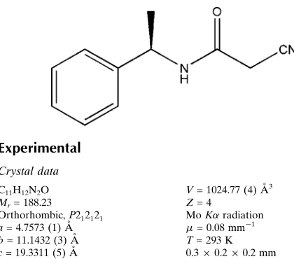
In the title compound, C11H12N2O, the dihedral angle between
the acetamide group and the benzene ring is 68.7 (1). In the crystal, N—H O and weak C—H O hydrogen bonds link the molecules into chains along thea-axis direction.
Related literature
For related structures, see: Resendeet al.(2003); Ga´lvezet al.
(2010).
Experimental
Data collection
Oxford diffraction Xcalibur Sapphire3 diffractometer Absorption correction: multi-scan
(CrysAlis PRO; Oxford Diffraction, 2010)
Tmin= 0.965,Tmax= 1.000
21018 measured reflections 1201 independent reflections 1097 reflections withI> 2(I)
Rint= 0.039
Refinement
R[F2> 2(F2)] = 0.033
wR(F2) = 0.079
S= 1.04 1201 reflections
128 parameters
H-atom parameters constrained max= 0.10 e A˚ 3
[image:1.610.47.257.459.644.2]min= 0.13 e A˚ 3
Table 1
Hydrogen-bond geometry (A˚ ,).
D—H A D—H H A D A D—H A
N2—H2 O1i
0.86 2.04 2.878 (2) 165
C2—H2A O1i 0.97 2.42 3.215 (2) 138
Symmetry code: (i)x 1;y;z.
Data collection: CrysAlis PRO(Oxford Diffraction, 2010); cell refinement: CrysAlis PRO; data reduction: CrysAlis RED (Oxford Diffraction, 2010); program(s) used to solve structure:SHELXS97 (Sheldrick, 2008); program(s) used to refine structure:SHELXL97 (Sheldrick, 2008); molecular graphics: ORTEP-3 for Widows (Farrugia, 2012); software used to prepare material for publication: PLATON(Spek, 2009).
MK acknowledges the help of Bahubali College of Engi-neering, Shravanabelagola for his research work. RK acknowledges the Department of Science & Technology for the single-crystal X-ray diffractometer sanctioned as a National Facility under Project No. SR/S2/CMP-47/2003.
Supplementary data and figures for this paper are available from the IUCr electronic archives (Reference: GK2561).
References
Farrugia, L. J. (2012).J. Appl. Cryst.45, 849–854.
Ga´lvez, J. A., Quiroga, J., Cobo, J. & Glidewell, C. (2010).Acta Cryst.C66, o521–o523.
Oxford Diffraction (2010). CrysAlis PRO and CrysAlis RED. Oxford Diffraction Ltd, Yarnton, England.
Structure Reports
Online
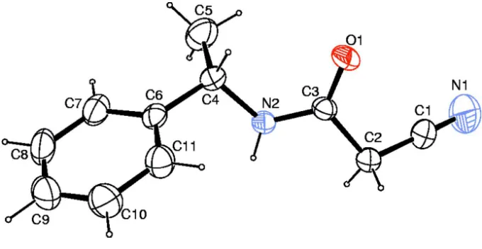
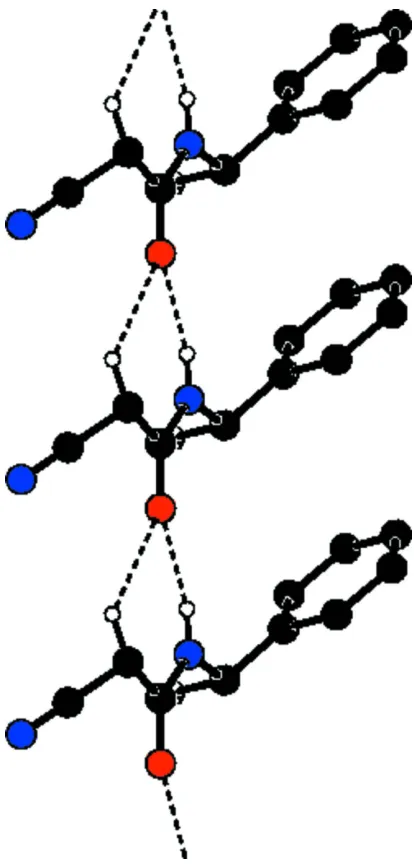
As part of our investigations on acetamide derivatives, the title compound has been prepared and its crystal structure is presented here.Bond lengths and angles in the title compound (Fig. 1) are comparable with the similar crystal structures (Resende et al., 2003; Gálvez et al., 2010). The dihedral angle between the acetamide group (C2/C3/O1/N2) and the benzene ring (C6—C11) is 68.7 (1)°. N—H···O and weak C—H···O hydrogen bonds link the molecules into chains along the a axis (Fig.2, Table 1). In the crystal, molecules are packed into layers parallel to the bc-plane (Fig.3).
S2. Experimental
The reaction of methyl 2-cyanoacetate (0.1 g, 0.01 mol) and (R)-1-phenylethanamine (0.1 g, 0.01 mol) were carried out in the presence of dilute acetic acid and the reaction mixture was allowed to stir at room temperature for 6-7 h in dry di-chloromethane (25 ml). The progress of the reaction was monitored by TLC. Upon completion, the solvent was removed under reduced pressure and residue was extracted with ethyl acetate. The compound was purified by successive
recrystallization from methanol (yield 83%). The melting range was found to be 393-395 K.
S3. Refinement
All H atoms were positioned geometrically and were treated as riding on their parent C/N atoms, with C—H distances of 0.93–0.98 Å and N—H distance of 0.86 with Uiso(H) = 1.2Ueq(C) or 1.5Ueq(methyl C). The 799 Friedel equivalents were
Figure 1
Figure 2
Figure 3
The packing arrangement of molecules viewed down the a axis. Hydrogen atoms have been omitted for clarity.
wR(F2) = 0.079
S = 1.04 1201 reflections 128 parameters 0 restraints
Primary atom site location: structure-invariant direct methods
neighbouring sites
H-atom parameters constrained w = 1/[σ2(F
o2) + (0.0368P)2 + 0.1643P]
where P = (Fo2 + 2Fc2)/3
(Δ/σ)max < 0.001
Δρmax = 0.10 e Å−3
Δρmin = −0.13 e Å−3
Special details
Experimental. CrysAlis PRO, Oxford Diffraction Ltd., Version 1.171.34.40 (release 27–08-2010 CrysAlis171. NET) (compiled Aug 27 2010,11:50:40) Empirical absorption correction using spherical harmonics, implemented in SCALE3 ABSPACK scaling algorithm.
Geometry. All esds (except the esd in the dihedral angle between two l.s. planes) are estimated using the full covariance matrix. The cell esds are taken into account individually in the estimation of esds in distances, angles and torsion angles; correlations between esds in cell parameters are only used when they are defined by crystal symmetry. An approximate (isotropic) treatment of cell esds is used for estimating esds involving l.s. planes.
Refinement. Refinement of F2 against ALL reflections. The weighted R-factor wR and goodness of fit S are based on F2,
conventional R-factors R are based on F, with F set to zero for negative F2. The threshold expression of F2 > 2sigma(F2) is
used only for calculating R-factors(gt) etc. and is not relevant to the choice of reflections for refinement. R-factors based on F2 are statistically about twice as large as those based on F, and R- factors based on ALL data will be even larger.
Fractional atomic coordinates and isotropic or equivalent isotropic displacement parameters (Å2)
x y z Uiso*/Ueq
H5C 0.3139 0.6939 0.0529 0.100* C6 −0.0760 (4) 0.73086 (16) 0.19678 (8) 0.0371 (4) C7 −0.2066 (5) 0.62085 (18) 0.20201 (11) 0.0527 (5) H7 −0.1722 0.5620 0.1689 0.063* C8 −0.3899 (5) 0.5972 (2) 0.25659 (13) 0.0670 (7) H8 −0.4768 0.5226 0.2595 0.080* C9 −0.4434 (5) 0.6819 (2) 0.30565 (12) 0.0675 (7) H9 −0.5676 0.6657 0.3416 0.081* C10 −0.3132 (6) 0.7905 (2) 0.30156 (11) 0.0640 (6) H10 −0.3473 0.8486 0.3351 0.077* C11 −0.1309 (4) 0.81478 (19) 0.24787 (10) 0.0514 (5) H11 −0.0427 0.8892 0.2459 0.062*
Atomic displacement parameters (Å2)
U11 U22 U33 U12 U13 U23
O1 0.0258 (6) 0.0575 (8) 0.0667 (8) 0.0020 (6) 0.0029 (6) 0.0128 (7) N1 0.1117 (19) 0.0878 (16) 0.0917 (15) 0.0037 (16) 0.0391 (15) 0.0382 (14) N2 0.0253 (7) 0.0399 (8) 0.0492 (8) 0.0027 (7) −0.0020 (6) 0.0118 (7) C1 0.0553 (12) 0.0439 (11) 0.0603 (12) −0.0003 (10) 0.0039 (11) 0.0130 (9) C2 0.0344 (9) 0.0414 (10) 0.0557 (10) 0.0029 (9) 0.0038 (9) 0.0109 (9) C3 0.0289 (9) 0.0391 (10) 0.0370 (8) 0.0009 (8) 0.0001 (7) 0.0019 (8) C4 0.0362 (10) 0.0374 (9) 0.0440 (9) 0.0049 (8) −0.0020 (8) 0.0059 (8) C5 0.0910 (18) 0.0519 (13) 0.0571 (12) 0.0138 (14) 0.0136 (13) −0.0012 (10) C6 0.0342 (9) 0.0363 (9) 0.0407 (9) 0.0025 (8) −0.0075 (8) 0.0088 (7) C7 0.0565 (12) 0.0385 (11) 0.0631 (12) −0.0027 (11) −0.0049 (11) 0.0081 (9) C8 0.0604 (15) 0.0514 (13) 0.0892 (16) −0.0123 (11) 0.0018 (14) 0.0289 (13) C9 0.0589 (14) 0.0799 (17) 0.0636 (13) 0.0067 (14) 0.0136 (13) 0.0301 (13) C10 0.0711 (15) 0.0698 (15) 0.0512 (11) 0.0076 (14) 0.0112 (12) 0.0047 (10) C11 0.0567 (12) 0.0470 (10) 0.0504 (10) −0.0064 (10) 0.0015 (10) −0.0007 (9)
Geometric parameters (Å, º)
N2—C4—C6 108.88 (14) C8—C9—H9 120.3 N2—C4—C5 109.82 (15) C10—C9—H9 120.3 C6—C4—C5 115.61 (16) C9—C10—C11 120.2 (2) N2—C4—H4 107.4 C9—C10—H10 119.9 C6—C4—H4 107.4 C11—C10—H10 119.9 C5—C4—H4 107.4 C10—C11—C6 121.52 (19) C4—C5—H5A 109.5 C10—C11—H11 119.2 C4—C5—H5B 109.5 C6—C11—H11 119.2 H5A—C5—H5B 109.5
C4—N2—C3—O1 0.1 (3) C5—C4—C6—C11 179.27 (18) C4—N2—C3—C2 −178.11 (15) C11—C6—C7—C8 0.9 (3) C1—C2—C3—O1 33.0 (3) C4—C6—C7—C8 179.84 (18) C1—C2—C3—N2 −148.74 (17) C6—C7—C8—C9 0.0 (3) C3—N2—C4—C6 148.29 (16) C7—C8—C9—C10 −0.7 (4) C3—N2—C4—C5 −84.2 (2) C8—C9—C10—C11 0.6 (4) N2—C4—C6—C7 124.55 (18) C9—C10—C11—C6 0.4 (3) C5—C4—C6—C7 0.4 (3) C7—C6—C11—C10 −1.1 (3) N2—C4—C6—C11 −56.6 (2) C4—C6—C11—C10 179.93 (18)
Hydrogen-bond geometry (Å, º)
D—H···A D—H H···A D···A D—H···A N2—H2···O1i 0.86 2.04 2.878 (2) 165
C2—H2A···O1i 0.97 2.42 3.215 (2) 138