2-Ethyl-5-triphenylmethyl-1,3-dioxane
Lin Yuan,aJiang-Hua Shi,aMin Zhangaand Seik Weng Ngb*
a
Department of Biology and Chemistry, Hunan University of Science and Engineering, Yongzhou Hunan 425100, People’s Republic of China, and
bDepartment of Chemistry, University of Malaya, 50603 Kuala Lumpur, Malaysia
Correspondence e-mail: [email protected]
Received 9 September 2010; accepted 14 September 2010
Key indicators: single-crystal X-ray study;T= 110 K; mean(C–C) = 0.002 A˚; Rfactor = 0.042;wRfactor = 0.114; data-to-parameter ratio = 17.1.
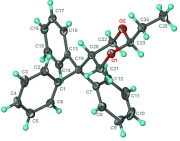
In the title compound, C25H26O2, the dioxane ring adopts a chair conformation with the two substituent groups occupying equatorial positions.
Related literature
For the crystal structure of 2,2-dimethyl-5-triphenyl-1,3-dioxane, see: Zhanget al.(2009).
Experimental
Crystal data
C25H26O2 Mr= 358.46
Monoclinic,P21=c a= 10.5401 (6) A˚
b= 13.3550 (8) A˚
c= 14.6044 (8) A˚ = 110.523 (1)
V= 1925.28 (19) A˚3
Z= 4
MoKradiation = 0.08 mm 1
T= 110 K
0.450.350.15 mm
Data collection
Bruker SMART APEX diffractometer 9493 measured reflections
4174 independent reflections 3047 reflections withI> 2(I)
Rint= 0.031
Refinement
R[F2> 2(F2)] = 0.042 wR(F2) = 0.114 S= 1.04 4174 reflections
244 parameters
H-atom parameters constrained
max= 0.29 e A˚ 3
min= 0.19 e A˚ 3
Data collection:SMART(Bruker, 2003); cell refinement:SAINT (Bruker, 2003); data reduction:SAINT; program(s) used to solve structure:SHELXS97(Sheldrick, 2008); program(s) used to refine structure: SHELXL97 (Sheldrick, 2008); molecular graphics: X-SEED (Barbour, 2001); software used to prepare material for publication:publCIF(Westrip, 2010).
We thank the Key Subject Construction Project of Hunan Province (No. 2006–180), the Key Scientific Research Project of Hunan Provincial Education Department (No. 08 A023, 05 C736), the NSF of Hunan Province (09 J J3028) and the University of Malaya for supporting this study.
Supplementary data and figures for this paper are available from the IUCr electronic archives (Reference: ZS2066).
References
Barbour, L. J. (2001).J. Supramol. Chem.1, 189–191.
Bruker (2003).SAINTandSMART. Bruker AXS Inc., Madison, Wisconsin, USA.
Sheldrick, G. M. (2008).Acta Cryst.A64, 112–122. Westrip, S. P. (2010).J. Appl. Cryst.43, 920–925.
Zhang, M., Yuan, X.-Y. & Liu, X.-M. (2009).Acta Cryst.E65, o304. Acta Crystallographica Section E
Structure Reports
Online
supporting information
Acta Cryst. (2010). E66, o2582 [doi:10.1107/S1600536810036767]
2-Ethyl-5-triphenylmethyl-1,3-dioxane
Lin Yuan, Jiang-Hua Shi, Min Zhang and Seik Weng Ng
S1. Comment
A previous study reported the crystal structure of 2,2-dimethyl-5-triphenylmethyl-1,3-dioxane (Zhang et al., 2009). Such
disubstituted 1,3-dioxanes are known from NMR studies to have substituents in equatorial rather than in axial
orientations on the six-membered ring. The the title compound, 2-ethyl-5-triphenylmethyl-1,3-dioxane analog (Scheme I,
Fig. 1), has similar features for the dioxane part, which adopts a chair conformation. The substitutent groups occupy
equatorial positions.
S2. Experimental
2-Triphenylmethyl-1,3-propanediol (0.24 g, 5.0 mmol), propionaldehyde (20 mmol) and p-toluenesulfonic acid (0.1 g)
were stirred in dichloromethane (20 ml) for a week. The solvent was evaporated and the residue was dissolved in ether
(20 ml) after which the solution was washed with water and 5% sodium bicarbonate (20 ml). The organic phase was dried
with anhydrous sodium sulfate. The solvent was evaporated and the product was recrystallized from ethyl acetate to give
1.0 g (yield 60%) of colorless crystals.
S3. Refinement
Carbon-bound H-atoms were placed in calculated positions (C—H = 0.95–0.99 Å) and were included in the refinement in
Figure 1
Thermal ellipsoid plot (Barbour, 2001) of the title compound at the 70% probability level.
2-Ethyl-5-triphenylmethyl-1,3-dioxane
Crystal data
C25H26O2 Mr = 358.46 Monoclinic, P21/c
Hall symbol: -P 2ybc
a = 10.5401 (6) Å
b = 13.3550 (8) Å
c = 14.6044 (8) Å
β = 110.523 (1)°
V = 1925.28 (19) Å3 Z = 4
F(000) = 768
Dx = 1.237 Mg m−3
Mo Kα radiation, λ = 0.71073 Å Cell parameters from 3994 reflections
θ = 2.6–27.0°
µ = 0.08 mm−1 T = 110 K Block, colorless 0.45 × 0.35 × 0.15 mm
Data collection
Bruker SMART APEX diffractometer
Radiation source: fine-focus sealed tube Graphite monochromator
ω scans
9493 measured reflections 4174 independent reflections
3047 reflections with I > 2σ(I)
Rint = 0.031
θmax = 27.1°, θmin = 2.1° h = −9→13
k = −17→12
Refinement
Refinement on F2
Least-squares matrix: full
R[F2 > 2σ(F2)] = 0.042 wR(F2) = 0.114 S = 1.04 4174 reflections 244 parameters 0 restraints
Primary atom site location: structure-invariant direct methods
Secondary atom site location: difference Fourier map
Hydrogen site location: inferred from neighbouring sites
H-atom parameters constrained
w = 1/[σ2(F
o2) + (0.0568P)2 + 0.2859P]
where P = (Fo2 + 2Fc2)/3
(Δ/σ)max = 0.001
Δρmax = 0.29 e Å−3
Δρmin = −0.19 e Å−3
Fractional atomic coordinates and isotropic or equivalent isotropic displacement parameters (Å2)
x y z Uiso*/Ueq
H18 0.4921 0.5253 0.3473 0.022* C19 0.64436 (13) 0.38587 (10) 0.29220 (9) 0.0162 (3) C20 0.48869 (13) 0.37838 (10) 0.23326 (9) 0.0163 (3) H20 0.4528 0.4483 0.2205 0.020* C21 0.45229 (13) 0.32618 (11) 0.13438 (10) 0.0191 (3) H21A 0.4950 0.3620 0.0933 0.023* H21B 0.4876 0.2568 0.1440 0.023* C22 0.41008 (13) 0.32455 (11) 0.28894 (10) 0.0186 (3) H22A 0.4418 0.2545 0.3020 0.022* H22B 0.4263 0.3582 0.3525 0.022* C23 0.24357 (14) 0.27454 (11) 0.14269 (9) 0.0178 (3) H23A 0.2791 0.2046 0.1563 0.021* C24 0.09327 (14) 0.27184 (11) 0.08743 (10) 0.0207 (3) H24A 0.0758 0.2405 0.0227 0.025* H24B 0.0578 0.3412 0.0765 0.025* C25 0.01923 (15) 0.21322 (12) 0.14283 (11) 0.0261 (3) H25A −0.0782 0.2131 0.1051 0.039* H25B 0.0354 0.2447 0.2066 0.039* H25C 0.0528 0.1442 0.1525 0.039*
Atomic displacement parameters (Å2)
U11 U22 U33 U12 U13 U23
C24 0.0198 (7) 0.0196 (7) 0.0204 (7) −0.0001 (6) 0.0042 (6) −0.0022 (6) C25 0.0193 (7) 0.0286 (8) 0.0286 (8) −0.0044 (6) 0.0064 (6) −0.0006 (7)
Geometric parameters (Å, º)
O1—C23 1.4125 (16) C13—C18 1.3899 (19) O1—C21 1.4345 (15) C13—C14 1.3994 (19) O2—C23 1.4181 (15) C13—C19 1.5509 (18) O2—C22 1.4355 (15) C14—C15 1.382 (2) C1—C6 1.3959 (19) C14—H14 0.9500 C1—C2 1.403 (2) C15—C16 1.385 (2) C1—C19 1.5465 (19) C15—H15 0.9500 C2—C3 1.387 (2) C16—C17 1.384 (2) C2—H2 0.9500 C16—H16 0.9500 C3—C4 1.387 (2) C17—C18 1.3925 (19) C3—H3 0.9500 C17—H17 0.9500 C4—C5 1.381 (2) C18—H18 0.9500 C4—H4 0.9500 C19—C20 1.5661 (18) C5—C6 1.387 (2) C20—C21 1.5264 (18) C5—H5 0.9500 C20—C22 1.5287 (19) C6—H6 0.9500 C20—H20 1.0000 C7—C8 1.395 (2) C21—H21A 0.9900 C7—C12 1.395 (2) C21—H21B 0.9900 C7—C19 1.5459 (19) C22—H22A 0.9900 C8—C9 1.396 (2) C22—H22B 0.9900 C8—H8 0.9500 C23—C24 1.5053 (18) C9—C10 1.381 (3) C23—H23A 1.0000 C9—H9 0.9500 C24—C25 1.523 (2) C10—C11 1.376 (2) C24—H24A 0.9900 C10—H10 0.9500 C24—H24B 0.9900 C11—C12 1.392 (2) C25—H25A 0.9800 C11—H11 0.9500 C25—H25B 0.9800 C12—H12 0.9500 C25—H25C 0.9800
C4—C5—C6 120.53 (14) C21—C20—C19 114.62 (11) C4—C5—H5 119.7 C22—C20—C19 113.35 (10) C6—C5—H5 119.7 C21—C20—H20 107.3 C5—C6—C1 121.34 (14) C22—C20—H20 107.3 C5—C6—H6 119.3 C19—C20—H20 107.3 C1—C6—H6 119.3 O1—C21—C20 110.30 (11) C8—C7—C12 117.57 (14) O1—C21—H21A 109.6 C8—C7—C19 121.37 (13) C20—C21—H21A 109.6 C12—C7—C19 120.78 (13) O1—C21—H21B 109.6 C7—C8—C9 120.98 (15) C20—C21—H21B 109.6 C7—C8—H8 119.5 H21A—C21—H21B 108.1 C9—C8—H8 119.5 O2—C22—C20 109.71 (10) C10—C9—C8 120.36 (16) O2—C22—H22A 109.7 C10—C9—H9 119.8 C20—C22—H22A 109.7 C8—C9—H9 119.8 O2—C22—H22B 109.7 C11—C10—C9 119.42 (15) C20—C22—H22B 109.7 C11—C10—H10 120.3 H22A—C22—H22B 108.2 C9—C10—H10 120.3 O1—C23—O2 110.18 (10) C10—C11—C12 120.42 (16) O1—C23—C24 109.22 (11) C10—C11—H11 119.8 O2—C23—C24 108.81 (11) C12—C11—H11 119.8 O1—C23—H23A 109.5 C11—C12—C7 121.26 (15) O2—C23—H23A 109.5 C11—C12—H12 119.4 C24—C23—H23A 109.5 C7—C12—H12 119.4 C23—C24—C25 111.50 (11) C18—C13—C14 117.58 (12) C23—C24—H24A 109.3 C18—C13—C19 123.54 (12) C25—C24—H24A 109.3 C14—C13—C19 118.71 (12) C23—C24—H24B 109.3 C15—C14—C13 121.43 (13) C25—C24—H24B 109.3 C15—C14—H14 119.3 H24A—C24—H24B 108.0 C13—C14—H14 119.3 C24—C25—H25A 109.5 C14—C15—C16 120.50 (13) C24—C25—H25B 109.5 C14—C15—H15 119.8 H25A—C25—H25B 109.5 C16—C15—H15 119.8 C24—C25—H25C 109.5 C17—C16—C15 118.78 (13) H25A—C25—H25C 109.5 C17—C16—H16 120.6 H25B—C25—H25C 109.5
![Crystal structure of 5′′ benzylidene 1′ methyl 4′ phenyltrispiro[acenaphthylene 1,2′ pyrrolidine 3′,1′′ cyclohexane 3′′,2′′′ [1,3]dioxane] 2,6′′ dione](data:image/gif;base64,R0lGODlhAQABAIAAAP///wAAACH5BAEAAAAALAAAAAABAAEAAAICRAEAOw==)