Table 1 Historical sequence of main applications of CA to elec-trophoretic protocols in different areas of research and clinical investigations
Year Application
1957 CA is used as an electrophoretic support (Kohn) 1971 Application to conventional electrophoresis of white cell
and red cell enzymes (Meera Khan)
1975 Application to isoelectric focusing of alpha-1-antitrypsin in human serum and 6}phosphogluconate dehydro-genase (Harada)
1984 Application to counterflow affinity isotachophoresis of antigens in biological fluids with low protein contents (Abelev and Karamova)
1992 Introduction of CA for protein transfer from polyacrylam-ide gels
1993 Introduction of protocols for reusing CA zone sharpening make it possible to obtain, in
par-ticular cases, much better results than when using CZE. Most promising is the combination of ITP with CZE where ITP serves as a preconcentration and pre-separation step for analysis of samples with com-plex matrices. Unfortunately, there is only one manual ITP}CZE system still commercially available.
Further Reading
Boc\ek P, Deml M, Gebauer P and DolnmHk V (1988)
Analyti-cal Isotachophoresis, pp. 5}237. Weinheim: VCH. Boc\ek P, Gebauer P, DolnmHk V and Foret F (1985) Recent
developments in isotachophoresis. Journal of Chromatography334: 157}195.
Everaerts FM, Beckers JL and Verheggen ThPEM (1976) Isotachophoresis.Theory,Instrumentation and
Applica-tions, Journal of Chromatography Library, vol. 6, pp. 7}282. Amsterdam: Elsevier.
Gebauer P and Boc\ek P (1997) Recent application and developments of capillary isotachophoresis. Elec-trophoresis18: 2154}2161.
Hirokawa T, Watanabe K, Yokota Y and Kiso Y (1993) Bidirectional isotachophoresis.Journal of Chromatog-raphy633: 251}259.
Hjalmarsson SG and Baldesten A (1981) A critical review of capillary isotachophoresis.CRC Critical Reviews in Analytical Chemistry11: 261}352.
Kaniansky D and MaraHk J (1990) On-line coupling of capillary isotachophoresis with capillary zone elec-trophoresis.Journal of Chromatography498: 191}204. Thormann W (1990) Isotachophoresis in open-tubular fused-silica capillaries. Impact of electroosmosis on zone formation and displacement.Journal of Chromatogra-phy516: 211}217.
Cellulose Acetate
G. Destro-Bisol, University ‘La Sapienza’, Rome, Italy
M. Dobosz and V. Pascali, Catholic University, Rome, Italy
Copyright^ 2000 Academic Press
The introduction of zone electrophoresis, pioneered by Konig in 1939, played a crucial role in the progress of electrokinetic separations. With this technique, molecules migrate as zones with sharp boundaries in a supporting medium immersed in a buffer solution under the application of an electricReld. Zone elec-trophoresis was quickly found to be superior in per-formance to Tiselius’s original technique of moving boundary electrophoresis and replaced it entirely}to be superseded in turn by displacement electrophoresis and isoelectric focusing (IEF). Interestingly, the term ‘zone electrophoresis’ wasRrst suggested by Tiselius himself.
KohnRrst used cellulose acetate (CA) as a support-ing medium for zone electrophoresis in 1957, as a su-perior substitute for plainRlter paper. Since then, CA has been used in many electrophoretic protocols, for both research and clinical investigations (Table 1). Nowadays CA electrophoresis is a widespread technique.
In this article we explain what CA is and why it is used in electrophoresis. This is followed by a brief overview of the uses of CA in various electrophoretic contexts. Finally, some recent and innovative applica-tions of CA in electrophoretic protocols are dis-cussed.
General Concepts
Preparation of CA
electrolytic solution. When shaped into gel sheets CA has better resistance to the dehydration involved in the dissipation of heat and is more easily handled. Thus, pre-gelled CA membranes (also referred to as Cellogel2+) are the Rrst choice of support for many electrophoretic applications. For better handling, some commercial versions of CellogelTMcome welded to an inert support of polyester plastic (Mylar2+). These commercial forms of CA pass practically un-changed through the entire separation}staining} de-staining cycle of a classical electrophoretic experi-ment.
There are several major factors accounting for the versatile electrophoretic properties of CA: (1) the cellulose chain length, which ranges from a few to millions of individual molecules; (2) the degree of acetylation (from 0.1% to 44%); (3) the pore size (between 50 A> and 10m), the random pore distribu-tion and the volume of the pores compared with the solid matrix (20% to 80%). The spatial coiling of cellulose molecules, the type and concentration of wetting agents and the presence of residual con-taminants may also be important factors.
CA as an Electrophoretic Medium
Migration of molecules through the CA matrix de-pends mainly on the nett charge on the molecule, the buffer pH and ionic strength and the intensity of the electric Reld. The difference in surface nett charge between the molecular species in a sample to be separated is perhaps the most important point to consider. Proteins are amphoteric, like their constitu-ent amino acids, and they may be charged positively or negatively depending on the pH of the solvent medium (the buffer solution, in an electrophoretic experiment).
In gel electrophoresis a sieving effect may affect the separation, depending on the critical relationship be-tween the spatial shape of a protein species and the pore size of the matrix medium. Because of the ex-tremely large cellulose matrix pores, the mobility of proteins in CA electrophoresis is a direct function of their surface net charge, whereas molecular weight and shape are less important. For example, the hu-man heavy-2 macroglobulin (Mr: 1 000 000; pI 5.9) moves faster than the much lighter haptoglobin (Mr 100,000; pI 6.1) in alkaline buffer solutions.
As in most electrophoretic protocols, to improve a CA separation the ideal buffer pH and ionic strength, strip temperature, voltage, current, elec-troosmosis and time of separation should be selected. The optimal ionic strength is between 0.01 and 0.1 (mequiv L\1). Although mobility is theoretically en-hanced at high temperature, proteins are easily heat
denatured so the separation temperatures should be kept below 503C. Moreover, since CA electrophoresis is traditionally carried out with no cooling, separ-ation voltages should not exceed 500 V (60 V per linear centimetre in gel strips), and the current should be adjusted to less than 2.5 mA cm\2. CA contains polar groups } hydroxy (OH\) and acetyl (CH3COO\) radicals} that become charged at the pH system and move towards the anode through the cellulose matrix. This produces a counter-reaction, displacing buffer toward the cathode and interfering with the separation of the molecules of interest (en-doosmosis). Prolonged separation times may thus lead to the creation of artefacts due to the combined effects of heat, buffer turbulence and the counter-diffusion of molecules. Running times should be al-tered accordingly.
A few fundamental properties make CA elec-trophoresis notably superior to elecelec-trophoresis using Rlter paper: (1) the CA matrix is homogeneous, microporous and chemically pure, reducing molecu-lar adsorption to a minimum; (2) instant heat dissi-pation occurs in the matrix, which does not need to be cooled; (3) the amount of protein needed is very small (1 mg or less); (4) the inherent buffering} staining}destaining system is very simple; (5) stained CA strips have no background; (6) the standard elec-trophoretic apparatus required is simple and inexpen-sive (Figure 1).
For most purposes}especially for routine clinical investigations}small-scale CA electrophoresis (with membranes(10 cm long) is widely used (Figures 2
and3). Larger scale membranes (usually 20 cm long) suit a variety of research analytical purposes and micropreparative applications.
CA in Electrophoretic Protocols
Conventional Electrophoresis
CA was originally introduced as a classical support for analytical zone electrophoresis but found a much broader range of applications. Essentially, it can now be used for both analytical and preparative purposes. Preparative applications exploit the speed of CA sep-arations, the absence of molecular interaction, and the easy recovery of biologically active substances from the matrix.
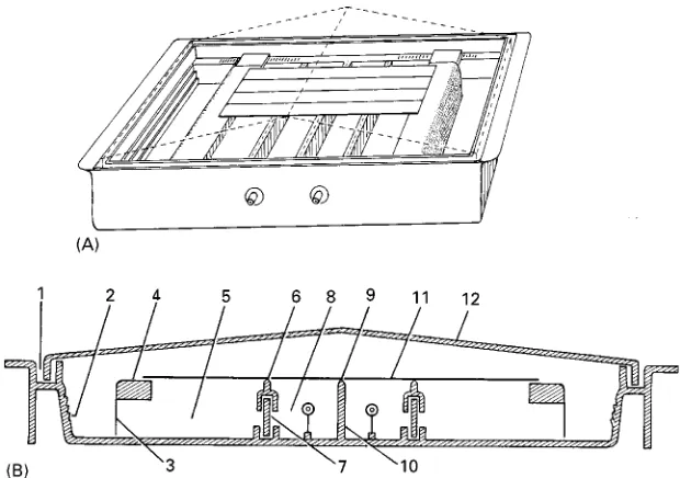
Figure 1 Description of a universal electrophoretic apparatus for CA electrophoresis (redrawn and modified from Kohn, 1957). The CA strips (11) are supported at each end by the shoulder pieces (4) and when taut are just clear of the top edge of the centre partition (10). The top edge of this centre partition is formed as a row of pyramids (9) which support the strip should it tend to sag. When using long strips, strip supports (6) may be fitted to the labyrinth partitions (7) that form the connections between the buffer compartments (5) and electrode compartments (8). Filter paper wicks (3) connect the CA strips to the buffer compartments. The internal sides of the tank are stepped all round (2) as an aid to buffer level checking. The lid (12) fits in a recess (1) moulded all round the tank.
Figure 2 Electrophoretic separation of human haemoglobin variants A, C and S. Ponceau red staining was used to visualize hemoglobin bands, and the anode was on top.
forensic purposes and for the biochemical character-ization and classiRcation of various pathogenic microorganisms such as Leishmania and Trypano-somaspecies.
In addition to one-dimensional electrophoretic methods, two-dimensional CA electrophoretic proto-cols are also available. A summary of important ap-plications is given inTable 2.
Detection and Quantitation
Any protein stain can be used with CA, provided that the solution does not contain a cellulose solvent. Aqueous staining solutions are preferred to alcoholic ones, since with the latter strips tend to shrink and curl unless they are passed through an aqueous bath.
Staining solutions for CA are less concentrated than those used in Rlter paper electrophoresis, and they can be repeatedly used with no appreciable loss of sensitivity.
A wide range of analytical applications can be listed with an impressive variety of fully compatible staining methods, including Coomassie blue brilliant, Ponceau red, Nigrosin, Schiff, gold and silver stain, different types of immuno-staining, and many differ-ent types of enzyme speciRc staining. A 5% (w/w) aqueous solution of acetic acid is a universal washing solution for reducing the background.
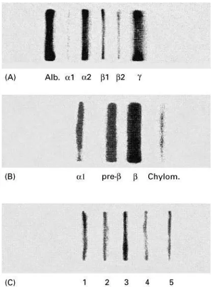
[image:3.568.135.434.582.688.2]Figure 3 Routine clinical electrophoretic separations on CA: (A) serum proteins; (B) lipoproteins; (C) Lactate dehydrogenase isoenzymes. Samples were obtained from healthy patients.
Table 2 Some recent applications of CA electrophoresis Year Application
1994 Introduction of thermocooling apparatus for CA IEF Sequential electrophoresis, with detection of 21 different
alleles in ESD-2 locus inDrosophila buzzatii 1995 Improved separation of apolipoproteins by use of
surfac-tant Tween 20
1996 Rapid screening of biochemical loci of rat
Highly sensitive detection of urinary proteins using col-loidal silver staining
1997 Detection of superoxide dismutase isozymes to distin-guish betweentsetse blood meals of human and non-human origin
CA electrophoresis used as the method of choice for alpha-thalassaemia screening
IEF on CA applied to the analysis of microheterogeneity of immunoglobulins and serum protein fraction Quantitative determinations can be carried out by elution or by scanning of the stained strips. Once stained, protein bands can be easily eluted from the membrane by an appropriate buffer system (a classi-cal system is Tris (2-amino-2-hydroxymethyl-propane-1,3}diol) or Barbitone elution buffer over Ponceau red stained bands). Alternatively, a solvent (e.g. chloroform}ethanol 9 : 1 v/v) can be used to dissolve the membrane and recover the protein
of interest. To enhance the recovery efRciency, gelled CA blocks (about 0.5 cm thick, instead of much thin-ner 0.5 mm supports) can be used.
Scanning is preferred to elution for routine clinical applications. To reduce background and increase sen-sitivity, CA strips should be cleared prior to scanning. As withRlter paper it is important to use oil with the same refractive index as the support. CA strips cleared with oil may be returned to their original dry state by using a solvent such as ether. By contrast, swelling agents such as acetic acid and dioxan used in conjunction with heat treatment, permanently clear CA.
Isoelectric Focusing
CA has ideal features to suit IEF separations. CA is virtually a non-sieving matrix enabling a quasi-free fractionation of macromolecules according to their respective isoelectric points ( pI, the pH at which there occurs an equal number of negative and positive surface charges). CA is easily soaked with very small amounts of carrier ampholyte species, allowing them to be eluted in due course with no damage to stained}destained proteins; this in turn allows den-sitometry measurements and storage.
Unfortunately, the combined effect of CA elec-troosmoticSow and the low ionic strength of com-mercial ampholines can seriously impair the resulting separation of proteins at their isoelectric points. To overcome these drawbacks, CA has been variously treated with surface active agents or with methylating agents. Such treatments can partly } if not wholly }reduce the osmoticSow. Also, a high concentration of carrier ampholytes should be used to cover broad pH ranges (8% v/v instead of the customary 2% v/v) and electrolyte additives at low concentration (such as 0.2 M lysine and 0.2 M acetic acid) should help stabilize narrow pH intervals. Untreated CA strips give better results when 5%-mercaptoethanol and 5 M urea are used as stabilizing agents.
Alternative strategies to circumvent electroosmo-sis, which differ in effectiveness, involve shortening the inter-electrode distance or using ‘chemical space-rs’ to Satten the pH gradients at the appropriate segment of separation. These devices may help to create high Reld strengths with low voltages. Re-cently, thermoelectric cooling has been used to stabil-ize CA IEF gradients.
Counter]ow Af\nity Isotachophoresis
[image:4.568.50.280.532.707.2]proteins are stacked as closely spaced, narrow bands between the ‘leading’ and the ‘trailing’ ions. Iso-tachophoresis on CA gels takes advantage of the absence of sieve effect in this matrix to study sets of interacting biological macromolecules, such as anti-gen/antibody and glycoprotein/lectin systems. How-ever, electroosmosis once again interferes with this application. Abelev and Karamova were able to over-come this drawback by demonstrating that the cath-odic counterSow, combined with the constantSow of liquid through the membrane, stabilizes separations. The counterSow may be also used as a ‘conveyer belt’ to move immunoreagents through antigens or anti-bodies immobilized onto the membrane. Abelev and Karamova used a discontinuous buffer system, in which the two buffers have the same cation and differ in the anion species (chloride as the leading ion and
-alanine as the trailing ion). Under these conditions, macromolecules are separated between the two an-ions.
Abelev and Karamova’s method was originally developed to analyse proteins in highly dilute bio-logical Suids such as urine, tears, and cerebrospi-nal and amniotic Suids, and it turned out to also be useful for detecting low levels of urinary mon-oclonal immunoglobulin light chains (Bence Jones protein) and alpha-fetoprotein in various pathologi-cal conditions.
CA as a Reusable Electrophoretic Support
CA separations are faster than those on other sup-ports, usually with no resolution loss. However, CA sheets cost considerably more than starch, agar, agarose or polyacrylamide gel sheets.
Recently, a wash method has been described that makes it possible to recycle CA strips. The procedure has been shown to work even after using the strips for analysis of a variety of erythrocyte isoenzymes, which notoriously expose the support matrix not only to the strain of the electricReld but also to many somewhat elaborate biochemical colorimetric treatment steps. Surprisingly, none of these stages seem to irreversibly affect the mechanical and physicochemical properties of the CA. In fact, after a variety of enzyme activity tests (adenosine deaminase, adenylate kinase, carbon-ic anhydrase, erythrocyte acid phosphatase, esterase D, glutathione peroxidase, glyoxalase 1, phosphog-lucomutase and 6-phosphogluconate dehydrogenase) Cellogel2+returns to its original features if soaked/ washed in water and methanol for a short time. In the course of double blind trials, no difference in band sharpness and resolution was noticed between new and used Cellogel2+ strips. The procedure can be repeated two or three times if care is taken to avoid
warping strips with absolute methanol soaking or rough handling.
Blotting Proteins from Polyacrylamide Gels to CA Sheets
Different electrophoretic species run in the same gel for the same time with the same electricReld settings. The end of a given experiment is currently set depend-ing on the speciRc requirements of the molecules to be separated, in zone electrophoresis as well as in IEF.
To achieve optimal resolution of different protein constituents of the same sample, various experiments are often carried out, only differing in voltage and duration. To save time, a simple method involves repeatedly blotting a polyacrylamide gel with CA sheets at various stages of separation. The blots ob-tained in this way can be sob-tained and the protein species made to show the optimal resolution.
The advantages that can be obtained from CA blots of the same acrylamide gel are great, the most out-standing being:
1. various stages of a single protein separation can be tested in one experiment, to improve the protocol; 2. common and rare variants of a single elec-trophoretic pattern can be detected, each under optimal separation;
3. several proteins can be analysed at optimal condi-tions in the same experiment;
4. all the allele products may be discriminated by isotacophoretic mechanisms (in non-equilibrium IEF) and isoelectric point (in true equilibrium IEF) within the same run.
Conclusion
Almost uniquely among the various supports for elec-trokinetic separations, CA electrophoresis is still in-tensively used for both research and routine applica-tions. The reasons for this long-lasting success are clear: simplicity of use, low cost, versatility and cost effectiveness. These same factors are likely to provide the general basis for the continuing use of CA in the future.
Acknowledgement
The drawing of Figure 1 was provided by Niccolo` Falchi of the Department of Animal and Human Biology, University of Rome ‘La Sapienza’.
Further Reading
Ambler J (1978) Isoelectric focusing of proteins on cellulose acetate gel membranes. Clinica Chimica Acta 85: 183d191.
Destro-Bisol G and Santini SA (1995) Electrophoresis on cellulose acetate and Cellogel: current status and per-spectives.Journal of Chromatography A698: 33d40. Golias TL (1971) Helena Laboratories Electrophoresis
Manual. Beaumont, Texas: Helena Laboratories. Grunbaum BJ, ed. (1980)Handbook for Forensic
Individ-ualization of Human Blood and Bloodstains. Gottingen: Sartorius.
Harada H (1975) Isoelectrofocusing in cellulose acetate membrane: the method and application.Clinica Chim-ica Acta63: 275d283.
Kohn J (1970) Electrophoresis and immunodiffusion tech-niques on cellulose acetate membrane. Methods in Medical Research12: 243d260.
Meera Khan P (1971) Enzyme electrophoresis on cellu-lose acetate gel: zymogram patterns in man}mouse and man}Chinese hamster somatic cell hybrids.
Archives of Biochemistry and Biophysics 145: 470d483.
Righetti PG (1976)Isoelectric Focusing,Theory,Methods and Applications. Amsterdam: Elsevier.
Schneider RG (1978) Methods for detection of hemoglobin variants and hemoglobinopathies in the routine clinical laboratory.CRC Critical Reviews in Clinical Laborat-ory Sciences9: 243d271.
Deoxyribonucleic Acid, Theory of Techniques for Separation
J. Noolandi, Xerox Research Centre of Canada, Mississauga, Ontario, Canada
Copyright^ 2000 Academic Press
Introduction
Separation of biochemical molecules can be carried out in gels or polymer solutions and, in speciRc cases, in free solution, using constant or variable electric Relds. Gels are used primarily in deep-dish containers, submerged in buffer, and polymer solutions are used in glass capillaries, with inner diameters less than 100m. Thin gels between two glass plates have been used for separating and sequencing single-stranded DNA molecules. We begin the theoretical discussion by considering the separation of double-stranded DNA molecules (dsDNA) in submarine gels under constant electric Reld conditions.
Geometrical Sieving Model for Small
DNA Molecules in a Constant Electric
Field
Ogston was the Rrst to calculate the fractional vol-ume available to a sphere of radius R in a gel of a given concentration. The gel itself was modelled as a random array made up ofRbres of radiusr. Within this description, a sphere with a radiusRrcannot pass through the network if the sphere is not allowed to deform. This geometrical model predicts that the electrophoretic mobility of DNA molecules, as a re-sult of molecular ‘sieving’, varies as:
0J
exp
!Rga
2
[1]whereRgis the radius of gyration of the DNA mol-ecule,is the free solution mobility,ais the average pore size of the gel, and the exponential dependence of the mobility arises from the assumption of Poisson statistics for the distribution of spaces in a random network of straightRbres. This model describes the mobility of small molecules when theyRrst encounter the gel Rbres as obstacles to molecular motion. The analysis of experimental data using eqn [1] is highly model dependent, but can provide some guidance for the development of new gel structures for more efR -cient electrophoretic separations of small molecules.
Entropic Trapping of Small DNA
Molecules
For DNA in the entropic size regime, the deformable molecules select the larger pores in order to maximize locally their conformational entropy. However, in order to accomplish this, they must squeeze through the narrow channels connecting the larger pores. The corresponding polyelectrolyte dynamics is dominated by an activation process in this regime, where the electrophoretic mobility is given by an inverse power law ('1) over a size range that is larger than for the Ogston regime, but smaller than for the beginning of reptation, which is discussed in the next section.