http://www.scirp.org/journal/ojmn ISSN Online: 2163-0585
ISSN Print: 2163-0569
DOI: 10.4236/ojmn.2018.82015 Apr. 11, 2018 174 Open Journal of Modern Neurosurgery
Localizing the Language Network with fMRI
and Functional Connectivity: Implications
for Pre-Surgical Planning
Victoria Lyn Ives-Deliperi
1, James Thomas Butler
1,21Divison of Neurology, Department of Medicine, University of Cape Town, Cape Town, South Africa 2Department of Neurology, University of Stellenbosch, Stellenbosch, South Africa
Abstract
Object: Functional MRI is frequently applied to lateralize language in pre-surgical planning, with potential to localize functionally important cortex too. Here we present BOLD signal activation maps and related functional connectivity, in response to three commonly administered fMRI language tasks. Methods: Datasets from 55 pre-surgical fMRI studies were analyzed. Verbal response naming, covert word generation and passive listening tasks were administered in all studies. Single-subject analyses, group analyses and region-of-interest analyses were conducted, and a multi-subject functional connectivity analysis was performed. Results: Single-subject analyses revealed that clinically important language regions were activated in all but three pa-tients using the panel of tasks. Group analyses revealed significant bilateral BOLD signal increases in anterior and posterior language regions in response to verbal response naming and bilateral signal increase in posterior language regions only in response to passive listening. Covert word generation acti-vated anterior language regions bilaterally and posterior language cortex in the dominant hemisphere. Functional connectivity analyses confirmed that activated regions were significantly correlated in all tasks. Conclusion: The findings of single-subject and group analyses add to the evidence supporting the use of a panel of fMRI tasks to map the language network for pre-surgical planning. Our findings support the additional use of functional connectivity analysis in routine language mapping to add to the localization value to fMRI. In addition, the results of our investigation demonstrate these three com-monly applied tasks reliably activate unique aspects of the language network, which advocates closer individual inspection, guided by the surgical interven-tion planned.
How to cite this paper: Ives-Deliperi, V.L. and Butler, J.T. (2018) Localizing the Lan-guage Network with fMRI and Functional Connectivity: Implications for Pre-Surgical Planning. Open Journal of Modern Neurosurgery, 8, 174-186.
https://doi.org/10.4236/ojmn.2018.82015
Received: January 17, 2018 Accepted: April 8, 2018 Published: April 11, 2018
Copyright © 2018 by authors and Scientific Research Publishing Inc. This work is licensed under the Creative Commons Attribution International License (CC BY 4.0).
DOI: 10.4236/ojmn.2018.82015 175 Open Journal of Modern Neurosurgery
Keywords
Brain Mapping, fMRI, Functional Connectivity, Language, Pre-Surgical Planning
1. Introduction
Neurosurgical intervention carriers the risk of adverse effects on cognitive and motor functioning. Localizing eloquent cortex ensures that functionally impor-tant brain regions are protected during surgery, while maximizing the area of resection for optimal outcome. Language is one of the most common functions to be mapped in neurosurgical planning.
fMRI is considered a reliable tool to identify the language-dominant hemi-sphere, with potential localization value too [1]. Lateralization of language by fMRI and Wada has been shown to be concordant in up to 86% of individuals
[2] [3]. Although direct cortical stimulation mapping (DCS) remains the gold
standard of localizing language cortex, novel approaches to improving the local-ization value of fMRI are encouraged to either expedite DCS or standalone when DCS is unfeasible or unsuccessful [4].
It is recommended that a panel of tasks be administered in fMRI language [5]. The aim of this study was to more closely inspect the variable activation patterns generated by three commonly administered tasks and the functional connec-tivity between activated regions for the benefit of clinicians tasked with pre-surgical language mapping and of interest to neurosurgeons planning resec-tions in the dominant hemisphere. The findings are aimed to inform the devel-opment of methods to localize specific language regions for surgical planning.
2. Materials and Methods
Functional MRI datasets for patients undergoing surgical resections from 2011 to 2016 were retrieved from a single neurosurgical unit. A possible 256 data sets were identified of which 55 fMRI studies included the administration of all three language tasks used for surgical planning in the unit, including verbal response naming, covert word generation and passive listening. Neither ethics approval nor patient informed consent was required for this retrospective study, as out-lined by the National Code for Clinical Trials, as all data analyzed were collected as part of routine diagnosis and treatment.
2.1. fMRI Data Acquisition
DOI: 10.4236/ojmn.2018.82015 176 Open Journal of Modern Neurosurgery angle = 8˚; field of view = 240 × 240; voxel size = 1 × 1 × 1 mm; matrix size = 192 × 192). During the fMRI protocol 110 functional volumes sensitive to blood oxygen level dependent contrast were acquired in all mapping tasks with a T2*-weighted gradient echo, echo planar imaging sequence (TR = 2000 ms, TE = 50 ms, 21 interleaved slices, 5 mm thick, gap 1.25 mm, matrix size 64 × 64, reso-lution 3.4 × 3.4 × 3.4mm3).
2.2. Scanning Protocol
The scan commenced with a 6-minute T1 sequence to generate structural data, followed by the three functional paradigms. Each task consisted of 10 alternating blocks of 20 s commencing with 20 s rest (total = 3:66 min). In all language tasks, stimuli in the control condition consisted of high and low tones to engage auditory processing and attention. Participants were instructed to listen to the tones attentively. In the active condition of the tasks stimuli were projected on a screen at the base of the bed and viewed through an overhead mirror in the head coil. Foam padding was used around the head to reduce motion.
In the active conditions of the tasks patients were asked to a) silently name objects being described by short sentence presented aurally and visually in verbal response naming, b) silently generate verbs associated with nouns presented au-rally and visually in word generation, and c) listen to a story presented auau-rally in passive listening.
2.3. fMRI Data Analyses
All fMRI analyses were performed using Brain Voyager QX (Brain Innovation, Maastricht, The Netherlands). Two dummy images were excluded from analysis in each task run. Images were motion corrected relative to the first volume with trilinear estimation and interpolation. Images were corrected for different slice acquisition times and linear trends, spatially smoothed using a Gaussian filter (FWHM 4 mm), and temporally smoothed with a high pass filter of 3 cycles/ point. Data sets were rejected if they exceeded movement criteria 3 mm dis-placement, 3.0 rotation within a functional run. Each subject’s functional data was co-registered to 1) his/her high-resolution native anatomical MRI for sin-gle-subject analyses, and 2) to their anatomical MRI normalized in Talairach space, rotated into the AC-PC plane, for the benefit of group analyses. The 3.4 × 3.4 × 3.4 mm3 fMRI voxels were interpolated during Talairach normalization to
3 × 3 × 3 mm3.
2.3.1. Whole-Brain Single-Subject and Group Analyses
For each task, a fixed-effect analysis of variance was performed using the general linear model (GLM) with one predictor for the active conditions convolved by the standard hemodynamic function. BOLD signal changes were examined by comparing active and control conditions. The voxel-wise threshold was set to p
DOI: 10.4236/ojmn.2018.82015 177 Open Journal of Modern Neurosurgery contiguous voxels, where the voxel size refers to the 1 × 1 × 1 mm3 resolution of
the iso-voxeled structural images.
2.3.2. Functional Connectivity Analyses
A region-of-interest analysis was performed on the group activation maps gen-erated by each task. The average beta-values in the time-series of all patients in all activated regions during task performance were extracted and entered into a spreadsheet. Beta-values were averaged and correlation analyses were performed on the averaged values in each language region. In addition to this, a subgroup analysis was conducted to investigate potential differences in functional connec-tivity between patients with left TLE, compared to right TLE.
2.3.3. Language Lateralization
Lateralization indices (LI) were calculated in single-subject and group activation map results for each task. All the active voxels in the left hemisphere were sub-tracted from active voxels in the right hemisphere, divided by the active voxels in the left hemisphere added to the active voxels in the right hemisphere, with cut-off values of 0.2/−0.2 used to confirm dominance [6]. Lateralization indices were also calculated from the BOLD activations maps generated in multi-subject analyses of each task and referred to here as groupLIs.
3. Results
The initial patient cohort consisted of 55 pre-surgical patients, 24 males and 31 females, ranging from 20 to 54 years (mean age ± standard deviation [SD], 35.6 ± 13 years). Right-hand dominance was confirmed in 52 patients, two were left-hand dominant and one was mixed-handed. Seizure disorders were present in all but three patients, the latter of whom were awaiting left-hemisphere tumor resections. Of the patients with seizures disorders, 34 were diagnosed with TLE, 22 of whom had dominant hemisphere TLE and 12 in which hippocampal scle-rosis was identified on MRI. Nine patients were diagnosed with frontal lobe epi-lepsy, in which electrographic disturbances in the dominant hemisphere were present in six, five were diagnosed with occipital lobe epilepsy, four of which showed dominant hemisphere abnormalities on EEG, and finally four with epi-leptogenic discharges over cortical dysplasias in the dominant hemisphere. Tu-mors in three of the patients were close to, but not directly infringing upon lan-guage cortex and there were no other patients with mass lesions. All patients with epilepsy were deemed suitable for surgery following long-term video-EEG monitoring, and had fMRI as part of the pre-surgical work-up. All patients were cooperative and able to perform the fMRI tasks.
DOI: 10.4236/ojmn.2018.82015 178 Open Journal of Modern Neurosurgery right-hemisphere dominant for language. These four patients were excluded from further group analyses.
3.1. Group Analyses
3.1.1. Verbal Response Naming
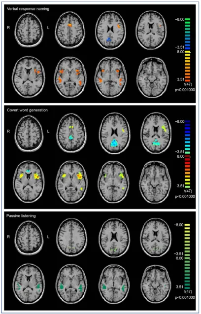
The BOLD signal activation map resulting from the group analysis of 51 patients who underwent verbal response naming revealed significant BOLD signal in-creases in frontal and temporal lobe language centers involved in expressive and receptive language functions (Figure 1). The regions included the posterior, in-ferior aspect of the frontal convexity (BA44/Broca’s area) in the left hemisphere
t(47) = 7.35, p= 0.001 and right hemisphere t(47) = 6.39, p = 0.001, the posterior middle frontal gyrus in the left hemisphere (Exner’s area) t(47) = 6.22, p = 0.001, the supplementary speech area (sensory motor area SMA; BA6) t(47) = 7.46, p = 0.001 and the posterior part of the superior temporal gyri (BA22/Wernicke’s area) bilaterally (t(47) = 6.36, p = 0.001 and t(47) = 6.95, p= 0.001) (Table 1). More intense and wider-spread activations were noted in the left hemisphere and while all the LIs in single-subjects analyses were above 0.1, only 42 of the 51 patients met the criteria for left-hemisphere dominance (LI >/= 0.2). The later-alization index for the group fell below the cut-off at 0.18. Significant signal de-creases were also noted in the precuneus (BA 7) t(47) = −8.21, p = 0.001. Al-though the task generated signal activation in the basal temporal region of the left hemisphere, this was only evident slightly below the threshold of significance (p = 0.0503).
3.1.2. Covert Word Generation
Activation maps generated for the group analysis of the covert word generation task overlapped that of verbal response naming, but only in the dominant hemi-sphere for receptive language function (Figure 1). Significant BOLD signal in-creases were evident in anterior language cortex bilaterally, left; t(47) = 9.29, p= 0.001 and right; t(47) = 6.16, p= 0.001, and in the posterior language region in the left hemisphere t(47) = 5.48, p= 0.001. Significant signal increase was also noted in SMA t(47) = 5.84, p = 0.001, and decreases in the precuneus t(47) = −9.46, p= 0.001 (Table 1). The LIs were greater than 0.2 in all but one patient and the group LI was 0.59.
3.1.3. Passive Listening
DOI: 10.4236/ojmn.2018.82015 180 Open Journal of Modern Neurosurgery Table 1. Functional MRI BOLD signal changes.
Talairach co-ordinates
Brain region BA Voxels Peak x Peak y Peak z Voxel T Verbal response naming
LH Broca’s area BA 44 3853 −48 6 11 7.35 RH Broca’s area BA 44 2766 45 11 3 6.39 LH Exner’s area BA 6 2104 −40 2 27 6.22 LH Wernicke’s area BA 22 2932 −48 −28 −3 6.36 RH Wernicke’s area BA 22 3306 45 −34 −3 6.95 Supplementary speech area BA 6 3370 0 9 43 7.46
LH 1942 −3 9 43 5.67
RH 1428 4 11 41 5.31
Precuneus BA 7 4141 3 −58 27 −8.21
LH 278 −9 −63 22 −4.38
RH 3863 3 −58 27 −5.66
Covert word generation
LH Broca’s area BA 44 12071 −48 8 9 9.29 RH Broca’s area BA 44 3119 33 17 6 6.16 LH Wernicke’s area BA 22 656 −51 −46 0 5.48 Supplementary speech area BA 6 584 −6 11 42 5.85
LH 301 −6 11 42 3.43
RH 283 4 10 42 2.75
Precuneus BA 7 19094 −6 −46 36 −9.46
LH 9942 −6 −46 36 −6.70
RH 9152 8 −57 27 −6.13
Passive listening
LH Wernicke’s area BA 22 5701 −54 −25 −3 7.18 RH Wernicke’s area BA 22 4201 45 −37 3 6.57 LH Precuneus BA 7 1283 −6 −76 24 −5.23 RH Precuneus BA 7 1190 18 −67 12 −5.21
Note: LH, left hemisphere; RH, right hemisphere; BA, Brodmann Area.
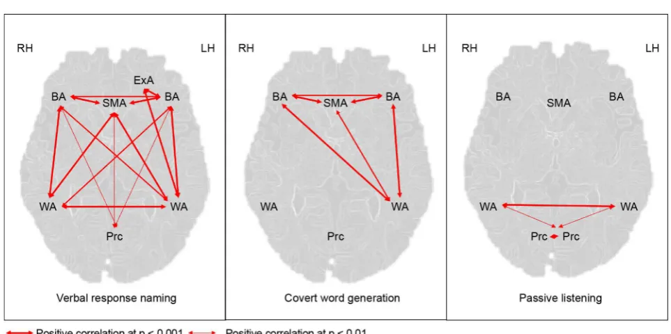
3.2. Functional Connectivity Analysis
Region of interest (ROI) analyses were conducted to further interrogate the group activation maps associated with each of the three language tasks.
DOI: 10.4236/ojmn.2018.82015 181 Open Journal of Modern Neurosurgery 0.96, p < 0.001) and Wernicke’s area in the ipsilateral hemisphere (r = 0.90, p < 0.001), Wernicke’s area in the contralateral hemisphere (r = 0.90, p < 0.001), SMA (r = 0.93, p < 0.001) and precuneus (r = 0.23, p < 0.05). There was a highly significant correlation between Exner’s area and Wernicke’s area in the ipsilat-eral hemisphere (r = 0.89, p < 0.001). Significant positive correlations were also evident between activations in Broca’s area in the right hemisphere and Wer-nicke’s areas in the left hemisphere (r = 0.81, p < 0.001) and right hemisphere (r
= 0.83, p < 0.001), and SMA (r = 0.86, p < 0.001) and precuneus (r = 0.38, p < 0.001). Significantly positive correlations were also evident between activations in Wernicke’s areas in the two hemispheres (r = 0.95, p < 0.001) and Wernicke’s area in the left hemisphere and SMA (r = 0.89, p < 0.001). Activations in Wer-nicke’s area in the right hemisphere was significantly correlated with those in SMA (r = 0.92, p < 0.001).
In response to covert word generation, activations in Broca’s area in the left hemisphere were significantly correlated with those in the right hemisphere (r = 0.95, p < 0.001), Wernicke’s area in the ipsilateral hemisphere (r = 0.90, p < 0.001) and SMA (r = 0.93, p < 0.001). Activations in Broca’s area in the right hemisphere was also strongly correlated with activation in Wernicke’s area in the left hemisphere (r = 0.86, p < 0.001) and SMA (r = 0.90, p < 0.001). Signifi-cant correlations were also evident between activations in Wernicke’s area in the left hemisphere and SMA (r = 0.88, p < 0.001).
Activations in Wernicke’s area in the left and right hemispheres in response to passive listening were significantly correlated (r = 0.96, p < 0.001) and activa-tions in Wernicke’s area in the left hemisphere were significantly correlated with those in the left and right precuneus (LH: r = 0.20, p < 0.05 and RH: (r = 0.27, p < 0.01). Bilateral precuneus activation were also highly correlated (r = 0.91, p < 0.001). These results are graphically depicted in Figure 2.
According to the subgroup analyses, there was no significant difference in connectivity within the language network between patients with dominant TLE and non-dominant TLE, although there was a trend towards weaker correlations between activated regions patients with left TLE.
4. Discussion
DOI: 10.4236/ojmn.2018.82015 182 Open Journal of Modern Neurosurgery Figure 2. Functional connectivity between activated regions in the three language tasks.
between the language regions showing significant BOLD signal increases in all three tasks are functionally connected, which supports the notion that the re-gions subserve language and 5) there were no significant differences in connectivity between language regions in patients with dominant TLE and non-dominant TLE.
4.1. Language Lateralization
Determining hemispheric laterality may be achieved by either calculating a LI or visual inspection of fMRI activation maps by experienced clinicians [4]. Lan-guage lateralization was deemed satisfactory by visual inspection in all 51 sin-gle-subject analyses in this study, across all tasks. Hemispheric dominance was confirmed by calculating LIs in 80% of the single-subject activation maps gener-ated by the verbal response naming and passive listening tasks, and in 100% of related activation maps generated by covert word generation. Verbal response naming and passive listening generate bilateral signal increases, however, activa-tions in the left hemisphere are more intense and wider spread than in the right hemisphere. Right hemisphere signal activations in anterior and posterior lan-guage cortex were more striking in the group analyses than in single-subject analyses, and consistent with findings from resting state connectivity analyses [9].
4.2. Language Localization
4.2.1. Anterior Language Regions
DOI: 10.4236/ojmn.2018.82015 183 Open Journal of Modern Neurosurgery verbal response naming and covert word generation reliably activated Broca’s area but only the former task engaged Exner’s areas. In four out of five patients who underwent DCS in our cohort, speech arrest occurred in a congruent loca-tion to fMRI activaloca-tions in Broca’s area. While Exner’s area is seldom a target for localization during DCS at our center, stimulation of the region in the DLPFC has shown to temporarily disrupt handwriting [11]. Verbal response naming and covert word generation both produced reliable activation in supplementary speech area (SMA), bilaterally in this investigation. This region is involved in initiating and sequencing of motor movements for speech and the extent of its involvement in language is highly variable between patients [8]. Although uni-lateral resections may produce transient aphasia, the condition recovers post-operatively and thus SMA cannot strictly be considered a critical language area [12] [13]. fMRI activations in this region are potentially informative nonetheless, in that the presence of BOLD signal changes in the contralateral healthy SMA and diminished BOLD signal in the ipsilateral SMA in patients with low grade glial tumours have been shown to correlate with the speed of recovery from the post-operative SMA syndrome [12].
4.2.2. Posterior Language Regions
The localization of critical receptive language regions in the temporal lobe is more challenging, both in fMRI and DCS. Primary areas targeted in mapping of receptive language include Wernicke’s’ area, Angular gyrus and basal temporal regions. DCS stimulation in the region of angular gyrus has been shown to in-terrupt handwriting and repetition and disrupt naming, comprehension, reading and repetition when applied to more basal regions of the temporal lobe, fusiform and parahippocampal gyri [4] [13].
Verbal response naming and covert word generation involve reading, com-prehension of the written sentence and naming and thus, predictably, activate the posterior language regions demonstrated in this investigation. Verbal re-sponse naming was the only task in the panel of three to activate the basal tem-poral language region, according to groups analyses, possibly related to the in-creased reading load of the task. Although the covert word generation task pro-duced signal activations in basal temporal region below the threshold of signifi-cant, activations in the regions were noted in a number of single-subject analyses in response to the task. Surgical resection of the basal temporal region, however, seldom produces lasting language deficits so, like SMA, may be considered a secondary language area [14]. The findings from the analyses of the passive lis-tening task were helpful in this investigation, in so far as that the location of sig-nal activations corroborated findings from the other tasks, and produced the most intense and widest activation in Wernicke’s area.
DOI: 10.4236/ojmn.2018.82015 184 Open Journal of Modern Neurosurgery question how best critical language cortex in the temporal lobe may be system-atically mapped. This warrants further investigation. The margins of cranioto-mies exposed the regions of temporal lobe activated in the fMRI tasks in only two of the five patients in our cohort who underwent awake speech mapping. Anomia was elicited in one of these patients when Wernicke’s area was stimu-lated.
BOLD signal decreases were evident in the precuneus in response to all three language tasks in this study, consistent with previous findings of reduced activity in the region during the performance of a variety of higher-order cognitive func-tions [15].
4.3. Functional Connectivity of Language Regions
Studies have shown reduced functional connectivity between language regions in patient with TLE compared to healthy subjects [16] [17]. This is likely due to in-tra-hemispheric and interhemispheric language reorganization in localized epi-lepsy [18]. Recent studies have also demonstrated reduced connectivity in the language network between patients with dominant and non-dominant TLE [9]. No significant differences in connectivity between the groups was evident in this study, perhaps due to our larger cohort.
5. Conclusions
The findings of this investigation are informative in so far as they may guide fMRI task selection and analysis in pre-surgical fMRI language mapping, guided by the proposed area for resection. Since verbal response naming generates widespread, bilateral BOLD signal increases in the language network, this task would be the most useful when there is a requirement to map the entire language network, for example in speech and language research. Should language laterali-zation be the primary requirement, then the task of choice would be covert word generation, which produces the most discrete activations in anterior and poste-rior language regions in the dominant hemisphere, with the most impressive lateralization value of the three tasks. It would be particularly useful to include a passive listening task when attempting to localize posterior language regions in the case of temporal lobe resections. The addition of functional connectivity analyses in fMRI studies helps to further corroborate the assumptions made about localization of eloquent cortex from task-based fMRI findings.
DOI: 10.4236/ojmn.2018.82015 185 Open Journal of Modern Neurosurgery protocol for the mapping white matter tracts involved in language, as well as DCS of these tracts, is also strongly recommended for the preservation of func-tion [19].
Disclosures
The authors report no conflict of interest concerning the materials or methods used in this study or the findings specified in this paper.
References
[1] Szaflarski, J.P., Binder, J.R., Gaillard, W.D., Golby, A.J., Holland, S.K., Ojemann, J.,
etal. (2017) Practice Guideline Summary: Use of fMRI in the Presurgical Evaluation of Patients with Epilepsy. Report of the Guideline Development, Dissemination, and Implementation Subcommittee of the American Academy of Neurology. Neurol-ogy, 88, 395-402. https://doi.org/10.1212/WNL.0000000000003532
[2] Abbott, D.F., Waites, A.B., Lillywhite, L.M. and Jackson, G.D. (2010) fMRI Assess-ment of Language Lateralization: An Objective Approach. Neuroimage, 50, 1446-1455. https://doi.org/10.1016/j.neuroimage.2010.01.059
[3] Janecek, J.K., Swanson, S.J., Sabsevitz, D.S., Hammeke, T.A., Raghavan, M., Roz-man, M., etal. (2013) Language Lateralization by fMRI and Wada Testing in 229 Patients with Epilepsy: Rates and Predictors of Discordance. Epilepsia, 54, 314-322.
https://doi.org/10.1111/epi.12068
[4] Benjamin, C.F., Walshaw, P.D., Hale, K., Gaillard, W.D., Baxter, L.C., Berl, M.M., et al. (2017) Presurgical Language fMRI: Mapping of Six Critical Regions. Human BrainMapping, 38, 4239-4255. https://doi.org/10.1002/hbm.23661
[5] Duncan, J.S. (2010) Imaging in the Surgical Treatment of Epilepsy. NatureReview Neurology, 6, 537-550. https://doi.org/10.1038/nrneurol.2010.131
[6] Seghier, M.L. (2008) Laterality Index in Functional MRI: Methodological Issues.
MagneticResonanceImaging, 26, 594-601.
https://doi.org/10.1016/j.mri.2007.10.010
[7] Smits, M., Visch-Brink, E., and Schraa-Tam, C.K. (2006) Functional MR Imaging of Language Processing: An Overview of Easy-to-Implement Paradigms for Patient Care and Clinical Research. RadioGraphics, 26, 145-159.
https://doi.org/10.1148/rg.26si065507
[8] Brennan, N.P., Peck, K.K. and Holodny, A. (2016) Language Mapping Using fMRI and Direct Cortical Stimulation for Brain Tumor Surgery: The Good, the Bad, and the Questionable. TopicsinMagneticResonanceImaging, 25, 1-10.
https://doi.org/10.1097/RMR.0000000000000074
[9] Pravata, E., Sestieri, C., Mantini, D., Briganti, C., Colicchio, G., Marra, C., etal. (2011) Functional Connectivity MR Imaging of the Language Network in Patients with Drug-Resistant Epilepsy. AmericanJournalofNeuroradiology, 32, 532-570.
https://doi.org/10.3174/ajnr.A2311
[10] Keller, S.S., Crow, T., Foundas, A., Amunts, K. and Roberts, N. (2009) Broca’s Area: Nomenclature, Anatomy, Typology and Asymmetry. BrainLanguage, 109, 29-48.
https://doi.org/10.1016/j.bandl.2008.11.005
[11] Roux, F.E., Dufor, O., Giussani, C., Wamain, Y., Draper, L., Longcamp, M. and Demonet, J.F. (2009) The Graphemic/Motor Frontal Area Exner’s Area Revisited.
DOI: 10.4236/ojmn.2018.82015 186 Open Journal of Modern Neurosurgery
[12] Krainik, A., Lehericy, S., Duffau, H., Capelle, L., Chainay, H., Cornu, P., etal. (2003) Postoperative Speech Disorder after Medial Frontal Surgery: Role of the Supple-mentary Motor Area. Neurology, 60, 587-594.
https://doi.org/10.1212/01.WNL.0000048206.07837.59
[13] Binder, J.R. (2015) The Wernicke Area. Neurology, 85, 2170-2175.
https://doi.org/10.1212/WNL.0000000000002219
[14] Lüders, H., Lesser, R.P., Hahn, J., Dinner, D.S., Morris, H.H., Wyllie, E., etal. (1991) Basal Temporal Language Area. Brain, 114, 743-754.
https://doi.org/10.1093/brain/114.2.743
[15] Shulman, G.L., Fiez, J.A., Corbetta, M., Buckner, R.L., Meizin, F.M., Raichle, M.E.,
et al. (1997) Common Blood Flow Changes across Visual Tasks: II. Decreases in Cerebral Cortex. JournalofCognitiveNeuroscience, 9, 648-663.
https://doi.org/10.1162/jocn.1997.9.5.648
[16] Kucukboyaci, N.E., Kemmotsu, N., Cheng, C.E., Girard, H.M., Tecoma, E.S., Iragui, V.J., etal. (2013) Functional Connectivity of the Hippocampus in Temporal Lobe Epilepsy: Feasibility of a Task-Regressed Seed-Based Approach. BrainConnectivity, 3, 464-474. https://doi.org/10.1089/brain.2013.0150
[17] Maccotta, L., He, B.J., Snyder, A.Z., Eisenman, L.N., Benzinger, T.L., Ances, B.M., et al. (2013) Impaired and Facilitated Functional Networks in Temporal Lobe Epi-lepsy. Neuroimage: Clinical, 2, 862-872. https://doi.org/10.1016/j.nicl.2013.06.011
[18] Gaillard, W.D., Berl, M.M., Moore, E.N., Ritzl, E.K., Rosenberger, L.R., Weinstein, S.L., etal. (2007) Atypical Language in Lesional and Nonlesional Complex Partial Epilepsy. Neurology, 69, 1761-1771.
https://doi.org/10.1212/01.wnl.0000289650.48830.1a