IN THE DEVELOPMENT
OF STARCH ACCUMULATING STRUCTURES
A thesis
submitted in partial fulfilment of the requirements for the Degree
of
Doctor of Philosophy in the
University of Canterbury by
Paula E. Jameson
DEDICATION
TABLE OF CONTENTS
LIST OF TABLES
LIST OF FIGURES
ABSTRACT
CHAPTER I INTRODUCTION
CHAPTER II CYTOKININ BIOASSAYS
2.1 Introduction
2.2 Materials and Methods
PAGE
iii vii
xii
xvi;; 1
41
41
42
2.2.1 Initiation of soybean callus clones 42
2.3
2.2.2 Maintenance of a soybean callus clone obtained from Plant Diseases Division,
DSIR, Auckland 43
2.2.3 Bioassays of plant extracts and
esti-mation of cytokinin-like activity 44
2.2.4 Soybean hypocotyl bioassay 45
Results 47
2.3.1 Initiation of soybean callus clones 47
2.3.2 Maintenance of callus clone obtained
from DSIR 54
2.3.3 Soybean hypocotyl bioassay 59
2.4 Discussion 61
64
64
64.
64
64
65
CHAPTER III CHROMATOGRAPHY SYSTEMS
3.1 Introduction
3.2 Materials and Methods
3.2.1 Chemicals
3.2.2 Paper chromatography
PAGE
CHAPTER III (Continued)
3.3 Results 66
3.4 Discussion 68
CHAPTER IV POTATO 71
4.1 Introduction 71
4.2 Materials and Methods 71
4.2.1 Preliminary experiments with mature
s.
tuberosum tubers 714.2.2 Controlled Growth Room Experiments
(Canterbury) 74
4.2.3 Potato Field Trial (Lincoln,
mid-Canterbury) 77
4.2.4 Controlled Environment Experiments~
Plant Physiology Division, DSIR,
Palmerston North 80
4.3 Results 88
4.3.1 Preliminary experiments with mature
S. tuberosum cultivar Ilam Hardy tubers 88
4.3.2 Controlled Growth Room Experiments
(Canterbury) 95
4.3.3 Potato Field Trial (Lincoln~
mid-Canterbury) 97
4.3.4 Controlled Environment Experiments, Plant Physiology Division, DSIR~
Palmerston North 111
4.3.4.1 PPD Experiment 1 111
4.3.4.2 Observations on the effects of different growth conditions on the tuberisation response of
S. andigena cv 165 141
4.4 Discussion 146
4.4.1 Preliminary Experiments 146
4.4.2 Plant Physiology Division Experiment One 155
4.4.3 Tuberisation response of S. andigena
PAGE
CHAPTER V TUBER FORMATION IN VITRO 180
5.1 Introduction 180
5.2 Materia1s and Methods 181
5.2.1 Tuberisation in vitro. Experiment 1 181 5.2.2 Tuberisation in vitro. Experiment 2 182 5.2.3 Tuberisation in vitro. Experiment 3 183
5.3 ResuHs 184
5.3.1 Tuberisation in vitro. Experiment 1 184 5.3.2 Tuberisation in vitro. Experiment 2 186 5.3.3 Tuberisation in vitro. Experiment 3 191
5.4 Discussion 193
CHAPTER VI WHEAT 200
6.1 Introduction 200
6.2 Materia1s and Methods 200
6.2.1 Wheat Fie1d Tria1 1 200
6.2.2 Wheat Fie1d Tria1 2 204
6.3 ResuHs 209
6.3.1 Wheat Fie1d Tria1 1 209
6.3.2 Wheat Fie1d Tria1 2 218
6.4 Discussion 254
CHAPTER VII IDENTIFICATION OF CYTOKININS IN WHEAT GRAINS 273
7.1 Introduction 273
7.2 Materia1s and Methods 275
7.2.1 Trimethy1si1y1ation 275
7.2.2 Permethy1ation 275
7.2.3 Gas chromatography and combined gas
PAGE
CHAPTER VII (Continued)
7.3 Results 277
7.3.1 Zeatin riboside 277
7.3.2 Zeatin 280
7.3.3 Quantification 287
7.4 Discussion 287
CHAPTER VI II GENERAL DISCUSSIONS AND CONCLUSIONS 291
ACKNOWLEDGEMENTS 294
REFERENCES 295
LIST OF TABLES
TABLE PAGE
2.1 Response of hypocotyl segments to different cytokinins 61
3.1 Soybean callus bioassays of authentic zeatin riboside (A) and zeatin (B) following partition chromatography
on Sephadex LH-20 using 35% ethanol as eluant 68
4.1 Potato Field Trial. List of harvest dates, total number of rows harvested and size categories to which
excised plant material was allocated 78
4.2 PPD Experiment 1. Harvest schedule detailing the number of pails and plants harvested after a given
time in the controlled environment rooms 81
4.3 PPD Experiment 1. Harvest schedule listing the categories excised stolon and tuber material were allocated to, the number of stolons or tubers ground prior to extraction and the fresh weight of material
extracted 83
4.4 PPD Experiment 1. Extraction schedule listing the number of stolons or tubers ground prior to extraction, and the fresh weight and dry weight of material
extracted 84
4.5 PPD Experiment 1: Starch and Soluble Sugar Analyses. Details of the analyses showing the dry weight, fresh weight and number of stolons or tubers ground per sample, and the dry weight extracted per replicate. The volumes to which the starch hydrolysates were diluted and the aliquots removed for incubation with
glucose oxidase are also indicated 85
4.6 PPO Experiment 2. Harvest schedule detailing the number of pails (and plants) harvested after a
specified time in each of the controlled environment
rooms 88
4.7 Soybean callus bioassay of petroleum ether- and butan-l-ol-soluble components of potato tuber extract. The butanol-soluble components were resolved by partition
chromatography on Sephadex LH-20 94
4.8 Field Trial 1. Comparison of the total cytokinin-like activity of stolons from harvest 1 and tubers of
diameter 4 to 7.5 mm from harvest 4 111
4.9 PPD Experiment 1. Percentage tuberisation of S. andigena
plants under long day conditions in controlled
TABLE
4.10 PPO Experiment 1. The number of plants harvested, total weight and number of stolons and tubers per plant and the total number of tubers per plant is
PAGE
presented for each harvest 113
4.11 PPO Experiment 1. Range of tuber sizes and the proportion of the total tuber number contributing to approximately 50% of the total fresh weight of
tubers 119
4.12 PPO Experiment 1. Starch content of stolons and
tubers less than 7.5 mm in diameter 119 4.13 PPO Experiment 1. Soluble sugar content of stolons
and tubers less than 7.5 mm diameter 122 4.14 PPO Experiment 1. Estimation of the cytokinin-like
activity present in 4 g fresh weight samples of
stolon and tuber material 133
4.15 PPO Experiment 1. Estimation of the cytokinin-like activity present in 8 g eq FW of stolon and tuber extracts. The activity was resolved by partition chromatography on Sephadex LH-20 eluted with 20%
ethanol 137
4.16 PPO Experiment 1. Comparision of the amount of cytokinin-like activity present in fraction 9 and fraction 12 following the extraction of 4 g eq FW and 8 g eq FW respectively of stolons and tubers. The activity was resolved by partition chromatography on Sephadex LH-20 eluted with 35% ethanol (4 g eq FW)
and 20% ethanol (8 g eq FW), respectively 139 4.17 PPO Experiment 1. Contribution from S. andigena tubers
of the size categories 10 to 15 mm and 15 to 20 mm diameter to the total number and weight of stolons and
tubers per plant 141
4.18 Response of S. andigena to long day and short day
conditions 141
4.19 Field harvest dates, storage conditions and length of storage of S. andigena tubers used in all controlled
environment experiments 142
4.20 Percentage tuberisation attained by S. andigena plants
grown from seed tubers with different storage histories 143 4.21 Growth of S. andigena under differing conditions in the
CE rooms (PPO). Plants were subjected to different day/
night temperature regimes 144
4.22 Response of S. andigena plants to differing amounts of photosynthetically active radiation (PAR). Seed tubers were planted within one week of Harvest., Oay and night
TABLE
4.23 A comparison of the response of S. andigena plants grown in different controlled growth rooms at Canterbury (experi'ment 3). Both light intensity
PAGE
and temperature differed between the rooms 146
5.1 Tuberisat;on in vitro. Experiment 1. Combinations of sucrose and kinetin with which the modified
Murashige and Skoog nutrient agar was supplemented 182
5.2 Tuberisation in vitro. Experiment 2. Culture
details listing the number of apical and medial segments incubated on Murashige and Skoog nutrient agar
supple-mented with different combinations of sucrose and kinetin 183
5.3 Tuberisation in vitro. Experiment 3. Culture details listing the number of whole segments and single-nodal segments of S. tziberos1Am and S. andigena transferred
onto Media 5 and 6 184
5.4 Tuberisation in vitro. Experiment 1. In vitro culture
of sprout segments removed from S. andigena tubers. 185
5.5 Tuberisation in vitro. Experiment 2. Tuber formation by medial sprout segments cultured on medium supple-mented with different concentrations of sucrose and
kinetin 188
5.6 Tuberisation in vitro. Experiment 3. Tuber formation by second transfer shoots obtained from sprout segments
cultured in vitro 192
6.1 Wheat Field Trial 1. Harvest schedule listing the dates, developmental stage relative to ear emergence and anthesis, the number of ears dissected in the field per sample, and the number dissected in the laboratory
as a "bulk sample" at each respective harvest 201
6.2 Wheat Field Trial 1. Extraction sequence detailing the number of grains extracted, the dry weight of sample extracted, the volume of 80% methanol used for extrac-tion of individual samples over each 24 hour period, and the amount of solvent used to wash the retained
residue following each filtration 203
6.3 Wheat Field Trial 2. Harvest schedule listing the date, developmental stage relative to ear emergence and
anthesis, the number of ears dissected in the field per sample, and the number dissected in the laboratory as
TABLE
6.4 Wheat Field Trial 2. Extraction sequence detailing the number of grai ns extracted, the dry weight of sample extracted, the volume of 80% methanol used for extraction of individual samples over each 24 hour period. and the amount of solvent used to wash the
PAGE
retained residue following each filtration 206
6.5 Wheat Field Tr$al 2. Starch and Soluble Sugar Analyses. Details of the analyses showing the dry weight, fresh weight and number of grains ground per sample, and the dry weight extracted per replicate. The volumes to which starch hydrolysates were
diluted and the aliquots removed for incubation with
glucose oxidase are also indicated 210
6.6 Wheat Field Trial 1. Quantification of cytokinin-like
activity in 500 wheat pistils collected at pollination 212
6.7 Wheat Field Trial 1. Quantification of cytokinin-like
activity in 500 wheat grains collected at anthesis 216
6.8 Wheat Field Trial 1. Quantification of cytokinin-like activity in 500 wheat grains collected three days
post-anthesis 216
6.9 Wheat Field Trial 1. Quantification of cytokinin-like activity in 500 wheat grains collected seven days
post-anthe~is 218
6.10 Wheat Field Trial 2. Quantification of cytokinin-like activity in extracts of 500 pistils collected at
pollination (nine days after ear emergence) 226
6.11 Wheat Field Trial 2. Quantification of cytokinin-like activity in extracts of 500 wheat grains collected at
anthesis (10 days after ear emergence) 230
6.12 Wheat Field Trial 2. Quantification of the cytokinin-like activity in extract of 500 wheat grains collected in the Ilbulk samplell
two days post-anthesis (12 days
after ear emergence) 233
6.13 Wheat Field Trial 2. Quantification of the cytokinin-like activity in extracts of 100 wheat grains" collected
two days post-anthesis (12 days after ear emergence) 233
6.14 Wheat Field Trial 2. Quantification of the cytokinin-like activity in extracts of 100 wheat grains collected two days post-anthesis (12 days after ear emergence).
The Sephadex LH-20 chromatography eluant was 20% ethanol 235
6.15 Wheat Field Trial 2. Quantification of the cytokinin-like activity collected in fractions 7 to 16 following the treatment of fractions 3 to 9 with S-glucosidase and rechromatography. Extracts of 100 grains from three samples collected two days post-anthesis were treated
TABLE
6.16 Wheat Field Trial 2. Comparison of the total cytokinin-like activity detected in fractions 3 to 9 before and after S-glucosidase treatment of extracts of 100 wheat
PAGE
grains collected two days post-anthesis 236 6.17 Quantification of the cytOkinin-like activity in
extracts of 100 wheat grains collected in the IIbu"lk sample" two days post-anthesis following extraction with either 80% methanol or Bieleski's solvent
cocktail 240
6.18 Wheat Field Trial 2. Quantification of the cytokin;n-like activity in extracts of 500 wheat grains collected
four days post-anthesis (14 days after ear emergence) 240 6.19 Wheat Field Trial 2. Quantification of the
cytokinin-like activity in extracts of 100 wheat grains collected four days post-anthesis (14 days after ear emergence) 241 6.20 Wheat Field Trial 2. Quantification of the
cytokinin-like activity in extracts of wheat grains collected four days post-anthesis (14 days after ear emergence). The
Sephadex LH-20 chromatography eluant was 20% ethanol 245 6.21 Wheat Field Trial 2. Quantification of the
cytokinin-like activity in extracts of 500 wheat grains collected
seven dayspost-anthesis (17 days after ear emergence) 249 7.1 Relative intensities of high mass ions in the mass
spectrum obtained from direct probe insertion of
1,000 ng trimethylsilylated zeatin riboside 277 7.2 Relative peak heights of ions diagnostic of TMS4
-zeatin riboside obtained following the injection of 1,000 ng of trimethylsnylated zeatin riboside standard and multiple ion monitoring of ions at
m/z 639, 624 and 536 280
7.3 Relative peak heights of ions diagnostic of TMS4
-zeatin riboside obtained following the injection of a 2 ~l aliquot of wheat sample and multiple ion monitoring of ions at A: m/z·639, 624 and 536;
and B: m/z 624, 550 and 549 . 284
7.4 Relative intensities of high mass ions in the mass spectrum obtained from direct probe insertion of
FIGURE 2.1 2.2 2.3 2.4 2.5 2.6 2.7 2.8 2.9 3.1 3.2 4.1 4.2
LIST OF FIGURES
Responses of callus cultured from soybean cultivars Kitamasume (clone A) and Okuhara to increasing kinetin concentrations and two agar concentrations
Response of callus cultured from soybean cultivars Kitamasume (clones A and B) and Okuhara to increasing kinetin concentrations
Soybean callus .bioassays terminated after 14, 21, 28 and 35 days
Comparison of soybean callus bioassays set up with callus passed through different numbers of sub-cultures
Comparison of soybean callus bioassays set up with
A 7 mg transplants
B 20 mg transplants
C callus maintained 7 days on medium devoid of cytokinin prior to starting the bioassay
Comparison of soybean callus bioassays using callus at different ages post-transfer and maintained on either kinetin or 6-benzylaminopurine
Regression lines calculated from the growth of callus on kinetin standards,
Response of callus clone Bap to different concentrations of zeatin, zeatin riboside and kinetin
Soybean hypocotyl bioassay. Response of hypocotyl segments from soybean cv Caloria to different kinetin concentrations
Mobilities of zeatin riboside and zeatin (in propan-2-01 - ammonia - water (10:1:1 v/v) on Whatman 3 MM chromatography paper. Compounds were detected- by the soybean callus bioassay
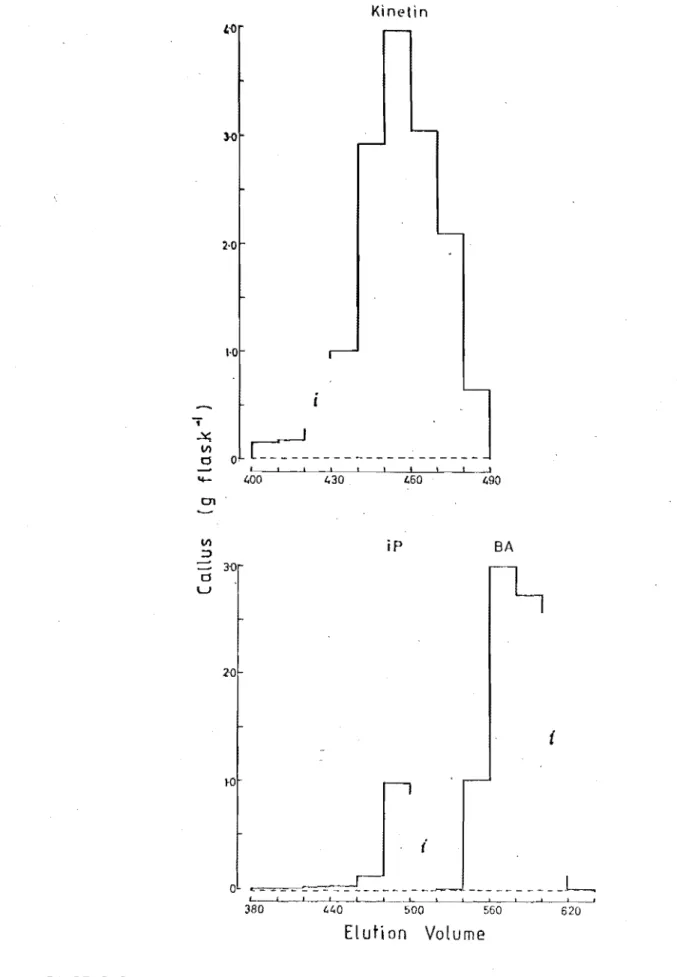
Elution volumes of kinetin, isopentenyladenine and 6-benzylaminopurine following partition chromatography on Sephadex LH-20 with 35% ethanol as eluant. Compounds were detected by the soybean callus bioassay
Soybean callus bioassay of paper chromatograms loaded with methanolic extract of mature tuber tissue
Soybean callus bioassay of paper chromatograms loaded with aqueous and butan-1-01 soluble components of mature tuber tissue extract
FIGURE PAGE 4.3 Soybean callus bioassay of paper chromatograms loaded
with methanolic extract and extract partially purified
by Oowex 50 (H+ form) cation exchange chromatography 92 4.4 Soybean callus bioassay of methanolic extract and of
extract partially purified by Oowex 50 (H+ form)
cation exchange chromatography 93
4.5 Soybean callus bioassay of paper chromatograms loaded with extract of Ilam Hardy "bolterll
plants. Tubers were removed from plants under SO conditions, stolons
from plants under LO conditions 96
4.6 Soybean callus bioassay of extract of
s.
andigena cv 165. Tubers were removed from plants under SOconditions, stolons from plants under LO conditions 98 4.7 Soybean callus bioassay of paper chromatograms loaded
with aqueous and butan-1-ol soluble components of
extract of
s.
andigena cv 165 tubers. 99 4.8 Potato Field Trial. Fresh weight increase withincreasing tuber size 101
4.9 Potato Field Trial. Soybean callus bioassay of paper chromatograms loaded with extract of
s.
tubeposum cvIlam Hardy seed tubers 102
4.10 Potato Field Trial. Soybean callus bioassay of extract
of S. tubeposum cv Ilam Hardy seed tubers 103 4.11 Potato Field Trial. Soybean callus bioassay of paper
chromatograms loaded with aqueous- and butanol-soluble
components of stolon tip extracts 104
4.12 Potato Field Trial. Soybean callus bioassay of stolon
extract 105
4.13 Potato Field Trial. Soybean callus bioassay of paper chromatograms loaded with aqueous- and butan-1-ol-soluble components of extracts of tubers less than 4 mm
diameter 106
4.14 Potato Field Trial. Soybean callus bioassay of paper chromatograms loaded with aqueous- and butan-1-ol-soluble components of extracts of tubers between 4.0
and 7.5 mm diameter 107
4.15 Potato Field Trial. Soybean callus bioassay of extract
of tubers 4 to 7.5 mm diameter 108
4.16 PPO Experiment 1. Growth analysis of the population of plants sampled between 21 and 84 days after transfer to controlled environment rooms. Relative rate of tuber weight increase per plant as a function of time after
FIGURE 4.17 4.18 4.19 4.20 4.21 4.22 4.23 4.24 4.25 4.26 4.27 4.28 4.29 4.30 4.31 4.32
PPD Experiment 1. Growth analysis of the population of plants sampled between 21 and 84 days after transfer to controlled environment rooms. Each size category of stolons and tubers is presented as a proportion of the total stolon and tuber weight per plant as a function of increasing plant age
PPO Experiment 1. Growth analysis of the population of plants sampled between 21 and 84 days after transfer to controlled environment rooms. Each size category of stolons and tubers is presented as a proportion of the total stolon and tuber number per plant as a function of increasing plant age
PPO Experiment 1. Increase in dry weight and fresh weight as a function of tuber diameter
PPO Experiment 1. Starch content of stolons and tubers as a function of tuber diameter
PPO Experiment 1. Soluble sugar content of stolons and tubers as a function of tuber diameter
PPO Experiment 1. Soybean callus bioassay of extracts of stolons tips (4 g eq FW)
PPO Experiment 1. Soybean callus bioassay of extracts of tubers 1. 5 to 3 mm di ameter (4 g eq FW)
PPO Experiment 1. Soybean callus bioassay of extracts of tubers 3 to 5 mm diameter (4 g eq FW)
PPO Exper'j ment 1. Soybean ca 11 us bi oassay of extracts of tubers 5 to 7.5 mm diameter (4 g eq FW)
PPO Experiment 1. Soybean callus bioassay of extracts of tubers 7.5 to 10 mm diameter (4 g eq FW)
PPO Experiment 1. Soybean callus bioassay of extracts of tubers 10 to 15 mm diameter (4 g eq FW)
PPO Experiment 1. Soybean callus bioassa¥ of extracts of tubers 15 to 20 mm diameter (4'g eq FW) ,
PPO Experiment 1. Soybean callus bioassa¥ of extracts of tubers 20 to 25 mm diameter (4 g eq FW)
PPO Experiment 1. Soybean callus bioassay of extracts of tubers 40 to 50 mm diameter (4 g eq FW)
PPO Experiment 1. Total cytokinin-like activity in potato tubers following the extraction of 4 g eq FW tissue.
PPD Experiment 1. Soybean callus bioassay of extracts of stolons and tubers of the sizes specified in the figure (8 g eq FW)
FIGURE
4.33 4.34 6.1 6.2 6.3 6.4 6.5 6.6 6.7 6.8 6.9 6.10 6.11 6.12 6.13PPO Experiment 1. Cytokinin-like activity in stolons and tubers expressed as kinetin equivalents (components)
PPO Experiment 1. Cytokinin-like activity in stolons and tubers expressed as kinetin equivalents (total)
Wheat Field Trial 1. Fresh weight and dry weight of grains as a function of days from ear emergence
Wheat Field Trial 1. Soybean callus bioassay of extracts of 500 wheat pistils collected 10 days after ear emergence (pollination)
Wheat Field Trial 1. Soybean callus bioassay of extracts of 500 grains collected 11 days after ear emergence
(anthesis)
Wheat Field Trial 1. Soybean callus bioassay of paper chromatograms loaded with extract of 500 grains
collected 11 days after ear emergence (anthesis)
Wheat Field Trial 1. Soybean callus bioassay of extracts of 500 wheat grains collected 14 days after ear emer-gence (3 days post-anthesis)
Wheat Field Trial 1. Soybean callus bioassay of extracts of 500 wheat grains collected 18 days after ear emer-gence (7 days post-anthesis)
Wheat Field Trial 1. Total Cytokinin-like activity in wheat grains between pollination and seven days post-anthesis (10 and 18 days after ear emergence)
Wheat Field Tri.al 2. Fresh weight and dry weight of grains as a function of time from ear emergence.
Wheat Field Trial 2. Starch content of developing wheat grains estimated either as the amount per grain or as a proportion of the total fresh or dry weight of the grain
Wheat Field Trial 2. Soluble sugar content of developing wheat grains estimated either as the amount per grain or as a proportion of the total fresh or dry weight of the grain
Wheat Field Trial 2. Soybean callus bioassay of 500 wheat piStils collected at ear emergence
Wheat Field Trial 2. Soybean callus bioassay of 500 wheat pistils collected nine days after ear emergence
(po 11 i nat; on)
Wheat Field Trial 2. Soybean callus bioassay of 500 wheat grains collected 10 days after ear emergence
FIGURE
6.14 Wheat Field Trial 2. Soybean callus bioassay of 500 wheat grains collected 12 days after ear emergence
PAGE
(2 days post-anthesis) 229
6.15 Wheat Field Trial 2. Soybean callus bioassay of 500 wheat grains collected in the IIbulk samplell 12 days
after ear emergence (2 days post-anthesis) 231
6.16 Wheat Field Trial 2. Soybean callus bioassay of extracts of 100 wheat grains collected 12 days after
ear emergence 232
6.17 Wheat Field Trial 2. Soybean callus bioassays of extracts of 100 wheat grains collected 12 days after ear emergence.
1. Extracts were resol ved by partition chroma-tography on Sephadex LH-20
2. Soybean callus bioassay of the first zone of acti vi ty in II P (fracti ons 3 to 9) after
hydrolysis with S-glucosidase and further
separation on Sephadex LH-20 234
6.18 Wheat Field Trial 2. Soybean callus bioassay of extracts of 100 wheat grains from the "bulkll
sample collected 12 days after ear emergence.
1. Samples were extracted with 80% methanol
2. Samples were extracted using Bieleski's procedure 237
6.19 Wheat Field Trial 2. Soybean callus bioassay of extracts of 500 wheat grains collected 14 days after ear
emer-gence (4 days after anthesis) 239
6.20 Wheat Field Trial 2. Soybean callus bioassay of extracts of 100 wheat grains collected 14 days after ear
emer-gence (35% ethanol) 242
6.21 Wheat Field Trial 2. Soybean callus bioassay of extracts of 100 wheat grains collected 14 days after ear emergence
(20% ethanol) 243
6.22 Wheat Field Trial 2. Soybean callus bioassay of extracts of 500 wheat grains collected 17 days after ear emergence
(7 days post-anthesis) 244
6.23 Wheat Field Trial 2. Soybean callus bioassay of extracts of 500 wheat grains collected 17 days after ear emergence 1. Soybean callus bioassay of fractions 3 to 9 after
hydrolysi s with S-gl ucosidase and further separ-ation on Sephadex LH-20
2. Soybean callus bioassay of the remaining non- 246 hydrolysed fraction
6.24 Wheat Field Trial 2. Soybean call~s bioassay of extracts of 500 wheat grains collected 21 days after ear emergence
FIGURE PAGE
6.25 Wheat Field Trial 2. Soybean callus bioassay of extracts of 500 wheat grains collected 28 days after ear emergence
(18 days post-anthesis) 248
6.26 Wheat Field Trial 2. Total cytokinin-like activity in wheat grains from ear emergence to maturity. The data
are expressed as kinetin equivalents per grain 250 6.27 Wheat Field Trial 2. Total cytokinin-like activity in
wheat grains from ear emergence to maturity. The data are expressed as kinetin equivalents either per gram.
dry weight or per gram fresh weight 251 6.28 Wheat Field Trial 2. Changes in the activity of the
cytokinin-like compounds in developing grains of wheat between ear emergence and maturity. The data are
expressed as kinetin equivalents per grain 252 6.29 ~~heat Field Trial 2. Changes in the activity of the
cytokinin-like compounds in developing grains of wheat between ear emergence and maturity. The data are
expressed in kinetin equivalents per unit dry weight 253 7.1 FlO trace from gas chromatography of 1,000 ng
trimethyl-silylated zeatin riboside standard. 278
7.2 Total ion current trace from combined gas chromatography - mass spectrometry of 1,000 ng trimethylsilylated
zeatin riboside standard 279
7.3 Multiple ion monitoring of ions characteristic of TMS4
-zeatin riboside. 1,000 ng TMS4-zeatin riboside standard
injected 281
7.4 Multiple ion monitoring of ions characteristic of TMS4
-zeatin riboside. 2 ul aliquot of tr1methylsilyated wheat
sample injected 282
7.5 FlO trace from gas chromatography of 1,000 ng
permethyl-ated zeatin standard 283
7.6 Multiple ion monitoring of ions characteristic of
Me3-zeatin. 100 ng permethylated zeatin standard injected 285 7.7 Multiple ion monitoring of ions characteristic of
Me3-zeatin. Samples of permethylated zeatin (A, 100 ng and B, 5 ng) and of permethylated wheat (1 ul C and 0) were
ABSTRACT
Cytokinin-like activity and soluble sugar and starch levels were monitored during wheat grain (Triticum aestivum L.) and potato tuber
(So~num spp) development. Cytokinin-like activity was resolved on
Sephadex LH-29 eluted with 35 or 20% ethanol and estimated in kinetin equivalents fr'om the soybean callus bioassay.
The cytokinin-like activity increased in tubers larger than 5 - 7.5 mm di ameter and reached a maximum in tubers 15 - 20 mm di ameter. The amount per tuber was greatest in the largest size category analysed
(40 - 50 mm diameter). The amount of sugar and starch per tuber also increased after tuber formation. There was a positive correlation between the highest concentration of cytokinin-like activity and the reported period of intense cell div;sionr
The cytokinin-like activity per pistil and per unit weight increased between ear emergence and pollination. On a per grain basis the activity increased to a high level 14 days after ear emergence (four days post-anthesis) but subsequently decreased to an undetectable level by 21 days. On a per unit weight basis the concentration was high but fluctuated between 10 and 14 days after ear emergence before
decreasing. The most polar components of the activity were the O-gluco-sides of zeatin and zeatin riboside. These showed a rapid increase between ear emergence and anthesis but subsequently decreased, whereas zeatin increased most rapidly following pollination and reached a maximum four days post-anthesis. Zeatin riboside' remained at a relatively low level at all stages of development. The highest level of cytokinin-like activity correlated with the reported onset of normal cell divisions in the endosperm. The amount of activity remained low as sugar and starch levels in the grain increased.
CHAPTER I
INTRODUCTION
The cytokinins have been implicated in the growth and development of plants since the liquid endosperm of the coconut (coconut milk) was first used as a source of unidentified growth factors to stimulate growth of excised tissues. Van Overbeek et aZ (1941) used coconut milk to supplement culture media in order to stimulate the in vitro growth of young embryos of Datura. Ball (1946) noted that if the medium was supplemented with coconut milk, callus composed almost purely of
parenchyma cells grew on the cut surface of stem segments taken subadjacent to the apex of TropaeoZum majus L seedlings. Caplin and Steward (1948) noted that the factor from coconut milk which promoted the growth of carrot cultures was heat stable and also stable to prolonged autoclaving. Since the activity decreased on dialysis, the active component(s) was considered to be of small molecular size. The IIgrowth factor" was detected in the watery endosperm of the coconut at all stages of development and while some activity was shown by parts of the immature embryo, none was shown by the solid endosperm (Steward and Caplin, 1952). In contrast, Mauney
et aZ (1952) suggested that most of the activity was derived from the coconut meat (the solid endosperm).
It was soon noted that the "growth factor" was not confined to coconut tissues. Steward and Caplin (1952) showed that extracts from
Zea mays in the milk stage, from the gelatinous content of immature fruits of JungZans regia (walnut) and also from the young gametophyte of
Ginko biZoba could all stimulate growth in the carrot assay.
The level of growth-stimulating activity in developing Zea mays
was monitored by Steward and Caplin (1952). Water extracts of Zea mays
reached in a few days a stage of development comparable to that which
Zea mays reached after a considerably longer period of time. The authors suggested more frequent sampling would be required to detect high levels of activity from wheat grains.
The condition which Steward and Caplin (1952) considered was
common to all plants extracted and which was conducive to the accumulation of the growth factor was the relatively delayed growth of the structure to be nourished (eg the embryo) and the relative precocious development of the nutritive material (eg the endosperm) at the expense of the nucellus.
Mauney et aL (1952), aware that inhibitory substances may be
masking growth stimulatory activity in plant extracts, tested a range of purification procedures and showed, for example, that yeast extract yielded an active fraction whereas previously the crude extract had been.
shown to be inactive (Steward et aL, 1952). Extracts of barley seed at the milky ripe stage, extract of Jack Pine seed and an extract of vascular tissue of tobacco were all found to duplicate the stimulatory effect of coconut meat extract on the growth of carrot callus (Mauney et aL, 1952).
If an auxin, indol-3yl acetic acid (IAA) was added to the medium in addition to a highly purified extract of coconut meat, the growth of carrot callus was increased considerably. This synergistic interaction between an auxin and the active component(s) of coconut meat extract had been noted previously by Capl in and Steward (1948) between IAA and coconut milk, and by Steward and Caplin (1951) between 2,4~dichlorophenoxy-acetic acid (2,4-0) and coconut milk.
Clear confirmation that a specific cell division inducing factor existed, and which was not an auxin, was provided by the cytological studies of Jablonski and Skoog (1954). Coconut milk, malt or water extracts from vascular tissues were shown to promote cell division in tobacco pith tissue, whereas IAA alone promoted growth solely by cell enlargement but not by cell division even though an increase in nuclear material occurred in the expanding cells.
In 1955a, Mi 11 er et aL i so 1 a ted a compound exhi bit; ng the same physiological activity and requirements as the unidentified coconut milk factor. The compound was isolated from herring sperm DNA and identified as 6-furfurylaminopurine. The structural assignment was verified by synthesis of an authentic sample (Miller et aL, 1955b). A four-year old
preparation of herring sperm was found to be active in the tobacco
DNA from calf thymus behaved similarly (Miller et at, 1955a). The trivial name "kinetin" was proposed for 6-furfurylaminopurine which was found to be capable of stimulating cell divisions in tobacco callus, but only in the presence of an auxin (Miller et at, 1955a).
Hall and de Ropp (1955) concluded from their experiments that kinetin was an artifact formed from natural components of DNA. Scopes
et at (1976) reinvestigated the experiments on kinetin formation from DNA constituents and concluded that kinetin was formed as a result of dehydration and migration of a deoxyribose moiety from the 9-position of adenine to the N6-position resulting in the formation of a 6-substituted
anri nopuri ne.
Das et at (1956) performed a detailed cytological study on the effects of kinetin and IAA on the processes of mitosis and cytokinesis in tobacco pith tissues cultured in vitro. Regardless of whether or not kinetin was added to the medium,.no mitoses occurred in tissue cultured without IAA. IAA alone induced some mitoses but very little cell division whereas the combination of kinetin and IAA not only increased the
frequency of mitoses but caused them to be followed by cytokinesis. Das et at (1956) supported the concept that in plants, growth by cell division was regulated by a proper balance between endogenous growth factors and suggested the term "kinin" as a generic name for all sub-stances which promoted cell division in a manner similar to that of kinetin.
It was soon shown that kinetin could elicit responses from plant tissues in addition to cell division. Many of these responses have been used subsequently to assay for kinetin-like compounds (eg Miller, 1963; Letham, 1967; Letham, 1978). Miller (1956, 1958) and Scott and Liverman (1956) found similarities in the effects of kinetin and red light on
certain plant responses. Both kinetin and red light were shown to promote leaf expansion. However, while the promotive effect of red light could be reversed by far-red light, that of kinetin or 6-beniylaminopurine could not be reversed by a s'inrilar exposure to far-red light (Scott and Liverman, 1956). Miller (1956) also showed that red light and kinetin both caused a reduction in the elongation of pea stem internodes and promoted lettuce seed germination (Miller, 1956, 1958). In addition, Hillman (1957) showed that kinetin could replace red light in promoting dark growth of Lemna minor L. Gorton et at (1957) showed that kinetin was active in promoting bud formation in a moss (Tortetta caespitosa)
span of detached Xanthium pennsylvanicum leaves. Moreover, cell division was not observed. Mothes et al (1959) noted the ability of kinetin to mobilise soluble nitrogen compounds towards areas of exogenous kinetin application. The leaf cells were not dividing.
Letham (1958) showed that both immature and mature tissues excised from apples were stimulated to grow when the medium was supplemented with coconut milk and 2,4-0. Using mature tissue, Letham showed that coconut milk could be replaced effectively by an aqueous extract of immature maize seeds or kinetin. In sterile culture Letham and Bollard (1961) showed that excision of apple seeds, which were known to yield active extracts, rendered the growth of apple fruitlet explants more dependent on the exogenous supply of a stimulant of cell division. The authors suggested that the developing fruit may depend, in part, on the developing seeds for a supply of such stimulants. However, Letham and Bollard (1961) showed that apple fruitlets still contained a cell division-promoting factor at a time when cell division had probably ceased and suggested that all cell divison was not solely regulated by the presence or absence of such stimulants.
A correlation between "kinin" levels and the frequency of cell division activity in developing fruit was suggested by Goldacre and Bottomley (1959). A high level of "kinin" activity was shown in apple fruitlets at the stage of cell division: a much lower level was observed at a later stage.
Letham and Bollard (1961) further detected cell division stimulants in several more mature fruitlets (eg plum, peach, pear) and showed the plum factor to be more similar to coconut milk than to kinetin in its in-ability to satisfy the non-photosynthetic red light requirement of
Spirodela oligorrhiza which is analogous to that of Lemna minor (Hillman, 1957). At a similar time, Miller (1961) showed that the factor promoting cell division in extracts from immature kernels of Zea mays was chemically distinct from kinetin. Water or alcohol extracts of Ze'a mays could
substitute very effectively for kinet-in in the growth of soybean (Glycine max) callus tissue. Miller (1961) applied Zea mays extract to a Oowex
ion exchange column prepared in the acidic form. Some of the activity was not retained by the column, while a portion was retained and subse-quently released by 6N ammonium hydroxide (NH40H). Miller suggested that
factor may differ from kinetin only in the nature of the substituent attached to the amino group and that this substituent was unsaturated with probably at least one hydroxyl component (Miller, 1962).
High concentrations of the maize factor were found in both kernels and cobs during the onset and development of reproductive organs (Witham and Miller, 1963). As the kernels matured the concentration of the factor decreased sharply (as previously noted by Steward and Caplin, 1952). However, unfertilised ears also contained high concentrations of the factor.
Letham (1963) suggested the use of the term Ucytokininu as a
generic name for all substances which promote cell division since the term Ilkininll was already in use with reference to factors causing muscle contraction.
Further correlations with cell division were suggested. In agree-ment with Goldacre and Bottomley (1959), Letham (1963) found that cell division-promoting activity in apple fruits was high during the period of cell division and declined considerably about the time cell division ceased; activity in plum extracts reached a maximum at the onset of cell division and declined subsequently. Bottomley et at (1963) detected
cytokinin activity in apples, quince, pear, plum and tomato. A more detailed analysis of the activity in apple and tomato fruits showed that the distribution of activity within these fruits followed the intensity of cell division in the component tissues. However, they noted that the extracts were unpurified so that differences in assayed activity may have been due to factors other than differences in the concentration of cyto-kinins. Subsequently, Zwar et at (1963) resolved the cell division
activity in apple extracts into five independent zones. The activity in coconut milk was resolved into four zones which matched four of the zones detected in the apple extracts by the tobacco stem pith culture bioassay. Zwar and Skoog (1963) found that while some of the active material
extracted from pea seedlings was retained by Dowex 50 (H+ form) and could be eluted from the column with ammonia or 4N HC1, a portion of the active material was not retained by the resin. Differential retention of active compounds was found also by Miller (1961) when purifying extracts of Zea mays grains. Zwar and Skoog (1963) suggested that this retention could
be used to distinguish at least two groups of cell division-promoting substances.
earlier by Miller (1962) zeatin was a 6-monosubstituted purine with an unsaturated side chain containing a hydroxyl group: 6, (4-hydroxy-3-methyl-but-trans-2-enyl) ami nopur;ne. Shaw and Wil son (1964) had
synthesized the trans-isomer of zeatin which allowed Letham et aZ (1964)
the unambiguous assignment of the trans-configuration for the geometrical isomers of zeatin. Zeatin was later synthesized by an alternative route (Cebalo and Letham, 1967; Letham et aZ, 1969) and a full account of
the evidence used to determine the structure of zeatin was presented by Letham et aZ (1964).
Letham (1966b) purified and tentatively identified zeatin riboside by comparison of the UV spectra and chromatographic behaviour on paper with synthetic zeatin riboside. Letham (1966c) tentatively identified a third cytokinin as 9-S-D-ribofuranosylzeatin-5'-phosphate. Although
its activity was less than that of zeatin in the carrot root tissue assay, it appeared to contri bute more to.'the tota 1 cytoki ni n acti vi ty of sweet corn extracts then either zeatin or zeatin riboside although zeatin appeared to account for most of the activity in plum fruitlets (Letham, 1966a). Letham and Miller (1965) collaborated to establish unequivocally that they were isolating the same compound from Zea mays kernels, zeatin.
Miller (1967) also detected three and possibly a fourth cytokinin in Zea mays. In agreement with Letham1s findings, zeatin, zeatin riboside and a phosphate derivative containing ribose were tentatively identified. Both qualitative and quantitative changes in the cytokinin components of maize kernels were monitored during maturation (Miller~ 1967). Little cytokinin-like activity was detected in maize kernels three days after pollination but by the sixth day both zeatin and zeatin riboside were detected. However, by the eleventh day the activity was highest in the nucleotide region. Calculated on either a per kernel or per unit fresh weight basis. the cytokinin-like activity had fallen to a low level by 21 days after pollination, leaving mature grains with little or no
activity. This was similar to the overall pattern determined previously by Steward and Caplin (1952) and Witham and Miller (1963).
effect of red light but did not inhibit germination when either kinetin or the maize factor was present. Both zeatin and kinetin inhibited pea stem elongation in the presence of auxin (Miller, 1956). The maize factor was found to be more effective than kinetin at low concentrations in the soybean callus test introducted by Miller (1961, 1962, 1963). Further differences were apparent in the tobacco pith test where kinetin alone tended to stimulate some growth, the maize factor alone was
ineffective. A combination of kinetin and IAA induced growth which was mainly due to cell division whereas zeatin and IAA stimulated both cell division and cell enlargement (Witham and Miller, 1965).
Witham and Miller (1965.) suggested that the maize factor may function in the accumulation of nutrients at various loci in the intact plant. Consequently, when present at a high level in a localised part of the plant as found in Zeamays grains after fertilisation, zeatin
might enhance the preferential transport of nutrients from the vegetative region of the plant to the developing reproductive structures.
By the mid-1960 ' s it was apparent that the cell division promoting activity detected in extracts of a number of tissues was not a single discrete compound but rather that a complex of compounds existed. The activity of zeatin had been shown to be sinrilar, but not identical, to kinetin (Witham and Miller, 1965) and responses to zeatin in addition to cell division had been recorded (Witham and Miller, 1965). Qualitative as well as quantitative changes of cytokiniris with plant development had been noted (Miller, 1967) and zeatin had been shown to interact syner-gistically with another naturally occurring plant growth regulator, the auxin, IAA (Witham and Miller, 1965).
To be classified as a cytokinin, a compound must lIin the presence of optimal auxin, induce cell division in tobacco pith and similar tiss~e cultures ( soybean callus, carrot secondary phloem); in its other activities a cytokinin also resembles kinetin, the first known cytokininll
(Letham, 1978). The definition of a cytokinin is therefore more a physio-logical one (Horgan, 1978W based on the ability of a compound to evoke a certain response in plant tissues under defined culture conditions
rather than one in which classification is in terms of chemical structure. The above definition (Letham, 1978 recognises the fact that cytokinins do elicit responses in plant tissue in addition to cell division.
Following the identification of zeatin (Letham et aZ, 1964, 1967), it became evident that the plant cytokinins were a group of structurally related compounds. So far all the naturally occurring cytokinins
Davey, 1979) in accordance with the premise by Skoog and Armstrong (1970) that "the structural requirements for high order cytokinin activity include an "intact adenine moiety with an N6-substituent of moderate
molecular length".
Coconut milk has been shown to contain zeatin riboside which was characterised unequivocally by Letham (1974), zeatin which was identified by mass spectrometry by van Staden and Drewes (1975) and a zeatin
glucoside (van Staden, 1976a). Van Staden suggested that the glycon was glucose and that the glucoside may be attached at position 3 of the purine ring. This assignment has been queried by Wang et aZ (1977) who suggested that the reported activity of the glucoside was indicative of the glucoside moiety being attached to the isopentenyl side chain, while Letham (1978) suggested that a characteristic ion (m/z 160) for zeatin
was absent from the spectrum reported by van Staden (1976a). The isolation of 1,3-diphenylurea from coconut milk proved to be an artifact of the extraction process (Shantz and Steward, 1955).
Cytokinin-like activity has been detected in a wide range of plant material by bioassay methods (see detailed reviews by Letham, 1978; van Staden and Davey, 1979). Letham (1978) discusses the sensitivity and assay times of 16 bioassays which are based on the ability of cytokinins to elicit a variety of responses depending on the type of tissue used. Cytokininbioassays will be discussed in Chapter 2.
Cytokinins occur in plant tissues not only as "free forms" soluble for example in 80% ethanol, but as forms bound to transfer RNA (see reviews by Burrows, 1975; Horgan, 1978; Laloue, 1978; Letham, 1978). The unequivocal identification of free (as distinct from tRNA-bound) endogenous cytokinins are relatively few. Letham (1978) describes and lists all unequivocally identified cytokinins known at the time of publication. Since then further cytokinins have been characterised, several indirectly as a result of metaboli~ studies.
Parker et aZ (1972) and Parker and Letham (1973} partially characterised a metabolite of zeatin as the 7-glucoside. The compound, assigned the trivial name "raphanatinll was a stable metabolite of [3H]-zeatin which was supplied to de-rooted radish seedlings. The
analogous 7-glucosideof6-benzylaminopurine (6-Ba~wasformed when [3H] -6-BAP was similarly supplied through the transpiration stream to de-rooted
radish seedlings (Parker et aZ, 1973). In this report, the 9-glucoside was also detected and unequivocally identified as
cotyledons. In addition, a third highly active but. minor metabolite (Metabolite C) was detected but not characterised (Wilson et aZ, 1974).
The complete structural assignment of raphanatin was reported as the 7-S-D-glucopyranoside of zeatin. The same configuration was assigned to the 7-glucoside of 6-BAP (Cowley et aZ, 1975; Duke et aZ, 1975; Cowleyetal,
1978). Both assignments had to be made with reference to unambiguously synthesized compounds because of the minute quantities of metabolites isolated (Cowley et aZ, 1978).
Metabolite C from de-rooted radish seedlings was isolated and identified as the 6-benzyl-amino-3-S-D-glucopyranosylpurine (Cowley
et aZ, 1975; Parker et aZ, 1975; letham et aZ, 1975). This was the first report of a compound isolated from plant tissues in which the glycosidic linkage was at position 3 of the purine ring.
When [3H] -zeatin was supplied to roots of intact Zea mays seedlings, to de-rooted seedlings and to a culture of embryonic tissue, the 9-gluco-side was detected as a prominent metabolite while the 7-gluco9-gluco-side was of minor significance (Parker et aZ, 1973; Parker and letham, 1974). The ring form of the sugar and configuration of the glycosidic linkage were confirmed as glucopyranose and S respectively by comparison with synthetic compounds (Cowley et aZ, 1978).
In a preliminary account of experiments in which [3H] -zeatin was fed through the transpiration stream to de-rooted lupin seedlings, Macleod et aZ (1975) detected 7- and 9-glycosides of zeatin, but the pr"inC"ipal metabolites were two new compounds. One was identified unequivocally by Parker et aZ (1978) and Duke et aZ (1978) as l-S- [6,4-hydroxy-3-methyl-but-trans-2-enylamino-purin-9-Yl] alanine and assigned
the trivial name lupinic acid by MacLeod et aZ (1975). This compound is regarded as the first from a natural source in which an amino acid is conjugated to a purine ring nitrogen atom. The identification of the second compound was confi rmed as a-S-D-gl u.copyranosyl zea ti n (Parker
et aZ, 1975). The a-glucoside was markedly more activ~ in the radish cotyledon assay than either the 9-glucoside or lupinic acid.
In equivalent experiments with [3H]-6-BAP and de-rooted PhaseoZus vuZgaris plants l-S-[6-benzylaminopurin-9-yl] alanine, ie the 9-alanine conjugate of 6-BAP, was identified as the principal metabolite. Tissues of different ages were investigated and in all tissues the alanine
conjugate was detected but the 3-, 7- and 9-glucosides of 6-BAP did not contribute appreciably to the radioactivity of the bean extract (letham
et aZ, 1979).
on the isopentenyl side chain of several metabolites of exogenously supplied zeatin. or zeatin riboside in mature leaves of two poplar
species. In this and a subsequent paper (Duke et at. 1979) the structures of the following compounds were characterised:
o-6-D-glucopyranosyl zeatin (OGZ).
O-B-D-glucopyranosyl-9-B-D-ribofuranosyl zeatin (OGZR). o-B-D-glycopyranosyl dihydro zeatin (OGDZ), and
o-B-D-glucopyranosyl-9-6-D-ribofuranosyl dihydro zeatin (OGDZR). The aompounds were characteri sed by compari son wi th unambi guous ly synthes i zed compounds. The presence of cis-zeatin-o-glucoside was attributed to the presence of cis-zeatin in the commercial sample of zeatin.
It became apparent that exogenously supplied zeatin and zeatin riboside could be metabolised to a variety of other cytokinins including glucosides. some of which had not been identified as endogenous
cytokinins. With the ability to synthesize these metabolites as well as their deuterated derivatives, Summons et at (1977) successfully
introduced the technique of combined gas chromatography-mass spectrometry (GCMS) using deuterium-labelled internal standards to assist in identi-fying and to quantify endogenous phytohormones. Raphanatin (7-B-D-glucopyranosyl zeatin) was subsequently identified as an endogenous cytokinin in radish seed (Summons et at, 1977).
In addition to quantifying zeatin riboside and zeatin Summons
et at (l979a) reported the first identification and quantification of zeatin-9-B-D-glucopyranosyl from immature grains of Zea mays. Scott
et at (1980~ subsequently identified this cytokinin in crown gall tissue.
After the O-glucosides were identified as metabolites of exo-genously supplied [3H]-zeatin or [3H]-zeatin riboside (Parker et at,
1975; Letham et at, 1977). Morris (1977) identified OGZ and OGZR from
Vinaea rosea crown gall tissue. Wang et at (1977) identified OGDZ in extracts from decapitated Phaseotus vutgaris plants. The dihydro derivative of zeatin riboside was identified as a min6r component in leaves of the same plant by Wang and Horgan (1978).
Using penta-deuterium labelled internal standards Summons
(1979b) accurately determined the amounts of OGZ, OGDZ, OGZR and OGDZR from pod walls and seeds of lupin. ZR was only detected in the seeds. DZR was identified in both pod walls and seed by GCMS. In addition, the other cytokinins quantified were unequivocally identified by complete mass spectral analysis.
unequivocally as endogenous cytokinins. This includes a recent report of the identification of lupinic acid in lupin pods and of dihydrolupinic acid in Lupinus Zuteus root nodules by Summons et aZ (1981). However, the 3-glucoside of zeatin has not been reported as either a metabolite of exogenously fed zeatin nor as an endogenous cytokinin in contrast to its detection as a metabolite of [3Hl-6-BAP (Cowley et aZ, 1975;
Parker et aZ, 1975; Letham et aZ, 1975).
Conventional GCMS and comparison with an authentic compound enabled Watanabe et aZ (1978b) to identify isopentenyladenosine (-iPA). They claim this to be the first unequivocal evidence of iPA in intact higher plants, ie the young shoots of hop plants (HumuZus ZupuZus L).
Previously iPA had been detected as a free nucleoside in an autonomous strain of tobacco tissue (Dyson and Hall, 1972). Letham (1978) suggested that identification of iPA from cotton ovules was achieved by Shindy and Smith (1975) but although gas chromatography retention times and
quantification of the trimethylsilyl-derivatives of iPA were reported, the identity of iPA was not authenticated by their combined GCMS system.
Hashizume et aZ (1979) independently developed the technique of quantifying endogenous cytokinins by mass spectroscopy and stable isotope dilution. Measurable quantitie~ of zeatin riboside and N6
-iso-pentenyl-2-methylthioadenosine (msiPA) were detected in extracts of
cabbage hearts. Although a full mass spectral analysis was not presented, this was the first indication of the presence of msiPA as a free
endogenous cytokinin. Previously it had been found only as a constituent of the tRNA of plants and bacteria (Letham, 1978).
Recently Chaves das Neves and Pais (1980) reported the presence in
zantesedechea aethiopica of the unusual cytokinin Q-hydroxy-benzyl-adenosine originally identified from PopuZus robusta leaves by Horgan
et aZ (1973). In addition Chaves das Neves and Pais (1980) identified 6-(O-hydroxybenzylamino)-2-methylthio-9-[3-D-glucofuranosyl purine in the same fruit.
The identification of cytokinins with the cis-configuration of the isopentenyl side chain was, until recently, confined to cytokinins bound to tRNA species (eg Horgan, 1978; Letham, 1978), and to those produced by bacterial cultures of Cornyebacterium fascians (eg Scarbrough
et aZ, 1973) and Agrobacterium tumefacians (eg Chapman et aZ, 1976).
the cis configuration to all the zeatin riboside isolated from potato tissue and identified by GCMS. The cis configuration was assigned on the basis of HPlC retention times. Further, Watanabe et aZ (1978c) reported the occurrence of cis-zeatin riboside as a free nucleoside in cones of the hop plant and have subsequently reported the presence of not only
cis- and trans-zeatin riboside but also cis- and trans-zeatin in the tops of the hop plant. This is the first report of the presence of free
cis-zeatin in plant tissues. Yokota et aZ (1981) reported the presence of both cis- and trans-zeatin riboside in extracts of DiZochos ZabZab
seeds. Hashizume et aZ (1978) reported the presence of
cis-zeatin riboside in the tops of tobacco plants while McCloskey et aZ
(1979), employing the stable isotope dilution technique, detected
approximately equal amounts of cis- and trans-zeatin riboside in bamboo shoots.
The presence of cis-zeatin riboside as a free endogenous cytokinin has been reported by four independent laboratories and cis-zeatin by one laboratory. It is likely, therefore, that both cytok"inins are part of the compliment of free endogenous cytokinins.
There are only two reports of the identification of cytokinins in potato tissues. The presence of cis-zeatin riboside was reported in
extract from above-ground tissues of the potato plant (Mauk and langille, 1978) and Arteca et aZ (1980) provided unequivocal evidence by HPlC and GCMS for the presence of zeatin and zeatin riboside in purified potato leaf extracts.
However, much of the early literature can be interpreted now as indicating the presence of endogenous cytokinins in potato tissues and isolated potato tuber tissue has been shown by numerous workers to be responsive to and able to metabolise applied cytokinins (eg Steward and Caplin, 1951; Chapman, 1955; Shantz et aZ, 1955; Okazawa, 1968; Deleuze et aZ, 1972; Fox et aZ, 1973; Macleod et aZ, 1976; letham
et aZ, 1979). In retrospect it is apparent that the response of potato tuber tissue to cytokinin was observed by Haberlandt (1913; cited by Das
etaz, 1956) in experiments in which a substance derived from the phloem (and now presumed to be cytokinin) was shown to diffuse through a thin film of agar and induce cell division in the parenchymatous tissue of potato tuber. He also demonstrated that an application of crushed cells promoted cell division (Haberlandt, 1914, 1921, cited by Das et aZ, 1956).
on a basal media supplemented with NAA and adenine.
There are innumerable examples of the detection of IIcytokinin-likell
activity from potato tissues following the bioassay of potato extracts. Cytokinin-like activity has been detected from potato tuber tissue
(eg Okazawa, 1969, 1970; Koda and Okazawa, 1977; Engelbrecht and
Bielinska-Czarnecka, 1972; Antis and Northcote, 1975; van Staden, 1976c). Langille and Forsline (1974) and Forsline and Langille (1975) reported the presence of cytokinin-like activity in both above- and below-ground portions of potato plants. Van Staden and Dimalla (1976, 1977a) extracted potato material exhibiting the IIlittle potatoll disorder and showed that parent tubers, internodal sprouts, stolon tips and little potatoes all contained cytokinin-like activity as did various tissues removed from potato plants grown in water culture under differing nitrogen regimes (Sattelmacher and Marschner, 1978a,b,c).
The cytokinin-like activity has been resolved into components exhibiting chromatographic properties similar to zeatin ribotide, zeatin glucoside, zeatin riboside, zeatin, isopentenyladenine, isopentenyl-adenosine and possibly the ribotide of isopentenylisopentenyl-adenosine (eg van
Staden, 1976c; van Staden and Dimalla, 1977a; Koda and Okazawa, 1977). A number of independent groups have suggested that the cytokinins are closely involved in the tuberisation process. Courduroux (1966, cited by Palmer and Smith, 1970) suggested that the cytokinins may be the st-imulus initiat-ing tuberisation. Furthermore, Mauk and Langille (1978) considered that zeatin riboside might indeed be the "tuber forming stimulusll
suggested by Courduroux (1966). Palmer and Smith (1969a) suggested that the cytokinins may be mobilising metabolites to the loci of tuber formation by creating a metabolic sink while Palmer and Smith (1969b, 1970) and Smith and Palmer (1970) considered that tuber formation stimulated in vitro by kinetin was at least partially a consequence of the stimulating effect of kinetin on starch synthe-sizing enzymes.
There are three schools of thought concerning tuber initiation. One implicates the movement of a specific tuberisation stimUlus to the site of tuber formation. A second concerns the nutrient balance within the whole plant, tuber formation resulting from the accumulation of substrates in the stolon apices. The third school implicates hormonal interactions. tuber initiation occurring only when a certain critical balance is attained.
a Mr O.F. Cooke and which was described as the response of the plant to the relative length of the day and night). Zimmerman and Hitchcock (1936) attempted to localise the mechanism which regulated tuberisation in
pl ants. Work"j ng with HeZianthus tuberosus the authors suggested that the growing stem tips rather than the entire plant regulated rhizome and tuber development and that the regulators were probably "chemical agents of a hormone-like nature manufactured in the stem tips and sent to other parts of the plant where they exerted a controlling influence on develop-ment". Hamner and Long (1939), also working with H. tuberosus suggested, in contrast to Zimmerman and Hitchcock that the stem tip was not the locus of the photoperiodic perception but that it was the leaves (even single leaves) which were respon~i.ve to short day lengths. Gfafting experiments, however, did support Zimmerman and Hitchcock·s suggestion that tuberisation may be controlled by a sUbstance with hormone-like properties.
Ito and Kato (1951) conducted experiments with potato sprouts and found that tubers formed at the base of an inverted sprout although the upright controls did not tuberise within the 14 days of the experiment. The authors suggested that tuberisation was due to the downward trans-location of a certain substance and that tuber growth occurred under conditions of decreased aux"j n supply, the "assumpti ve sUbstance" bei ng intimately related with auxin and possibly being a precursor. Mes and Menge (1954) however, noted that in tissue culture tubers sometimes formed at the tips of upright stems which was not in agreement with the hypothesis that the "stimulus·· was translocated in a downward direction.
It was Gregory (1956) who showed that the inducing stimulus
controlling tuber formation in cv Kennebec was also a graft transmissible factor. Gregory suggested that plants grown under conditions favourable to tuber formation be considered to be in the induced state while plants grown under conditions leading to complete absence of tubers be
considered to be in the non-induced state. Gregory found that cuttings from apical segments of plants which had been grown under inducing condi ti ons tuberi sed more rapi dl y than those from sub-api ca 1