2 ABSTRACT Alekhya Yechoor
Determining the Fatty Acid Substrate Preferences of Long-Chain Acyl-CoA Synthetase Isoforms (Under direction of Rosalind A. Coleman)
Before a fatty acid can be used in a cell, it must first be converted to its active form coenzyme A (CoA). This activation is catalyzed by a group of enzymes known as acyl-CoA synthetases, which use the energy of ATP to add a acyl-CoA group to the fatty acid to create fatty acyl-CoA. By controlling the synthesis of fatty acyl-CoAs, long-chain acyl-CoA
synthetases (ACSL) can regulate fatty acid uptake and metabolism by selective activation of fatty acids. Activated fatty acids can be channeled to numerous downstream pathways after their conversion into acyl-CoA. The control over this fatty acid channeling towards different
downstream pathways is not clear, but may vary depending on the isoform of the ACSL enzyme used to synthesize the acyl-CoA. Five different isoforms of ACSL (1,3,4,5,6) exist, each with varying roles in the body5. With each isoform, there is likely to be a distinct fatty acid
preference and metabolic fate for the generated fatty acyl-CoA8,9,10,11. We expect each ACSL isoform to have differing chain-length and saturation preferences for its substrates. To better understand the substrate preferences of each isoform we used engineered expression vectors containing genes for each ACSL isoform along with a FLAG tag to produce purified
recombinant enzyme. These expression vectors were transformed into E. coli and induced with IPTG to make recombinant protein. The FLAG-ACSL enzyme produced was affinity purified using a FLAG column and then used in an indirect spectrophotometric assay with different substrates to determine ACSL isoform substrate preference. The specific activity for each isoform was calculated with fatty acids of varying chain-length and saturation, to give
3
4
ACKNOWLEDGEMENTS
5
TABLE OF CONTENTS
LIST OF FIGURES……….7
Chapter 1. INTRODUCTION 1.1 Lipid Metabolism………..……….8
1.2 Acyl-CoA Synthetase………..………..9
1.3 Eicosanoids…..………10
2. SPECIFIC AIMS AND HYPOTHESIS………...21
3. METHODS 3.1 Transformation of pFLAG-CTC-ACSL Plasmids……….……….22
3.2 Miniprep and Sequencing of pFLAG-CTC-ACSL Plasmids………...22
3.3 Recombinant Protein Induction………….………....22
3.4 Lysis of Bacterial Pellets…….………...23
3.5 Western Blot……….………..24
3.6 Separation of Membrane and Soluble Proteins……….…………...24
3.7 ACS Specific Activity Assay……….………....25
3.8 Affinity Column Chromatography……….………25
3.9 Indirect ACS Assay……….………...26
4. RESULTS 4.1 Discovering Problems with ACSL Purification……..………28
4.2 Determining Source of Problems with ACSL Activity…………..…………30
4.3 Activity with New Protocol………31
4.4 Indirect ACS Assay……..………..33
5. DISCUSSION 5.1 Developing Protocol for Purified Protein………..………35
6
7
LIST OF FIGURES Figure
1. Reaction catalyzed by Acyl-CoA synthetase………9
2. Coenzyme A Structure……….10
3. Reaction schematic for indirect ACS assay……….13 4. Omega-6 series and omega-3 series of polyunsaturated fatty
acids……….15 5. Eicosanoid biosynthesis pathways using arachidonic acid, and sample structures….16 6. The effects of using ω-3 polyunsaturated fatty acids as precursors for eicosanoid
synthesis………..19 7. Functions of selected eicosanoids and drugs that target the eicosanoid synthesis
pathway………..20 8. Protein expression of FLAG-ACSL4 from LS2226 strain in unpurified and purified
membrane and soluble fractions………28 9. Low activity of FLAG-ACSL4 as shown by the indirect ACS assay and radioactive
specific activity assay using total particulate from mouse liver as positive
control...….29 10.Negligible specific activity of ACSL4 and EV lysates with palmitate compared to
positive control of total particulate from mouse liver ……….30 11.Specific activity of ACSL isoforms with palmitate ………31 12.Protein expression for FLAG-ACSL5 lysate, column flow through, and purified
fractions………32 13.Protein dependent indirect ACS assay of purified FLAG-ACSL5 from Fraction 2 with
8 CHAPTER 1 INTRODUCTION 1.1 Lipid Metabolism
Lipid metabolism involves the synthesis, transport, and degradation of lipids. One class of lipids is fatty acids, which are carboxylic acids with a long hydrocarbon chain. Various types of fatty acids exist, and their properties can vary depending on chain length and degree of unsaturation. Depending on these properties, fatty acids play numerous roles in the body. Fatty acid derivatives are used as hormones and intracellular messengers; they are used to synthesize phospholipids and glycerolipids, and perhaps most significantly, they provide a crucial source of fuel for the body1.
Most fatty acids can be synthesized in the body, but some, such as linoleic acid and alpha-linolenic acid, must be obtained from the diet. These fatty acids are usually stored in adipose tissue as triacylglycerols, which are glycerols with three uncharged esters.
9
density lipoproteins (VLDL). They can subsequently be delivered to cells for use in cellular structure and function1. Before a fatty acid can be used in a cell, it must first be converted to its active form acyl-CoA. This conversion is done by acyl-CoA synthetase, which uses the energy of ATP to add a CoA group to the fatty acid.
1.2 Acyl-CoA Synthetase
Long-chain acyl-CoA synthetase is an enzyme that converts a fatty acid of 16-22 carbons in length into an acyl-CoA product. This reaction is catalyzed in a two-step process involving ATP:
1) Fatty acid+ATP acyl-AMP+PPi 2)Acyl-AMP+CoA Acyl-CoA+AMP
The reaction including structures is shown in Figure 1.
10
As shown in Figure 1, the enzyme first attaches ATP to the negatively charged fatty acid. This creates an acyl-ATP intermediate. Then, pyrophosphate is released leaving acyl-AMP. This reaction provides the energy to create the high energy thioester linkage in the next step of the reaction. The CoA synthetase creates a thioester linkage between the CoA and the acyl-AMP. The AMP is then released and yields the final acyl-CoA product3. This fatty acyl-CoA product is considered ‘activated’ because the complex Coenzyme A group, rather than the carboxyl group in the ‘unactivated’ fatty acid, preserves the high energy potential for esterification. The structure for the Coenzyme A group is shown in Figure 2.
Figure 2: Coenzyme A Structure3
By regulating the synthesis of acyl-CoA, long-chain acyl-CoA synthetases (ACSL) play significant roles in the body. Of its many roles, one of the major functions of ACSL is fatty acid channeling4. ACSL can regulate fatty acid uptake and metabolism of fatty acids by selective activation. The fatty acids can be channeled to numerous downstream pathways after their
11
toward different pathways is not very clear, but it may vary depending on the isoform of the ACSL enzyme used to synthesize the acyl-CoA4.
Five different Acsl genes are present in mammals, each encoding a different isoform of ACSL. These isoforms are: ACSL-1, -3, -4, -5, -6. Though encoded by different genes, each of the ACSL isoforms converts long-chain fatty acids to acyl-CoA. Each isoform has different spliced variants that differ in the 5′-UTR regions, the first encoding exon, alternative coding-exons, and two exchangeable motifs located near the ATP-binding site6. These variations of the isoforms can create slight differences in structure that cause the distribution in cell to vary. For example, all ACSL isoforms except ACSL1 encode variants that produce different N-termini. These differences in N-termini can cause variations in how the enzyme interacts with the plasma membrane and subcellular membranes6. ACSL isoforms are thought to be mainly membrane proteins, however some have been detected in the cytosol of cells7. Research is ongoing to determine the actual cellular localization of ACSL isoforms.
Apart from difference in cellular localization, each of these isoforms can also vary in distribution in the body. For example, Acsl4 mRNA expression is highest in the adrenal gland and other steroidogenic organs, Acsl6 mRNA is most abundant in brain and skeletal muscle, and
Acsl5 is expressed most abundantly in small intestine7. This mRNA data may not directly
correspond to protein expression; differences in expression throughout the body can be indicative of differences in functions in the body4.
12
preference depending upon chain length and degree of saturation. For example, ACSL1 exhibited highest activity with palmitic acid, palmitoleic acid, oleic acid or linoleic acid as a substrate8. ACSL3 exhibited highest activity with myristic acid, arachidonic acid or eicosapentaenoic acid as substrate9. ACSL4 exhibited highest activity with arachidonic acid, or eicosapentaenoic acid as substrate10. ACSL5 had the highest activity with palmitic acid, palmitoleic acid, oleic acid, linoleic acid or α-linoleic acid as substrate11. ACSL6 had the highest activity with palmitic acid, palmitoleic acid, arachidonic acid or eicosapentaenoic acid as substrate8. Though the studies conducted in the Yamamoto lab are often used as references for substrate preference, some limitations in the studies make the results obtained inaccurate. For example, the purification process to obtain ACSL may not have been sufficient to produce purified ACSL. Therefore, the results obtained regarding substrate preferences may not accurately reflect the preference of each ACSL isoform due to the presence of other proteins. In addition, the differences in activity were all measured relative to activity with palmitate, so rather than specific activity, relative activity was measured. Relative activity may not be sufficient to quantify or qualify the substrate preferences of each ACSL isoform.
13
measured at an absorbance of 334 nm and stoichiometry can be used to calculate the activity of ACSL.
Figure 3: Reaction schematic for indirect ACS assay
Obtaining accurate data for substrate preference of the different ACSL isoforms is challenging, due to the difficulty of mimicking a cellular environment. However, determining substrate preference can suggest the function of each enzyme. Since ACSL plays a key role in lipid metabolism by converting long-chain fatty acids to their activated form, understanding the function of the enzyme can be useful when the mechanisms by which inflammation pathways, and disorders such as diabetes, occur can be better understood12.
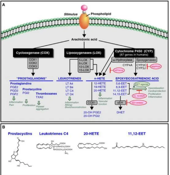
1.3 Eicosanoids
14
levels of free EETs and reduced esterified EETs, indicating that that ACSL4 in this model normally shuttles EETs towards incorporation into glycerophospholipids13. Since arachidonic acid is the primary precursor for eicosanoids, determining which isoforms have high specific activity with arachidonic acid, the regulation of synthesis of eicosanoids can be better
understood. In addition, ACSL may play a role in activating lipid metabolites, such as eicosanoids, allowing for their incorporation into phospholipids13.
Eicosanoids are a class of signaling molecules derived from ω3 and ω6 polyunsaturated fatty acids. The term encompasses several different compounds, including prostaglandins, leukotrienes, and thromboxanes14. They play significant roles in the body, by acting as local hormones and participating in intracellular signaling cascades15. They ensure gastric integrity, maintain renal function, regulate smooth muscle contraction, and control blood vessel
contractility16. Eicosanoids are critical in inflammation and immunity, and researching eicosanoid synthesis and function is important for understanding multiple disease processes, including diabetes and cardiovascular disease.
Biosynthesis of eicosanoids occurs in all cells except erythrocytes. Eicosanoids are synthesized from PUFAs that are derived from either linoleic or alpha-linolenic fatty acid. The most common fatty acid used for the synthesis of eicosanoids is the 20 carbon fatty acid
arachidonic acid (C20:4ω6), which is derived from linoleic acid. Other common fatty acids used to synthesize eicosanoids are eicosapentaenoic acid (C20:5ω3), and dihomo-gamma-linolenic acid (C20:3ω6)13
15
Figure 4: omega-6 series and omega-3 series of polyunsaturated fatty acids14
The eicosanoids formed depend on the type of fatty acid used for synthesis. Based on the type of fatty acid present in the diet, different or even opposing effects on bodily functions can be produced, as different eicosanoids will be synthesized. The biosynthesis of eicosanoids is controlled, as they are only synthesized when required. When a stimulus, such as trauma, or cytokine signaling occurs, phospholipase A2 translocates to the cell membrane, where it cleaves
the 20-carbon fatty acid from the sn-2 position of a phospholipid14. Another mechanism
involves the cleavage of a fatty acid from diacylglycerol by diacylglycerol lipase15. Once the 20-carbon fatty acid is released, it can be converted into an eicosanoid through different pathways.
To discuss the different pathways of eicosanoid synthesis, arachidonate will be used as the example, as it is the most common precursor for eicosanoid biosynthesis. The two major pathways for eicosanoid synthesis are the lipoxygenase (LOX) pathway, and the cyclooxygenase pathway (COX). Another pathway is the cytochrome p450 pathway, which produces
16
Figure 5: Eicosanoid biosynthesis pathways using arachidonic acid, and sample structures16
The lipoxygenase pathway produces leukotrienes, which are important mediators of inflammation pathways. For the synthesis of leukotrienes, the fatty acid, in this case arachidonic acid, binds to 5-lipoxygenase-activating protein (FLAP). FLAP enables arachidonic acid to interact with lipoxygenase. The enzyme lipoxygenase then converts arachidonate into 5-hydroperoxyeicosatetraenoic acid (5-HPETE). 5-HPETE is converted into leukotriene-A4, which can be further converted into other leukotrienes15. Other lipoxygenase enzymes can transform fatty acids into other HPETEs and further into hydroxyeicosatetraenoic acids (HETES), or
17
to peroxisome proliferator-activated receptors (PPARs) to produce responses, like neutrophil migration and bronchoconstriction18.
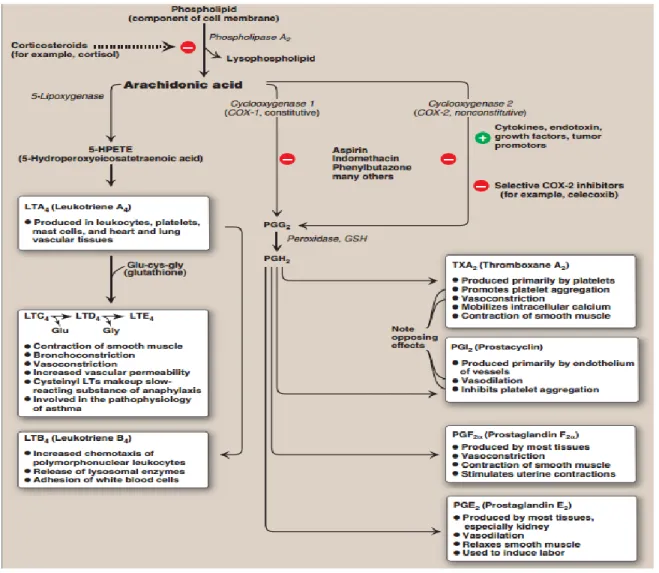
The cyclooxygenase pathway converts arachidonate into either thromboxanes or prostaglandins. The enzyme prostaglandin H2 synthase, which has cyclooxygenase and
peroxidase activities, utilizes arachidonate to produce prostaglandin H2. Two forms of this COX
enzyme are present in humans: COX-1 and COX-2. COX-1 is responsible for basal levels of prostaglandins and is expressed in gastric mucosa, kidney, platelets, and vascular endothelial cells. COX-2 is activated in response to inflammation, and increases prostaglandin production primarily in monocytes and macrophages19. The final product of the reaction is prostaglandin H2.
This molecule can be transformed into thromboxane A2 in platelets, and is prothrombotic20.
Prostaglandin H2 can also be converted into prostaglandins, which usually act via G-protein
coupled receptors. Prostaglandins can also bind to PPARs to affect nuclear transcription of proteins19. Prostaglandins have varying effects, such as contraction of smooth muscle tissue, and inhibition of platelet aggregation that depend on the type of prostaglandin formed, and the receptors on the target tissue.
18
While many types of HETEs can be synthesized, the primary metabolite of the ω-hydroxylase pathway is 20-HETE, which is synthesized in the vascular smooth muscle cells15. 20-HETE plays an essential role in inflammation, as it is pro-inflammatory and stimulates the production of cytokines in endothelial cells. It also promotes angiogenesis by controlling cell proliferation, migration, and survival in endothelial cells17. HETEs are also important
vasoconstrictors. In contrast, EETs, which are also regulators of blood vessel diameter, are generally vasodilators. EETs are synthesized in endothelial cells, as well as in hepatocytes, cardiac myocytes and other cell types expressing CYP epoxygenase17. Once synthesized, EETs are rapidly metabolized by soluble epoxide hydroxylase (sEH) into dihydroxyeicosatrienoic acids (DHETs), which are inactive. EETs can also be incorporated into phospholipids, and undergo beta-oxidation and chain elongation17. The functions of EETs are not completely understood, but they seem to inhibit apoptosis, encourage cell proliferation and promote angiogenesis21. Pancreatic physiology also seems to be affected by EETs, as increasing exogenous EETs can reduce glucose-stimulated insulin secretion, and alter beta-cell function, though this is not fully understood13.
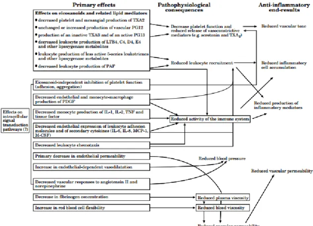
Many different eicosanoid molecules can be produced via the three major pathways. Within the each pathway, the different classes of molecules synthesized can produce varying physiological responses. The roles of eicosanoids are further diversified depending on whether they have been synthesized with arachidonic acid or eicosapentaenoic acid (EPA) as the precursor. For example, the use of EPA instead of arachidonic produces thromboxane A3
(TXA3), which is weaker than the TXA2 produced from arachidonate. Therefore, using EPA may
19
summarized in Figure 6. Understanding the effects of lipid availability on eicosanoid production is therefore critical, as the type of lipid used for eicosanoid synthesis can result in very different physiological responses.
Figure 6: The effects of using ω-3 polyunsaturated fatty acids as precursors for eicosanoid synthesis14
20
shows some of the functions of some important eicosanoids, as well as pathways that are targeted by drugs. Overall, eicosanoids play many roles in the body in nearly every organ system.
21 CHAPTER 2
SPECIFIC AIMS AND HYPOTHESIS
Specific Aim One: To obtain purified FLAG-ACSL for all rat ACSL isoforms.
Specific Aim Two: To determine the different substrate preferences of each ACSL isoform.
22 CHAPTER 3
METHODS
3.1 Transformation of pFLAG-CTC-ACSL Plasmids
Recombinant plasmids of rat pFLAG-CTC-ACSL1, –ACSL3, –ACSL4, –ACSL5 and ACSL-6 were engineered previously E. coli strain DH5-α was transformed by taking 100 μL of competent cells and mixing with 5 μL of plasmid pFLAG-CTC-ACSL DNA. The cells were incubated on ice for 30 minutes then heat shocked at 37˚C for 2 minutes. The cells were then returned to ice for 1 minute. Cells were inoculated into 200 μL of Super Optimal broth with 20 mM glucose (SOC media) and shaken at 37˚C for 1 hour. Cultures were then plated on Luria-Bertani (LB) agar plates supplemented with 50 μg/mL ampicillin and X-Gal and grown overnight at 37˚C.
3.2 Miniprep and Sequencing of pFLAG-CTC-ACSL Plasmids
Ampicillin colonies were selectively picked from each plate and grown in 2 mL LB broth supplemented with 50 μg/mL ampicillin (LB-Amp) overnight at 37˚C in a shaking incubator. Plasmid DNA was harvested from the cultures using the Qiagen QIAprep Spin Miniprep Kit using the manufacturer’s protocol was followed and 700 ng of plasmid DNA from each sample was sent to the UNC-CH Genome Analysis Facility to confirm that the plasmids contained the proper FLAG-ACSL inserts.
3.3 Recombinant Protein Induction
23
overnight at 37˚C in a shaking incubator. Primary cultures were then subcultured into 500 mL LB medium with 50 μg/mL ampicillin and grown in a 37˚C shaking incubator23
. The absorbance (OD600) was measured hourly for each subculture until the OD600 was approximately 0.7. Then
cultures were transferred into 250 mL centrifuge bottles and centrifuged for 15 minutes at 5,000 rpm at 4˚C in a Sorvall ST 16R rotor23
.
The pellets were resuspended in 500 mL Terrific Broth medium with 50 μg/mL ampicillin and 1 mM isopropyl β-D-1-thiogalactopyranoside (IPTG). The bacteria was induced at 25˚C for 16 hours, or until the final absorbance was approximately 2. Once this absorbance was reached, the cultures were again pelleted for 15 minutes at 5,000 rpm at 4˚C.
3.4 Lysis of Bacterial Pellets
Pellets were resuspended in 10 mL/g pellet cold breaking buffer containing 50 mM
HEPES/NaOH, pH 7.4, 0.5 M NaCl, 5% glycerol, 0.05% n-dodecyl β-D-maltoside (DDM), 0.05% N,N-Dimethyldodecylamide-N-oxide (LDAO), 1 mM phenylmethanesulfonyl fluoride (PMSF), 1 mM ATP, 5 mM MgCl2, a protease inhibitor tablet (Roche), and 10 mg/mL lysozyme
24 3.4 Western Blot
Protein was quantified from the cell extract samples using the Thermoscientific Pierce BCA Protein Assay kit. To visualize protein expression, 100 μg of protein from each of the cell extracts were resolved on an 8% SDS-PAGE gel at 100V for 1.5 hours. Resolved proteins were transferred onto a nitrocellulose membrane at 120V for 1 hour. To ensure transfer, protein bands were visualized with Ponceau S. The membrane was blocked in 5% non-fat dairy milk for one hour and then incubated for one hour with monoclonal mouse anti-FLAG primary antibody in the blocking solution (Sigma, dilution 1:5000). The membrane was washed with TBS-Tween (0.05% v/v) and incubated for 45 minutes with goat anti-mouse horseradish peroxidase secondary antibody (dilution 1:10000). This membrane was washed again with TBS-Tween (0.05% v/v) and placed on ECL substrate from Thermoscientific Pierce for 5 minutes. Chemiluminescence was detected by exposure to film for 1 minute.
3.5 Separation of Membrane and Soluble Proteins
A sucrose gradient was prepared to separate the soluble and membrane fractions of ACSL recombinant protein. In disposable Beckman Ultracentrifuge tubes, 0.5 mL of 5% w/v sucrose solution was layered on top of 2 mL 55% w/v sucrose solution. The cell extract was carefully layered on top of these sucrose solutions. The prepared samples were subjected to
25 3.6 ACS Specific Activity Assay
Activity of the FLAG-ACSL proteins obtained were quantified (nmol fatty acid/mg protein/min) using [14C] palmitate. This assay was conducted to quickly check for activity of the proteins before purification. A reaction mix (contained 290 mM Tris pH 7.4, 13 mM MgCl2, 8.3 mM
DTT, 17 mM ATP, 0.5 mM CoA, 5μM [14C] palmitate in 0.5 mM Triton X-100 in EDTA)with amounts of Medium 1(10 mM Tris pH 7.4, 1 mM EDTA, 0.25 M sucrose, 1 mM DTT), so that once protein was added the volume of protein and Medium 1 would equal 80 μL. Protein amounts of 20μg, 40μg, or 60μg were added to start the reaction. A positive control of total particulate from mouse liver was also used to compare specific activities.
The 10-minute reactions were conducted at room temperature, and stopped with 1 mL Dole’s reagent (isopropanol:heptane:1 M H2SO4, 80:20:2, by volume). The unesterified fatty acid was
removed with two washes with 2 mL heptane and 0.5mL distilled water. This mix was centrifuged at 1500 rpm for 5 minutes to improve phase separation. The organic phase was discarded and radioactivity was quantified in 600 μL of the aqueous phase by liquid scintillation using Ecolite. All specific activity assays used a positive control of wild-type liver total
particulate from mouse, which was obtained by Dr. Eric Klett on 7/11/05.
3.7 Affinity Column Chromatography
26
The cell extract samples used for purification were prepared by adding an equal volume of 1xTBS, 1% (w/v) Triton X-100 and incubating on ice for 20 minutes. The samples were centrifuged at 2700 rpm for 5 minutes at 4˚C to remove DNA and debris that could clog the column.
The sample was run through the prepared column three times and the final flow-through was stored. The column was then washed twice with two column volumes of 1xTBS to remove unbound proteins. To elute the FLAG proteins, 5 mL of 100 μg/mLsolution of FLAG peptide (Sigma) was prepared from a 5 mg/mL stock solution of FLAG peptide and 50mM Tris-HCl pH 7.4. For fraction 1a, 0.5mL of FLAG peptide was added and the protein collected. This was repeated for fraction 1b. For fractions 2-5, 1 mL of FLAG peptide was added and the fractions collected. These fractions were aliquoted and used for Western blots and specific activity assays that were done as previously described. They will also be used for an indirect spectrophotometric assay using NADH.
3.8 Indirect ACS Assay
An indirect ACS assay was used to sensitively measure the activity of each ACSL isoform with various substrates. In the spectrophotometric assay, the activity of ACSL was measured by coupling the reaction of ACSL with the reactions of adenylate kinase, pyruvate kinase and lactate dehydrogenase and following the oxidation of NADH at 334nm with a recording spectrophotometer (Beckman DU640). The reaction mix contained 100 mmol Tris-HCl buffer pH 8.0, 10 mmol ATP, 15 mmol MgC12, 5 mmol dithiothreitol, 150 mM KCl, 1.0 mM potassium
27
triethanolamine pH 8.2. An NADH standard curve was first constructed for use in calculations of ACSL activity using NADH concentrations of 0 M, 0.01128 M, 0.0564 M, 0.1128 M, and
0.2256 M. Absorbance was measured at 334 nm at room temperature.
28 CHAPTER 4
RESULTS
4.1 Discovering Problems with ACSL Purification
To obtain purified protein for use in the indirect ACS assay, I originally used a protocol by Caviglia, Li and others22. Protein expression of FLAG-ACSL4 in the rat LS2226 strain was measured before purification, and after separating the membrane and soluble fractions with the sucrose gradient. In Figure 8A, the expected band at 75 kDa (actual size is 7440 kDa) was visible in the membrane and soluble fractions. Expression was also measured in each fraction after column purification. Fraction 1a is the first 0.5 mL of eluate after the addition of FLAG peptide. Fraction 1b is the next 0.5 mL, and fractions 2-5 are sequential 1 mL fractions of eluate. After column purification, as shown in Figure 8B, much fainter bands were visible at 75 kDa in membrane fractions 1b, and 2, and in soluble fractions 1b, 2 and 3, indicating protein loss during purification.
A
B
Figure 8: Protein expression of FLAG-ACSL4 from LS2226 strain in unpurified (A) and purified (B) membrane and soluble fractions
Membrane Soluble
Membrane Soluble
29 0 0.5 1 1.5 2 2.5 3 3.5 4
0 10 20 30 40 50 60 70 80 90
10 0 11 0 12 0 13 0 14 0 15 0 16 0 17 0 18 0 A b s at 334 n m Time (seconds)
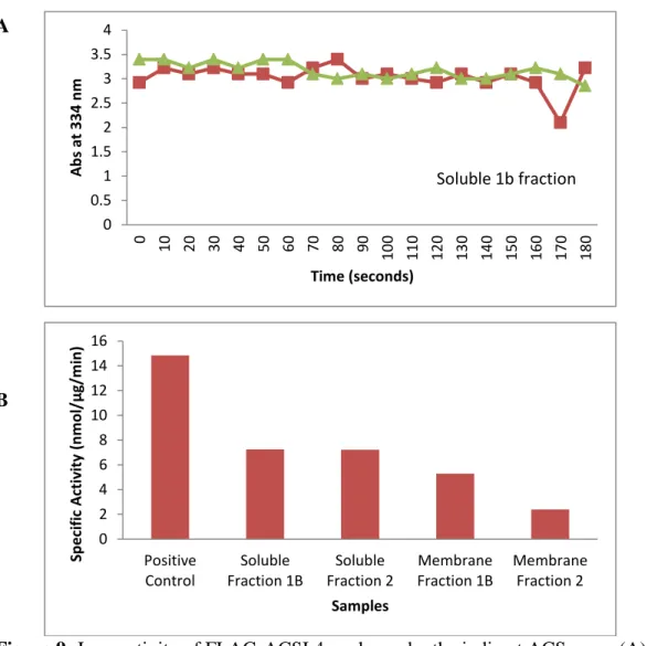
Soluble 1b fraction
0 2 4 6 8 10 12 14 16 Positive Control Soluble Fraction 1B Soluble Fraction 2 Membrane Fraction 1B Membrane Fraction 2 Sp e ci fi c A ctiv ity (n m o l/ μ g/ m in ) Samples
The indirect ACS assay was performed using 0.5 mg of protein from both membrane fraction 1b and soluble fraction 1b with arachidonic acid as substrate. However, as seen in Figure 9A, the results were not as expected; instead of declining progressively, absorbance remained fairly constant for both soluble and membrane fractions, indicating low activity. To confirm, a
radioactive specific activity assay was performed with palmitate as substrate. As shown in Figure 9B, activity compared to the positive control of mouse liver total particulate was low. Though the activity was low, the presence of activity with the radioactive ACS assay indicates that there may have been some problem with the indirect assay, such as oxidation of the arachidonate.
A
B
Figure 9: Low activity of FLAG-ACSL4 as shown by the indirect ACS assay (A) and
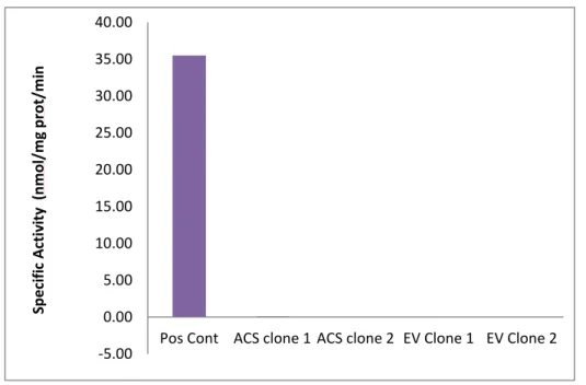
30 -5.00 0.00 5.00 10.00 15.00 20.00 25.00 30.00 35.00 40.00
Pos Cont ACS clone 1 ACS clone 2 EV Clone 1 EV Clone 2
Sp e ci fi c A ctiv ity (n m o l/ m g p ro t/ m in
4.2 Determining Source of Problems with ACSL Activity
To determine where the loss of enzyme activity occurred, activity of bacterial lysates was measured, instead of that of purified samples. The activity of the protein produced by bacteria containing the ACSL4 plasmid was compared to that containing an empty vector. Two clones were selected for each strain. In Figure 10, the results show that activity of the ACSL4 lysate was not different from that of the empty vector, and was much lower than that of the total particulate from mouse liver positive control, meaning the protein was inactive.
Figure 10: Negligible specific activity of ACSL4 and EV lysates with palmitate compared to positive control of total particulate from mouse liver
31 32.99 23.17 19.99 0 5 10 15 20 25 30 35
Positive Control ACSL3 Lysate ACSL 3 Extract
Sp e ci fi c A ctiv ity (n m o l/ mg /m in ) Samples 4.3 Activity with New Protocol
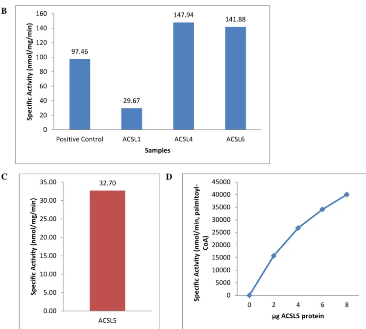
To resolve the problems with activity, the sequences of each FLAG-ACSL plasmid were confirmed and transformed into the DH5-α strain of E. coli. Upon the advice of Concetta DiRusso (University of Nebraska), we tried an overnight induction at 25˚C instead of 37˚C and the lysis buffer mentioned in the methods section was used. A radioactive specific activity assay was conducted with palmitate as substrate to check for activity. In Figure 11A, the activity of both the ACSL3 lysate and extract using the new protocol was comparable to that of the positive control of total particulate from mouse liver. In Figure 11B, the cell extracts of each ACSL isoform were used to measure specific activity. Compared to the positive control of mouse liver total particulate, the specific activity of the protein formed using the new protocol was high. ACSL4, and ACSL6 were particularly high, as they were higher than the positive control. As seen in Figure 11C and Figure 11D, ACSL5 had activity, and with increasing protein
concentrations showed increased activity, as expected. This result means that with the new protocol, the recombinant protein produced was active.
32 97.46 29.67 147.94 141.88 0 20 40 60 80 100 120 140 160
Positive Control ACSL1 ACSL4 ACSL6
Sp e ci fi c A ctiv ity (n m o l/ m g/ m in ) Samples 32.70 0.00 5.00 10.00 15.00 20.00 25.00 30.00 35.00 ACSL5 Sp e ci fi c A ctiv ity (n m o l/ m g/ m in ) 0 5000 10000 15000 20000 25000 30000 35000 40000 45000
0 2 4 6 8
Sp e ci fi c A ctiv ity (n m o l/ m in , p al m ito yl -Co A )
μg ACSL5 protein B
C D
Figure 11: Specific activity of ACSL isoforms with palmitate (A) Specific activity of ACSL 3 lysate and extract compared to total particulate from mouse liver positive control (B) Specific activity of ACSL 1, -4, -6 extracts compared to total particulate from mouse liver (C) Specific activity of ACSL5 (D) Protein dependent specific activity of ACSL 5
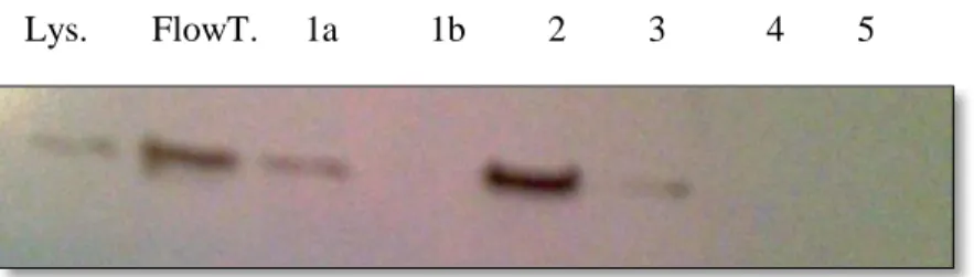
The membrane fraction of FLAG-ACSL5 was purified in the affinity chromatography column and fractions were collected. Protein expression in the lysate, column flow through, and purified fractions was measured using a Western blot with an anti-FLAG antibody. Fraction 1a is the first 0.5 mL of eluate after the addition of FLAG peptide. Fraction 1b is the next 0.5 mL, and
33
lysate, and the flow through, fraction 1a, 2 and 3. The flow through and fraction 2 had the highest expression.
Figure 12: Protein Expression for FLAG-ACSL5 lysate, column flow through, and purified fractions
4.4 Indirect ACS Assay
Since fraction 2 showed the best protein expression, it was used for the indirect ACS assay. Protein amounts of 0.5, 1.5, 2, 25, and 3 μg were used with oleic acid as substrate. For palmitic acid as substrate, protein amounts of 0, 1, 2, and 2.4 μg were used from fraction 2. Absorbance was measured at 334nm for 3 minutes to measure the oxidation of NADH to NAD+. The initial and final absorbances were used to calculate the specific activity. In Figure 13A, activity increased linearly with increasing concentrations of protein with oleate. In figure 13B, with palmitate as substrate, activity increased quickly up to 1 μg of protein, after which the specific activity and increased much more slowly. In figure 13C, a comparison shows that with palmitate, 1 μg of ACSL5 produced an activity of 27 nmol NAD+
/min, while with oleate, more than 2.25 μg of protein was required to reach the same level of activity. ACSL5 seems to prefer palmitate over oleate as substrate, particularly at lower concentrations of protein, but at higher
34
y = 11.073x R² = 0.9951 0
10 20 30 40
0 0.5 1 1.5 2 2.5 3 3.5
Sp e ci fi c A ci tiv ity (n m o l N A D +/m in )
μg FLAG-ACSL5 protein
0 10 20 30 40
0 0.5 1 1.5 2 2.5 3
Sp e ci fi c A ctiv ity (n m o l N A D +/m in )
μg FLAG-ACSL5 protein
0 5 10 15 20 25 30 35 40
0 1 2 3 4
Sp e ci fi c A ctiv ity (n m o l N A D +/ m in )
μg FLAG-ACSL5 protein
Palmitate
Oleate A
B
C
35 CHAPTER 5
DISCUSSION
5.1 Developing Protocol for Purified Protein
ACSL plays a key role in lipid metabolism by selectively activating fatty acids and channeling them towards different pathways5. The five ACSL isoforms present in mammals show differences in tissue localization, and cellular localization, which are indicative of the varying functions of each isoform7. Of particular interest are the differing substrate preferences of each isoform, as activity of each isoform seems to be affected by the lipid environment. By determining the substrate preferences of each ACSL isoform, the function of each isoform can be better understood. Learning more about ACSL isoforms, their functions, and regulation can be useful when studying inflammatory pathways, and disorders in which lipid metabolism is disturbed.
36
In this study, we attempted to resolve the problems that were present in other
experimental designs by using purified ACSL and quantitatively measuring activity through an indirect ACS assay.
The purification process proved to be extremely difficult. The protocol used by Caviglia, Li and others for ACSL purification was used, but proved ineffective for this experiment22. Though FLAG-ACSL protein was produced, the enzyme showed low, or no activity. After much trial and error, the problem was discovered. The induction procedure caused loss of enzyme activity, as the high temperature induction for 16 hours was perhaps creating an environment in which the protein could not fold properly into its active form. In addition, the LS2226 strain originally used may not have been able to support the plasmid, and the protein produced may have been toxic and killed the cells. Another possible explanation is the aggregation of the ACSL enzyme into inclusion bodies, which could have prevented the enzyme from interacting with the fatty acid substrate. The problem was resolved with the use of the DH5-αE. coli strain, a 25˚C induction temperature and a lysis buffer containing detergents, such as DDM and LDAO to keep proteins solubilized and maintain activity.
This new protocol proved to be effective in producing active ACSL protein for each ACSL isoform. Even after purification, the ACSL5 produced remained active, though some protein and activity seems to have been lost throughout the purification process. This loss of might be avoided by loading the sample more times onto the column. Some loss of activity, however is, likely unavoidable.
37
different concentrations of protein. Particularly at low concentrations of protein, ACSL5 seemed to prefer palmitic acid over oleic acid. More trials need to be conducted to confirm this result, but these results indicate that ACSL5 differs in activity when substrates available are changed.
The protocol developed can be used to further assess the activity of ACSL5 and the other ACSL isoforms with different fatty acid substrates. Due to time constraints and the amount of time spent optimizing the purification protocol, the hypothesis could not be confirmed and will have to be explored in future studies.
5.2 Limitations
The protocol developed is an improvement to other studies, due to its ability to provide sensitive, quantitative measurements of ACSL activity. However, this purification procedure is not without limitations. The plasmids used for FLAG-ACSL production produce rat ACSL isoforms. Therefore, the results obtained regarding enzyme activity cannot necessarily be directly applied to humans since the amino acid sequence of rat and human ACSL5 is only 81% identical. In addition, an unavoidable loss of protein activity during the purification process occurred due to the time required to purify the protein. Though the purification was conducted at 4˚C, some loss of activity still occurred. Therefore, when measuring enzyme activity, the results obtained may not reflect true enzyme activity.
38
are not present in the cellular environment. In addition, concentrations of chemicals used in the indirect assay may not reflect cellular levels. For example, the 10 mmol ATP and 600 μM CoA were purposely in excess, so that the reaction was not limited by reactant availability. In addition, the concentrations of different fatty acids can vary greatly in the body based on diet. For example, values provided by Mayo Clinic Laboratories provide a large range of 650-3,500 nmol/mL for serum values of oleic acid and 1,480-3,730 nmol/mL of palmitic acid25. The concentration of 250 μmol/mL oleic acid and palmitic acid used for the indirect ACS assay are well above the ranges, so these may not reflect physiological values.
5.3 Future Studies
Due to the amount of time used to obtain purified ACSL isoforms that retained activity, the different substrate preferences of each ACSL isoform could not be measured. ACSL5 seems to show a preference for palmitic acid over oleic acid, but further trials with wider ranges of protein concentrations need to be conducted to confirm this preference and determine quantify the activity of the enzyme. Other fatty acids with varying chain-length and saturation also need to be tested.
39
concentrations should also be used to determine the conditions in which a certain fatty acid is preferred.
40
REFERENCES
1. Berg, J. M., Tymoczko, J. L., & Stryer, L. (2002). Biochemistry. “Chapter 22: Fatty Acid Metabolism”. WH Freeman, New York.
2. "MetaCyc: Reaction: 6.2.1.3." MetaCyc. <http://biocyc.org/META/NEW-IMAGE?type=REACTION&object=RXN-9623>.
3. Brandt, Mark. "Fatty Acid Breakdown." Rose-Hulman Institute of Technology. 2000. <http://www.rose-hulman.edu/~brandt/Chem331/Lipid_Breakdown.pdf>
4. Ellis, J. M., Frahm, J. L., Li, L. O., & Coleman, R. A. (2010). Acyl-coenzyme A synthetases in metabolic control. Current Opinion in Lipidology, 21(3), 212-217.
5. Fujimoto, Y., Itabe, H., Kinoshita, T., Homma, K. J., Onoduka, J., Mori, M., Yamaguchi, S., Makita, M., Higashi, Y, Yamashita, A. & Takano, T. (2007). Involvement of ACSL in local synthesis of neutral lipids in cytoplasmic lipid droplets in human hepatocyte HuH7. Journal
of lipid research,48(6), 1280-1292.
6. Soupene, Eric, and Frans A. Kuypers. Mammalian Long-Chain Acyl-CoA Synthetases. Experimental Biology and Medicine 233.5 (2008): 507-521.
7. Li, Lei O., Eric L. Klett, and Rosalind A. Coleman. Acyl-CoA synthesis, lipid metabolism and lipotoxicity. Biochimica et Biophysica Acta (BBA)-Molecular and Cell Biology of
Lipids 1801.3 (2010): 246-251.
8. Iijima, H., Fujino, T., Minekura, H., Suzuki, H., Kang, M. J., & Yamamoto, T. (1996). Biochemical Studies of Two Rat Acyl‐CoA Synthetases, ACS1 and ACS2. European
41
9. Fujino, T., Kang, M. J., Suzuki, H., Iijima, H., & Yamamoto, T. (1996). Molecular characterization and expression of rat acyl-CoA synthetase 3.Journal of Biological
Chemistry, 271(28), 16748-16752.
10.Kang, M. J., Fujino, T., Sasano, H., Minekura, H., Yabuki, N., Nagura, H., Ilijima, H. & Yamamoto, T. T. (1997). A novel arachidonate-preferring acyl-CoA synthetase is present in steroidogenic cells of the rat adrenal, ovary, and testis. Proceedings of the National Academy
of Sciences, 94(7), 2880-2884.
11.Oikawa, E., Iijima, H., Suzuki, T., Sasano, H., Sato, H., Kamataki, A., Nagura, H., Kang, M., Fujino, T., Suzuki, H. & Yamamoto, T. T. (1998). A novel acyl-CoA synthetase, ACS5, expressed in intestinal epithelial cells and proliferating preadipocytes. Journal of
Biochemistry, 124(3), 679-685.
12. Van Herpen, N. A., and V. B. Schrauwen-Hinderling. Lipid accumulation in non-adipose tissue and lipotoxicity. Physiology & behavior 94.2 (2008): 231-241.
13.Klett, E. L., Chen, S., Edin, M. L., Li, L. O., Ilkayeva, O., Zeldin, D. C., Newgard, C.B. & Coleman, R. A. (2013). Diminished acyl-CoA synthetase isoform 4 activity in INS 832/13 cells reduces cellular epoxyeicosatrienoic acid levels and results in impaired glucose-stimulated insulin secretion. Journal of Biological Chemistry.
14. De Caterina, R., and G. Basta. n-3 Fatty acids and the inflammatory response—biological background. European Heart Journal Supplements 3.suppl D (2001): D42-D49.
15."Synthesis of Eicosanoids." Rennsselaer Polytechnic Institute, 2008.
<http://www.rpi.edu/dept/bcbp/molbiochem/MBWeb/mb2/part1/prostag.htm>. 16.Harvey, Richard A., and Denise R. Ferrier. Prostaglandins and Related Compounds.
42
17. Panigrahy, D., Kaipainen, A., Greene, E. R., & Huang, S. (2010). Cytochrome P450-derived eicosanoids: the neglected pathway in cancer. Cancer and Metastasis Reviews, 29(4), 723-735.
18.Dahlén, S. E., Björk, J., Hedqvist, P., Arfors, K. E., Hammarström, S., Lindgren, J. A., & Samuelsson, B. (1981). Leukotrienes promote plasma leakage and leukocyte adhesion in postcapillary venules: in vivo effects with relevance to the acute inflammatory
response. Proceedings of the National Academy of Sciences, 78(6), 3887-3891.
19. "Introduction to the Eicosanoids." Eicosanoid Synthesis and Metabolism: Prostaglandins,
Thromboxanes, Leukotrienes, Lipoxins.
<http://themedicalbiochemistrypage.org/eicosanoids.php>.
20.Katagiri, H., Ito, Y., Ishii, K. I., Hayashi, I., Suematsu, M., Yamashina, S., Murata, T., Narumiya, S., Kakita, A. & Majima, M. (2004). Role of thromboxane derived from COX‐1 and‐2 in hepatic microcirculatory dysfunction during endotoxemia in
mice. Hepatology, 39(1), 139-150.
21."Eicosanoids Regulation and Control." Advanced Computing Lab at St. Edward's University. <http://www.cs.stedwards.edu/chem/Chemistry/CHEM43/CHEM43/Eicosanoids/REGCNTR L.HTML>.
22.Harvey, Richard A., and Denise R. Ferrier. Prostaglandins and Related Compounds.
Biochemistry. Philadelphia: Lippincott/Williams & Wilkins, 2005. 215.
23.Caviglia, J. M., Li, L. O., Wang, S., DiRusso, C. C., Coleman, R. A., & Lewin, T. M. (2004). Rat long chain acyl-CoA synthetase 5, but not 1, 2, 3, or 4, complements Escherichia coli
43
24.Hosaka, K., Mishinia, M., Tanaka, T., Kamiryo, T., & Numa, S. (1979). Acyl‐Coenzyme‐A Synthetase I from Candida Lipolytica. European Journal of Biochemistry, 93(1), 197-203. 25."Test ID: FAPEP Fatty Acid Profile, Essential, Serum." Mayo Clinic Mayo Medical