1
A Systematic Review of the Harms of Lung Cancer Screening with Low Dose
Computed Tomography
By
Cody Schwartz
A Master‟s Paper submitted to the faculty of
the University of North Carolina at Chapel Hill
in partial fulfillment of the requirements for
the degree of Master of Public Health in
the Public Health Leadership Program.
Chapel Hill
2011
__________________________
Advisor:
____________________________________
Date
_________________________________
Second Reader:
_________________________________
2 Abstract:
Background: Lung cancer causes the majority of cancer-related deaths in the United States. Screening for lung cancer with low dose CT (LDCT) has become a prominent topic. This modality has the potential to contribute to a decrease in lung cancer associated mortality. Preliminary data from a large randomized controlled trial have shown a 20% decrease in mortality. Currently, no guidelines endorse LDCT screening for lung cancer. The harms of screening for lung cancer have not been fully characterized in a focused systematic review. Purpose: To explore and characterize the harms associated with low dose CT screening for lung cancer in high risk populations by evaluating the current evidence. This information will assist policy makers and clinicians as they weigh the risks and benefits of screening high risk populations for lung cancer.
Data Sources: MEDLINE, EMBASE, Cochrane Library, reviews, reference lists, experts Study Selection: Studies were included that evaluated low dose screening for lung cancer. Those that characterized harms were central to this review.
Data Extraction: Data were abstracted to abstraction forms. Studies were graded according to methods used by the United States Preventive Services Task Force (USPSTF).
Data Synthesis: Studies reported variable rates of abnormal results with positivity of 8 to 45%. A range of 4 to 55% of total invasive procedures and none to 40% of surgeries were performed for benign indications. Incidental findings were reported in 0.77 to 62% of participants. Of the included studies, 30% discussed anxiety, 55% discussed overdiagnosis, 50% reported morbidity and mortality of workup, and 35% reported false negative cases. False positives at baseline ranged from 86 to 98%.
3 unavailable in English were included so this review has a potential for publication and language bias.
Conclusions: The harms of low dose CT screening for lung cancer are poorly reported in screening trials. Even though harms are incompletely characterized in the included studies, these harms are likely clinically significant. Clinicians must weigh the risks and benefits of the screening procedure if they are to implement mass screening for lung cancer with LDCT. Introduction:
Background
To date, lung cancer screening has not proven useful. Systematic reviews have not
shown a mortality benefit to screening for lung cancer1-4. No current guidelines recommend
mass screening for lung cancer, and the American College of Chest Physicians recommends
that individuals at high risk should only undergo screening as part of a clinical trial5,6. However,
preliminary data from the NLST randomized screening trial show a 20% reduction in mortality
for those who received a LDCT screening exam compared to the control group7. While this data
is encouraging in the fight against lung cancer, a good quality screening program requires that the benefits outweigh the harms. No current systematic review has been designed to fully characterize the harms of lung cancer screening with LDCT. In order for clinicians, policy makers, and patients to make informed decisions about screening, a systematic review of screening harms is required. This evidence will help clinicians, policy makers, and patients balance the benefits and risks of screening in a population at risk for lung cancer. Due to the absence of a current systematic review, I will attempt to evaluate the harms of LDCT screening for lung cancer in this review.
Burden of suffering
Lung cancer is the leading cause of overall cancer death in the United States and
worldwide8-10. According to the American Cancer Society (ACS), lung cancer is expected to
4 2007, the most recent year for complete data available from the Centers for Disease Control
(CDC), lung cancer was responsible for 6.5% of all US deaths11. In 2010, the ACS predicted
222,520 new cases of lung cancer will be diagnosed in the United States, 15% of all cancer
diagnoses8. In perspective, lung cancer causes more deaths per year than stroke (136,000),
lower respiratory disease (128,000), and accidents (124,000)12. If lung cancer mortality was an
individual category, it would be the third leading cause of death in the US, behind only heart disease and all other cancers.
Worldwide, the ACS and the International Agency for Research on Cancer (IARC) estimated about 1.6 million new cases were diagnosed in 2008, representing about 13% of all
cancer diagnoses9,10. Deaths from lung cancer totaled 1.4 million in 2008, representing 18% of
all cancer deaths9,10. Lung cancer is the leading cause of cancer death in males, and is second
in females behind only breast cancer9,10.
Lung cancer incidence in males has decreased in the United States over the past
decade, but has increased among females from 1991 to 200613,14. During the same period in
the US, mortality rates have decreased among males but have increased and remained stable
since 2003 for females13,14. Even though rates among males have decreased, absolute
incidence and mortality remain higher for males compared to females. Tobacco use patterns are likely contributing to the divergent trend observed between sexes. As a result, prevention strategies to decrease lung cancer incidence and mortality among both groups remain a priority. Prevention Strategies
The primary risk factor for developing lung cancer is tobacco use15. In 2000, tobacco
use was responsible for 435,000 deaths in the United States16. This represents 18.1% of all US
deaths in that year. These deaths include not only lung cancer, but other cancers, respiratory illnesses, and cardiovascular diseases associated with tobacco use. According to the CDC and a 2004 report from the Surgeon General, tobacco use is attributable to 80-90% of lung cancer
5
nonsmoking males over their lifetime15. Female smokers are 13 times as likely to develop lung
cancer as nonsmoking females over their lifetime15. Former male smokers are 9 times as likely
to develop lung cancer as never smokers15. Former female smokers are 5 times as likely to
develop lung cancer as female never smokers15. After 20 years of tobacco cessation, lung
cancer risk decreases to about 2 times that of never smokers but never reaches the risk of a
never smoker18. Lung cancer risk from tobacco exposure is dose dependent, so the more one
smokes, the greater the risk of developing cancer19. While other risk factors play a role in
developing lung cancer, this demonstrates that the main priority for prevention of lung cancer deaths is tobacco cessation and use prevention.
As tobacco use contributes the majority of risk for developing lung cancer17, prevention
of lung cancer is straightforward. Smoking cessation and use prevention is the priority. While the prevalence of smoking in the US has decreased since the 1980s according to data from the National Health Interview Survey, 20.6% of adults, or 46.6 million people, were current smokers
in 200920. An estimated 23.5% of men and 17.9% of women were current smokers in 2009 and
an additional 21.9% of the population was considered a former smoker20. This shows that
42.5% of the US population was either a current or former smoker in 2009, which demonstrates the significant proportion of individuals at increased risk for developing lung cancer compared to never smokers. While prevention of tobacco use is a priority, individuals remain at risk for lung cancer long after they quit smoking as cancer takes many years to develop. Thus, lung cancer screening has been proposed as a tool to reduce the burden of suffering.
In addition to primary prevention, screening for early disease has been proposed to combat lung cancer mortality. While the greatest reduction in the burden of suffering would likely come from a reduction in smoking rates, early detection could further reduce the burden of suffering if screening randomized controlled studies demonstrate a reduction in lung cancer mortality.
6 Screening has the potential to detect early stage cancers which are more amenable to cure than late stage cancers. Most lung cancers present with symptoms and are diagnosed in
an advanced stage when prognosis is poor21. Only 15% of lung cancers were diagnosed in a
local stage in 200613. The ideal screening test would decrease mortality by improving the
probability of cure from early detection of early stage cancers. To date, a mortality benefit for lung cancer screening has not been shown. Even if a mortality benefit is eventually shown, screening for early stage cancers can only be effective if the benefits outweigh the harms. Burden of testing, incidental findings, cost, workup morbidity and mortality, anxiety, surgical morbidity, radiation exposure, and screening biases need to be taken into account when
weighing the risks and benefits of screening2,22. However, because the prognosis of late stage
cancers is dismal, even a small benefit in discovering and treating early stage cancers is likely to afford a mortality advantage. As a result of this poor life expectancy, screening has been proposed as a possible mechanism to decrease lung cancer mortality.
Lung cancer screening dates back to the 1960s when researchers studied chest x-ray and sputum cytology as screening modalities. Early trials published in the 1960s and 1970s that evaluated chest x-ray and sputum cytology showed no mortality differences for the
intervention group versus controls23-29. Observational and experimental studies evaluating chest
x-ray published in the 1980s and 1990s also failed to demonstrate a mortality benefit30-33.
These studies were criticized for lack of adjustment for screening biases, study design problems
including lack of power to detect mortality differences, and contamination in control groups34.
Due to these problems, one arm of the current Prostate Lung Colon Ovarian (PLCO) Cancer screening randomized trial is designed to address mortality differences by comparing annual
chest x-ray with usual care for early detection of lung cancer34. This study sought to enroll
7
non-smokers two annual repeat scans34. Final results of this trial are currently unavailable, but
are expected soon in 2012.
Two up to date systematic reviews have explored the efficacy of lung cancer screening with chest x-ray for a reduction in mortality. Seven controlled trials comparing chest x-ray with usual care were included in a Cochrane systematic review, six were randomized and one was
non-randomized1. This review reported a pooled analysis of no mortality benefit and suggested
a possible mortality increase in the screening group. A second systematic review also found no
mortality benefit from screening with chest x-ray compared to controls2. Together these reviews
and the chest x-ray screening arm of the PLCO trial will likely address the issue of the efficacy of lung cancer screening with chest x-ray.
Low dose CT screening for lung cancer was proposed in a 1990 study by Naidich et al35
who compared the feasibility of conventional CT versus low dose CT of the chest. Observational studies evaluating low dose CT screening for lung cancer followed soon
thereafter. LDCT screening has been shown to detect early stage lung cancers in uncontrolled
observational studies36-46. Observational studies and short randomized trials of LDCT have
been evaluated in systematic reviews which have demonstrated no mortality benefit to
screening2-4,47. Due to the screening biases of length time, lead time, and overdiagnosis bias
inherent in observational studies, it is difficult to discern whether any observed survival
differences are due to actual reductions in mortality from screening or due to bias22. Studies
without a control group may overestimate survival and only a randomized controlled trial comparing screening with LDCT to a control group can validly evaluate any mortality
difference22. However, published data from completed randomized controlled trials are not yet
available.
The NLST study comparing LDCT screening versus chest x-ray in a population with at
least 30 pack-year history smokers will be published soon in 201148,49. The NELSON trial from
8
will be published soon thereafter in 201250. These trials will address the question of whether or
not there is a mortality benefit to LDCT screening for lung cancer. However, the goal of this review is to evaluate the harms of LDCT screening. As the characterization of harms likely does not suffer from the same screening biases as mortality evidence, this review will include
observational studies, randomized trials, and systematic reviews when available. Summary
The best method to decrease mortality from lung cancer is to prevent it from occurring in the first place. As mentioned, the majority of risk of developing lung cancer is attributed to smoking. Thus, tobacco cessation programs and use prevention have the greatest likelihood of decreasing lung cancer mortality. But for those who already have a smoking history or are at greater risk for lung cancer, an intervention that decreases lung cancer mortality may benefit the high risk population. Researchers are evaluating LDCT screening as a viable option.
Studies have evaluated individual harms of LDCT screening for lung cancer, but have not fully addressed all harms including, but not limited to, burden of testing, invasive
procedures, anxiety, false positives, radiation exposure, and incidental findings. This review will attempt to characterize the harms of screening for lung cancer with LDCT. This will help
clinicians, policy makers, and patients weigh the risks and benefits of screening in order to make informed decisions.
This review will attempt to answer the following key questions: Key Questions
(1) What is the evidence for harms associated with LDCT screening? (burden of testing, incidental findings, false positives, false negatives, testing burden, adverse effects of treatment, radiation exposure)
(1a) What is the evidence for potential biases in screening for lung cancer? (length time, lead time, overdiagnosis)
9 Methods:
As published evidence thus far has shown no mortality benefit, I focused this review on the harms of screening. To address the focused question of identifying and quantifying the harms in lung cancer screening, a brief search of systematic reviews was conducted to
determine if a review was indicated. No systematic review has been published that addresses potential harms of lung cancer screening with LDCT.
Data Sources and Searches
I searched the MEDLINE, EMBASE, and CENTRAL online databases from inception to 6/1/2011 to identify relevant articles. I used the MeSH terms of “lung neoplasm,” “mass
screening”, "tomography, x-ray computed," and “tomography, spiral computed” plus keyword
searches of low dose CT and screening harms. Complete search terms are provided in the appendix. I also hand searched relevant reviews and reference lists of included studies for articles. An attempt to locate gray literature was made in a search of clinicaltrials.gov for trials currently underway and in consultation with experts. An experienced research librarian was consulted in formulating the search strategy.
Study Selection
This review will include original research papers that are relevant to answer the key questions of screening harms. One author reviewed the initial full set of identified titles and abstracts. If it was not clear from the title or abstract if the study fit inclusion criteria, it was included for full text review to maintain a sensitive search. If multiple articles were published on one study, all relevant data was abstracted for our outcomes of interest. Reference lists of included studies were searched for relevant articles.
10 identification of primary studies and were not included in the analysis. If systematic reviews quantified harms, I commented on these in the discussion section of this paper. We included any population of current or former smokers who have received a low dose CT scan for the purpose of screening for lung cancer. Studies were eligible for any duration of follow up as potential screening harms can be immediate, such as false positives or anxiety of awaiting results. Studies conducted in any setting that has the potential for lung cancer screening were eligible, including primary care and specialty settings. I required the study to measure at least one of the primary outcomes harm as described in the PICOTS in Table 1. Excluded articles included editorials, non-systematic reviews, case reports, and case series. Studies were excluded if they did not evaluate low dose CT as the intervention screening modality, such as those that evaluated chest x-ray or sputum cytology.
Data Extraction and Quality Assessment
I collected data from the included studies into evidence tables using a standard data collection form. Information collected included study design, population, intervention,
comparators, and outcome measures as described in Table 1. Studies reporting incomplete outcomes were reported in the analysis. No attempt was made to contact the primary authors
to obtain data. I assessed quality using the USPSTF methodology51. One author completed
quality assessment for each study. Data Synthesis and Analysis
I reported data for each of our primary outcomes if available in the included studies. A qualitative synthesis was conducted for each of the primary outcomes of interest due to the heterogeneity of study populations. Heterogeneity was not formally assessed using statistical methods due to the variability of eligibility criteria and differences in study protocols across the included studies. I used the PRISMA reporting guidelines for systematic reviews to preserve
11 PICOTS:
Category Inclusion criteria
Population Current and former tobacco smokers, including groups identified in the
literature as high risk, low risk, and those whose risk is not defined, no restriction on numbers of pack-years
- Former users as defined in literature
Intervention Screening with low dose CT scan, any interval
Comparators Usual care, chest x-ray, sputum cytology, different combinations of
these
Outcomes Primary outcomes:
- Harms of screening/workup
- Testing burden
- Invasive procedures (including biopsies, thoracotomies, VATS)
- False positives
- False negatives
- Radiation exposure
- Incidental findings
- Anxiety
- Unnecessary procedures
- Repeat imaging – especially associated with radiation exposure,
HRCT, PET
- Overdiagnosis
- Quality of life
Secondary outcomes:
- Smoking cessation rates of abnormal result
Timing of effect Harms: any time period
Timing of search Since MEDLINE inception – 1966 to present
Setting Primary care, specialty care, inpatient setting, or other relevant lung
cancer screening settings
Study Design SRs, RCTs, non-randomized trials, prospective cohort studies
12 Results:
A total of 655 titles and abstracts were identified and reviewed using our search strategy. Of these, 215 were included for full text review. Articles were excluded for wrong interventions, poor outcome reporting, duplicate publications, wrong design, poor quality, and inability to
obtain an English translation, special populations. Nine articles were reviewed from searching
reference lists. On final inclusion, 46 articles from 20 studies were included in this review. Six randomized controlled trials and 14 cohort studies were included. Eight systematic reviews and ten articles from special populations (asbestos, nuclear plants) were identified. Most
randomized trials only reported baseline findings. A flow diagram of the results of our search strategy is shown in Figure 1.
Summary of Study Descriptions
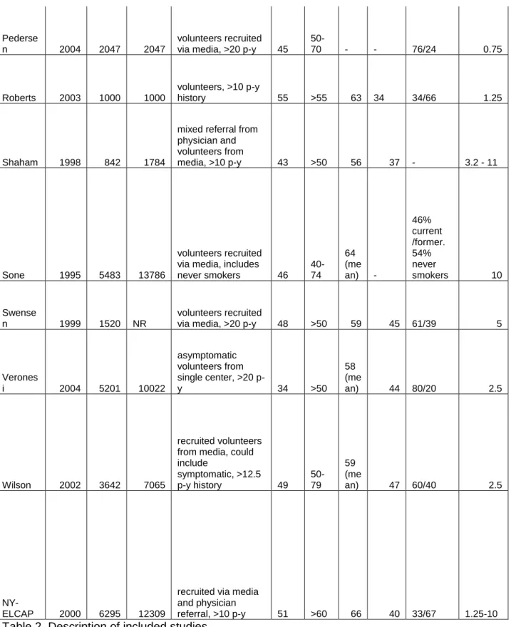
Characteristics of included studies are summarized in Table 2. A total of 94,521 low dose CT screening exams were performed for 46,946 participants in the included studies. Median age ranged from 53 to 67 years old. Median smoking history ranged from 30 to 54 pack-years. Low dose CT slice thickness ranged from 0.75 to 11 mm.
The total number of lung cancers diagnosed was 845 with 67% of these diagnosed at baseline screening. Baseline positivity ranged from 5 to 53%. Stage I disease was diagnosed in 38 to 93% of lung cancers with 17 to 85% of all cancers representing adenocarcinomas. Lung cancer outcomes are summarized in Table 3. Study descriptions and lung cancer outcomes are
provided in text form in the appendix. Quality Summary of Included Studies
13 undergoing a CT scan versus a chest x-ray or usual care. Generalizability was difficult to
determine due to inadequate description of recruiting strategies inhibiting the ability to determine the source population. A final issue included poor reporting of target outcomes, especially for incident scans. Complete quality assessments for individual studies are reported in the appendix.
Summary of Target Outcomes
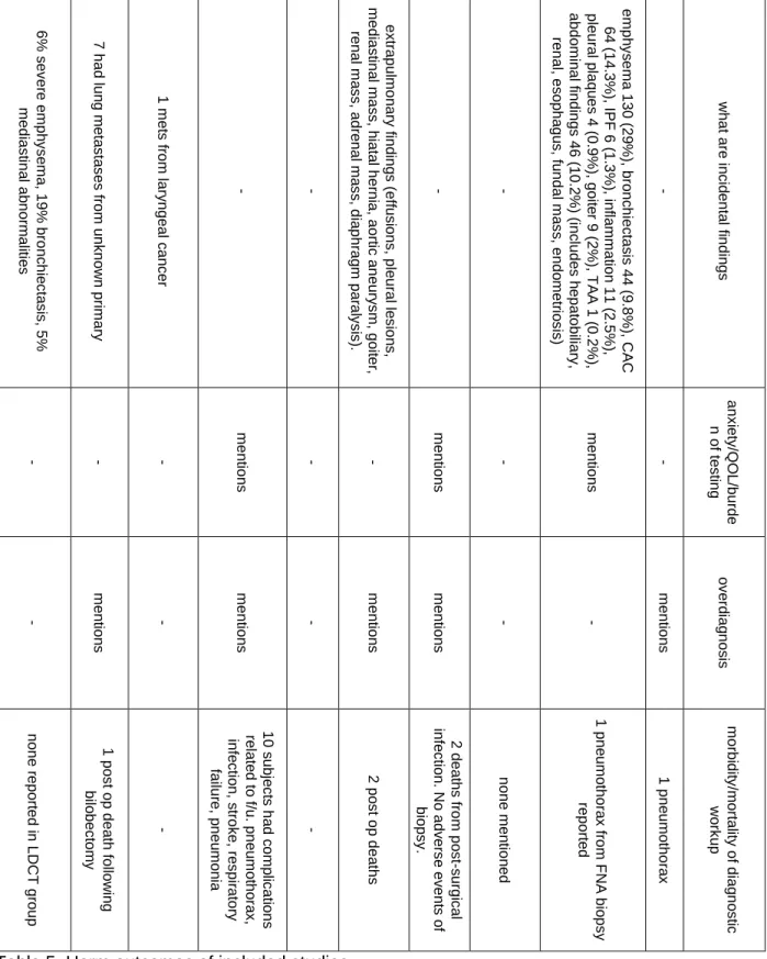
Total positivity of low dose CT screening ranged from 8 to 45%. For the studies reporting workup, 5 to 28% of those screened at baseline received some further workup as a result of a screening test. A range of 4 to 55% of total invasive procedures were performed for benign lesions. None to 40% of surgical procedures were performed for benign lesions. In the studies that reported incidental findings, 0.77 to 62% of those screened had abnormal findings other than lung nodules. Studies reported on extra-pulmonary tumors, emphysema,
bronchiectasis, lymphadenopathy, aneurysms, coronary calcifications, renal abnormalities, and adrenal abnormalities. 30% of the included studies mentioned anxiety or burden of testing and 55% of included studies mentioned overdiagnosis as potential harms to lung cancer screening. Half of the studies reported observed morbidity and mortality for diagnosis or treatment. 35% of studies reported false negative cases. The false positive rate of baseline LDCT ranged from 86 to 98%. Target outcomes for studies are summarized in Table 5 and are individually addressed in the text below.
Depiscan - Blanchon54
14 abnormalities. No morbidity or mortality was reported in the LDCT screening group. No false negative cases were reported. The false positive rate of LDCT was 95% at baseline.
Colorado - Garg55
Total positivity rate was 33%. 28% of LDCT screened participants received further workup. Invasive procedures and surgeries were not reported. Incidental findings were not reported, but one case of metastatic laryngeal cancer was mentioned. Morbidity, mortality, and overdiagnosis were not mentioned as potential harms. No false negative cases were reported. The false positive rate was 90%.
DANTE - Infante56-58
Total positivity rate was 28% for baseline and incident screens combined. 18% of those screened received some form of further workup. Of those who screened positive, 57% received a high resolution CT and 16% received a PET scan. Ninety-six invasive procedures were performed, 11% were for benign lesions. 13% of thoracotomies were performed for benign lesions. Five of the 46 total thoracotomies were required for non-lung cancer lesions including mesothelioma, esophageal cancer, complex aspergilloma, empyema, and leiomyoma.
Incidental findings were reported in 3% of subjects. These included effusions, pleural lesions, mediastinal masses, hiatal hernias, aortic aneurysms, goiters, renal and adrenal masses, and diaphragmatic paralysis. Two post-operative deaths were reported. Overdiagnosis was mentioned as a potential harm of screening. One false negative case was detected on sputum cytology. The false positive rate was 86% at baseline.
ELCAP - Henschke36,37
15 no thoracotomies were performed for benign lesions. Incidental findings were not reported in the primary articles. Morbidity, mortality, and other potential harms of screening were not reported. Five false negative cases were reported as four lung cancers and one interim cancer were visible on baseline exam. The false positive rate was 88%.
A follow study conducted by Anderson et al examined whether negative screening
results influenced smoking cessation behaviors on a subpopulation of those in the ELCAP
study59. The study found that negative scans were not associated with a lower likelihood of
prolonged abstinence among baseline smokers or a higher likelihood of relapse among former smokers over 6 years of follow up compared with those with a false positive result. In other words, negative scans were not associated with adverse smoking behavioral changes. However, smoking status was not collected uniformly throughout the study period and could influence measurement of results. In addition, 39% of those who reported quitting during the study period were excluded from analysis because of missing data.
A second follow up study evaluated smoking behaviors among a subpopulation of 313
individuals from the ELCAP sample60. Cancer anxiety and perceived health benefits of quitting
were found to be predictors of smoking cessation following LDCT exam. However, the result of the screening exam, whether positive or negative, was not statistically significantly associated with smoking cessation.
NY-ELCAP61
post-16 operative deaths of workup or treatment were observed. No complications were mentioned. Incidental findings were not reported in the primary article, however the study reported four cancers detected on biopsy that were not lung cancer, including 2 cases of metastatic colorectal cancer and 2 cases of lymphoma. The study mentions overdiagnosis as a potential harm to lung cancer screening. The study did not report any false negative cases. The false positive rate of LDCT screening was 89% in this study.
A companion paper analyzed the frequency of mediastinal masses detected in both the
ELCAP and NY-ELCAP populations62. The authors reported a prevalence of 0.77% of those
screened who had any mediastinal mass. These included masses in the thymus, thyroid, esophagus, and trachea. Other incidental findings were noted but not reported in this paper. Matsumoto - Sone43,63,64
Total positivity rate was 4.3% for baseline and incident screens combined. 4.9% of those at baseline, 3.8% of those screened at first repeat, and 3.3% of those screened at second repeat received further workup. I could not determine who received further diagnostic imaging. Seventy-two invasive procedures were reported, all were surgical biopsies. 22% of these surgeries were performed for non-cancerous lesions. Incidental findings were not reported. The study reported 24 false negative cases. No complications were mentioned. The false positive rate was 92%.
A follow article reported 10 year follow up of the study cohort64. Main outcomes in this
article included survival of patients diagnosed with CT detected lung cancer. However, the study did include an estimated overdiagnosis rate of 13% using a formula which included the volume doubling time of the tumor and the patient‟s age.
ALCA – Sobue/Kaneko42,65
17 imaging. Seventy-one total invasive procedures were performed, 49% were for benign lesions. Twenty-one total surgeries were performed with a 29% rate for benign disease. Incidental findings were not reported in the primary articles. Two deaths from post-surgical infection were recorded. There were no adverse events of biopsy observed. Anxiety and overdiagnosis were mentioned as potential harms to screening. No false negatives were reported. The false positive rate was 95%.
ITALUNG – Lopes Pegna66
Total positivity rate was 30%. 26% of LDCT screened participants received further workup. Fifty-two invasive procedures were performed, 4% were for benign lesions. One of the 18 surgeries performed was for a benign lesion, a rate of 5.5%. Incidental findings were not reported. Morbidity, mortality, and overdiagnosis were not mentioned as potential harms. No false negative cases were reported. The false positive rate was 95%.
COSMOS - Veronesi67-69
Total positivity rate was 53% for baseline screening and could not be determined for incident screening. 9.7% of those screened received further workup including diagnostic testing and biopsy. This study reported 104 surgeries with 14% of those for benign lesions.
Procedures included 5 VATS wedge resections, 9 open wedge resections, and 1 open
lobectomy. This study did not report incidental findings. The study mentions radiation exposure and overdiagnosis as potential harms to LDCT screening for lung cancer. Major post-operative morbidity was reported in 4.6% of cancer resections. No peri-operative morbidity occurred in surgeries for benign disease and no post-operative mortality was observed among surgeries. The study reported 24 false negative cancers that had a prevalent lesion on baseline. The false positive rate was 98% at baseline.
Munster - Diederich39,40,70
18 invasive procedures performed at baseline were for benign lesions. Three surgical procedures were performed at baseline, but I could not determine the proportion of total surgeries that were performed. This study did not report incidental findings but reported on seven cases of lung metastases from unknown primary malignancies. Overdiagnosis was mentioned as a potential harm to screening for lung cancer. One post-operative death was observed following
bilobectomy for cancer treatment. Four false negative cases were reported as abnormal baseline scans but were diagnosed symptomatically within screening intervals. The false positive rate on baseline scan was 96%.
Hitachi - Nawa41
Total positivity rate was not reported and I could not determine who received further workup. At baseline, 6.8% received diagnostic CT whereas 2.7% received diagnostic CT at repeat screening. Seventy-one invasive procedures were performed in total, but it was not clear how many were for benign lesions. Seventeen of 57 surgeries, or 30%, were performed for a benign lesion. The authors reported extrapulmonary tumors as incidental findings. These were tumors in the thyroid, parathyroid, mediastinum, and chest wall. No complications of workup were reported. The false positive rate at baseline was 98%.
Milan - Pastorino71
Total positivity rate was 15% for baseline and repeat screening combined. I could not determine who received further workup. 6% of those screened at baseline and 3.4% of those screened at repeat screening had further diagnostic imaging performed. Total invasive
19 Mayo Clinic - Swensen44,45,72,73
Total positivity rate was 51% for baseline screening and 74% of those screened had at least one non-calcified nodule (NCN) detected on any screen over the 5 total screening exams. I could not determine who received further diagnostic imaging or invasive diagnostic
procedures. At 3 year follow up, 8 of 39 surgeries were performed for benign lesions, a rate of 21%. At 5 year follow up, 15 surgeries were performed for benign lesions out of an unknown total. This study reported that 79% of those screened had either a pulmonary lesion or an incidental finding after 3 years of follow up. The most common extra-pulmonary findings included lymphadenopathy, aortic aneurysms, renal abnormalities, and adrenal abnormalities. In addition, 18 extra-pulmonary tumors were detected including cases of renal cell carcinoma, breast cancer, lymphoma, carcinoid, ovarian cancer, pheochromocytoma, and metastases. The study mentions both anxiety of workup and overdiagnosis as potential harms to LDCT screening for lung cancer. The study reported 5 false negative cases, 1 sputum detected case that was visible on baseline scan and 4 cancers detected on incident scans that were visible on prior scans. The false positive rate was 96% at baseline.
A follow up article by Cox et al reported on smoking changes of 97% of the Mayo study
participants with smoking data at baseline74. 14% of baseline smokers quit at one year and
10% former smokers were smoking at one year. A longer duration of cessation was associated with abstinence at one year for former smokers. A positive screening result was not associated with cessation and a negative screening result was not associated with continued smoking or relapse. The study was not designed to determine if screening was associated with any change as there was no non-screened control group.
A second follow up article by Townsend et al evaluated the relationship between
smoking cessation and results of screening exam over three annual screening exams75. This
20 baseline lung function, and abnormal CT findings on prior screening exam. For those who received 3 abnormal exams, 42% reported quitting smoking. For those who received 2 abnormal exams, 28% reported quitting. For those with one abnormal exam, 24% quit. For those without an abnormal exam, 20% quit. It appears this association is dose dependent. Longer duration of abstinence was the only predictor of maintaining abstinence among former smokers.
Another follow up study by Crestanello et al more closely examined thoracic surgery
procedures in the Mayo cohort through 200273. The article reported a total of 60 surgical
procedures in 55 patients performed for diagnosis and treatment. Lobectomy was the most common procedure followed by wedge resection. Ten surgeries were performed for benign disease, a rate of 18.1%. One lobectomy and nine limited resections were performed for benign lesions. Complications occurred in 27% of surgeries including air leak, arrhythmia, pneumonia, ileus, stroke, depression, vocal cord paralysis. One death (1.7%) occurred post-operatively from an intracerebral hemorrhage. A total of 79 nodules were resected, 39% of these nodules had benign pathology.
PLuSS - Wilson76,77
21 A companion paper explored healthcare usage after LDCT screening in a subpopulation
of 400 participants in the PLuSS cohort78. There were no observed differences between
groups, however data showed an increase in outpatient visits over the first 6 months following the screening exam, regardless of whether the participant received a positive or negative result. Visits declined to pre-screening levels over the next 6 months. These data show that a
temporary increase in visits occurred following screening, but this increase was not sustained past 6 months.
A follow article presented results exploring the relationship of emphysema diagnosed on
CT with airflow obstruction76. This paper found that 24% of those screened had mild to
moderate/severe emphysema on LDCT. It is unclear if these results are clinically significant, however emphysema was found to be independently associated with lung cancer.
PALCAD - MacRedmond79,80
Total positivity rate was 25% for baseline and incident screens combined. I could not determine who received further workup or the total number of invasive procedures performed. Five invasive procedures were performed for benign lesions. One of 4 surgeries was performed for a benign lesion. Incidental findings were well reported. 62% had any incidental finding on baseline screen with 49% having a clinically significant finding requiring further workup. Most common findings included emphysema (29%), coronary calcification (14%), bronchiectasis (10%), and abdominal abnormalities (10%). One complication of workup was reported as a pneumothorax for a FNA biopsy. The false positive rate at baseline was 98%.
LSS – Gohagan81,82
22 performed at baseline, 39% of these for a benign diagnosis. Eighteen biopsies and resections were performed after repeat screening and it was unclear how many of these were for benign lesions. Ten participants had complications related to follow up including pneumothorax, infection, stroke, respiratory failure, and pneumonia. The study mentioned anxiety and overdiagnosis as potential harms to lung cancer screening. Incidental findings were not reported in the primary articles, however a follow up letter reported proportions of incidental findings described below. No false negative cases were reported. The false positive rate at baseline was 91%.
Incidental findings of the Lung Screening Study were reported in a letter by Pinsky et
al83. Of those who received a LDCT screening exam, 16% had a nodule or mass, 17% had a
granuloma, 29% had scarring or fibrosis, 25% had COPD, 10% had pleural fibrosis, 0.32% had pleural fluid, 1.5% had a bone or soft tissue abnormality, 14% had a cardiac abnormality, and 19% had coronary artery calcifications. It is unclear if these incidental findings were clinically significant.
A follow up study by Croswell et al examined the cumulative risk for false positive
screening exams84. This study defined a false positive result as a positive LDCT screen with
negative workup or one year without a lung cancer diagnosis. The primary outcome showed that the cumulative probability of obtaining a false positive screening test was 33% (CI: 31, 35%) after two LDCT screening exams in this population.
Toronto – Menezes85
23 pneumothorax recorded as a complication of a biopsy. The false positive rate at baseline was 91%.
Israel - Shaham86
Total positivity rate was 8.2% for baseline and incident screens combined. I could not determine who received further workup. There were 16 reported invasive procedures
performed, all were biopsies of lung lesions. Two of the 12 total surgeries were performed for a benign lesion for a benign surgical rate of 17%. Of note, these surgeries were against
recommended protocol. Incidental findings were not reported. The study did not report complications of workup or resection. The false positive rate at baseline was 88%. DLCST – Pedersen87
Total positivity rate was 8.7% as only baseline results have been reported. I could not determine who received further medical evaluation after screening. There were 40 total
invasive procedures performed with 25% of invasive procedures performed for a benign lesion. Eleven surgeries were performed, 2 of these (18%) were performed for benign lesions.
Incidental findings were not recorded. One post-operative death was reported following
lobectomy for adenocarcinoma, but no morbidity or mortality was reported for work up. No false negative cases were reported. The false positive rate at baseline was 91%.
A companion article by Ashraf et al sought to determine smoking cessation changes in
this trial88. Primary measures were cessation and relapse rates at one year. No statistically
24 Toronto – Roberts89
Total positivity rate was 26% for baseline screening. The study states that 100% of those who screened positive had further workup, with 98% receiving further imaging. A total of 26 invasive procedures were performed for workup with 15% of these performed for benign lesions. The authors reported 3 surgeries for workup of which none were performed for benign indications. The majority of cancers were diagnosed with CT-guided biopsy. The authors reported 6 incidental findings consisting of one abnormal pulmonary vein, one mediastinal cyst, one mild lymphadenopathy, one thickened diaphragm, one carcinoid tumor, and one
25 Figure 1. Flow diagram of search strategy.
Title review led to exclusions: n= 440 Studies identified from
reference lists n= 9
Full text retrieved and assessed for eligibility n = 215
Full text review led to exclude n= 96
*Not relevant
publication type n= 62 *Not relevant design n= 8
*No outcomes of interest reported n= 19 *Wrong intervention n=7
Publications meeting inclusion criteria n=119
46 articles from 20 studies included in review
Excluded n= 73 *Poor outcome reporting n= 31
*Poor quality trial n= 13 *Duplicate publication n=9
*Unable to obtain English translation n=2 *Systematic review n=8 *Special population n=10
26
Author
year starte d
# scree ned with LDCT at baseli ne
total # LDCT screeni ng tests perfor
med population
fem ales (%)
age rang e
me dia n age (ye ars)
smoki ng histor y (medi an p-y)
% current/fo rmer/nev er smokers
LDCT slice thicknes s (mm)
Blancho
n 2002 336 336
subjects were under care of GP
recruiters, asymptomatic >15 cigs/day for >20 yrs
(quit <15 yrs prior) 29
50-75 56 30 64/36 1.5
Diederic
h 1995 817 2542
asymptomatic volunteers recruited by media, >20 p-y
history 28 >40 53 45 - 5
Garg 2001 92 92
veterans, included those with COPD and sputum atypia in previous cohort or >30 p-y. recruited
via telephone. 3
50-80 - - - 5
Gohaga
n 2000 1586 2984
volunteers recruited via mass mailing and referral, >30 p-y (quit w/in 10 yrs). From PLCO centers not already in PLCO
trial 41
55-74 - 54 58/42 5
Hensch
ke 1992 1000 2184
asymptomatic volunteers recruited from physician offices and
27
Infante 2001 1276 3612
male volunteers recruited via GPs and mass
mailings/media, >20 p-y (quit <10 yrs) 0
60-74 65 47 56/44 5
Sobue/K
aneko 1993 1611 9502
subjects from a dues paying organization that provides lung cancer screening to its members, includes never
smokers. 12
40-79 - - 62/25/14 10
Lopes
Pegna 1406 1406
recruited by sending invitation letters to registered patients of GPs, >20 p-y
history 35
55-69
61 (me
an) 39 65/35 1-3
MacRed
mond 449 1371
recruited by media, residents of community served by hospital, >10 p-y
smoking at 45 yo 50 >50 56 45 68/32 10
Meneze
s 2003 3352 6924
volunteers or referred by
physician, >10 p-y 54 >50 60 30 NR 1-1.25
Nawa 1998 7956 13524
Hitachi employees, includes never
smokers 21
50-69 - -
62% current/fo rmer, 38% never
smoker 10
Pastorin
o 2000 1035 2031
asymptomatic volunteers recruited
28
Pederse
n 2004 2047 2047
volunteers recruited via media, >20 p-y 45
50-70 - - 76/24 0.75
Roberts 2003 1000 1000
volunteers, >10 p-y
history 55 >55 63 34 34/66 1.25
Shaham 1998 842 1784
mixed referral from physician and volunteers from
media, >10 p-y 43 >50 56 37 - 3.2 - 11
Sone 1995 5483 13786
volunteers recruited via media, includes never smokers 46
40-74
64 (me an) -
46% current /former. 54% never
smokers 10
Swense
n 1999 1520 NR
volunteers recruited
via media, >20 p-y 48 >50 59 45 61/39 5
Verones
i 2004 5201 10022
asymptomatic volunteers from single center, >20
p-y 34 >50
58 (me
an) 44 80/20 2.5
Wilson 2002 3642 7065
recruited volunteers from media, could include
symptomatic, >12.5 p-y history 49
50-79
59 (me
an) 47 60/40 2.5
NY-ELCAP 2000 6295 12309
recruited via media and physician
referral, >10 p-y 51 >60 66 40 33/67 1.25-10
29
Author
year start ed
# scree ning tests perfor med
# lung canc ers total
description of positive test
# basel ine positi ve (%)
basel ine lung canc ers detec ted
preval ence
repe at posit ive
incid ent lung canc ers detec ted
% sta ge I
% adenocarc inoma and BAC
Blancho
n 2002 336 8
any NCN without benign features
152 (45%
) 8 2.38% - -
38
% 63%
Diederic
h 1995 2552 29
any NCN without benign features
378 (46%
) 12 1.35% 89 (13
%) 10 56
% 41%
Garg 2001 92 3
1-6 NCN without benign features
30 (33%
) 3 3.26% - - - -
Gohaga
n 2000 2984 40
NCN >3 mm or other
suspicious abnormalities
325 (21%
) 30 1.89% 360 (26
%) 8 48
% 60%
Hensch
ke 1992 2184 36
1-6 NCN without benign features
233 (23%
) 27 2.70% 30 (2.5
%) 7 82
% 68%
Infante 2001 3612 63
abnormality of malignancy including NCN >10 mm or smaller with suspicious features. Negative if NCN <5mm
199 (16%
) 28 2.19% 152 (4.2
%) 19 65
30
Kaneko/
Sobue 1993 9502 36
those who were requested to undergo thin section CT scan based on suspicion of benign tumor/inflamma tion/lung cancer
186 (12%
) 14 0.87% 721 (9.1
%) 22 78
% 67%
Lopes
Pegna 1406 21
at least 1 solid NCN >=5mm or nonsolid >= 10mm or part solid NCN
426 (30%
) 21 1.42% - -
48
% 48%
MacRed
mond 1371 6 Any NCN
105 (23%
) 2 0.45% 6 (1.3
%) 3 50
% 17%
Meneze
s 2003 6924 65
baseline: >1 NCN >=5mm or non-solid >= 8mm. Repeat: growth or new NCN
600 (18%
) 56 1.67% 92 6 65
% 68%
Nawa 1998 13524 41
Non-calc SPN >=8 mm without benign features
2099 (26%
) 37 0.45% - 4 85
% 85%
Pastorin
o 2000 2031 22 NCN >= 5 mm 199 (19%
) 11 1.06% 99 (10
%) 11 77
% 77%
Pederse
n 2004 2047 17
NCN>=5mm and growing nodules
179 (8.7
%) - 0.83% - -
53
% 71%
Roberts 2003 1000 20
>=1 solid/part solid NCN >=5 mm or nonsolid >= 8 mm.
256 (26%
) 20 2% - -
75
% 70%
Shaham 1998 1784 14
baseline: 1-6 NCN >=5mm at first. Then adjusted to 1-6 solid/part solid NCN >=5mm or >1 non solid >=8 mm. repeat: new and/or growth
102 (12%
) 12 1.43% 45
(5%) 2 86
31
Sone 1995 13786 60
positive result included scan read as possible cancer, probable cancer, or small nodule <3 mm
279 (5.1
%) 22 0.40% 309 (3.7
%) 34 88
% 85%
Swense
n 1999 NR 68 any NCN
780 (51%
) 31 2.04% 773
(NR) 34 57
% 53%
Verones
i 2004 10022 92 any NCN
2754 (53%
) 55 1.06% 45 (0.9
%) 36 66
% 68%
Wilson 2002 7065 80 any NCN
1477 (41%
) 53 1.46% 42% 24 50
% NR
NY-ELCAP 2000 12309 124
baseline: at least 1 solid/part solid NCN >= 5 mm or nonsolid NCN >= 8 mm. incident: any new NCN regardless of size or any growth.
906 (14%
) 101 1.60% 361 (6.0
%) 20 93
% 60%
32
Nawa Pas
to rin o Pe d e rs e n Rob e rts Sh a h a m So n e Swen s e n Ve ro n e s i Wi ls o n NY -EL CAP Au th o r p ro s p e c tiv e c o h o rt p ro s p e c tiv e c o h o rt RC T (L DC T v s n o s c re e n in g a n n u a l) p ro s p e c tiv e c o h o rt p ro s p e c tiv e c o h o rt p ro s p e c tiv e c o h o rt p ro s p e c tiv e c o h o rt p ro s p e c tiv e c o h o rt p ro s p e c tiv e c o h o rt p ro s p e c tiv e c o h o rt d e s ig n na na 2 0 5 2 in e a c h g ro u p , c e n tr a l c o m p u te r p e rm u te d b lo c k s o f 1 0 . a d e q u a te . Unc le a r c o n c e a lm e n t
na na na na na na na
34 2 re a d e rs in d e p e n d e n tly r e a d a ll c a s e s . Di s c re p a n c ie s re s o lv e d a t wee k ly c o n fe re n c e s . 2 ra d s i n d e p e n d e n tl y . Di s c re p a n c ie s r e s o lv e d b y 3 rd ra d . re a d b y 2 r a d s (u n k n o wn i n d e p e n d e n c e ). Di s c re p a n c y b y c o n s e n s u s . u n c le a r wh o /h o w m a n y re a d s c a n s c h a n g e d d e fi n iti o n o f a p o s iti v e re s u lt d u ri n g t h e t ri a l, u n c le a r wh e n . Al s o c h a n g e d s lic e t h ic k n e s s whi c h w o u ld a ff e c t in te rp re ta ti o n o f im a g e s . O n e ra d io lo g is t re a d a ll s c a n s re a d b y 1 o f 4 ra d io lo g is ts , th e n 2 n d r e a d e r re a d s u s p ic io u s o r c lin ic a lly s ig n ifi c a n t s c a n s . Un m a s k e d t o f irs t re a d . re a d b y 1 o f 4 ra d io lo g is ts . u s e d b o th c o m p u te r a id e d d e te c ti o n a n d m a n u a l d e te c ti o n . Rad io lo g is t in te rp re te d a n d l e s io n s iz e v a lid a te d b y 2 ra d s a t c o n fe re n c e . 1 /2 ra d s re a d in iti a l s c a n . Co n fe re n c e re v ie we d m o d e ra te /h ig h s u s p ic io n s tu d ie s . U s e d c o m p u e r a lg o ri th m to c la s s ify p re -b io p s y lu n g c a r is k 2 ra d s re a d i n d e p e n d e n tl y . Un m a s k e d t o p a rti c ip a n t wi th in t h e s tu d y m e a s u re m e n t o f o u tc o m e s poor fa
ir fair fair fair
u n c e rta in : la rg e p ro p o rt io n o f fe m a le s we re n e v e r s m o k e rs , m a jo rity o f to ta l p o p u la ti o n we re n e v e r s m o k e rs fa
ir fair fair
fa ir. Pt s p a id f o r wo rk u p th e m s e lv e s g e n e ra liz a b ilit y fa
ir fair fair fair fair fair fair fair fair fair
35 Bl a n c h o n Die d e ri c h G a rg G o h a g a n Hen s c h k e In fa n te Ka n e k o /So
bue Lo
36 6 2 1 /7 6 5 (8 1 % ) wi th b a s e lin e d a ta d u e t o wi th d ra wal . Wi th d ra wal s m o re l ik e ly to b e i n CXR g ro u p a n d y o u n g e r 6 6 8 /8 1 7 (8 2 % ) re tu rn e d f o r re p e a t s c a n a t 1 y r all 9 2 ra n d o m iz e d t o CT g ro u p u n d e rwe n t b a s e lin e s c a n . 9 6 % a n d 8 6 % a d h e re n c e i n L DC T g ro u p a t b a s e lin e a n d 1 y r re s p e c ti v e ly 8 4 1 /1 0 0 0 ( 8 4 % ) h a d a n n u a l r e p e a t s c a n , 1 1 7 we re c o n s id e re d d ro p o u ts ( 1 2 % ) 2 8 1 1 t o ta l ra n d o m iz e d , 2 4 7 2 ( 8 8 % ) u n d e rwe n t b a s e lin e e x a m . U n c le a r a d h e re n c e a t fo llo w u p 1 1 8 0 /1 6 1 1 (7 3 % ) u n d e rwen t 1 s t re p e a t. 4 4 5 (2 7 % ) a t 4 y e a rs . 1 4 0 6 /1 6 1 3 (8 7 % ) ra n d o m iz e d to L DC T u n d e rwen t b a s e lin e s tu d y 4 1 3 (9 2 % ) c o m p le te d 2 y r f o llo w u p 2 6 8 6 /3 3 5 2 (8 0 % ) re tu rn e d fo r a n n u a l re p e a t a d h e re n c e /d ro p o u ts 6 (1 % ) o f th o s e i n CXR g ro u p g iv e n L DC
T - -
37 2 ra d io lo g is ts r e a d e a c h s c a n in d e p e n d e n tl y , d is c re p a n c y b y c o n s e n s u s s c a n s r e a d b y 1 o f 2 ra d io lo g is ts , u n c le a r a b o u t m a s k in g re a d b y 1 c h e s t ra d io lo g is t, s u s p ic io u s s c a n s r e a d in d e p e n d e n tl y b y 2 n d ra d io lo g is t, c o n s e n s u s f o r d is c re p a n c y . N o m e n ti o n o f m a s k in g re a d b y ra d io lo g is t a t e a c h s ite . Res u lts r e c o rd e d o n s ta n d a rd iz e d f o rm s . Un c le a r o f m a s k in g o r d is c re p a n c y in re a d s . e a c h s c a n r e a d b y 2 ra d io lo g is ts m a s k e d to o th e r' s r e a d . As s e s s e d re a d e r a g re e m e n t 2 c h e s t ra d io lo g is ts re a d e a c h s c a n i n d e p e n d e n tl y , c o n s e n s u s f o r d is c re p a n c y . 2 ra d io lo g is ts o r c h e s t p h y s ic ia n s re a d e a c h s c a n . 2 n d r e a d e r n o t b lin d e d t o f irs t in te rp re ta tio n . 3 rd r e a d e r d e te rm in e d re c o m m e n d a tio n fo r t h in s e c ti o n CT. Com p u te r a id e d d ia g n o s is wa s i n tr o d u c e d i n m id d le o f tr ia l. re a d b y 2 r a d s i n d e p e n d e n tl y . Dis c re p a n c y b y c o n s e n s u s . p u lm o n o lo g is t was n o t bli n d e d t o g ro u p . re a d b y 2 r a d s i n d e p e n d e n tl y . Dis c re p a n c y b y c o n s e n s u s . I-EL CAP p ro to c o l Not re p o rte d . Th u s d o n o t k n o w who re a d s c a n s m e a s u re m e n t o f o u tc o m e s fa ir - o n ly 4 1 % GP s p a rti c ip a te d i n re c ru itm e n t, b u t th is wo u ld o n ly a ff e c t g e n e ra liz a b ilit y if G P s s e rv e d d iff e re n t p o p u la ti o n s fa ir p o o r (m a n y a lre a d y i d e n tifi e d wit h s p u tu m a ty p ia in p rio r s tu d y ) fa
ir fair fair
p o o r: m e m b e rs h ip i n a n o rg a n iz a tio n fa
ir fair fair
g e n e ra liz a b ilit y fa
ir fair fair
good fa
ir fair fair fair fair fair
s tu d y q u a lity
38 Nawa Pa s to rin o Pe d e rs e n Rob e rts Sh a h a m So n e Swen s e n Ve ro n e s i Wi ls o n NY -EL CAP Au th o r
13524 2031 2047 1000 1784 13786
-
10022 7065 12309
# s c re e n in g te s ts p e rfo rm e d
41 22 17 20 14 60 68 92 80 124 tota
l lung c a s - 1 5 % (o f to ta l s c a n s ) 179 (8 .7 % )
26% 8.2
0 % 588 (4 .3 % )
- 2799
(2
8
%
)
41% 10% % t
o ta l p o s iti v e
- - 29% -
2 0 3 (2 4 % )
- 51% 53% 41% -
% o f b a s e lin e p a rti c ip a n ts w ith a n y NCN o n a n y s c a n (e x c lu d in g b e n ig n ) ( in c lu d e s < 5 mm)
- - - 26% -
4 .9 % a t b a s e lin e , 3 .8 % a t 1 s t re p e a t, 3 .3 % a t 2 n d re p e a t - 5 0 4 (9 .7 % ) 8 2 1 (2 2 % ) a t b a s e lin e . 8 % a t b a s e lin e , 3 % a t re p e a t % o f to ta l s c re e n e d re c e iv in g a n y f u rth e r work u p - - - 100% - 9 5 % a t b a s e lin e , 9 7 % a t 1 s t re p e a t, 9 5 % a t 2 n d re p e a t
- 18% 56%
39 6 .8 % a t b a s e lin e , 2 .7 % a t re p e a t 6 1 (5 .9 % ) a t b a s e lin e , 3 4 (3 .4 % ) a t re p e a t - 2 5 0 (9 8 % ) - - - - 8 2 1 b a s e lin e (2 2 % ) - # /% re c e iv in g d ia g n o s tic im a g in g (DXC T, HRC T, PET) 71
- 40 26 16 72 - 104 -
1 5 8 b io p s ie s in v a s iv e p ro c e d u re s f o r d ia g n o s tic w o rk u p - - 1 0 (2 5 % ) 4 (1 5 % ) 2 22% 1 5 (CD) 1 5 (1 4 % ) - 7 (6 % ) a t b a s e lin e , 2 ( 8 % ) a t r e p e a t o f 1 3 4 re c o m m e n d e d . 2 4 n o n -re c o m m e n d e d a ll f o r b e n ig n . T o ta l 3 3 ( 2 1 % ) # , % i n v a s iv e p ro c e d u re s f o r b e n ig n
57 27 11 3 12 72
- 104 90 - tota
l s u rg e rie s 1 7 (3 0 % ) 6 (2 2 % ) 2 (1 8 % ) a p p e a rs n o n e 2 (1 7 % ) 1 6 (2 2 % ) 1 5 t o ta l. 8 (2 1 % a t 3 y r fo llo w u p ) 1 5 (1 4 % ) 3 6 (4 0 % ) n o l o b e c to m ie s (u n c le a r a b o u t o th e r p ro c e d u re s ) # , % s u rg ic a l (t h o ra c o to m y o r VAT S) f o r b e n ig n o f to ta l s u rg e rie s
- - - 6 - -
41 Bl a n c h o n Die d e ri c h G a rg G o h a g a n Hen s c h k e - EL CAP I In fa n te Ka n e k o /So b u e L o p e s Pe g n a M a c Re d m o n d M e n e z e s Au th o r
336 2552 92 2984 2184 3612 9502 1406 1371 6924
# s c re e n in g te s ts p e rfo rm e d
8 29 3 40 36 63 36 21 6 65
to ta l l u n g c a s 45% 4 6 7 (1 8 % ) 3 0 (3 3 % ) 6 8 5 (2 3 % ) 2 6 3 (2 6 % ) 3 5 1 (2 8 % ) (i n d iv id u a ls wi th p o s iti v e re s u lt) 9 0 7 (9 .5 % ) o f to ta l s c re e n in g te s ts 4 2 6 (3 0 % ) 1 1 1 (2 5 % ) 6 9 2 (1 0 % ) % t o ta l p o s ti v e 1 5 2 (4 5 % ) 57% 33%
- 23% - - - 25% -
42 - n o t c le a r 2 6 (2 8 % ) 3 0 9 (1 9 % ) a t b a s e lin e , 3 3 2 (2 4 % ) a t re p e a t 1 8 0 (1 8 % ) a t b a s e lin e , 2 8 ( 3 .3 % ) a t re p e a t 2 2 6 (1 8 % ) 1 9 2 (1 2 % ) a t b a s e lin e , 7 7 0 (9 .8 % ) a t re p e a t 3 6 6 (2 6 % )
- 600
% o f to ta l s c re e n e d re c e iv in g a n y f u rth e r wo rk u p - n o t c le a r 100% (9 8 % ) a t b a s e lin e , (9 5 % ) a t re p e a t 7 7 % a t b a s e lin e , 9 3 % a t re p e a t 2 2 6 (6 4 % ) 1 9 2 (1 0 3 % ) a t b a s e lin e , 7 7 0 (1 0 7 % ) a t re p e a t 3 6 6 (8 6 )% - 100% % o f p o s iti v e s r e c e iv in g a n y fu rth e r w o rk u p - n o t c le a r - 2 3 2 (7 3 % ) a t b a s e lin e , 1 4 0 (4 0 % ) a t re p e a t 1 8 0 a t b a s e lin e ( 7 7 % ) 1 9 9 HRC T (5 7 % ), 5 7 PET (1 6 % ) 1 8 1 (9 7 % ) a t b a s e lin e , 7 1 9 (9 9 .7 % ) a t re p e a t 366 - 1 2 (2 % ) # /% re c e iv in g d ia g n o s tic im a g in g (DXC T, HRC T, PET ) 12 - - 7 5 a t b a s e lin e , 3 2 a t re p e a t. 3 1 (1 3 % ) a t b a s e lin e , 9 (3 0 % ) a t re p e a t 96 2 2 a t b a s e lin e , 4 9 a t re p e a t 52 - 1 6 b io p s ie s , 1 8 s u rg e rie s , 1 8 b ro n c h
- 78
43 3 (2 5 % ) 4 (2 0 % ) - 2 3 (8 % ) a t b a s e lin e , NR a t re p e a t 4 (1 3 % ) (3 a g a in s t re c o m m e n d a tio n ) a t b a s e lin e . 1 (1 1 % ) a t re p e a t. 6 (1 1 % ) 8 (3 6 % ) a t b a s e lin e , 2 7 (5 5 % ) a t re p e a t 2 (3 .8 % ) 5 (NR) 1 6 (2 1 % ) # , % i n v a s iv e p ro c e d u re s f o r b e n ig n - - - 4 6 a t b a s e lin e . 1 8 a t re p e a t.
- 46 21 18 4 NR
to ta l s u rg e ri e s 3 (3 8 % ) t h o ra c o to m ie s 3 (u n c le a r) - 1 8 a t b a s e lin e . U n c le a r a t re p e a t. n o t h o ra c o to m ie s f o r b e n ig n 6 (1 3 % ) o f th o ra c o to m ie s 6 (2 9 % ) (VAT S a n d t h o ra c o to m ie s ) 1 (5 .5 % ) 1 (2 5 % ) - # , % s u rg ic a l (t h o ra c o to m y o r VATS) fo r b e n ig n o f t o ta l s u rg e rie s
- - - - - 3% - -
44 6 % s e v e re e m p h y s e m a , 1 9 % b ro n c h ie c ta s is , 5 % m e d ia s tin a l a b n o rm a liti e s 7 h a d lu n g m e ta s ta s e s fro m u n k n o wn p ri m a ry 1 m e ts f ro m la ry n g e a l c a n c e r - - e x tra p u lm o n a ry f in d in g s (e ff u s io n s , p le u ra l l e s io n s , m e d ia s tin a l m a s s , h ia ta l h e rn ia , a o rt ic a n e u ry s m , g o ite r, re n a l m a s s , a d re n a l m a s s , d ia p h ra g m p a ra ly s is ). - - e m p h y s e m a 1 3 0 (2 9 % ), b ro n c h ie c ta s is 4 4 (9 .8 % ), CAC 6 4 (1 4 .3 % ), IPF 6 ( 1 .3 % ), in fl a m m a tio n 1 1 ( 2 .5 % ), p le u ra l p la q u e s 4 (0 .9 % ), g o ite r 9 (2 % ), T AA 1 ( 0 .2 % ), a b d o m in a l f in d in g s 4 6 ( 1 0 .2 % ) (i n c lu d e s h e p a to b ilia ry , re n a l, e s o p h a g u s , fu n d a l m a s s , e n d o m e tr io s is ) - wha t a re in c id e n ta l f in d in g s - - - m e n tio n s - - m e n tio n s - m e n tio n s - a n x ie ty /QO L /b u rd e n o f te s ti n g - m e n tio n s - m e n tio n s - m e n tio n s m e n tio n s - - m e n tio n s o v e rd ia g n o s is n o n e r e p o rt e d i n L DC T g ro u p 1 p o s t o p d e a th f o llo w in g b ilo b e c to m y - 1 0 s u b je c ts h a d c o m p lic a ti o n s re la te d t o f/ u . p n e u m o th o ra x , in fe c ti o n , s tro k e , re s p ira to ry fa ilu re , p n e u m o n ia - 2 p o s t o p d e a th s 2 d e a th s fro m p o s t-s u rg ic a l in fe c ti o n . No a d v e rs e e v e n ts o f b io p s y . n o n e m e n ti o n e d 1 p n e u m o th o ra x f ro m F NA b io p s y re p o rte d 1 p n e u m o th o ra x m o rb id ity /m o rta lity o f d ia g n o s ti c work u p
45 Discussion:
This systematic review attempted to characterize the harms associated with lung cancer screening. Overall, harm outcomes were not uniformly reported across included studies. In addition, harms were difficult to quantify as most studies did not report information or reported incomplete information on harms. However, data from the included studies show that the harms of screening for lung cancer with low dose CT are not trivial. Harms should warrant discussion when deciding whether to initiate mass screening for lung cancer even if a mortality benefit is observed in good quality randomized controlled trials. For a successful screening program, the benefits of undergoing the screening test should outweigh the potential harms.
Healthcare usage
Healthcare usage following screening exam was addressed in the PLuSS study only78.
Results showed increases in outpatient visits over the first 6 months following screening and then a decline to pre-screening levels by 12 months. This short term increase could be due to distress of testing, participation in a study, and a motivated volunteer population. Measures were self-reported so one must interpret these results with caution as recall and social biases may also play a role in reporting. Further studies should explore the relationship of healthcare usage post screening as this is likely to play a role in the burden of testing and burden on the healthcare system.
Distress/anxiety/quality of life
Psychological factors and quality of life were mentioned in a third of included
studies42,71,72,81,89. Distress and anxiety were usually briefly mentioned in discussion as a
potential psychological harm to screening, especially when a false positive result occurs. None of the included studies quantified distress on a validated scale. In the NELSON trial for which results are not yet published, the psychological effects of screening are specifically addressed in
46
In a study conducted by Bunge et al, the authors examined distress in 351 subjects
before and after LDCT screening in the NELSON randomized trial91. Participants were
surveyed 1 day before screening and 6 months after using the Impact of Event Scale (IES). Statistically significant results were observed for participant‟s perceived risk of developing lung
cancer before and after screening. 14.5% perceived their risk for lung cancer as high 1 day before screening whereas 10.5% perceived their risk to be high 6 months later (p<0.01). Of note, all individuals here received a negative screening result so the results may be explained by reassurance in the screening group. From this study, it is unclear how those with a false positive result would perceive their risk after screening.
In a second study, the authors surveyed 351 participants who were randomized to the
LDCT screening arm of the NELSON trial90. Health related quality of life (HRQoL) and
discomfort were measured 1 day before, within one week after, and 6 months post screening with the 12–item Short Form and the EuroQol questionnaire. This study found that 87 to 99% of
participants reported no discomfort related to the LDCT screening procedure and HRQoL did not significantly change over time, but 46% of participants reported at least some discomfort in waiting for results and 51% of participants reported dreading the screening exam results. The authors concluded that LDCT screening demonstrated no adverse effects on quality of life but discomfort was observed in waiting for results. Of note, this study excluded positive screening exams so does not measure the discomfort of false positives.
In a further study, 733 participants in the NELSON trial were surveyed for health related
quality of life, anxiety, and lung cancer specific distress before and after screening92. Quality of
47 thus could not determine the effect of a false positive result on quality of life, anxiety, or lung cancer specific distress.
From these results, it is likely that distress, quality of life, and anxiety are likely to be important factors that should be addressed before implementing a LDCT screening program. This includes providing timely results and how to handle the psychological effect of an
indeterminate result. None of these three studies addressed false positive results, which likely increase anxiety and lung cancer specific distress even further than an indeterminate result. False positives and false negatives
One third of studies reported any false negative findings. False negatives were not uniformly defined. I included false negatives as they were defined in the primary studies. This definition included cancers detected on incident screen that were visible on prior screening exam, interim cancers that were visible on prior exam, and any cancers missed on CT but diagnosed by other means. Veronesi and Sone reported high numbers of false negative cases,
24 and 18 respectively, whereas other studies reported fewer than 5 cases43,69. In a follow up
study, Li et al describes the errors in interpretation and detection as the primary causes for
missing these lung cancers93.
I was able to determine the false positive rate for all included studies. I defined false positives as screening exams that were interpreted as positive, according to the study definition of a positive test, but did not result in a diagnosis of lung cancer. For the majority of studies, at least 9 out of 10 positive screening tests were false positives at baseline. In addition, Croswell
et al showed that the probability of having a false positive result after 2 total scans was 33% in
the LSS population84. This study was conducted in a high risk population with >30 pack-years
48 These results show the poor predictive value of LDCT in screening for lung cancer. By changing the definition of a positive result, investigators hoped to decrease the false positive
rate as was done in the ELCAP study94. However, the false positive rates in I-ELCAP and
NY-ELCAP were still 90% and 88%, respectively38,61. The lowest false positive rate at baseline was
86% in the DANTE trial where the definition of a positive result was any abnormality of
malignancy which included NCN ≥10mm or smaller with suspicious features56. This definition
has a greater cutoff than other definitions as most studies defined a positive result as any NCN ≥5mm. Thus one would expect a smaller degree of false positives with an increased size cutoff.
Furthermore, in the DANTE trial there were 13 interim cancers diagnosed because of symptoms out of 63 total, representing a higher proportion of symptom diagnosed cancers than other
studies56. These data demonstrate that LDCT screening is not without false negative exams
and has a high proportion of false positive cases, even when a strict definition of a positive result is used in a high risk population.
Surgeries for benign disease
49 Morbidity and mortality of workup
Complications were incompletely reported as only half of the studies reported observed morbidity and mortality for diagnosis or treatment. Studies reported complications of biopsy including pneumothorax, infection, respiratory failure, and stroke. Nine total deaths related to follow up were reported in total. In a follow up article to the Mayo study, the authors reported
complications of workup and treatment73. The complication rate in this study was 27%, a value
higher than that reported in any other study. The Lung Screening Study reported the next greatest number of complications related to follow up with 10 total complications, representing a
much lower percentage than that in the Mayo study81,82. From this information, it is likely that
complications of workup are underreported in the included studies and further studies should seek to report complications of work up including non-surgical and surgical biopsy. This information is vital to quantifying harms of lung cancer screening workup.
Incidental findings
Seven out of the 20 included studies reported any incidental findings. Clinically relevant findings were reported at a rate of 7 to 49% among the seven studies. Thus, IFs were
underreported and variable. The ELCAP study reported a prevalence of 0.77% of mediastinal
masses in the study cohort62 whereas MacRedmond reported that 62% of those screened had
an incidental finding on LDCT screening79,80. It appeared that only the PALCAD study
completely reported incidental findings, including those that did and did not require further
workup79,80. The Mayo study reported only clinically relevant findings but I could not determine
the proportion of those screened with an incidental finding44,45,72.
Incidental findings were explored as part of the NELSON study95. In 1929 participants