ARTIGO ORIGINAL
42
Revista Científica da Ordem dos Médicos www.actamedicaportuguesa.com
Prevalence of Chromosomal Abnormalities
in Spontaneous Abortions or Fetal Deaths
Estudo da Prevalência de Anomalias Cromossómicas em Abortamentos
Espontâneos ou Mortes Fetais
1. Faculdade de Medicina. Universidade do Porto. Porto. Portugal.
2. Departamento de Ginecologia e Obstetrícia. Faculdade de Medicina. Universidade do Porto / Centro Hospitalar S. João. Porto. Portugal. 3. Departamento de Genética. Faculdade de Medicina. Universidade do Porto. Portugal.
Recebido: 05 de Agosto de 2013 - Aceite: 04 de Outubro de 2013 | Copyright © Ordem dos Médicos 2014
Raquel BASTOS1, Carla RAMALHO2, Sofia DÓRIA3 Acta Med Port 2014 Jan-Feb;27(1):42-48
RESUMO
Introdução: O abortamento espontâneo é um dos problemas mais frequentes da gravidez, estimando-se que afete, pelo menos, 25% das mulheres que tentam engravidar. O objetivo principal deste trabalho foi o estudo da prevalência das anomalias cromossómicas em perdas gestacionais, analisando a sua relação com a idade materna, idade gestacional e história de abortamentos prévios.
Material e Métodos: Realizou-se um estudo retrospetivo em 401 casos de perdas de gravidez que efetuaram análise citogenética e anátomo-patológica, entre janeiro de 2008 e junho de 2012, no Centro Hospitalar de S. João.
Resultados: Dos 401 casos enviados para estudo citogenético foi possível obter informação sobre o complemento cromossómico em 333 casos, dos quais 72,7% apresentaram cariótipo normal e 27,3% cariótipo anormal. As aneuploidias representaram 92,3% das cromossomopatias identificadas, sendo as trissomias as mais frequentes, associadas a uma idade materna avançada e a uma idade gestacional precoce. Não se verificou concordância entre os resultados da análise anátomo-patológica e citogenética.
Discussão e Conclusão: A prevalência de anomalias cromossómicas, no primeiro trimestre, foi semelhante entre os casos de abor-tamento esporádico e recorrente. Com o aumento da idade materna observou-se um aumento das trissomias, com um incremento médio de 7,4% no risco de ocorrência por ano. Não foi estabelecida uma correlação cariótipo-patológica significativa. A contaminação materna da amostra foi o principal entrave à determinação exata da prevalência de anomalias cromossómicas. As técnicas de cito-genética molecular já disponíveis podem colmatar esta e outras limitações da técnica convencional.
Palavras-chave: Abortamento Espontâneo/genética; Aberrações Cromossómicas; Anomalias Cromossómicas; Infertilidade Feminina/ genética; Portugal.
ABSTRACT
Introduction: Spontaneous abortion is one of the most frequent problems of pregnancy, estimated to affect, at least, one in every four women who tries to conceive. The main purpose of this work was to study the prevalence of chromosomal abnormalities in gestational losses, evaluating their relation with maternal age, gestational age and previous abortion history.
Material and Methods: Retrospective study of 401 pregnancy loss cases that have gone through cytogenetic and anatomopathologic analysis between January 2008 and June 2012, in Centro Hospitalar de S. João, Oporto.
Results: Of the 401 cases sent to cytogenetic study, it was possible to obtain information about the chromosomal complement in 333 cases, of which 72.7% showed normal karyotype, and 27.3% abnormal karyotype. Aneuploidies represented 92.3% of the identified chromosomopathies, with trisomies being the most frequent, related with an advanced maternal age and an early gestational age. There was no agreement between the results of the cytogenetic and the anatomopathologic analysis.
Discussion/Conclusion: The prevalence of chromosomal abnormalities, during the first trimester, is similar between sporadic and recurrent miscarriages. With increased maternal age, trisomies, the most frequent type of aneuploidy, are more likely to occur, with a mean increment in probability of 7.4% per year. A significant karyotype-pathological correlation was not established. Maternal contami-nation is the main obstacle to the accurate determicontami-nation of the prevalence of chromosomal abnormalities. The molecular cytogenetic techniques already available can overcome the limitations of the conventional technique.
Keywords: Abortion, Spontaneous/genetics; Chromosome Aberrations; Chromosome Disorders; Infertility, Female/genetics; Portugal.
INTRODUCTION
Spontaneous miscarriage is a common problem during pregnancy and it is estimated to affect 25 to 50% of women trying to get pregnant, although only 15% of the cases are clinically detected.1,2 It is known that most miscarriages
oc-cur over the first weeks of pregnancy, especially until the 14th week.1 Although most miscarriages are sporadic, 1-3%
of the couples present recurrent miscarriage, defined as consecutive loss of three or more pregnancies, with a sig-nificant psycho-social impact.1,3,4
Several illnesses have been considered as a cause for sporadic miscarriage, with chromosomal conception abnormalities representing approximately 50-60% of the
cases.2,5,6 Most of these abnormalities are numerical (95%)
and trisomy is the most prevalent (60%), more frequently trisomy 16, followed by polyploidy and X monosomy.1,2
As regards recurrent miscarriage, the literature is not so consensual as to the frequency of chromosomal abnormalities, some stating that they occur with a similar frequency as sporadic miscarriage7-10 while others argue for
a lower frequency.11,12
Maternal age increase is related with an increase in chromosomal conception abnormalities and consequently with spontaneous miscarriages.2,10,13,14 Over the years and
worldwide a progressive maternity delay with a significant
anos
35
35 ano
s a p rom
ov
er
as
ciê
ncia s bio méd
ica
s
A
CT A MÉ
DI
CA
P
ORT UGUE
SA
ARTIGO ORIGINAL
35
35 ano
s a p rom
ov
er
as
ciê
DI
CA
PO
RTUG
UESA
1979 - 2014
increase in the age of the first pregnancy is becoming evident. In Portugal, during a 10 year period (1998-2008), an increase in the number of deliveries above 40 (from 1.8% to 3.2%), as well as a change in the age group in which most cases occurred (from 25-29 to 30-34) have been observed.15 This increase in maternal age is related
with a higher spontaneous miscarriage and foetal loss rate, following a J-curve with a steep rise at the age of 35.16 This
fact represents an increase in the proportion of abnormal foetal karyotypes: 51-54% in women aged below 35 and 74-76% in older women.10,17
Taking into account the chances at the time of fertilisation, male and female embryos would be expected in equal numbers. Nevertheless, there is a distortion at the time of delivery, with a higher number of male foetus in an approximate relation of 1.0518,19 (1.053 in Portugal, during
2011).20
Therefore, taking into account the huge impact of chromosomal abnormalities in the success of pregnancy, our study aimed mainly to identify the most common chromosomal abnormalities related with spontaneous miscarriages or foetal losses and to assess their relation to gestational age, maternal age and previous miscarriages. In addition, we have also assessed the relative number of miscarriages per gender and found a relation of these results with cytogenetic and histopathological examinations.
MATERIAL AND METHODS
Our study involved a retrospective analysis of the results obtained through cytogenetic and histopathological examination of spontaneous miscarriage or foetal loss products, at the Centro Hospitalar de S. João between
January 2008 and June 2012.
Clinical records were consulted and information obtained on mother’s age and past obstetric history, gestational age at the time of pregnancy loss and results of genetic and histopathological examination of miscarriage products. This study was approved by the Ethics Committee of the
Centro Hospitalar de São João/Faculdade de Medicina da
Universidade do Porto (FMUP).
The criteria for cytogenetic examination were the following: termination of pregnancy; foetal loss and late pregnancy loss; repeated early pregnancy loss (more than 2) and miscarriage in a couple with a known chromosomal abnormality. For the study of spontaneous miscarriages, the terminations of pregnancy were excluded, in order to avoid a possible bias in the results. Beyond those meeting the previously described criteria, our group of patients also included spontaneous miscarriages occurring in primigravidae, as part of a previous research project. Taking into account the gestational age at the time of miscarriage, the products that were examined were assigned according to the gestational trimester: the first until the 12th week, the second between 13th and 24th and the third from the 24th week of pregnancy. Three age groups were also defined for the assessment of the impact of maternal age on the results: younger than 35, between 35 and 39 and above 40 years of age.
The decision was taken to establish six groups taking into account the results suggested by the histopathological examination, in order to establish a correlation between the histopathological and cytogenetic information (normal or abnormal karyotype: (1) probable chromosomal abnormality; (2) without an apparently adequate implantation; (3) apparent growth disorganization of foetus/foetal tissue; (4) probable infection; (5) signs of foetal growth restriction; (6) other indications.
The genetic study involved carrying out the Multiplex Ligation-dependent Probe Amplification (MLPA) technique, using the commercial kit P095 (MRC Holland, Amsterdam, The Netherlands) and GTL-banding karyotyping, according to previously established protocols in the Department of Genetics at the FMUP, where this study was carried out.21,22
Data statistical study used Microsoft Office Excel 2007 and IBM SPSS Statistics 20 software. We used the Student t-test to validate the sample regarding distribution and dispersion, as well as to test significance between variables, together with the chi-square test, as appropriate. A logistic
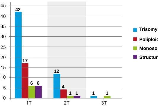
Figure 1 - Absolute number of chromosomal abnormalities identified in each pregnancy trimester 0
5 10 15 20 25 30 35 40 45
Trisomy Poliploidy Monosomy Structural
1T 2T 3T
42
17
6 6 12
4
ARTIGO ORIGINAL
44
Revista Científica da Ordem dos Médicos www.actamedicaportuguesa.com
regression model was also used for the multivariable analysis and risk modelling. We considered as significant p
values < 0.05.
RESULTS
According to the above criteria, a cytogenetic examination was carried out in 401 spontaneous miscarriage or foetal loss products, from which 251 occurred in the first, 118 in the second and only 32 occurred in the third trimester of pregnancy.
The chromosomal analysis was not conclusive, as cells failed to grow in culture in 20.0% (80/401) of the cases. This failure occurred in 21.5% (54/251) from the first trimester cases, 12.7% (15/118) from the second and 34.4% (11/32) from the third. In 12 of these 80 cases, the analysis using the MLPA technique allowed for identification of aneuploidies or male genetic material and these cases were included in our sample. Therefore, from the 401 products sent for genetic examination, we were able to obtain information on
the chromosomal complement in 333 (83.0%), from which 72.7% (242/333) presented a normal karyotype and 27.3% (91/333) an abnormal karyotype.
Considering the cases with a normal karyotype, numerical abnormalities were found in 92.3% (84/91). Trisomy was the most common chromosomal abnormality (55/84; 65.5%). Trisomy 16 (n = 17; 30.9%) was the most
frequent abnormality, followed by trisomy 18 (n = 12;
21.8%) and trisomy 21 (n = 8; 14.5%). Twenty-one cases
of polyploidy were also identified (25.0%) and seven cases with X-chromosome monosomy (8.3%) (Fig.1). In addition, seven structural abnormalities were found and are described in Table 1. The inherited abnormality was found to have a maternal origin in two of these cases and the remaining were de novo abnormalities.
We found that 78.0% (71/91) of chromosomal abnormalities occurred in the first trimester, 19.8% (18/91) in the second and 2.2% (2/91) in the third. A positive correlation was found between foetal maturation and the
Figure 2 - Distribution of the total number of spontaneous miscarriages or foetal losses per week of pregnancy
Table 1 - Description of the structural abnormalities found and their clinical information
Structural Abnormalities Maternal age Recurrence Trimester
45,X[26]/46,X,del(X)(q)[22]/46,XX[2] < 35 No 1st
46,XX,der(8)t(6;8)(q13;p23,1)mat < 35 Yes 1st
46,XX,dup(15)(q12q13)mat 35 – 39 Yes 1st
45,XX,dup(1q),der(13;14) 35 – 39 No 2nd
46,XX, der(22) 35 – 39 No 1st
46,XX,i(8)(q11,1) < 35 No 1st
47,XX,mar[3]/46,XX[27] < 35 No 1st
Number of miscarriages
0
2 4
6 8 10
12 14
16 18 20
Total Week of pregnancy 1
0
2
0
3
0
4
0
5
2
6
5
7
6
8
19
9
15
10
11
11
6
12
6
13
5
14
3
15
1
16
3
17
3
18
3
19
0
20
0
21
1
22
0
23
0
24
0
25
1
26
0
27
0
28
0
29
0
30
0
31
0
32
0
33
1
34
0
35
0
36
0
37
0
38
0
39
ARTIGO ORIGINAL
presence of chromosomal abnormalities (p < 0.001) and
half of these occurred (45/91) between the 8th and the 10th week of pregnancy (Fig. 2).
From the 333 cases in our study, 71 were recurrent and 262 were from sporadic miscarriage products. We did not find any significant difference in chromosomal 1st trimester abnormality prevalence between these two groups (36.2%
vs. 33.8%, respectively; p > 0.05). Nevertheless, maternal
age was 32.4 in the group of sporadic miscarriage and 34.8 in the group of recurrent miscarriage (p = 0.002).
Maternal age was on average 32.9 (standard deviation: 5.8; 99% Confidence Interval (CI) [32.1 – 33.8]);185 (55.6%) mothers were aged below 35, 106 (31.8%) between 35 and 39 and 42 (12.6%) above 40 years of age. The younger age group presented 22.2% (41/185) of chromosomal abnormalities, 31.1% (33/106) in the 35-39 age group and 40.5% (17/42) in the group above 40. A positive correlation was identified between maternal age increase and the presence of an abnormal karyotype (p = 0.02), with a mean increase of 7.4% (95% CI: [2.6
– 12.4]; p = 0.002) in the risk of occurrence of these
abnormalities for every additional year of age in the mother. . Nevertheless, when considering the diverse chromosomal abnormalities detected, this association was only found in the presence of trisomy (p < 0.01). There was
no association between maternal age increase and a higher risk of miscarriage in the presence of polyploidy (p = 0.939)
or X-monosomy (p = 0.568). Taking into account the most
common trisomies, we found a higher mean maternal age in the presence of trisomy 21 (38.3; p < 0.005) than in the
presence of trisomy 16 (33.1; p < 0.005) or 18 (34.3; p <
0.005).
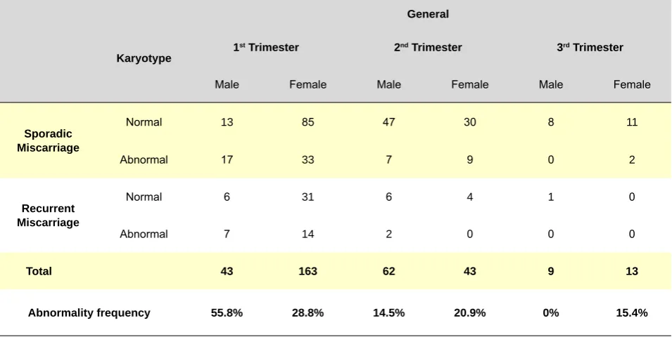
Considering the results, we found that 219/333 (65.8%) were female and 114/333 (34.2%) were male. Taking into account the gestational age, this female predominance was
only found in the first trimester: 163 (79.1%) were female and 43 (20.9%) were male. In the second trimester, female/ male ratio was 41.0% vs. 59.0% and in the third trimester it
was 59.1% vs. 40.9%.
We did not find any differences on chromosomal abnormality prevalence between male and female foetus (28.9% vs. 26.5%; p = 0.19) except when the results
were stratified per trimester: a higher number of abnormal karyotypes were found in male cases in the first trimester of pregnancy (55.8% vs. 28.8%; p = 0.01). The comparison
between the groups of sporadic and recurrent miscarriage regarding the relation between male and female foetus is presented in Table 2.
The histopathological examination of miscarriage products suggested the presence of a chromosomal abnormality in 89/333 (26.7%) cases. From these, karyotype chromosomal abnormalities were only found in 39 (43.8%) cases, with no agreement between the results of both examinations (p = 0.608). We also found 74 cases
with signs suggesting an abnormal foetal implantation, 42 cases of infection, 29 cases of disorganized growth and 15 of foetal growth restriction. The group that included a lower proportion of chromosomal abnormalities (4/42; 9.5%) was the one where miscarriage was possibly due to an infectious cause.
DISCUSSION
The identification of the cause in a pregnancy loss is challenging. Aneuploidies must be examined, as they are usually a cause of sporadic miscarriage, namely in the first trimester.21,23 Although most chromosomal abnormalities
are sporadic and therefore stand a small chance of re-emergence, their identification is reassuring for the couple and has a prognostic value regarding the success of the next pregnancy. Several studies suggested a better
Table 2 - Distribution by trimester of foetal gender and corresponding chromosomal abnormality frequency in pregnancy losses
Karyotype
General
1st Trimester 2nd Trimester 3rd Trimester
Male Female Male Female Male Female
Sporadic Miscarriage
Normal 13 85 47 30 8 11
Abnormal 17 33 7 9 0 2
Recurrent Miscarriage
Normal 6 31 6 4 1 0
Abnormal 7 14 2 0 0 0
Total 43 163 62 43 9 13
ARTIGO ORIGINAL
46
Revista Científica da Ordem dos Médicos www.actamedicaportuguesa.com
prognosis in a subsequent pregnancy when a miscarriage is due to aneuploidy than in the presence of a normal-karyotype.11,23,24 In addition, when there is a structural
abnormality as a cause of spontaneous miscarriage, the presence of possible chromosomal abnormalities in the parents must be ruled out, in order to provide the best possible genetic/reproductive counselling.25
In our study, karyotype was not obtained in a high percentage of cases (20.0%). This fact is described in literature as one constraint related to cell-culture technique and foetal karyotype determination, with reported culture cell failure rates up to 40%.22 This high cell-failure rate may
be due to factors like prolonged in utero retention time,
bacterial or fungal contamination during sample collection or even sample contamination with maternal cells.26 Therefore,
the higher failure percentage obtained in the third trimester in our group of patients may be due to cell failure related to a long time interval elapsed between foetal loss and the reception of material for cytogenetic analysis.
In this regard, currently there are new techniques allowing for alternative testing. In the absence of a karyotype determination due to culture failure or due to external contamination (conventional technique), other techniques may be requested from the Genetics Laboratory, such as array Comparative Genomic Hybridization (aCGH) and Quantitative Fluorescence PCR (QF-PCR) techniques. As long as a DNA sample is kept for subsequent studies, these may provide information regarding the chromosome complement of the foetal product,.
Recently, Morgen et al.27 suggested a diagnostic
protocol which has proved to be effective. These authors proposed that the most suitable study protocol of foetal products related with miscarriages should comprise an initial QF-PCR and, in the case of a normal result, an aCGH technique should follow, in order to overcome the two major constraints related with cell-culture i.e. culture failure and maternal contamination. This protocol worked efficiently, allowing for the detection of a higher number of cases with a chromosomal abnormality and the recognition of most maternal contamination situations.
According with expectations, numerical errors were 92.3% of the identified abnormalities, with trisomies being the most prevalent (65.5%). The higher frequency of trisomy 16, followed by 18 and 21 is in line with the evidence available from the literature.24,28 We found a
prevalence increase of these abnormalities with maternal age increase, with a mean 7.4% annual increment of the risk of occurrence. Taking into account its relevant contribution for spontaneous miscarriage, progressive maternity delay observed in Portugal as in the rest of the world has become an increasing concern.. Regarding polyploidies and monosomies, also frequent causes of miscarriage, we did not find any correlation with maternal age, as found by Rolnik et al.28
Regarding structural abnormalities, due to their high recurrence rate, is it extremely important to provide the couple with adequate information and genetic counselling
for future pregnancies. Therefore, karyotype of both parents must be obtained, in order to identify the hereditary or de novo nature of the abnormality found in the pregnancy loss.
The incidence of chromosomal abnormalities in the first trimester was lower than expected (50-60%)2,5,6 in sporadic
(33.8%) when compared to recurrent miscarriage (36.2%). The presence of maternal contamination may have contributed to these values, as demonstrated by a female foetal predominance with a normal karyotype in the whole sample (161/242; 66.5%). This result is in line with what has been described: approximately 30 to 40% of 46,XX karyotype obtained by a conventional technique may have a maternal origin.29,30
Therefore, the presence of a higher number of chromosomal abnormalities in male foetus may be related to excessive maternal contamination in our study, which may have masked the results. This result underlines the importance of carrying out a study to rule out maternal contamination every time early miscarriage products are sent for cytogenetic examination.
We did not identify any differences in the prevalence of chromosomal abnormalities from the first trimester, between sporadic and recurrent miscarriage groups. These results are in line with some previous studies9,10 and contradict
others showing a lower abnormality rate in the case of recurrent miscarriage.11,12
A reduction in chromosomal abnormality incidence was evident with pregnancy progression: 78.0% occurred in the first trimester, 19.8% in the second and only 2.2% in the third. Therefore, these abnormalities have shown to be inversely proportional to pregnancy age, with a lower odd´s
ratio as the end of pregnancy approaches.1,3,31,32 This could
be interpreted as a process of early natural selection and other possible causes must be considered for miscarriages occurring after 14 weeks of pregnancy.
Kano et al19 raise the possibility that, in recurrent
miscarriage without any chromosomal abnormality or other known aetiology, foetal loss is predominantly female. However, in this study, the higher proportion of female foetus with a normal karyotype found in the first trimester, in sporadic as well as in recurrent miscarriage, may once more be explained by the presence of maternal contamination in the studied samples. The typical immaturity of miscarriage products in the first months of pregnancy may make it difficult to ensure the origin of the studied material.
ARTIGO ORIGINAL
of inflammatory cells are easily identifiable, this probably explains a lower diagnostic error found in this group. On the contrary, the diagnostic precision in the remaining groups may be affected by the subjectivity of the criteria that we used. To the best of our knowledge, this is actually the first study comparing the results obtained with cytogenetic vs.
histopathological examination.
We consider that the size of our sample (333 cases of spontaneous miscarriage or foetal loss) has shown to be a strong point, ensuring the reliability of the results obtained. We should also emphasize that, in the 12 cases in which we only obtained information regarding chromosome complement by MLPA, it is not possible to exclude other chromosomal abnormalities. Although this examination has the advantage of requiring only a small DNA sample and constitutes a rapid and inexpensive test for 13, 18, 21, X and Y chromosomal aneuploidy detection,22 it does not
allow for analysis of other karyotype abnormalities, namely chromosome 16 trisomy, commonly involved as cause of spontaneous miscarriage. Even so, the use of the MLPA technique demonstrated its utility by confirming most of the results obtained by karyotype analysis, as well as allowing for the exclusion of some of the most common chromosomal abnormalities in the cases of culture failure.
The inclusion of sporadic and recurrent miscarriage products in the whole sample was very useful as it allowed for the simultaneous study of chromosomal abnormality prevalence in both situations.
CONCLUSIONS
Pregnancy loss, regardless of the time when it occurs, is an event with a very important impact on the couple attempting pregnancy.. Apart from a possible prognostic value for a subsequent pregnancy, karyotype analysis may bring important psychological relief to the couple and avoid other investigations.28 Even in the case of sporadic
miscarriage, chromosomal abnormalities are commonly implicated most often until the 14th week of pregnancy, with a similar prevalence to the one observed in the case of recurrence. In the specific case of identification of a structural abnormality, it may have its origin in one of the parents and therefore it is crucial to provide all the information and genetic counselling to the couple. Karyotype analysis of every miscarriage product, recurrent or not, will give comfort to the couple and will help them to decide as regards a new pregnancy attempt.
We are aware that an advanced maternal age is also one of the major factors in pregnancy failure. Therefore,
with the current maternity delay and aneuploidy-related miscarriage probability increase, it is necessary for the doctor to previously inform patients of this progressive risk with advancing age.
We suggest a combination of cytogenetic analysis techniques allowing for the study of the whole karyotype or, at least, of the most frequently involved aneuploidies as a cause of spontaneous miscarriage, in order to obtain an efficient study of the chromosomal abnormalities, and in this way avoiding cell culture high failure rate.
Taking into account the high maternal contamination rate involved in this kind of study, maternal DNA additional analysis becomes crucial. In this way it will be possible to ensure the total exclusion of maternal cells from the sample, allowing for the precise prevalence of chromosomal abnormalities to be known and for the study of a possible difference between female and male rate of foetal loss. We wish to emphasize that this study was based on retrospective data analysis and that, in order to solve the constraints of MLPA technique, the Genetics Department included QF-PCR technique in the study of first trimester miscarriages. Therefore, it was possible not only to exclude maternal contamination, but also to additionally study the number of copies for 15, 16 and 22 chromosomes. As in the latter method, the aCGH technique does not require a cell culture, and has the additional advantage of allowing for the total chromosomal complement analysis, with a much higher resolution than when conventional karyotype analysis is performed.
Therefore, the adoption of a new diagnostic protocol for the genetic study of pregnancy losses including QF-PCR and aCGH techniques to replace the conventional technique (karyotype) seems to be the most adequate methodology and in addition to perform with a more efficient cost-benefit ratio.
CONFLICTS OF INTEREST
The authors declare that there were no conflicts of interest in writing this manuscript.
FINANCIAL SOURCES
There were no external financial sources for the writing of this manuscript.
ACKNOWLEDGEMENTS
The authors wish to thank the essential contribution of João Barbosa in data statistical analysis, without which this study would not be possible.
REFERENCES
1. Warren JE, Silver RM. Genetics of pregnancy loss. Clin Obstet Gynecol. 2008;51:84-95.
2. Rai R, Regan L. Recurrent miscarriage. Lancet. 2006;368:601-11. 3. Stephenson M, Kutteh W. Evaluation and management of recurrent
early pregnancy loss. Clin Obstet Gynecol. 2007;50:132-145. 4. Toth B, Jeschke U, Rogenhofer N, Scholz C, Wurfel W, Thaler CJ, et al.
Recurrent miscarriage: Current concepts in diagnosis and treatment. J
Reprod Immunol. 2010;85:25-32.
5. Robberecht C, Pexsters A, Deprest J, Fryns JP, D’Hooghe T, Vermeesch JR. Cytogenetic and morphological analysis of early products of concep-tion following hystero-embryoscopy from couples with recurrent preg-nancy loss. Prenat Diagn. 2012;32:933-42.
ARTIGO ORIGINAL
48
Revista Científica da Ordem dos Médicos www.actamedicaportuguesa.com
in young couples. Eur J Obstet Gynecol Reprod Biol. 2012;161:182-6. 7. Stephenson MD, Awartani KA, Robinson WP. Cytogenetic analysis of
miscarriages from couples with recurrent miscarriage: A case-control study. Hum Reprod. 2002;17:446-51.
8. Morikawa M, Yamada H, Kato EH, Shimada S, Yamada T, Minakami H. Embryo loss pattern is predominant in miscarriages with normal chro-mosome karyotype among women with repeated miscarriage. Hum Re-prod. 2004;19:2644-7.
9. Marquard K, Westphal LM, Milki AA, Lathi RB. Etiology of recur-rent pregnancy loss in women over the age of 35 years. Fertil Steril. 2010;94:1473-7.
10. Grande M, Borrell A, Garcia-Posada R, Borobio V, Munoz M, Creus M, et al. The effect of maternal age on chromosomal anomaly rate and spectrum in recurrent miscarriage. Hum Reprod. 2012;27:3109-17. 11. Ogasawara M, Aoki K, Okada S, Suzumori K. Embryonic karyotype
of abortuses in relation to the number of previous miscarriages. Fertil Steril. 2000;73:300-4.
12. Sullivan AE, Silver RM, LaCoursiere DY, Porter TF, Branch DW. Re-current fetal aneuploidy and reRe-current miscarriage. Obstet Gynecol. 2004;104:784-8.
13. Morikawa M, Yamada H, Kato EH, Shimada S, Sakuragi N, Fujimoto S, et al. Live birth rate varies with gestational history and etiology in women experiencing recurrent spontaneous abortion. Eur J Obstet Gynecol Re-prod Biol. 2003;109:21-6.
14. Cleary-Goldman J, Malone FD, Vidaver J, Ball RH, Nyberg DA, Com-stock CH, et al. Impact of maternal age on obstetric outcome. Obstet Gynecol. 2005;105:983-90.
15. Santos V, Moura M, Pinto JP, Almeida V, Maio J. Características sócio--demográficas das puérperas e seguimento da gravidez: o que mudou em 17 anos? Acta Acta Med Port. 2011;24:877-84.
16. Nybo Andersen AM, Wohlfahrt J, Christens P, Olsen J, Melbye M. Mater-nal age and fetal loss: Population Based Register Linkage Study. BMJ. 2000;320:1708-12.
17. Sugiura-Ogasawara M, Ozaki Y, Kitaori T, Suzumori N, Obayashi S, Su-zuki S. Live birth rate according to maternal age and previous number of recurrent miscarriages. Am J Reprod Immunol. 2009;62:314-9. 18. Del Fabro A, Driul L, Anis O, Londero AP, Bertozzi S, Bortotto L, et
al. Fetal gender ratio in recurrent miscarriages. Int J Womens Health. 2011;3:213-7.
19. Kano T, Mori T, Kimura A. Gender ratio distortion in abortuses and live births from patients with recurrent spontaneous abortion. Am J Reprod Immunol. 2009;62:125-7.
20. Instituto Nacional de Estatística. Lisboa: INE; 2012. [consultado 2012 Dez 13]. Disponível em: http://www.ine.pt.
21. Doria S, Carvalho F, Ramalho C, Lima V, Francisco T, Machado AP, et al. An efficient protocol for the detection of chromosomal abnormalities in spontaneous miscarriages or foetal deaths. Eur J Obstet Gynecol Re-prod Biol. 2009;147:144-50.
22. Carvalho B, Doria S, Ramalho C, Brandao O, Sousa M, Matias A, et al. Aneuploidies detection in miscarriages and fetal deaths using multiplex ligation-dependent probe amplification: An alternative for speeding up results? Eur J Obstet Gynecol Reprod Biol. 2010;153:151-5.
23. Sugiura-Ogasawara M, Ozaki Y, Katano K, Suzumori N, Kitaori T, Mizu-tani E. Abnormal embryonic karyotype is the most frequent cause of recurrent miscarriage. Hum Reprod. 2012;27:2297-303.
24. Carp H, Toder V, Aviram A, Daniely M, Mashiach S, Barkai G. Karyotype of the abortus in recurrent miscarriage. Fertil Steril. 2001;75:678-82. 25. Nagaishi M, Yamamoto T, Iinuma K, Shimomura K, Berend SA, Knops
J. Chromosome abnormalities identified in 347 spontaneous abortions collected in Japan. J Obstet Gynaecol Res. 2004;30:237-41.
26. Dória S. Mecanismos genéticos subjacentes ao abortamento espon-tâneo. Porto: Departamento de Genética, Faculdade de Medicina da Universidade do Porto; 2010.
27. Morgen EK, Maire G, Kolomietz E. A clinical algorithm for efficient, high-resolution cytogenomic analysis of uncultured perinatal tissue samples. Eur J Med Genet. 2012;55:446-54.
28. Rolnik DL, Carvalho MH, Catelani AL, Pinto AP, Lira JB, Kusagari NK, et al. Cytogenetic analysis of material from spontaneous abortion. Rev Assoc Med Bras. 2010;56:681-3.
29. Karaoguz MY, Nas T, Konac E, Ince D, Pala E, Menevse S. Is cyto-genetic diagnosis of 46,Xx karyotype spontaneous abortion specimens erroneous? Fluorescence in situ hybridization as a confirmatory tech-nique. J Obstet Gynaecol Res. 2005;31:508-13.
30. Jarrett KL, Michaelis RC, Phelan MC, Vincent VA, Best RG. Microsatel-lite analysis reveals a high incidence of maternal cell contamination in 46,Xx products of conception consisting of Villi or a combination of Villi and membranous material. Am J Obstet Gynecol. 2001;185:198-203. 31. Wapner RJ, Lewis D. Genetics and metabolic causes of stillbirth. Semin
Perinatol. 2002;26:70-4.