© 2020 by the Serbian Biological Society How to cite this article: Zhang X, Sun Z, Zou Y.LncRNA NEAT1 exacerbates non- 243 small cell lung cancer by upregulating EIF4G2 via miR-582-5p sponging. Arch Biol Sci. 2020;72(2):243-52.
LncRNA NEAT1 exacerbates non-small cell lung cancer by upregulating EIF4G2 via
miR-582-5p sponging
Xin Zhang1, Zhonghua Sun2 and Yuepei Zou3,*
1Beijing Sport University, Xinxi Ave 48, Haidian District, Beijing 100084, China
2Medical Department, Affiliated Hospital of Shandong University of Traditional Chinese Medicine, Jingshi Road 16369, Jinan
250011, China
3Zhengzhou University, Science Ave. 100, Gaoxin District, Zhengzhou 450001, China
*Corresponding author: [email protected]
Received: February 18, 2020; Revised: April 16, 2020; Accepted: April 17, 2020; Published online: April 23, 2020
Abstract: In this study, we aimed to elucidate the role of long non-coding RNA nuclear enriched abundant transcript 1 (lncRNA NEAT1) in non-small cell lung cancer (NSCLC). Quantitative real-time polymerase chain reaction (qRT-PCR) was employed to detect the abundance of NEAT1, microRNA-582-5p (miR-582-5p) and eukaryotic translation initiation factor 4 gamma 2 (EIF4G2). Proliferation, apoptosis, metastasis and glycolytic metabolism were determined by 3-(4,5-dimeth-ylthiazol-2-yl)-2,5-diphenyltetrazolium bromide (MTT) flow cytometry, transwell assays and fluorescence-based glucose and lactate assay kits. The targets of NEAT1 and miR-582-5p were predicted by the starBase website, and dual-luciferase reporter and RNA immunoprecipitation (RIP) assays were performed to verify these predictions. Western blot analysis was conducted to detect the protein expression of EIF4G2. A xenograft tumor model was built to clarify the role of NEAT1 in vivo. Results showed that NEAT1 interference inhibited proliferation, metastasis and glycolysis, and facilitated the apop-tosis of NSCLC cells. MiR-582-5p was a functional target of NEAT1, and the biological influence of NEAT1 intervention on NSCLC cells was alleviated by transfection with anti-miR-582-5p. MiR-582-5p could bind to EIF4G2 messenger RNA (mRNA); it exerted its antitumor role in NSCLC cells by inhibiting EIF4G2. EIF4G2 was regulated by NEAT1/miR-582-5p signaling. NEAT1 accelerated NSCLC tumor growth via the miR-582-NEAT1/miR-582-5p/EIF4G2 axis in vivo. In conclusion, NEAT1 affected NSCLC by elevating their malignant potential via the miR-582-5p/EIF4G2 axis.
Keywords: NEAT1; miR-582-5p; EIF4G2; metastasis; glycolysis
Abbreviations: long non-coding RNA (lncRNA); nuclear enriched abundant transcript 1 (NEAT1); non-small cell lung cancer (NSCLC); eukaryotic translation initiation factor 4 gamma (EIF4G); eukaryotic initiation factor 4F (EIF4F)
INTRODUCTION
As a major histopathological type of lung cancer, non-small cell lung cancer (NSCLC) accounts for 70%-80% of lung cancer cases [1,2]. Tumor cells consume glucose and metabolize it into lactate even in the presence of oxygen, and this phenomenon is termed the Warburg effect [3]. The suppression of glycolysis could impede the development of cancers [4]. Although the treat-ment strategy has been improved, the prognosis of NSCLC patients remains dismal owing to metastasis and relapse [5]. Therefore, it is urgent to uncover the potential mechanism behind the proliferation, motility and glycolysis of NSCLC cells.
MicroRNAs (miRNAs) are another class of ncRNAs that contain 19-24 nucleotides. MiRNAs could regulate cellular biological functions by inducing degradation or by repressing the translation of messenger RNAs (mRNAs) by direct combination [15,16]. The crucial role of miRNAs in diverse cancers has been reported [17-19]. Herein, we focused on the role of miR-582-5p in NSCLC. It was shown that miR-582-5p suppressed the progression of NSCLC via mitogen-activated protein kinase kinase kinase 2 (MAP3K2) [20], and that miR-582-5p restrains the proliferation and invasion of NSCLC cells via Notch homolog 1, translocation-associated (NOTCH1) [21]. In the present study, we found a new regulatory mechanism involving miR-582-5p in NSCLC.
Eukaryotic translation initiation factor 4 gamma 2 (EIF4G2) is a component of eukaryotic initiation factor 4F (EIF4F). The protumor role of EIF4G2 in multiple cancers has been described in previous stud-ies [22,23]. It was demonstrated that miR-379 could inhibit cisplatin resistance of NSCLC cells through downregulation of EIF4G2, suggesting a protumor role of EIF4G2 in NSCLC [24]. However, the molecular mechanism involving EIF4G2 in NSCLC remains largely unexplored.
In the current study, we examined the expression profile of NEAT1 in NSCLC, and loss-of-function experiments were employed to investigate the role of NEAT1 in NSCLC. The lncRNA-miRNA-mRNA signal network was then examined in order to study the working mechanism of NEAT1 in NSCLC.
MATERIALS AND METHODS Tissue samples
Paired NSCLC tissues (n=40) and adjacent non-tumor tissues (n=40) were obtained from NSCLC patients in the Affiliated Hospital of Shandong University of Traditional Chinese Medicine who underwent surgi-cal resection. The tissues were snap-frozen in liquid nitrogen to detect the expression of NEAT1, miR-582-5p and EIF4G2. The procedures in this study were approved by the Ethics Committees of the Affiliated Hospital of Shandong University of Traditional Chinese Medicine. Written informed consent was provided from all subjects before radical resection.
Cell culture
NSCLC cell lines (A549 and H1299) and the normal human lung epithelial cell line BEAS-2B were purchased from BeNa Culture Collection (Beijing, China). All cell lines were grown in Dulbecco’s Modified Eagle Medium (DMEM; Gibco, Carlsbad, CA, USA) sup-plemented with 10% fetal bovine serum (FBS; Gibco), 10% penicillin (100 U/mL) and 10% streptomycin (100 μg/mL) at 37°C in a humidified incubator with 5% CO2.
Quantitative real-time polymerase chain reaction (qRT-PCR)
TRIzol reagent (Invitrogen, Carlsbad, CA, USA) was used for RNA extraction from tissues and cells. RNA concentration was measured using NanoDrop 2000 (NanoDrop Technologies, Wilmington, DE, USA). Complementary DNA (cDNA) was synthesized us-ing a High-Capacity cDNA Reverse Transcription Kit (Applied Biosystems, Foster City, CA, USA) and an All-in-One miRNA First stand cDNA Synthesis Kit (GeneCopoeia, Rockville, MD, USA). The amplifica-tion reacamplifica-tion was conducted using SYBR® Premix Ex
TaqTM II (Takara, Osaka, Japan). The PCR reaction
system and conditions were as follows: PCR reaction system (25 μL): SYBR® Premix Ex TaqTM (2×; 12.5 μL),
forward primer (10 μM; 0.5 μL), reverse primer (10 μM; 0.5 μL), DNA template (2 μL), dH2O (9.5 μL). PCR condition: 95°C for 30 s followed by 35 cycles of 95°C for 5 s and 60°C for 30 s. The abundance of NEAT1, miR-582-5p and EIF4G2 was calculated using the 2−ΔΔCt
Cell transfection
NEAT1 specific small interfering RNA (si-NEAT1 #1 and si-NEAT1 #2) and siRNA negative control (si-NC), NEAT1 specific short hairpin RNA (sh-NEAT1) and control (sh-NC), NEAT1 ectopic expression plasmid (NEAT1), EIF4G2 ectopic expression plasmid (EIF4G2) and control (vector), miR-582-5p mimic (miR-582-5p) and miR-NC, miR-582-5p inhibitor (anti-miR-582-5p) and control (anti-miR-NC) were obtained from Ge-nepharma (Shanghai, China). Transfection was carried out using Lipofectamine 3000 (Invitrogen, USA).
3-(4,5-Dimethylthiazol-2-yl)-2,5-diphenyltetrazolium bromide (MTT) assay
After transfection for 0 h, 24 h, 48 h or 72 h, NSCLC cells were incubated with 10 µL MTT (Invitrogen, USA) for 4 h. The absorbance at 490 nm was detected by a microplate reader.
Flow cytometry for apoptosis analysis
The apoptotic rate of NSCLC cells with specific treat-ment was analyzed by flow cytometry using an An-nexin V apoptosis detection kit (BD PharmingenTM,
San Diego, CA, USA). The apoptotic NSCLC cells were identified by a FC-500 flow cytometer (Beckman Coulter, Pasadena, CA, USA).
Transwell assays
To measure the invasive ability of NSCLC cells, tran-swell upper chambers (Costar, Corning, NY, USA) were pre-coated with Matrigel (BD PharmingenTM).
NSCLC cells (2×10⁴ cells per well) suspended in 100
μL serum-free medium were added into the pre-coated upper chambers; 500 μL medium supplemented with 10% fetal bovine serum (FBS) was added to the lower chambers. After incubation for 24 h, invaded NSCLC cells were stained with 0.5% crystal violet (0.5%; Sigma, USA). The number of invaded NSCLC cells in five random fields was counted using an optical microscope. To detect the migration capacity of NSCLC cells, a cell suspension was added to un-coated upper chambers following the same procedures.
Glucose uptake and lactate production analysis
Glycolysis of NSCLC cells was analyzed by measuring glucose uptake and lactate production using fluores-cence-based glucose and lactate assay kits (BioVision, Milpitas, California, USA).
Dual-luciferase reporter assay
The candidate targets of NEAT1 and miR-582-5p were predicted by the starBase database. The sequence of NEAT1 and the 3’ untranslated region (3’UTR) of EIF4G2 mRNA, including the binding sites with miR-582-5p, was cloned into pGL3 vector (Promega, Madison, WI, USA), generating NEAT1-WT and EIF4G2 3’UTR-WT. The corresponding mutant-type reporter vectors were termed NEAT1-MUT and EIF4G2 3’UTR-MUT. Luciferase activity was measured in A549 and H1299 cells cotransfected with miR-NC or miR-582-5p and the above reporter plasmid through the Dual-Luciferase Reporter Assay System (Promega, Madison, WI, USA).
RNA immunoprecipitation (RIP) assay
The RIP assay was performed in order to extract RNA-RNA/protein complexes with the Magna RIP RNA-binding protein immunoprecipitation kit (Mil-lipore, Billerica, MA, USA).
Western blot assay
Xenograft tumor model
This study was approved by the Animal Research Committee of the Affiliated Hospital of Shandong University of Tra-ditional Chinese Medicine. BALB/c-nu mice (5 weeks old) were purchased from the Chinese Academy of Medical Sciences (Beijing, China). A549 cells (2×10⁶) stably
transfected with sh-NC or sh-NEAT1 were suspended in 100 μL phosphate buffered saline (PBS). The back region of nude mice (n=5) was subcutaneously inoculated with A549 cells. The volume of NSCLC tumors was calculated using the following formula of volume=π×(length×width2)/6. Mice
were killed after 35 days inoculation and the tumors were resected and weighed.
Statistical analysis
The data are presented as the mean±standard deviation (SD). Student’s t-test was utilized to compare the differences between two groups, while the differences among mul-tiple groups were assessed using one-way or two-way analysis of variance (ANOVA) followed by Tukey’s test. The survival curve was generated using the Kaplan-Meier plot and log-rank test. The linear relationship was evaluated by Spearman’s correlation coefficient. A P value less than 0.05 was considered statistically significant.
RESULTS
NEAT1 is aberrantly upregulated in NSCLC tissues and cells
Abnormal upregulation in the expression of NEAT1 was observed in NSCLC tumor samples when compared with that in ad-jacent normal tissues (Fig. 1A). As shown in Fig. 1B, high expression of NEAT1 was associated with the poor survival rate of NSCLC patients. To uncover the molecular mechanism of NEAT1 in NSCLC, two
Fig. 1. NEAT1 is aberrantly upregulated in NSCLC tissues and cells. A – The expression of NEAT1 measured in NSCLC tissue specimens (n=40) and adjacent normal tissue specimens (n=40) by qRT-PCR. B – The survival curve of NSCLC patients generated using the Kaplan-Meier plot and log-rank test. C – qRT-PCR employed to determine the abundance of NEAT1 in NSCLC cell lines, including A549 and H1299 and the normal human lung epithelial cell line BEAS-2B. *P<0.05.
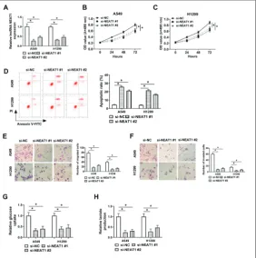
Fig. 2. NEAT1 potentiates the malignancy of NSCLC cells. A549 and H1299 cells were transfected with si-NC, si-NEAT1 #1 or si-NEAT1 #2. A – The enrichment of NEAT1 examined in A549 and H1299 by qRT-PCR. B, C – MTT assay employed to assess the proliferation of NSCLC cells. D – The apoptotic rate of NSCLC cells evaluated by flow cytometry. E, F – Transwell assays conducted to detect the capaci-ties of migration and invasion of NSCLC cells. G, H – Aerobic glycolysis of NSCLC cells analyzed using fluorescence-based glucose and lactate assay kits. *P<0.05.
NEAT1 potentiates the malignancy of NSCLC cells
qRT-PCR revealed that the knockdown efficiencies of two NEAT1 small interfering RNAs (si-NEAT1 #1 and si-NEAT1 #2) in NSCLC cells were high (Fig. 2A). Loss-of-function experiments were conducted to explore the biological functions of NEAT1 in NSCLC. NEAT1 intervention significantly restrained the proliferation of NSCLC cells (Fig. 2B and C). The apoptotic rate of NSCLC cells was increased with the transfection of si-NEAT1 #1 or si-NEAT1 #2 (Fig. 2D), suggested that NEAT1 suppressed the apoptosis of NSCLC cells. The motility of NSCLC cells with NEAT1 interference was also assessed by performing a transwell migration assay and a transwell invasion assay. As mentioned in Fig. 2E and F, NEAT1 interference notably inhibited the migration and invasion of NSCLC cells. Cancer cells
obtained energy mainly through glycolysis instead of oxidative phosphorylation [3]. The influence of NEAT1 silencing on the glycolysis of NSCLC cells was also evaluated by measuring glucose uptake and lactate production. NEAT1 interven-tion caused decreased consumpinterven-tion of glucose and production of lactate (Fig. 2G and H), demonstrated that NEAT1 could enhance the glycolysis of NSCLC cells. Taken together, we can conclude that NEAT1 promoted the proliferation, metastasis and glycolysis and suppressed the apoptosis of NSCLC cells. Si-NEAT1 #1 was chosen for the subsequent experiments owing to its higher interference efficiency in NSCLC cells.
NEAT1 can sponge miR-582-5p
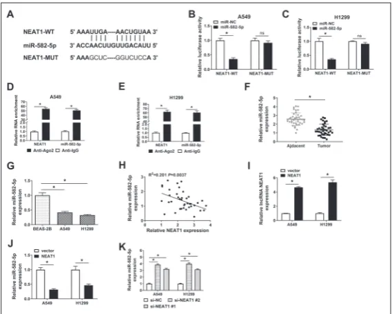
Studies have reported that lncRNAs could serve as miRNAs sponges to modulate the levels of target miRNAs and match-ing downstream genes [25]. LncRNA NEAT1-miRNAs interactions were inves-tigated using starBase online software, and miR-582-5p was chosen for the following experiments due to its tumor suppressor role in NSCLC [20, 21]. The binding se-quence between NEAT1 and miR-582-5p predicted by starBase database is shown in Fig. 3A. A dual-luciferase reporter assay revealed that luciferase activity was prominently decreased in miR-582-5p and the NEAT1-WT cotransfected group as compared with that in the miR-NC and NEAT1-WT cotransfected group; cotransfection of NC or miR-582-5p made no difference in the luciferase activity of NEAT1-MUT group (Fig. 3B and C), suggesting that miR-582-5p could directly interact with NEAT1 through putative binding sites. Besides, NEAT1 and miR-582-5p were both enriched in the Anti-Ago2 group (Fig. 3D and E), which indicated that NEAT1 could bind to the RNA-induced silencing complex (RISC), likely through a target relationship with miR-582-5p. Subsequently, we detected the expression of miR-582-5p in NSCLC tissues and cells. As indicated in Fig. 3F and G, the level of miR-582-5p was conspicuously decreased in NSCLC tissues and cells. A prominent
Fig. 3. NEAT1 can sponge miR-582-5p. A – The binding sites between NEAT1
and miR-582-5p predicted by starBase database; mutant binding sites in NEAT1 are shown in red. B, C – The target relationship between NEAT1 and miR-582-5p confirmed by the dual-luciferase reporter assay. D, E – RNA immunoprecipitation (RIP) assay applied to validate the combination between NEAT1 and miR-582-5p.
F, G – The expression of miR-582-5p detected in NSCLC tissues (n=40) and cells and corresponding normal tissues (n=40) and BEAS-2B cells by qRT-PCR. H – The linear relationship between miR-582-5p and NEAT1 analyzed by Spearman’s correlation coefficient. I – The overexpression efficiency of NEAT1 overexpression plasmid was evaluated by qRT-PCR. J – The abundance of miR-582-5p meas-ured in A549 and H1299 cells transfected with vector or NEAT1 by qRT-PCR.
negative correlation between the ex-pression of miR-582-5p and NEAT1 in NSCLC tissues was observed (Fig. 3H). The expression of NEAT1 was elevated with the transfection of NEAT1 ectopic expression plasmid, as compared with that in the vector group (Fig. 3I). The accumulation of NEAT1 caused a significant re-duction in the expression of miR-582-5p in NSCLC cells (Fig. 3J), and the intervention of NEAT1 elevated the enrichment of miR-582-5p in NSCLC cells (Fig. 3K). In summary, lncRNA NEAT1 suppressed the expression of miR-582-5p through direct interaction.
NEAT1 promotes the
progression of NSCLC through sponging miR-582-5p
Rescue experiments were conducted to explore whether NEAT1 exerted its protumor role through sponging miR-582-5p in NSCLC cells. The upregulation of miR-582-5p expres-sion caused by NEAT1 interference was counteracted by the introduc-tion of anti-miR-582-5p (Fig. 4A). The proliferation of NSCLC cells was impeded by the silencing of NEAT1, and the cotransfection of anti-miR-582-5p partly recovered the proliferation of NSCLC cells (Fig. 4B and 4C). The promoting effect of si-NEAT1 #1 transfection on the apoptosis of NSCLC cells was attenuated by the addition of anti-miR-582-5p (Fig. 4D). The number of migrated or invaded NSCLC cells was greatly decreased in the si-NEAT1 #1 group, while the metastasis ability was regained in the si-NEAT1 #1 and the anti-miR-582-5p cotransfected groups (Fig. 4E and F). The addition of anti-miR-582-5p also recovered
Fig. 5. EIF4G2 is a direct target of miR-582-5p in NSCLC cells. A – Complementary sites between miR-582-5p and EIF4G2 predicted by starBase bioinformatic software. B, C – Luciferase activity measured in NSCLC cells transfected with miR-NC or miR-582-5p and EIF4G2 3’UTR-WT or EIF4G2 3’UTR-MUT. D, E – The target relationship between miR-582-5p and EIF4G2 verified by the RIP assay. F – mRNA expression of EIF4G2 in NSCLC tissues (n=40) and adjacent normal tissues (n=40) determined by qRT-PCR.
G – Western blotting carried out to detect the protein level of EIF4G2 in NSCLC tissues (n=3) and matching non-tumor tissues (n=3). H – Protein expression of EIF4G2 was examined in NSCLC and BEAS-2B cell lines by Western blotting. I – The correlation between miR-582-5p and EIF4G2 mRNA analyzed using Spearman’s correlation coef-ficient. J, K – The protein level of EIF4G2 in NSCLC cells transfected with NC, miR-582-5p, anti-miR-NC or anti-miR-582-5p determined by Western blot analysis. *P<0.05.
Fig. 4. NEAT1 promotes the progression of NSCLC through sponging of miR-582-5p.
glucose uptake and lactate produc-tion in NSCLC cells, which were held back by the intervention of NEAT1 (Fig. 4G and H). These results indicated that NEAT1 fa-cilitated the malignant potential of NSCLC cells through suppression of miR-582-5p.
EIF4G2 is a direct target of miR-582-5p in NSCLC cells
The targets of miR-582-5p were also predicted by the starBase on-line website. Among the candidate targets of miR-582-5p, EIF4G2 was selected due to its promoting role in proliferation and metastasis of cancer cells [22,23]. The comple-mentary sequence in miR-582-5p and the 3’UTR of EIF4G2 mRNA is shown in Fig. 5A. As can be seen in Fig. 5B and C, the accumulation of miR-582-5p markedly reduced luciferase activity in the EIF4G2
3’UTR-WT group when compared to the EIF4G2 3’UTR-MUT group, suggesting direct interaction between miR-582-5p and EIF4G2. To further confirm this combination, the RIP assay was employed. MiR-582-5p and EIF4G2 were substantially enriched when using Ago2 antibody (Fig. 5D and E), demonstrating that miR-582-5p could bind to EIF4G2. The mRNA and protein expression of EIF4G2 was aberrantly up-regulated in NSCLC tissues relative to adjacent normal tissues (Fig. 5F and G). Also, striking upregulation in the abundance of EIF4G2 protein was observed in NSCLC cells in comparison to BEAS-2B cells (Fig. 5H). There was a significant inverse correlation between the levels of miR-582-5p and mRNA expression of EIF4G2 in NSCLC tissues (Fig. 5I). To further test the regulatory relationship between EIF4G2 and 582-5p, miR-582-5p mimic (miR-miR-582-5p) and miR-miR-582-5p inhibitor (anti-miR-582-5p) were transfected into NSCLC cells. As mentioned above (for Fig. 5J and K), miR-582-5p transfection significantly decreased the protein level of EIF4G2, with the protein level of EIF4G2 being markedly elevated after transfection with anti-miR-582-5p in the two NSCLC cell lines. These findings
revealed that miR-582-5p could negatively regulate the expression of EIF4G2 through direct interaction.
EIF4G2 accumulation reverses the biological functions of miR-582-5p on NSCLC cells
MiR-582-5p and EIF4G2 were cotransfected into NSCLC cells to address the question of the biological role of EIF4G2 in the miR-582-5p-mediated influ-ence of NSCLC cells. As shown in Fig. 6A and B, the downregulation of EIF4G2 protein expression caused by transfection with miR-582-5p was alleviated by the addition of EIF4G2. MiR-582-5p overexpression sup-pressed the proliferation, while accelerating apoptosis of NSCLC cells, and these effects were partly coun-teracted by the introduction of EIF4G2 (Fig. 6C-E). The overexpression of EIF4G2 also ameliorated the inhibitory effects of miR-582-5p accumulation on migration, invasion and glycolysis of NSCLC cells (Fig. 6F-K). Taken together, miR-582-5p inhibited the proliferation, metastasis and glycolysis while promot-ing apoptosis of NSCLC cells through a decrease in the level of EIF4G2.
NEAT1 elevates the expression of EIF4G2 by the sponging miR-582-5p in NSCLC cells
The interference with NEAT1 downregulated the ex-pression of EIF4G2, and the level of EIF4G2 was partly recovered in the si-NEAT1 #1 and the anti-miR-582-5p cotransfected group (Fig. 7A and B). The expression of EIF4G2 mRNA was positively correlated with the expression of NEAT1 (Fig. 7C). Collectively, lncRNA NEAT1 could sequester miR-582-5p from EIF4G2 mRNA, thus upregulating the expression of EIF4G2 at the post-transcriptional level in NSCLC cells.
NEAT1 accelerates tumor growth of NSCLC via the miR-582-5p/EIF4G2 axis in vivo
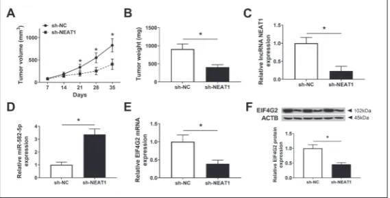
We validated the role of NEAT1 using a murine xenograft model. A549 cells stably expressing sh-NC or sh-NEAT1 were sub-cutaneously injected into the back region of nude mice. As can be seen in Fig. 8A and B, NSCLC tumors were smaller in the sh-NEAT1 group relative to the sh-NC group. The resected tumor tissues were used to measure the expression of NEAT1, miR-582-5p and EIF4G2. The abundance of NEAT1 was declined significantly in the sh-NEAT1 group in comparison to the sh-NC group (Fig. 8C). The level of miR-582-5p exhibited a reverse trend with respect to NEAT1 (Fig. 8D). Moreover, the mRNA and protein expression of EIF4G2 were decreased in the sh-NEAT1 group relative to the sh-NC group (Fig. 8E and F). In summary, NEAT1 contributed to the malignancy of NSCLC through the miR-582-5p/EIF4G2 axis in vivo.
DISCUSSION
The identification of crucial molecules that are involved in the occurrence and development of NSCLC is urgently need to improve the prognosis of NSCLC patients. We found that NEAT1 was abnormally upregulated in NSCLC tissues and cells, which is consistent with previous findings [12,26-29]. High expression of NEAT1 is related to the poor survival rate of NSCLC patients. Knockdown experiments revealed that NEAT1 acceler-ated the proliferation, metastasis and glycolysis, while restricting apoptosis of NSCLC cells. The protumor role of NEAT1 is in agreement with published articles, thus, NEAT1 facilitates the development of breast cancer via miR-448 and zinc finger E-box-binding homeobox 1 protein (ZEB1) [30], and NEAT1 con-tributes to the progression of colorectal cancer via the enzyme probable ATP-dependent RNA helicase DDX5 also known as DEAD box protein 5 or RNA helicase p68 (DDX5) [31].
Fig. 8. NEAT1 accelerates tumor growth of NSCLC via miR-582-5p/EIF4G2 axis
in vivo. A – The size of the tumor was recorded every 7 days using the following method: volume =π×(length×width2)/6. B – Tumors were resected from nude
mice after injection for 35 days, and the weights of the tumors in the sh-NC (n=5) and the sh-NEAT1 groups (n=5) were measured using an analytical balance.
C-F – The expression of NEAT1, miR-582-5p and EIF4G2 mRNA and protein in tumor tissues determined by qRT-PCR and Western blot analysis. *P<0.05.
LncRNAs regulate cellular physiological and patho-logical processes mainly by functioning as miRNAs sponges [32]. It was shown that LncRNA-00319 ag-gravated ovarian cancer by upregulating the nucleus accumbens associated protein (NACC1) via miR-423-5p sponging [33]. LncRNA XLOC_006390 potentiates the malignancy of cervical cancer cells through miR-331-3p and miR-338-miR-331-3p sponging [34]. MiR-582-5p serves as a tumor suppressor in many cancers, including hepatocellular carcinoma, colorectal carcinoma, gastric cancer and NSCLC [20,21,35-37]. MiR-582-5p sup-presses the proliferation and metastasis of colorectal cancer cells via Ras-related protein Rab27a [36], and impedes the proliferation and motility of NSCLC cells [21]. MiR-582-5p was found to be a direct target of NEAT1, and functional experiments showed that NEAT1 undermined NSCLC by sponging miR-582-5p.
EIF4G contains two forms, including EIF4G1 and EIF4G2 [38]. EIF4G is a member of EIF4F, which regulates the initiation process of mRNA transla-tion [39]. The dysregulatransla-tion in mRNA translatransla-tion causes initiation and progression of multiple cancers [40,41]. MiR-379 hinders the malignant potential of osteosarcoma cells by inhibiting EIF4G2 [22]. MiR-379 contributes to cisplatin chemosensitivity of NSCLC cells by suppressing EIF4G2 [24]. The dual-luciferase reporter and RIP assays confirmed that EIF4G2 was a direct target of miR-582-5p in NSCLC cells. Moreo-ver, the accumulation of EIF4G2 counteracted the inhibitory effect of miR-582-5p overexpression on the malignancy of NSCLC cells. Subsequently, we found that NEAT1 could enhance the expression of EIF4G2 through the sponging of miR-582-5p in NSCLC cells. The function of the NEAT1/miR-582-5p/EIF4G2 axis was also verified in a xenograft tumor model in vivo, and the depletion of NEAT1 in tumor cells blocked the growth of NSCLC tumors, at least in part, through miR-582-5p/EIF4G2 signaling.
In summary, NEAT1 potentiated the malignant potential of NSCLC cells through the miR-582-5p/ EIF4G2 axis in vitro and in vivo. The NEAT1/miR-582-5p/EIF4G2 axis could provide a new diagnostic and prognostic marker in NSCLC treatment.
Acknowledgments: The authors would like to thank the technical staff of the animal lab at Affiliated Hospital of Shandong Univer-sity of Traditional Chinese Medicine. They also wish to thank the
pathology department of Affiliated Hospital of Shandong University of Traditional Chinese Medicine for providing clinical samples.
Author contributions: All authors were involved in the design, experiments and analysis of the study.
Conflict of interest disclosure: The authors declare that they have no competing interests.
REFERENCES
1. Rosell R, Karachaliou N. Lung cancer: Maintenance therapy and precision medicine in NSCLC. Nat Rev Clin Oncol. 2013;10:549-50.
2. Margaritora S, Cesario A, Cusumano G, Dall’armi V, Porzi-ella V, Meacci E, Lococo F, D’Angelillo R, Congedo MT, Granone P. Pneumonectomy with and without induction chemo-radiotherapy for non-small cell lung cancer: short and long-term results from a single centre. Eur Rev Med Pharmacol Sci. 2013;17:29-40.
3. Warburg O. On the origin of cancer cells. Science. 1956;123:309-14.
4. Gill KS, Fernandes P, O’Donovan TR, McKenna SL, Dodda-kula KK, Power DG, Soden DM, Forde PF. Glycolysis inhi-bition as a cancer treatment and its role in an anti-tumour immune response. Biochim Biophys Acta. 2016;1866:87-105. 5. Siegel RL, Miller KD, Jemal A. Cancer statistics, 2016. CA
Cancer J Clin. 2016;66:7-30.
6. Mercer TR, Dinger ME, Mattick JS. Long non-coding RNAs: insights into functions. Nat Rev Genet. 2009;10:155-9. 7. Wang R, Dong HX, Zeng J, Pan J, Jin XY. LncRNA DGCR5
contributes to CSC-like properties via modulating miR-330-5p/CD44 in NSCLC. J Cell Physiol. 2018;233:7447-56. 8. Li X, Zhang X, Yang C, Cui S, Shen Q, Xu S. The lncRNA
RHPN1-AS1 downregulation promotes gefitinib resistance by targeting miR-299-3p/TNFSF12 pathway in NSCLC. Cell Cycle. 2018;17:1772-83.
9. Nie W, Ge HJ, Yang XQ, Sun X, Huang H, Tao X, Chen WS, Li B. LncRNA-UCA1 exerts oncogenic functions in non-small cell lung cancer by targeting miR-193a-3p. Cancer Lett. 2016;371:99-106.
10. Choudhry H, Albukhari A, Morotti M, Haider S, Moralli D, Smythies J, Schodel J, Green CM, Camps C, Buffa F, Ratcliffe P, Ragoussis J, Harris AL, Mole DR. Tumor hypoxia induces nuclear paraspeckle formation through HIF-2alpha depen-dent transcriptional activation of NEAT1 leading to cancer cell survival. Oncogene. 2015;34:4482-90.
11. An J, Lv W, Zhang Y. LncRNA NEAT1 contributes to pacli-taxel resistance of ovarian cancer cells by regulating ZEB1 expression via miR-194. Onco Targets Ther. 2017;10:5377-90.
12. Sun SJ, Lin Q, Ma JX, Shi WW, Yang B, Li F. Long non-cod-ing RNA NEAT1 acts as oncogene in NSCLC by regulatnon-cod-ing the Wnt signaling pathway. Eur Rev Med Pharmacol Sci. 2017;21:504-10.
cell lung cancer proliferation and metastasis. Biomed Phar-macother. 2018;103:1507-15.
14. Li S, Yang J, Xia Y, Fan Q, Yang KP. Long Noncoding RNA NEAT1 Promotes Proliferation and Invasion via Targeting miR-181a-5p in Non-Small Cell Lung Cancer. Oncol Res. 2018;26:289-96.
15. O’Connell RM, Rao DS, Baltimore D. microRNA regu-lation of inflammatory responses. Annu Rev Immunol. 2012;30:295-312.
16. Baltimore D, Boldin MP, O’Connell RM, Rao DS, Taganov KD. MicroRNAs: new regulators of immune cell develop-ment and function. Nat Immunol. 2008;9:839-45.
17. Bayraktar R, Van Roosbroeck K. miR-155 in cancer drug resistance and as target for miRNA-based therapeutics. Can-cer Metastasis Rev. 2018;37:33-44.
18. Sha HH, Wang DD, Chen D, Liu SW, Wang Z, Yan DL, Dong SC, Feng JF. MiR-138: A promising therapeutic target for cancer. Tumour Biol. 2017;39.
19. Yong-Ming H, Ai-Jun J, Xiao-Yue X, Jian-Wei L, Chen Y, Ye C. miR-449a: a potential therapeutic agent for cancer. Anti-cancer Drugs. 2017;28:1067-78.
20. Wang LL, Zhang M. miR-582-5p is a potential prognostic marker in human non-small cell lung cancer and functions as a tumor suppressor by targeting MAP3K2. Eur Rev Med Pharmacol Sci. 2018;22:7760-7.
21. Liu J, Liu S, Deng X, Rao J, Huang K, Xu G, Wang X. MicroRNA-582-5p suppresses non-small cell lung cancer cells growth and invasion via downregulating NOTCH1. PLoS One. 2019;14(6):e0217652.
22. Xie X, Li YS, Xiao WF, Deng ZH, He HB, Liu Q, Luo W. MicroRNA-379 inhibits the proliferation, migration and invasion of human osteosarcoma cells by targetting EIF4G2. Biosci Rep. 2017;37(3):BSR20160542.
23. Chai Y, Xie M. LINC01579 promotes cell proliferation by acting as a ceRNA of miR-139-5p to upregulate EIF4G2 expression in glioblastoma. J Cell Physiol. 2019;234:23658-66.
24. Hao GJ, Hao HJ, Ding YH, Wen H, Li XF, Wang QR, Zhang BB. Suppression of EIF4G2 by miR-379 potentiates the cis-platin chemosensitivity in nonsmall cell lung cancer cells. FEBS Lett. 2017;591:636-45.
25. Rong D, Sun H, Li Z, Liu S, Dong C, Fu K, Tang W, Cao H. An emerging function of circRNA-miRNAs-mRNA axis in human diseases. Oncotarget. 2017;8:73271-81.
26. Han D, Wang J, Cheng G. LncRNA NEAT1 enhances the radio-resistance of cervical cancer via miR-193b-3p/CCND1 axis. Oncotarget. 2018;9:2395-409.
27. Ding N, Wu H, Tao T, Peng E. NEAT1 regulates cell pro-liferation and apoptosis of ovarian cancer by miR-34a-5p/ BCL2. Onco Targets Ther. 2017;10:4905-15.
28. Li X, Wang X, Song W, Xu H, Huang R, Wang Y, Zhao W, Xiao Z, Yang X. Oncogenic Properties of NEAT1 in Prostate
Cancer Cells Depend on the CDC5L-AGRN Transcriptional Regulation Circuit. Cancer Res. 2018;78:4138-49.
29. Wu F, Mo Q, Wan X, Dan J, Hu H. NEAT1/hsa-mir-98-5p/ MAPK6 axis is involved in non-small-cell lung cancer devel-opment. J Cell Biochem. 2019;120:2836-46.
30. Jiang X, Zhou Y, Sun AJ, Xue JL. NEAT1 contributes to breast cancer progression through modulating miR-448 and ZEB1. J Cell Physiol. 2018;233:8558-66.
31. Zhang M, Weng W, Zhang Q, Wu Y, Ni S, Tan C, Xu M, Sun H, Liu C, Wei P, Du X. The lncRNA NEAT1 activates Wnt/beta-catenin signaling and promotes colorectal cancer progression via interacting with DDX5. J Hematol Oncol. 2018;11:113.
32. Zhang G, Pian C, Chen Z, Zhang J, Xu M, Zhang L, Chen Y. Identification of cancer-related miRNA-lncRNA bio-markers using a basic miRNA-lncRNA network. PLoS One. 2018;13:e0196681.
33. Du W, Feng Z, Sun Q. LncRNA LINC00319 accelerates ovarian cancer progression through miR-423-5p/NACC1 pathway. Biochem Biophys Res Commun. 2018;507:198-202. 34. Luan X, Wang Y. LncRNA XLOC_006390 facilitates cervi-cal cancer tumorigenesis and metastasis as a ceRNA against miR-331-3p and miR-338-3p. J Gynecol Oncol. 2018;29:e95. 35. Zhang Y, Huang W, Ran Y, Xiong Y, Zhong Z, Fan X, Wang
Z, Ye Q. miR-582-5p inhibits proliferation of hepatocellu-lar carcinoma by targeting CDK1 and AKT3. Tumour Biol. 2015;36:8309-16.
36. Zhang X, Zhang Y, Yang J, Li S, Chen J. Upregulation of miR-582-5p inhibits cell proliferation, cell cycle progression and invasion by targeting Rab27a in human colorectal car-cinoma. Cancer Gene Ther. 2015;22:475-80.
37. Jin Y, Tao LP, Yao SC, Huang QK, Chen ZF, Sun YJ, Jin SQ. MicroRNA-582-5p suppressed gastric cancer cell prolif-eration via targeting AKT3. Eur Rev Med Pharmacol Sci. 2017;21:5112-20.
38. Gradi A, Imataka H, Svitkin YV, Rom E, Raught B, Morino S, Sonenberg N. A novel functional human eukaryotic trans-lation initiation factor 4G. Mol Cell Biol. 1998;18:334-42. 39. Caron S, Charon M, Cramer E, Sonenberg N,
Dusanter-Fourt I. Selective modification of eukaryotic initiation factor 4F (eIF4F) at the onset of cell differentiation: recruitment of eIF4GII and long-lasting phosphorylation of eIF4E. Mol Cell Biol. 2004;24:4920-8.
40. Mazan-Mamczarz K, Zhao XF, Dai B, Steinhardt JJ, Per-outka RJ, Berk KL, Landon AL, Sadowska M, Zhang Y, Lehrmann E, Becker KG, Shaknovich R, Liu Z, Gartenhaus RB. Down-regulation of eIF4GII by miR-520c-3p represses diffuse large B cell lymphoma development. PLoS Genet. 2014;10:e1004105.