ARTIGO DE REVISÃO
RESUMO
A terapêutica farmacológica consiste, frequentemente, num equilíbrio entre os efeitos benéficos e prejudiciais dos fármacos. As reações alérgicas a fármacos são reações adversas mediadas por mecanismos imunológicos e não relacionadas com as ações farmacológicas do fármaco. Podem ser classificadas quer com base na apresentação clínica, quer no mecanismo imunológico subjacente. Embora pouco comuns, as reações alérgicas a fármacos são imprevisíveis, podendo ser graves e potencialmente fatais. O objetivo da presente revisão da literatura foi disponibilizar aos clínicos de diversas áreas médicas uma ferramenta de trabalho para uma melhor abordagem dos seus doentes com suspeita de alergia a fármacos. Foi conduzida de forma não sistemática e procura descrever a complexidade das reações alérgicas a fármacos, desde a fisiopatologia à heterogeneidade da apresentação clínica. Foi dado especial destaque aos fármacos mais frequentemente envolvidos, à classificação das reações e aos fatores de risco. Apesar de todos os avanços nesta área desafiante e complexa da alergologia e imunologia clínica, a alergia a fármacos não está ainda completamente compreendida e estabelecida. A farmacogenética trouxe um contributo excecional, embora apenas para um número muito limitado de fármacos esteja definida uma associação farmacogenética. São necessários estudos adicionais que permitam obter respostas mais diretas na abordagem de cada caso individual de alergia a fármacos.
Palavras-chave: Efeitos Colaterais e Reações Adversas Relacionados a Medicamentos; Farmacogenética; Hipersensibilidade a Medicamentos/etiologia; Hipersensibilidade a Medicamentos/imunologia
Overview of Drug Allergy: From Immunogenetic Basis to
Practice
Alergia a Fármacos: Da Imunogenética à Clínica
1. Allergy and Clinical Immunology Department. Centro Hospitalar São João. Porto. Portugal.
2. MedInUP-Center for Drug Discover and Innovative Medicines. Faculty of Medicine. University of Porto. Porto. Portugal. 3. Biomedicine Department. Faculty of Medicine. University of Porto. Porto. Portugal.
4. Public Health and Forensic Sciences, and Medical Education Department. Faculty of Medicine. University of Porto. Porto. Portugal. 5. I3S- Instituto de Investigação e Inovação em Saúde. University of Porto. Porto. Portugal.
Autor correspondente: Eunice Dias de [email protected]
Recebido: 17 de dezembro de 2017 - Aceite: 09 de agosto de 2018 | Copyright © Ordem dos Médicos 2018
Eunice DIAS DE CASTRO1,2, Fabrícia CAROLINO1, Laura RIBEIRO3,4,5, Josefina R. CERNADAS1 Acta Med Port 2018 Oct;31(10):581-588 ▪ https://doi.org/10.20344/amp.10092
ABSTRACT
Drug therapy is often a balance between the beneficial and harmful effects of drugs. Drug allergic reactions are adverse reactions mediated by immunological mechanisms and usually not related to the pharmacological actions of the drug. They can be classified based either on the clinical presentation or the underlying immunological mechanism. Although uncommon, drug allergic reactions are unpredictable and can be very severe, even life threatening. The aim of this review was to provide clinicians from different medical specialties with a working tool to improve management of their patients with suspected drug allergy. It was conducted as a non-systematic review, and attempts to describe the complexity of drug allergy. The information included ranges from pathophysiology to the heterogeneous clinical presentation, with a special focus on the drugs most frequently involved, as well as a classification of reactions and risk factors. Despite all advances in this challenging and complex field of allergy and clinical immunology, drug allergy is not yet fully established and understood. An exceptional contribution was brought by pharmacogenomics, even though a specific pharmacogenetic association has only been defined for a very limited number of drugs. Further studies are needed to obtain clearer answers when managing each individual case of drug allergy.
Keywords: Drug Hypersensitivity/etiology; Drug Hypersensitivity/immunology; Pharmacogenetics; Drug-Related Side Effects and Adverse Reactions
INTRODUCTION
Drug therapy is often a balance between the beneficial and harmful effects of drugs. Despite the intensive research in the field, adverse drug reactions (ADRs) remain a major problem. An ADR has been defined by the World Health Or-ganization as any noxious, unintended and undesired effect of a drug occurring at doses normally used for prevention, diagnosis or treatment.1 It has been estimated that ADRs account for 3% to 6% of all hospital admissions and oc-cur in 10% to 15% of hospitalized patients, contributing to morbidity and mortality.2,3 A widely used classification
sys-tem divides ADRs in type A (predictable, common, related to the pharmacological properties of the drug), and type B (unpredictable, uncommon, unrelated to the pharmacolog-ical actions of the drug). The first type comprises approxi-mately 80% of all ADRs and includes drug-induced toxicity,
side effects and drug interactions.4-7 Drug allergic reactions
(DARs) are those mediated by immunological mechanisms and belong to type B. In practice, based on the clinical pre-sentation alone, it is often difficult to differentiate between immune and non-immune mediated reactions. Therefore, the term drug hypersensitivity is applied when an immuno-logical mechanism cannot be demonstrated in drug reac-tions that clinically look like allergic. Drug hypersensitivity reactions (DHRs) comprise 15% of all ADR. DARs, although less common (estimated to represent a small percentage of all ADRs), can be very complex and potentially severe, even life-threatening.1,4
ARTIGO DE REVISÃO The aim of this review of the current literature was to provide clinicians from different medical specialties with a working tool to improve management of their patients with suspected drug allergy (DA). It was conducted as a non-systematic review, and attempts to describe the com-plexity of DA. The information included ranges from patho-physiology to clinical presentation, with a special focus on the drugs most frequently involved, as well as classification of reactions and risk factors.
1 – Classification
A consensus classification is mandatory to guide and validate the diagnostic work-up. DARs can be classified based on the clinical presentation or the underlying immu-nological mechanism (Table 1).
Clinically, DARs are classified depending on the time elapsed between drug administration and the onset of symptoms: immediate (usually occurring up to one hour; could be between 1 to 6 hours: accelerated reactions) and non-immediate (at any time, after one hour and up to sever-al days of drug administration).4
Any of the four immunologic mechanisms proposed by Gell and Coombs may underlie DARs, being the IgE and T-cell-mediated the most common.4,9 Type I, also known as
immediate reactions (IRs), are mediated by drug-specific IgE antibodies. Type II (cytotoxic) and Type III (immune complex) are mediated by drug-specific IgG or IgM
anti-bodies. Type IV are mediated primarily by T cells4-6,9-12 and
have been recently classified in 4 subtypes, according to cytokine patterns and the preferential activation of different immunocytes.9
2 – Clinical Presentation
2.1 – Immediate reactions (IRs): IRs present as isolat-ed symptoms (urticaria, angioisolat-edema, conjunctivitis, rhinitis, bronchospasm) or as a severe reaction such as anaphy-laxis. Urticaria/angioedema and anaphylaxis are the most common. The IgE-mediated allergy to β-Lactam (βL) antibi-otics is the paradigmatic example.4,13,14
2.2 – Non-immediate reactions (NIRs): The skin is the most frequently involved organ, with a wide range of clini-cal presentations. Maculopapular exanthema (MPE) and delayed urticaria are the most common.4,6,10,13 Fixed drug
eruption (FDE), acute generalized exanthematic pustulosis (AGEP), erythema multiforme (EM) and eczema are other presentations.12
Both skin and other organs can be involved, as in drug rash with eosinophilia and systemic symptoms (DRESS)/ drug-induced hypersensitivity syndrome (DiHS), vasculitis and Stevens-Johnson syndrome (SJS)/ toxic epidermal necrolysis (TEN).4,6,10,13.
Mild eruptions usually occur one to few days after the drug treatment is started, while most severe reactions often
Table 1 - Classification of drug allergies (adapted from 4)
Type Type of immune response Pathophysiology Timing of reaction Clinical symptoms Typical chronology of the reaction
I IgE Mast cell and basophil degranulation Immediate
Anaphylaxis Urticaria/Angioedema
Bronchospasm Rhinitis
Immediate, usually up to one hour, but could occur between 1h to 6h
after the last drug intake
II IgG and complement IgG and complement-dependent cytotoxicity
Non-immediate
Cytopenia 5 – 15 days after the start ofthe eliciting drug
III complement or FcRIgM or IgG and immune complexesDeposition of Serum sicknessUrticaria Vasculitis
7 – 8 days after the start of the eliciting drug for serum sickness/
urticaria; 7 – 21 days for vasculitis
IV a Th1 (IFN-gamma) Monocytic inflammation Eczema 1 – 21 days after the start ofthe eliciting drug
IV b Th2 (IL-4 and IL-5) inflammationEosinophilic DRESS/ DiHSMPE 1 to several days after the start of the eliciting drug for MPE; 2 – 6 weeks for DRESS/DiHS
IV c (perforin, granzyme B, Cytotoxic T cells FasL, granulysin)
Keratinocytic death mediated by CD4 or CD8
MPE FDE SJS/TEN
1 – 2 days after the start of the eliciting drug for FDE; 4 – 28 days for SJS/TEN
IV d T cells (IL-8/CXCL8) InflammationNeutrophilic AGEP elicitting drug, but can be longer1 –2 days after the start of the
ARTIGO DE REVISÃO begin later on (SJS/TEN: 4 - 21 days; DRESS/DiHS: 2 - 6
weeks).4
DRESS is an unusual DAR characterized by the pres-ence of morbilliform rash, atypical lymphocytosis, eosino-philia, fever and other organ involvement, usually liver.12,15 A
minimum criterion of rash, fever, hepatitis and lymphocyto-sis has been proposed for DiHS.16
SJS and TEN, the most severe type of reactions affect-ing the skin, are characterized by extensive epidermal de-tachment and mucous membrane erosion, including oral, conjunctival and anal.10,12 Although uncommon (estimated
prevalence: 5 - 6 cases and 1 - 2 cases per million patients for SJS and TEN, respectively),17 the morbidity and
mortali-ty is high (5% - 10% mortalimortali-ty for SJS16,17 and 30% - 50% for
TEN).12,16-18 Several authors support that SJS and TEN are
a single disease with common causes and physiopathology, but different spectrums of severity according to the exten-sion of epidermal detachment (< 10% : SJS; 10% – 30% : SJS – TEN overlapping; > 30% : TEN).10,12,16-18 Drugs
cau-sing SJS/TEN overlap with those caucau-sing DRESS/DiHS: aromatic amine anticonvulsants, sulfonamides antibiotics, nonsteroidal anti-inflammatory drug (NSAIDs) and antiret-roviral agents. Allopurinol and lamotrigine were also associ-ated with SJS/TEN.10,12,16,17
EM is characterized by the presence of target-shaped lesions and, although less severe, can be an early presen-tation of SJS/TEN. Any of these reactions contraindicate the re-administration of the culprit drug.12
3 – Pathogenesis and Physiopathology
3.1 – Chemical basis: For a drug to become an antigen able to elicit an immune response, two main mechanisms have been proposed: 1. The drug, a chemically reactive small-molecule, must bind irreversibly to a protein, gene-rating antigens (hapten concept); 2. The drug, chemically inert, needs to be converted into reactive metabolites before binding irreversibly to proteins (pro-hapten concept).4-5,19-22
For T-cell mediated reactions, the role of a carrier-pro-tein/hapten has not been fully defined as it has been for IgE-mediated reactions.4,19
An alternative hypothesis is that some drugs might also originate a direct reversible interaction with the T-cell recep-tors or HLA-molecules, activating T cells by pharmacological interaction (p-i concept).9 According to this hypothesis, the
drug eliciting an immunological response is not dependent on its structural features nor metabolism. Chemically inert drugs are able to directly activate T-cell receptors.4-5,11,20,21
Cross-reactivity between drugs is an immunological reaction that occurs on exposure to different drugs with a similar molecular structure. This can happen even without any previous exposure to the cross-reacting drug, allowing to predict, to some extent, the risk of reactivity to chemically related drugs.11
3.2 – Immunopathological mechanisms: It has been proposed that drug-protein conjugates might be processed and presented by antigen-presenting cells to naive T cells,
after drug intake, inducing tolerance or effector responses.24 In the last case, the immune system develops either im-mediate T-helper2 (Th2)- type responses, im-mediated by spe-cific IgE antibodies, or non-immediate Th1-type responses, mainly mediated by specific T cells.5,10 Alternatively, T cells
could be directly stimulated by the drug.9
3.2.1 – Immediate reactions: IRs develop as a result of IgE production. At an initial sensitization phase, B-cells proliferate and differentiate into plasma cells, in the pres-ence of specific Th2-cells. Drug-specific IgE are then pro-duced and bind to the high-affinity FcRI receptors on the surface of mast cells and basophils. On subsequent drug exposure, the drug antigen cross-links IgE on the surface of mast cells, activating them and inducing the release of pre-formed mediators (e.g., histamine, tryptase, TNF-α) and the production of new ones (e.g., leukotrienes, prostaglandins, cytokines). The sensitization phase is usually asymptomatic and may have occurred during an earlier drug treatment.4,5
3.2.2 - Non-immediate reactions: The majority are me-diated by T lymphocytes.4 Most of the information available relates to the specific effector immune response mediated by T cells. Little is known about the initial steps mediated by the innate immune system, mainly by dendritic cells.10,23 It
has been proposed that these cells can process the drug antigen as a first step to stimulate naive T cells.4 The an-tigen is internalized and transported to the regional lymph nodes, where it is presented to naive T cells. Alternatively, it may stimulate directly pathogen-specific T cells, without priming by dendritic cells. Antigen-specific T cells migrate to target organs and on re-exposure to the drug, are activated to secrete cytokines.
Other immune cells are involved in NIRs, fitting into the four subtypes of Type IV reactions9: IVa). Th1 cells produce
interferon-γ-activated macrophages, typically in eczema; IVb). Th2 cytokines induce the production of antibodies by B cells and the eosinophil responses, mainly in MPE and DRESS; IVc). CD4+ and CD8+ T cells produce cytotoxic mediators leading to keratinocyte apoptosis in MPE and massive apoptosis in SJS/TEN; IVd). neutrophil recruitment and T-cell-induced activation by the production of a chemo-kine, CXCL8, mainly in AGEP.
The histopathology findings in SJS/TEN show detach-ment of a large portion of the epidermis.12 Previously, Fas-FasL interaction and perforin-granzyme B were the path-ways reported as basic effectors.12,16 More recently,
granu-lysin was described as a key effector responsible for the death of keratinocytes.16,18,24 Granulysin concentrations in
blister fluid seem to correlate with the severity of SJS/TEN24 and high granulysin serum levels may be a useful early dia-gnostic biomarker.18
ARTIGO DE REVISÃO involve the formation of immune complexes (IC), a common event in a normal immune response, usually asymptoma-tic. On rare occasions, IC bind to endothelial cells and lead to IC deposition with complement activation in small blood vessels, resulting in serum sickness syndrome (SSS), drug-induced lupus erythematosus or vasculitis.5
3.3 – Pharmacogenetics: The discovery of strong as-sociations between certain severe reactions, mostly NIRs, and HLA-B alleles has allowed a great progress in DA. 4,16-18,20,21 The association of HLA alleles with SJS/TEN has been
reported, for the first time, more than 25 years ago.25 Since then, specific HLA alleles have been found to be associa-ted with this disease.17 A strong association between
car-bamazepine (CBZ)-induced SJS/TEN and HLA-B*1502 has been described in a Chinese population, where this allele was present in all patients suffering from CBZ-induced SJS. Subsequently this association was also found in Indianand Thai, but not in Japanese nor in European patients.16,21 This
association seems to be phenotype-specific (SJS/TEN)4 and is stronger than any other described so far.17,21
In northern Europeans, the presence of HLA-A*3101 has been associated with a wide spectrum of CBZ-induced reactions (MPE, DRESS/DiHS, SJS/TEN).26
Other important association include HLA-B*5801 and SJS/TEN or DRESS/DiHS with allopurinol, in Asian16 and
European populations.16,27
Finally, the carriage of HLA-B*5701 has been strong-ly associated withflucloxacillin-induced liver injury16,21 and
with abacavir hypersensitivity syndrome, a severe multi-or-ganic reaction.16,21,28,29 This association was higher among
Caucasians, where the allele was present in 94.4% cases (positive predictive value ≥ 70%; negative predictive value: 95% - 98%).29 International HIV treatment guidelines
re-commend the HLA-B*5701 screening prior to the abacavir treatment.30
Recent pharmacogenomic studies evolving from a candidate-gene approach to the genome-wide associa-tion study (GWAS) brought great advances in the disco-very of genes associated with inter-individual differences in drug response (mainly genes predisposing to ADRs but also genes responsible for drug efficacy). The HLA system has been a major focus for type B reactions, particularly the more severe immune reactions.16,17 A number of
poly-morphisms located on chromosome 6 have been found in association with SJS/TEN induced by allopurinol31 and aba-cavir.32 In IRs, some polymorphisms in cytokine genes have been weakly associated with βL-induced anaphylaxis.33-35
This greater knowledge has made some DARs quite predictable.16,20,21
3.4 – Risk factors: There are few identified factors in-fluencing the risk of sensitization and the severity of DARs. These are the chemical structure of the molecule, the na-ture of drug exposure (dose, route, frequency and duration), the presence of co-factors (e.g. stress, infections), genetic predisposition, immune status and female gender.1,5,20 In
the balance between drug and individual factors (Fig. 1), a disturbed immunologic status associated with the deve-lopment of recurrent infections, decreases the threshold to induce a response. Moreover, the HLA genotype can dictate the way in which a drug can cause allergy.22
Anaphylaxis has been associated with certain drugs (NSAIDs, radiocontrast media, antibiotics, opioids, peri-operative drugs) in patients with mast cell disorders. How-ever, data are scarce and evidence for an association is limited. Nevertheless, mastocytosis should be ruled out in cases of severe anaphylaxis.36
3.4.1 – The role of viruses: Viral infections are the main cause of skin reactions that can mimic DARs if the drug, usually an antibiotic, is taken simultaneously. Viruses can also interact with drugs and immune system, leading to allergic reactions such as the mild ampicillin exanthema linked to the Epstein-Barr virus (EBV) infection or DRESS/ DiHS.4,10,20-22,37,38 DRESS/DiHS is the best studied DAR
associated with viral infection and has been linked to the reactivation of human herpes virus (HHV)-6. EBV, cytomeg-alovirus and other HHV, even days or weeks after the dis-continuation of the drug.4,10,16,22,38,39
The interaction with the immune system can occur at several points: drug metabolism; drug presentation to T cells, by dendritic cells; and effector response (cytokine and chemokine production).10
4 – Allergic reactions to specific drugs 4.1 – Antibiotics
4.1.1 – β-Lactams: βLs are still the most frequent cause
of DARs. Benzylpenicillin was the first βL implicated, but amoxicillin has progressively become the most common culprit. A wide range of manifestations can occur, reflecting different underlying immunological mechanisms. They can induce IRs, mediated by IgE (usually urticaria/angioedema and anaphylaxis) and also NIRs (mainly MPE). Severe NIRs (AGEP, SJS/TEN, DRESS) can also occur.6,13
Figure 1 – Drug Allergy: a balance between drug factors and patient biology (adapted from 21)
p-i concept: Pharmacological interaction with immune receptors. Nature of Drug
Exposure
Drug Allergy
Individual factors: - Genetics - Disease - Co-factors
Chemistry:
- Hapten/pro-hapten - p-i concept
ARTIGO DE REVISÃO βLs clearly induce immunological reactions due to
hap-ten-carrier formation which occur through the nucleophilic opening of the βL ring and the generation of reactive in-termediates.6,13,22 Recent studies have pointed out the
rele-vance of the three-dimensional shape of the βL, as well as its inherent chemical reactivity, in determining the selectivity of the covalent binding.20 The role of side chains that
distin-guish different penicillin compounds as relevant allergenic determinants is also widely accepted, particularly in IRs to aminopenicillins and cephalosporins. Thus, cephalosporins with a similar side-chain should be avoided in patients with IgE-mediated reactions to penicillin.13,40 Moreover, in cases
of IRs to penicillin, skin testing with alternative drugs (cepha-losporins, carbapenems, aztreonam) is recommended prior to its administration. If negative, the drug should be given by increasing doses in an appropriate setting.13
In NIRs to aminopenicillins, both core structure and the whole molecule (core structure plus the amino-benzyl group of the side-chain) are recognized by T cells, although the latter plays a predominant role.13 Despite the fact that cross-reactivity between penicillins, cephalosporins and carbapenems for T-cell reactions is very rare, it also de-mands investigation.13,41
4.1.2 – Sulfonamides: Sulfonamides are defined as drugs with a SO2-NH2 moiety. Sulfonamide antibiotics also contain an aromatic amine (N4 position) and a substitu-ted ring (N1 position).40 After βLs, sulfonamide antibiotics
(namely sulfamethoxazole-SMX) are the most common cause of DARs.40,42 SMX usually cause cutaneous NIRs
and rarely IgE-mediated reactions12,22,40 through the direct
activation of T cells by covalent binding or acting as pro-hapten, respectively, as SMX is a chemically inert drug.21,22,43,44 MPE is the most common presentation, but
SJS/TEN were also described.12,22,40,42
About 40-80% of HIV patients treated with trimethoprim (TMP)-SMX develop a generalized MPE, usually accompa-nied by fever, while the incidence of skin rashes to TMP-SMX in healthy subjects is only 3% to 5%. The increased risk in HIV patients is probably related to immunologic and metabolic factors and to the frequent exposure to TMP-SMX.42
The N4 aromatic amine is critical for the development of NIRs to sulfonamide antibiotics and the N1 substituted ring appears to be important for IgE-mediated reactions. As non-antibiotic sulfonamides lack these structural compo-nents, cross-reactivity with sulfonamide antibiotics is not ex-pected.12,40,42 Conversely, all sulfonamide antibiotics should
be considered to be cross-reactive.11
4.1.3 – Fluoroquinolones: This widely used class of broad-spectrum antibiotics can induce reactions mediated by IgE (hapten-carrier formation) and T cells (p-i mecha-nism).6,45-47 that are estimated to occur in 2% to 3% of the
treated patients.45 IgE-mediated reactions are more com-mon and usually severe, with anaphylaxis as the most fre-quent presentation. Urticaria/angioedema can also occur. NIRs are less frequent and include MPE, FDE, vasculitis, AGEP and SJS/TEN.6,45-47 Moxifloxacin induces more
se-vere reactions6 and is the main culprit, followed by
cipro-floxacin and levocipro-floxacin.6,46 As cross-reactivity between
these antibiotics is common avoidance of all quinolones is advisable.12,48
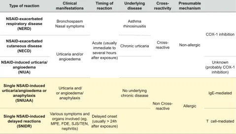
4.2 – Nonsteroidal anti-inflammatory drugs: NSAIDs
are responsible for 21% to 25% of reported ADRs, including immunological and non-immunological reactions. Depen-ding on the clinical presentation and the presumable under-lying mechanism, hypersensitivity to NSAIDs is classified in 2 groups and 5 subgroups (Table 2).49 In the first group
(≥ 75% of cases),6 the putative mechanism is the
inhibi-tion of ciclooxigenase-1, hence hypersensitivity to multiple NSAIDs is observed regardless of their chemical structure and/or anti-inflammatory potency.49
The second group involves the selective reactions, probably with an underlying immunological mechanism: a) IgE-mediated is the proposed mechanism in cases of ur-ticaria, angioedema and anaphylaxis induced by a single NSAID or a group of chemically related drugs.49,50
Pyrazolo-nes, paracetamol, ibuprofen, diclofenac and naproxen are the most common.49 The last three compounds have a
he-teroaryl acetic group, presumably carrying a higher risk of anaphylaxis (OR 19.7);51 b) NIRs, probably T-cell mediated, were also reported to be induced by a single NSAID or a group of chemically related drugs.49-51 Cutaneous reactions,
particularly MPE are the most frequent reaction. NSAIDs are the main cause of FDE and can also induce SJS/TEN (particularly oxicams).49
Diagnostic and management guidelines for children and adolescents with NSAIDs hypersensitivity, including a modified classification, were recently published considering specific clinical and epidemiological features.52
4.3 – Neuromuscular blocking agents (NMBAs): Im-mediate HRs during the perioperative period have been increasingly reported.53 Most are mediated by IgE and less frequently related to direct stimulation of histamine re-lease.53-55
IgE mechanism causes perioperative anaphylaxis (es-timated incidence of 1:10 000 to 1:20 000) and any drug administered in this period can potentially induce it.53 Dif-ferent populations exhibit difDif-ferent patterns of sensitiza-tion.7 NMBAs are the most common cause in Europe (50%
- 70%), followed by antibiotics and latex. While latex is be-coming a less common culprit, chlorhexidine is gaining im-portance and sugammadex is an emerging cause.54 Suxamethonium is the most frequent reported culprit, with a recent increment of rocuronium, vecuronium and pancuronium.53,56,57 Sensitization to NMBAs seems to
de-mand the presence of a substituted ammonium ion. In many cases, the reaction may occur at the first exposure, since a prior sensitization to another compound with a substituted ammonium ion (e.g. pholcodine) may have occurred.5,54,55,57
ARTIGO DE REVISÃO A new mast cell receptor MRGPRX2 (human Mas-rela-ted G-protein-coupled receptor member X2) was discov-ered and related with reactions to NMBAs not IgE-mediat-ed,54-55 which could explain the cases of perioperative
ana-phylaxis with negative skin testing (mast cell direct activa-tion through this receptor).54
4.4 – Radiocontrast media: In the past the ionic high-osmolar RCM induced a high incidence of IRs due to the nonspecific release of vasoactive mediators.6 Despite
the introduction of nonionic (NI) low-osmolar RCM, hyper-sensitivity reactions (HSRs) are still a matter of concern. A recent European multicenter study suggests that at least 50% of the HRs to NI-RCM are caused by an immunological mechanism. Cross-reactivity was frequent among NI-RCM with a very similar chemical structure.58 It is estimated that NI-RCM can cause IRs and NIRs in about 1% - 3% of ap-plications.59 IRs are mainly anaphylaxis, whereas NIRs
pre-dominantly manifest as mild skin eruptions occurring hours to days after RCM administration.58-62
4.5 – Biological modifiers: The biologic agents have
been recently developed and are increasingly used. They comprise proteins such as cytokines and monoclonal anti-bodies (mAbs)6,40 that differ from other drugs as they have
high molecular-weight with a great immunogenic potential.40
Three major groups of mAbs are in use: chimeric (-ximab), humanized (-zumab) and human antibodies (-mumab). They can induce reactions through different immuno-logical mechanisms.63,64 Clinical phenotypes include IRs
(type I; infusion-related reactions, cytokine release, mixed
reactions), type III and type IV delayed reactions.64
IgE-mediated reactions to basiliximab, infliximab and rituximab have been reported.6,7,42 IgE antibodies to
cetu-ximab specifically for alpha-1,3-galactose have been found in the majority of anaphylactic reactions.7,65 Rare delayed
anaphylaxis has been reported after exposures to omali-zumab, trastuomali-zumab, dacliomali-zumab, infliximab and basili-ximab.12
Some patients present IgG antibodies to biologics that may block the effect of the drug or be involved in the devel-opment of HSRs.64
NIRS are rare but have been described, after rituximab (vasculitis, SSS)7 and infliximab (SSS, SJS, DiHS).40
Desensitization allows a safe reintroduction of first-line biologic agent.64,66
4.6 – Antineoplastic agents: HSRs to antineoplastic agents are an increasing problem.67,68 Any cytostatic can
potentially expose the patient to the risk of an immune reaction. They can elicit either immediate (urticaria, bron-chospasm, dyspnea, thoracic/abdominal pain, fever, ana-phylaxis) or NIRs (macular/MPE, vasculitis). The severity of reactions ranges from mild symptoms to life-threatening anaphylaxis.67
HSRs are more common with platinum compounds (cis-platin, carbo(cis-platin, oxaliplatin), epipodophyllotoxins (teni-poside, etoposide), asparaginase, taxanes (paclitaxel) and procarbazine. Doxorubicin and 6-mercaptopurine are rare culprits. HSR to carboplatin and oxaliplatin are particular-ly frequent (incidence: 12% - 17%), with more than 50% of the reactive patients developing moderate to severe
Table 2 - Classification of Hypersensitivity Reactions Induced by NSAIDs(adapted from 55)
Type of reaction manifestationsClinical Timing of reaction Underlying disease reactivityCross- Presumable mechanism
NSAID-exacerbated respiratory disease
(NERD)
Bronchospasm Nasal symptoms
Acute (usually immediate to several hours after exposure)
Asthma rhinosinusitis
Cross-reactive Non-allergic
COX-1 inhibition NSAID-exacerbated
cutaneous disease (NECD)
Urticaria and/or angioedema
Chronic urticaria
NSAID-induced urticaria/ angioedema
(NIUA)
Unknown (probably COX-1
inhibition)
Single NSAID-induced urticaria/angioedema or
anaphylaxis (SNIUAA)
Urticaria and/ or angioedema/
anaphylaxis
No underlying chronic disease
Non
Cross-reactive Allergic
IgE-mediated
Single NSAID-induced delayed reactions
(SNIDR)
Various symptoms and organs involved (eg. MPE, FDE, SJS/TEN,
nephritis)
Delayed onset (usually > 24h
after exposure) T cell-mediated
ARTIGO DE REVISÃO symptoms.69
Most reactions occur during the treatment (platinum derivatives and taxanes), although some appear hours af-ter. Reactions to taxanes usually manifest during the first few minutes of the first or second infusion, whereas acute reactions to platinum agents usually occur after several cy-cles.65,68,70-72
Since these drugs are usually the first line therapy, pa-tients can be desensitized71 when no equally effective
al-ternative drugs are available. The desensitization should follow the general considerations for these procedures pu-blished in a consensus paper for IRs73and NIRs.74
CONCLUSION
This exhaustive review, although the limitation of being non-systematic, pointed out that despite all advances, drug allergy is not yet fully established and understood. An ex-ceptional contribution was brought by pharmacogenomics, even though a specific association has only been defined for a very limited number of drugs. Further investigation is needed to obtain clearer answers when managing each in-dividual case of DA. The development of new biomarkers and a ‘tailored-made’ medicine is probably the future. As knowledge in this field moves forward, new updates will be required.
CONFLICTS OF INTEREST
The authors declare they do not have any conflicts of interest and that they did not receive any financial support.
REFERENCES
1. Tanno LK, Torres MJ, Castells M, Demoly P, on behalf of the Joint Allergy Academies. What can we learn in drug allergy management from World Health Organization’s international classifications? Allergy. 2018;73:987–92.
2. Gomes ER, Demoly P. Epidemiology of hypersensitivity drug reactions. Curr Opin Allergy Clin Immunol. 2005;5:309-16.
3. Pirmohamed M, James S, Meakin S, Green C, Scott AK, Walley TJ, et al.
Adverse drug reactions as cause of admission to hospital: prospective analysis of 18,820 patients. BMJ. 2004;329:15–9.
4. Demoly P, Adkinson NF, Brockow K, Castells M, Chiriac AM, Greenberger PA, et al. International Consensus on drug allergy. Allergy. 2014;69:420– 37.
5. Schnyder B, Pichler WJ. Mechanisms of drug-induced allergy. Mayo Clin Proc. 2009;84:268-72.
6. Doña I, Barrionuevo E, Blanca-Lopez N, Torres MJ, Fernandez TD, Mayorga C,et al. Trends in hypersensitivity drug reactions: more drugs, more response patterns, more heterogeneity. J Investig Allergol Clin Immunol. 2014;24:143-53.
7. Rubio M, Bousquet PJ, Demoly P. Update in drug allergy: novel drugs with novel reaction patterns. Curr Opin Allergy Clin Immunol. 2010;10:457–62.
8. Johansson SG, Bieber T, Dahl R, Friedmann PS, Lanier BQ, Lockey RF, et al. Revised nomenclature for allergy for global use: Report of the Nomenclature Review Committee of the World Allergy Organization. J Allergy Clin Immunol. 2003;113:832-6.
9. Pichler WJ. Delayed drug hypersensitivity reactions. Ann Intern Med. 2003;139:683–93.
10. Torres MJ, Mayorga C, Blanca M. Nonimmediate allergic reactions induced by drugs: pathogenesis and diagnostic tests. J Investig Allergol Clin Immunol. 2009;19:80-90.
11. Pichler WJ, Adam J, Daubner B, Gentinetta T, Keller M, Yerly D. Drug hypersensitivity reactions: pathomechanism and clinical symptoms. Med Clin North Am. 2010;94:654-64.
12. Shepherd GM. Immune reactions to drugs and diagnostic agents. Mt Sinai J Med. 2011;78:717–29.
13. Blanca M, Romano A, Torres MJ, Fernandez J, Mayorga C, Rodriguez J, et al. Update on the evaluation of hypersensitivity reactions to betalactams. Allergy. 2009;64:183–93.
14. Martin-Serrano A, Barbero N, Agundez J, Vida Y, Perez-Inestrosa E, Montanez M. New advances in the study of IgE drug recognition. Curr Pharm Des. 2016;22:6759-72.
15. Cho YT, Yang CW, Chu CY. Drug reactions with eosinophilia and systemic symptoms (DRESS): an interplay among drugs, viruses, and immune system. Int J Mol Sci. 2017;18:1243.
16. Phillips EJ, Mallal SA. Pharmacogenetics of drug hypersensitivity. Pharmacogenomics. 2010;11:973–87.
17. Perkins JR, Ayuso P, Cornejo-Garcia JA, Raneac JA. The study of severe cutaneous drug hypersensitivity reactions from a systems biology perspective. Curr Opin Allergy Clin Immunol. 2014;14:301–6. 18. Chung W, Hung S. Genetic markers and danger signals in
Stevens-Johnson syndrome and toxic epidermal necrolysis. Allergol Int.
2010;59:325-32.
19. Pichler WJ, Beeler A, Keller M, Lerch M, Posadas S, Schmid D,et al.
Pharmacological interaction of drugs with immune receptors: the p-i concept. Allergol Int. 2006;55:17-25.
20. Pichler WJ, Naisbitt DJ, Park BK. Immune pathomechanism of drug hypersensitivity reactions. J Allergy Clin Immunol. 2011;127:S74-81. 21. Pirmohamed M, Ostrov DA, Park BK. New genetics finding lead the
way to a better understanding of fundamental mechanisms of drug hypersensitivity. J Allergy Clin Immunol. 2015;136:236-44.
22. Faulkner L, Meng X, Park BK, Naisbitt DJ. The importance of hapten– protein complex formation in the development of drug allergy. In: Wolters Kluwer Health|Lippincott Williams & Wilkins; 2014: Volume 14 - Number 4.
23. Lutz MB, Schuler G. Immature, semi-immature and fully mature dendritic cells: which signals induce tolerance or immunity? Trends Immunol. 2002;23:445-9.
24. Chung WH, Hung SI, Yang JY, Su SC, Huang SP, Wei CY, et al. Granulysin is a key mediator for disseminated keratinocyte death in Stevens-Johnson syndrome and toxic epidermal necrolysis. Nat Med. 2008;14:1343-50.
25. Roujeau JC, Huynh TN, Bracq C, Guillaume JC, Revuz J, Touraine R. Genetic susceptibility to toxic epidermal necrolysis. Arch Dermatol. 1987;123:1171–73.
26. McCormack M, Alfirevic A, Bourgeois S, Farrell JJ, Kasperaviciute D, Carrington M, et al. HLA-A*3101 and carbamazepine induced hypersensitivity reactions in Europeans. N Engl J Med. 2011;364:1134– 43.
27. Lonjou C, Borot N, Sekula P, Ledger N, Thomas L, Halevy S,et al. A European study of HLA-B in Stevens– Johnson syndrome and toxic epidermal necrolysis related to five high-risk drugs. Pharmacogenet Genom. 2008;18:99–107.
28. Mallal S, Phillips E, Carosi G, Molina JM, Workman C, Tomazic J,et al.
HLA-B*5701 screening for hypersensitivity to abacavir. N Engl J Med. 2008;358:568–79.
29. Davis CM, Shearer WT. Diagnosis and management of HIV drug hypersensitivity. J Allergy Clin Immunol. 2008;121:826-32.
30. Chaponda M, Pirmohamed M. Hypersensitivity reactions to HIV therapy. Br J Clin Pharmacol. 2011;71: 659–71.
31. Tohkin M, Kaniwa N, Saito Y, Sugiyama E, Kurose K, Nishikawa J,et al.
A whole-genome association study of major determinants for allopurinol-related Stevens–Johnson syndrome and toxic epidermal necrolysis in Japanese patients. Pharmacogenom J. 2013;13:60–6.
32. Colombo S, Rauch A, Rotger M, Fellay J, Martinez R, Fux C,et al. The HCP5 single-nucleotide polymorphism: a simple screening tool for prediction of hypersensitivity reaction to abacavir. J Infect Dis. 2008;198:864–7.
33. Guglielmi L, Fontaine C, Gougat C, Avinens O, Eliaou JF, Guglielmi P, et al. IL-10 promoter and IL4-Ralpha gene SNPs are associated with immediate beta-lactam allergy in atopic women. Allergy. 2006;61:921– 7.
ARTIGO DE REVISÃO JL,maculopapular eruption, urticaria and drug reaction with eosinophilia et al. Comparison of cytokine gene polymorphism in drug-induced and systemic symptoms (DRESS). J Eur Acad Dermatol Venereol. 2014;28:491-9.
35. Oussalah A, Mayorga C, Blanca M, Barbaud A, Nakonechna A, Cernadas J, et al. Genetic variants associated with drugs-induced immediate hypersensitivity reactions: a PRISMA-compliant systematic review. Allergy. 2016;71:443-62.
36. Bonaddona P, Pagani M, Aberer W, Biló MB, Brockow K, Oude Elberink H, et al. Drug hypersensitivity in clonal mast cell disorders: ENDA/ EAACI position paper. Allergy. 2015;70:755-63.
37. Caubet JC, Kaiser L, Lemaitre B, Fellay B, Gervaix A, Eigenmann PA. The role of penicillin in benign skin rashes in childhood: a prospective study based on drug rechallenge. J Allergy Clin Immunol. 2011;127:218– 22.
38. Camous X, Calbo S, Picard D, Musette P. Drug reaction with eosinophilia and systemic symptoms: an update on pathogenesis. Curr Opin Immunol. 2012;24:730–5.
39. Cho YT, Yang CW, Chu CY. Drug reactions with eosinophilia and systemic symptoms (DRESS): An interplay among drugs, viruses, and immune system. Int J Mol Sci. 2017;18:1243.
40. Khan DA, Solensky A. Drug allergy. J Allergy Clin Immunol. 2010;125:S126-37.
41. Dias de Castro E, Leblanc A, Sarmento A, Cernadas JR. An unusual case of delayed-type hypersensitivity to ceftriaxone and meropenem. Eur Ann Allergy Clin Immunol. 2015;47:225-7.
42. Dibbern DA, Montanaro A. Allergies to sulfonamide antibiotics and sulfur-containing drugs. Ann Allergy Asthma Immunol. 2008;100:91-100. 43. Sanderson JP, Naisbitt DJ. Park K. Role of bioactivation in drug-induced
hypersensitivity reactions. AAPS J 2006;8:E55-64.
44. Schnyder, Naisbitt DJ, Pichler WJ. Recognition of SMX and its reactives metabolites by drug-specific CD4+ Tcells from allergic individuals. J Immunnol. 2004;164:6647-54.
45. Schmid DA, Depta JP, Pichler WJ. T cell-mediated hypersensitivity to quinolones: mechanisms and cross-reactivity. Clin Exp Allergy. 2006;36:56-9.
46. Blanca-López N, Ariza A, Doña I, Mayorga C, Montañez MI, Garcia-Campos J,et al. Hypersensitivity reactions to fluoroquinolones: analysis of the factors involved. Clin Exp Allergy. 2013;43:560-67.
47. Schmid DA, Pichler WJ. Hypersensitivity reactions to quinolones. Curr Pharm Des. 2006;12:3316-26.
48. Manfredi M, Severino M, Testi S, Nacchia D, Ermini G, Pichler WJ. Detection of specific IgE to quinolones: mechanisms and cross-reactivity. J Allergy Clin Immunol. 2004;113:155-60.
49. Kowalski ML, Asero R, Bavbek S, Blanca M, Blanca-Lopez N, Bochenek G, et al. Classification and practical approach to the diagnosis and management of hypersensitivity to nonsteroidal anti-inflammatory drugs. Allergy. 2013;68:1219-32.
50. Ortega N, Doña I, Moreno E, Audicana MT, Barasona MJ, Berges-Gimeno MP,et al. Practical guidelines for diagnosing hypersensitivity reactions to nonsteroidal anti-inflammatory drugs. J Investig Allergol Clin Immunol. 2014;24:308-23.
51. Quiralte J, Blanco C, Delgado J, Ortega N, Alcntára M, Castillo R,et al. Challenge-based clinical patterns of 223 Spanish patients with nonsteroidal anti-inflammatory-drug-induced-reactions. J Investig Allergol Clin Immunol. 2007;17:182-8.
52. Kidon M, Blanca-Lopez N, Gomes E, Terreehorst I, Tanno L, Ponvert C, et al. EAACI/ENDA Position Paper: Diagnosis and management of hypersensitivity reactions to non- steroidal anti-inflammatory drugs (NSAIDs) in children and adolescents. Pediatr Allergy Immunol. 2018;29:469-80.
53. Mertes PM, Laxenaire MC. Allergy and anaphylaxis in anaesthesia. Minerva Anestesiol. 2004;70:285-91.
54. Hsu Blatman KS, Hepner DL. Current knowledge and management of hypersensitivity to perioperative drugs and radiocontrast media. J Allergy Clin Immunol Pract. 2017;5:587-92.
55. Spoerl D, Nigolian H, Czarnetzki C, Harr T. Reclassifying anaphylaxis to neuromuscular blocking agents based on the presumed patho-mechanism: IgE-mediated, pharmacological adverse reaction or “innate hypersensitivity”? Int J Mol Sci. 2017;18:1223.
56. Dong SW, Mertes PM, Petitipain N, Hasdenteufel F, Malinovsky JM. Hypersensitivity reactions during anesthesia. Results from the ninth French survey (2005-2007). Minerva Anestesiol. 2012;78:868-78. 57. Mertes PM, Aimone-Gastin I, Guéant-Rodriguez RM,
Mouton-Faivre C, Audibert G, O’Brien J, et al. Hypersensitivity reactions to neuromuscular blocking agents. Curr Pharm Des. 2008;14:2809-25. 58. Brockow K, Romano A, Aberer W, Bircher AJ, Barbaud A, Bonadonna P,
et al. Skin testing in patients with hypersensitivity reactions to iodinated contrast media – a European multicenter study. Allergy. 2009;64:234– 41.
59. Brockow K. Contrast media hypersensitivit -scope of the problem. Toxicology. 2005;209:189–92.
60. Kvedariene V, Martins P, Rouanet L, Demoly P. Diagnosis of iodinated contrast media hypersensitivity: results of a 6-year period. Clin Exp Allergy. 2006; 36:1072–7.
61. Laroche D, Dewachter P, Mouton-Faivre C, Clément O. Immediate reactions following ICM injection: results of a 3-year prospective multicenter survey. Contrast Med Mol Imaging. 2006;1:81.
62. Gomez E, Ariza A, Blanca-Lopez N, Torres MJ. Non-immmediate hypersensitivity reactions to contrast media. Curr Opinion Allergy Clin Immunol. 2013;1345-53.
63. Pichler JW. Adverse side-effects to biological agents. Allergy 2006;61:912-20.
64. Isabwe G, Sanchez L, Castells M Management of adverse reactions to biologic agentes. Allergy Asthma Proc. 2017;38:409–18.
65. Wickner PG, Hong D. Immediate drug hypersensitivity. Curr Allergy Asthma Rep. 2016;16:49.
66. Castells M. Rapid drug desensitization for hypersensitivity reactions to chemotherapy and monoclonal antibodies in the 21st century. J Investig Allergol Clin Immunol. 2014;24:72-9.
67. Pagani M. The complex clinical picture of presumably allergic side effects to cytostatic drugs: symptoms, pathomechanism, reexposure and desensitization. Med Clin N Am. 2010;94:835-52.
68. Giavina-Bianchi P, Patil SU, Banerji A. Immediate hypersensitivity reaction to chemotherapeutic agents. J Allergy Clin Immunol Pract. 2017;5:593-9.
69. Limsuwan T, Castells MC. Outcomes and safety of rapid desensitization for chemotherapy hypersensitivity. Expert Opin Drug Saf. 2010;9:39–53. 70. Castells M. Hypersensitivity to antineoplastic agents. Curr Pharm Des.
2008;14:2892-901.
71. Aroldi F, Prochilo T, Bertocchi P, Zaniboni A. Oxaliplatin-induced hypersensitivity reaction: underlying mechanisms and management. J Chemother. 2015;27:63-6.
72. Picard M. Management of hypersensitivity reactions to taxanes. Immunol Allergy Clin North Am. 2017;37:679-93.
73. Cernadas JR, Brockow K, Romano A, Aberer W, Torres MJ, Bircher A, et al. General considerations on rapid desensitization for drug hypersensitivity - a consensus statement. Allergy. 2010;65:1357–66. 74. Scherer K, Brockow K, Aberer W, Gooi JH, Demoly P, Romano A, et al.