Human innate responses and adjuvant activity of TLR ligands in vivo
in mice reconstituted with a human immune system
Liang Cheng
a, Zheng Zhang
a, Guangming Li
a, Feng Li
a, Li Wang
a, Liguo Zhang
b, Sandra M. Zurawski
c,d,e,
Gerard Zurawski
c,d,e, Yves Levy
d,e, Lishan Su
a,⇑a
Lineberger Comprehensive Cancer Center, Department of Microbiology and Immunology, University of North Carolina at Chapel Hill, Chapel Hill, NC 27599, United States bKey Laboratory of Infection and Immunity, Institute of Biophysics, Chinese Academy of Sciences, Beijing 100101, China
c
Baylor Institute for Immunology Research, Dallas, TX 75204, United States d
Vaccine Research Institute, Université Paris-Est, Faculté de Médecine, INSERM U955, Créteil, France e
Assistance Publique-Hôpitaux de Paris, Groupe Henri-Mondor Albert-Chenevier, Service d’immunologie clinique, 94010 Créteil, France
a r t i c l e i n f o
Article history:
Received 25 April 2017
Received in revised form 31 August 2017 Accepted 17 September 2017
Available online 25 September 2017
Keywords:
TLR ligands Adjuvant Humanized mice Human innate response CD40-targeting HIV vaccine Cytotoxic T cells
a b s t r a c t
TLR ligands (TLR-Ls) represent a class of novel vaccine adjuvants. However, their immunologic effects in humans remain poorly defined in vivo. Using a humanized mouse model with a functional human immune system, we investigated how different TLR-Ls stimulated human innate immune response in vivo and their applications as vaccine adjuvants for enhancing human cellular immune response. We found that splenocytes from humanized mice showed identical responses to various TLR-Ls as human PBMCs in vitro. To our surprise, various TLR-Ls stimulated human cytokines and chemokines differently in vivo compared to that in vitro. For example, CpG-A was most efficient to induce IFN-aproduction in vitro. In contrast, CpG-B, R848 and Poly I:C stimulated much more IFN-a than CpG-A in vivo. Importantly, the human innate immune response to specific TLR-Ls in humanized mice was different from that reported in C57BL/6 mice, but similar to that reported in nonhuman primates. Furthermore, we found that different TLR-Ls distinctively activated and mobilized human plasmacytoid dendritic cells (pDCs), myeloid DCs (mDCs) and monocytes in different organs. Finally, we showed that, as adjuvants, CpG-B, R848 and Poly I:C can all enhance antigen specific CD4+T cell response, while only R848 and
Poly I:C induced CD8+cytotoxic T cells response to a CD40-targeting HIV vaccine in humanized mice,
cor-related with their ability to activate human mDCs but not pDCs. We conclude that humanized mice serve as a highly relevant model to evaluate and rank the human immunologic effects of novel adjuvants in vivo prior to testing in humans.
1. Introduction
The most effective vaccines are live attenuated vaccines such as the yellow fever vaccine YF-17D[1] and smallpox vaccine[2,3], providing long-lasting protective immunity with a single adminis-tration. It has become clear that, by activating pathogen-encoded pattern recognition receptors (PRRs) on immune effector cells, these vaccines can efficiently activate the innate immune system to induce efficient antigen-specific humoral and cytotoxic T
lymphocytes (CTL) responses [4,5]. Conversely, recombinant
antigen-based vaccines are often poorly immunogenic and need
co-administered adjuvants to enhance the protective immunity especially the CTL response[5].
Toll like receptor (TLRs) represent an important type of PRRs that can sense the microbial components named pathogen-associated molecular patterns (PAMPs) [6,7]. TLRs are expressed by various cells, especially by the innate immune cells such as monocytes, myeloid dendritic cells (mDCs) and plasmacytoid dendritic cells (pDCs) [6,7]. Stimulation of these innate immune cells with natural or synthetic TLR ligands (TLR-Ls) results in up-regulation of co-stimulatory molecules, enhanced expression of MHC class II molecules, and production of inflammatory cytoki-nes[8,9]. Thus, natural ligands or synthetic agonists for TLRs are being developed as potential new vaccine adjuvants[10,11]. For
example, Monophosphoryl Lipid A (MPLA), a derivative
lipopolysaccharide which acts through TLR4, has been approved Abbreviations:LN, lymph node; mDC, myeloid dendritic cell; MPLA,
monophos-phoryl lipid A; pDC, plasmacytoid dendritic cell; TLR-Ls, Toll-like receptor ligands.
⇑ Corresponding author.
for clinic application as a component of AS04 adjuvant in Cervarix vaccine against cervical cancer and the vaccine against hepatitis B virus[12].
Although the adjuvant effects of TLR-Ls are promising, their immunological effects in vivo in humans are still poorly under-stood. Mouse models serve as the most widely used tools for mech-anistic study and preclinical evaluation of TLR-Ls adjuvants. However, fundamental differences exist between human and mouse since the two species diverged between 65 and 75 million years ago, and knowledge gained from mouse studies does not always apply to humans[13,14]. For example, preclinical toxicol-ogy study in mice did not provide any indication that fialuridine would be hepatotoxic to human beings[15], but 5 of 15 clinical trial participants died and the other two required a liver transplant after receiving a nucleoside analogue fialuridine treatment due to acute liver failure. The species-specific expression of a mitochon-dria nucleoside transporter in human but not in mouse is probably responsible for the human-specific liver toxicity caused by fialuri-dine[16]. Recently, it is reported that fialuridine induced acute liver damage in human-mouse liver chimeric TK-NOG mice[17].
The immune system of human also has diverse differences from mouse [13]. One obvious difference is that bronchus-associated lymphoid tissue is only developed in mice but not in healthy humans. This has possibly evolved because mice live so much clo-ser to the ground where they experience a higher dose of patho-gens [13,18]. It has also been reported that the distribution of several TLRs in innate immune cells is quite different between
human and mouse [13]. TLR9 in mouse is widely expressed on
pDC, mDC, B cells and also expressed in monocyte/macrophage lin-eage cells[19,20], whereas in humans, it is preferentially expressed on pDCs and B cells[21,22]. TLR8, which is expressed on mDC and macrophage, can respond to ssRNA stimulation in human but this is not functional in mice[23]. Moreover, TLR10, whose ligands are as yet unknown, is widely expressed in humans but not in mice
[24]. The discrepancies in TLRs distribution between mouse and human immune cells may limit the translation of findings into human clinical applications, when based on mouse work.
Another commonly used tool for evaluating the adjuvant effects of the TLR-Ls is the human peripheral blood mononuclear cells (PBMCs) in vitro culture system. However, this cell culture system is not useful to study non-circulating cells that also respond to TLR-Ls stimulation in vivo[25]. The other fundamental limitation of the human PBMCs in vitro culture system is that it cannot authenti-cally reflect the cell-cell interaction environment in vivo. The dynamics and accessibility of the drugs to the cells should also be different in vivo compared to that in vitro. It is also difficult to evaluate vaccine adjuvant activity in inducing human T and B cell responses in vitro.
Mice reconstituted with a functional human immune system provide a valuable platform to study the development and func-tions of human immune cells, and more importantly, to investigate human immune response to pathogens, vaccines and other stimu-lations in vivo[14]. We and others have shown that injection of human CD34+hematopoietic stem cells into the immunodefecient
BALB/c Rag2 /
c
c/ mice or NOD-scidc
c/ (NSG) mice as well asNOD-Rag2 /
c
c/ (NRG) mice can reconstitute all major human
myeloid and lymphoid subsets, including monocytes, mDCs, pDCs, T cells and B cells[26–31]. In this study, we used the humanized NRG mice as an in vivo model to explore how the human immune system responds to different PAMPs, specifically, how various TLR-Ls differentially stimulate human innate immune response and regulate adaptive CD4+helper T cell and importantly cytotoxic
T lymphocyte (CTL) response to CD40-targeting HIV candidate ther-apeutic vaccine in vivo. We demonstrate that human leukocytes developed in humanized mice respond similarly to TLR-Ls stimula-tion of human PBMCs in vitro. When tested in vivo, however,
TLR-Ls induce a significantly different profile of human cytokines and chemokines compared to that induced in vitro. We show that, in humanized mice, various TLR-Ls differentially activate distinct human immune cells in different lymphoid organs. Importantly, humanized mice respond to TLR-Ls stimulation differently from C57BL/6 mice[25]but similarly to that observed in nonhuman pri-mates[32]. Finally, we show that, consistent with their different abilities to activate mDCs, Poly I:C and R848 (but not CpG-B) were able to enhance antigen-specific CTL responses to a CD40-targeting HIV candidate vaccine in humanized mice. Our study indicates that various TLR-Ls differentially activate human innate immune cells to enhance antigen-specific cellular immune responses in human-ized mice. The humanhuman-ized mouse model thus provides a unique platform to evaluate the immunologic effects of novel adjuvants in vivo, prior to human testing.
2. Materials and methods
2.1. Ethics statement
The report followed NIH research ethics guidelines. For the con-struction of humanized mouse, human fetal liver was obtained from elective or medically indicated termination of pregnancy through a non-profit intermediary working with outpatient clinics (Advanced Bioscience Resources, Alameda, CA). The use of the tis-sue in the research had no influence on the decision regarding ter-mination of the pregnancy. Informed consent of the maternal donor is obtained in all cases, under regulation governing the clinic. We were provided with no information regarding the iden-tity of the patients, nor is this information traceable. The project was reviewed by the University’s Office of Human Research Ethics, which has determined that this submission does not constitute human subjects research as defined under federal regulations [45 CFR 46.102 (d or f) and 21 CFR 56.102(c)(e)(l)] and does not require IRB approval. The University of North Carolina at Chapel Hill Insti-tutional Animal Care and Use Committee (IACUC) has reviewed and approved this research. All animal experiments were con-ducted following NIH guidelines for housing and care of laboratory animals and in accordance with The University of North Carolina at Chapel Hill with protocols approved by the institution’s Institu-tional Animal Care and Use Committee (IACUC ID: 14-100).
2.2. Construction of humanized mice
We constructed humanized NRG (NOD-Rag2 /
c
c/ ) mice by
reconstitution with human fetal liver (17 to 22 weeks of
gesta-tional age) derived CD34+ hematopoietic progenitor cells
(Advanced Bioscience Resources, Alameda, CA) similarly as previ-ously reported[30]. Humanized BLT mice were generated accord-ing to a previous report[33]. Briefly, 6 to 8 weeks old NRG mice were sub-lethally irradiated and anesthetized the same day, and 1-mm3fragments of human fetal thymus were implanted under
the recipient kidney capsule. CD34+hematopoietic progenitor cells purified from fetal liver of same donor were injectedretro-orbital within 3 h. Human immune cell engraftment was detected by flow cytometry 12 weeks after transplantation. All animal studies were approved by the University of North Carolina Institutional Animal Care and Use Committee (IACUC).
2.3. TLR-L treatment in vitro and in vivo
CpG-A (ODN 2216), CpG-B (ODN 2006), CpG-C (ODN 2395), R848, MPLA and Poly I:C used in this study were all purchased
from InvivoGen. 1106 total human PBMCs or splenocytes of
for in vitro stimulation. Cells were stimulated with 5mg/ml of CpG-A, CpG-B, CpG-C, R848, Poly I:C and 2mg/ml of MPLA for 24 h and supernatants were collected for cytokine/chemokine detections. For in vivo treatment, humanized mice were treated with 50mg/ mouse of CpG-A, CpG-B, CpG-C, R848, Poly I:C or 20mg/mouse of MPLA through i.p. injection.
2.4. Detection of cytokines/chemokines
Human IFN-
a
was detected by enzyme-linked immunosorbentassay using the human IFN-
a
pan ELISA kits purchased from Mab-tech. A high sensitivity immunology multiplex assay (Luminex) (Millipore, Billerica, Massachusetts, USA) was used to measure human IL-12, IL-6, IFN-c
, TNF-a
, IL-1b, IP-10, MCP-1, IL-8, IL-4 and IL-10 in plasma of humanized mice or cell culture supernatant according to the manufacturer’s instructions.2.5. Flow cytometry
For surface staining, single cell suspensions prepared from peripheral blood, spleen, or bone marrow of humanized mice were stained with surface markers and analyzed on a CyAn ADP (Dako). For intracellular cytokine staining, cells were first stained with sur-face markers, and then permeabilized with cytofix/cytoperm buffer (BD Bioscience), followed by intracellular staining. FITC-conjugated anti-human CD40, CD56, PE-conjugated anti-human CD303, PE/ Cy5-conjugated human CD4, CD86, PE/Cy7-conjugated anti-human CD3, HLA-DR, PB-conjugated anti-anti-human CD4, CD14, IL-2,
APC-conjugated anti-human CD123, CD11c, TNF-
a
andAPC/Cy7-conjugated anti-human CD45 were purchased from Biolegend.
Pacific orange-conjugated anti-mouse CD45, PE/Texas
red-conjugated anti-human CD3, CD19, CD8 and LIVE/DEAD Fixable Aqua (LD7) Dead Cell Stain Kit were purchased from Invitrogen. Data were analyzed using Summit4.3 software (Dako).
2.6. Vaccination and antigen-specific T cell response detection
Recombinant anti-human CD40 antibody fused to 5 HIV peptide regions (
a
CD40-HIV5pep) was produced as previously reported[34], except that the HIV peptide and flexible linker sequences were reconfigured and the CD40 binding variable regions were changed to a human framework. These changes had no impact on CD40 binding or efficacy in expansion of antigen-specific T cells in vitro. Humanized BLT mice were intramuscularly (half dose) and intraperitoneally (half dose) injected with 10mg
a
CD40-HIV5pep alone or with 50mg of each TLR-L two times at 3 week intervals. Splenocytes from vaccinated humanized mice were collected 10 days after the second vaccination and stimulated ex vivo with 5 specific HIV long peptides[34]plusa
-CD28 for 12 h. Brefeldin A was added during the last 4 h of stimulation and IL-2, TNF-a
expression by CD4+ and CD8+ T cells were detected byintra-cellular staining. IFN-
c
production was detected by ELISPOT after 24 h of stimulation with 5 specific HIV long peptides plusa
-CD28.2.7. Statistics
Statistical analysis was performed using the two-tailed, unpaired Student’s t test (*p < 0.05; **p < 0.01; ***p < 0.001; ns,
non-significant) using GraphPad Prism 5 software (GraphPad Soft-ware, La Jolla, CA).
3. Results
3.1. Human PBMCs and human leukocytes developed in humanized NRG mice respond similarly to TLR-L stimulation in vitro
We reconstituted newborn NRG mice with human fetal liver derived CD34+hematopoietic stem/progenitor cells and detected the development of human leukocytes 3 months after human cell transplant. All major human CD45+leukocyte subsets including T
cells (CD3+), B cells (CD19+), NK cells (CD3 CD56+), monocytes/
macrophages (CD3 CD19 HLA DR+CD14+), mDCs (CD3 CD19
HLA DR+CD14 CD11c+) and pDCs (CD3 CD19 HLA DR+CD303+)
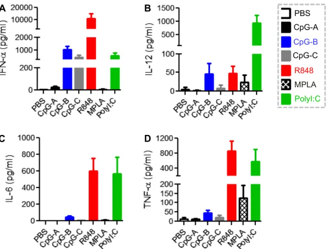
were developed in humanized NRG mice (Supplemental Fig. 1). In order to test whether the innate immune cells reconstituted in humanized NRG mice were functional, we stimulated splenocytes isolated from humanized mice in vitro with the TLR9-Ligands CpG-A, CpG-B, CpG-C[35,36], the TLR7/8-L R848[37], the TLR4-L MPLA [38] and the TLR3-L Poly I:C [25]. Human cytokines and chemokines were measured as functional readout for activation of immune cells. In parallel, results were also obtained from human PBMCs stimulated with the same individual TLR-Ls in vitro. As shown inFig. 1A, various TLR-Ls differentially stimu-lated the production of cytokines including IFN-
a
, IL-6, TNF-a
and IL-12 from splenocytes of humanized mouse. Importantly, the cytokine induction profile from splenocytes of humanized mice was comparable to that induced from human PBMCs in vitro (Fig. 1B). These results indicate that functional human innate immune cells are developed in humanized NRG mice 3 months after transplantation with human hematopoietic stem/progenitor cells.3.2. TLR-Ls stimulate different human cytokine production profiles in vivo compared to that in human PBMCs in vitro
To assess the early effects of TLR-Ls on systemic human immune responses in vivo, we treated humanized NRG mice with various TLR-Ls. We monitored the dynamics of each human cyto-kine and chemocyto-kine level at different time points after TLR-Ls treatment in vivo and the peak values were shown (Fig. 2and Sup-plemental Fig. 2A). Distinct TLR-Ls stimulated human innate immune response differentially as indicated by inducing different amounts of human IFN-
a
, IL-12, IL-6, TNF-a
, IP-10, MCP-1 andIL-8 in the serum after treatment (Fig. 2 and Supplemental
Fig. 2A). Among the TLR-Ls tested, CpG-B, R848 and Poly I:C were more potent than other TLR-Ls to stimulate production of IFN-
a
and other proinflammatory cytokines and chemokines (Fig. 2andSupplemental Fig. 2A).
To our surprise, we found that the array of human cytokines and chemokines stimulated by specific TLR-Ls in vivo was significantly different from that induced from human PBMCs or splenocytes of humanized mice in vitro (Figs. 1 and 2). As shown in Fig. 1B,
CpG-A was most efficient in stimulating IFN-
a
productionin vitro (16,218 ± 5861 pg/ml). However, it only induced a minimal amount of IFN-
a
(25 ± 9 pg/ml) in vivo (Fig. 2A). In contrast, R848 induced a low level of IFN-a
(162 ± 84 pg/ml) in vitro (Fig. 1B), while it robustly induced IFN-a
(10801 ± 4275 pg/ml) in vivo (Fig. 2A). Also in contrast to CpG-A, both CpG-B and Poly I:Cinduced very low levels of IFN-
a
from human PBMCs in vitro(Fig. 1B). However, CpG-B and Poly I:C both stimulated much more
IFN-
a
production in humanized mice (1039 ± 276 pg/ml and522 ± 265 pg/ml respectively) than CpG-A (25 ± 9 pg/ml)
(Fig. 2A). TLR4-L MPLA stimulated no significant production of IFN-
a
both in vitro and in vivo (Figs. 1Band2A).C and R848 stimulated similar level of IL-12 while CpG-B did not induce significant level of IL-12 in vitro (Fig. 1B). We found that in vivo Poly I:C induced around 20-fold higher IL-12 production than R848 or CpG-B (Fig.2B). MPLA stimulated a low level of IL-12 in vitro and in vivo (Figs. 1B and2B).
The proinflammatory cytokines IL-6, TNF-
a
and chemokines IP-10, MCP-1 and IL-8 induced by distinct TLR-Ls also varied between in vivo and in vitro systems (Figs. 1, 2andSupplemental Fig. 2). The data above indicate that different TLR-Ls distinctively stimulate cytokine and chemokine production in humanized NRG mice in vivo, but these profiles are different to those observed from human PBMCs, as well as splenocytes from the humanized mouse, in vitro. These data suggest that the immunological effect of TLR-Ls in vitro does not predict their effect in vivo.3.3. Kinetics of human cytokine production in vivo after TLR-Ls treatment
The results above showed that CpG-B, R848 and Poly I:C are more efficient than other TLR-Ls in stimulating production of human proinflammatory cytokines and chemokines in vivo in humanized mice. We further characterized the induction kinetics of human cytokines and chemokines by these three TLR-Ls in humanized mice. IFN-
a
was robustly induced by R848 at 1 h after injection, and reached peak level at 4 h (Fig. 3A). After 48 h, the IFN-a
level was only slightly reduced (Fig. 3A). In contrast, CpG-B and poly I:C stimulated slower and lower IFN-a
expression and the IFN-a
level became undetectable after 48 h (Fig. 3A). However, IL-12 induced by Poly I:C was higher and persisted longer in theplasma (Fig. 3B). Both Poly I:C and R848 induced higher level of IL-6 than CpG-B (Fig. 3C). Induction of other cytokines such as TNF-
a
, IFN-c
and IL-1b; and chemokines such as IP-10, MCP-1, and IL-8 also showed different kinetics after CpG-B, R848 or Poly I:C treatment (Fig. 3andSupplemental Fig. 3). All the TLR-Ls tested induced only very low levels of anti-inflammatory cytokine IL-10 and they did not induce detectable Th2 cytokine IL-4 ( Supplemen-tal Fig. 3).3.4. Humanized mice respond to TLR-Ls stimulation differently from C57BL/6 mice but similarly to that reported in nonhuman primates
Mouse models have been widely used for mechanistic study and preclinical evaluation of TLR-Ls adjuvants. Since the expres-sion of several TLRs in immune cells is quite different between human and mice[13], we investigated whether TLR-Ls differen-tially activate human and mouse immune response in vivo. It was reported that CpG-B induced a high level of mouse cytokines IL-6, TNF-
a
, IL-12 and IFN-c
in C57BL/6 mice[25]. In marked con-trast, our result showed that, in comparison with Poly I:C and R848, CpG-B only induced very low levels of human IL-6, TNF-a
, IL-12 and IFN-c
in humanized mice (Fig. 3 and Supplemental Fig. 3). Study in nonhuman primates such as Rhesus macaques, which has closer genetic relationship to human, also indicated that CpG-B induced low levels of IL-6, TNF-a
, and IFN-c
[32] when administered in vivo. Importantly, CpG-B therapy in mildly asth-matic human patients also did not stimulate TNF-a
productionThese results indicate that the immunological effect of CpG-B in humanized mice, but not in mice, is more relevant to the situation in humans.
Poly I:C treatment in C57BL/6 mice leads to higher amounts of mouse IFN-
a
production than R848 and CpG-B[25]. In contrast, we found here that R848 was the superior human IFN-a
inducer in humanized mice (Fig.3A). Similar to the result observed in Rhesus macaques[32], R-848 treatment induced rapid and abundant pro-duction of pro-inflammatory cytokines in humanized mice (Fig. 3and Supplemental Fig. 3). Taken together, these results indicate that mouse and human immune system respond differentially to in vivo administered TLR-Ls stimulation. Thus, knowledge about the immunological effect of TLR-Ls in mouse is not always applicable to humans. The similarity of immune responses to specific TLR-Ls between humanized mice and nonhuman primates support the use of humanized mouse model to study the human immunological effects of TLR-Ls, in vivo.
In summary, different TLR-Ls showed variable abilities to stim-ulate production of human cytokines and chemokines in vivo in humanized mice. Importantly, the human cytokine and chemokine
production profile induced by TLR-Ls in vivo was quite different compared to that induced in vitro from human PBMCs or spleno-cytes of humanized mouse. The data indicate that the immunologi-cal effect of TLR-Ls on human PBMC in vitro cannot simply apply to that in vivo. Results also showed that humanized mice responded to TLR-Ls stimulation differently from C57BL/6 mice, but similarly to that in nonhuman primate (Table 1). Thus, humanized mice pro-vide a unique platform to study the human immunological effect of TLR-Ls and novel adjuvants in vivo.
3.5. Different TLR-Ls distinctively activate and mobilize human monocytes/ macrophages, mDCs and pDCs in peripheral blood, spleen and lymph nodes
We further characterized the effects of TLR-Ls in vivo by mea-suring the expression level of surface activation markers (CD40, CD86, HLA-DR) on various human innate immune cells in different organs. We found that CpG-B preferentially activated CD303+pDCs
in the peripheral blood, spleen and mesenteric lymph nodes
(mLNs) while it had minimal effect on CD14+ monocytes/
macrophages and CD11c+ mDCs in peripheral blood and spleen
(Fig. 4A–C). However, monocytes/macrophages from the mLNs expressed a higher level of this activation marker in the CpG-B treated group than control mice (Fig. 4A). Poly I:C was most potent in activating mDCs, but not pDCs, in the peripheral blood, spleen, and mLNs (Fig. 4A–C). It significantly up-regulated the expression of CD40, CD86 and HLA-DR on monocytes/macrophages from trea-ted mLNs but not peripheral blood or spleen (Fig. 4A). Since mono-cytes do not express TLR-9 and TLR-3, their activation by CpG-B and Poly I:C in the mLN may be indirect. R848 triggered the activa-tion of a broader spectrum of immune cells, including pDCs, mDCs and also monocytes/macrophages from peripheral blood, spleen and mLNs (Fig. 4A–C).
In addition, we found that frequency of monocytes and mDCs decreased in the peripheral blood and spleen 24 h after R848 and Poly I:C treatment. In contrast, R848 and Poly I:C treatment increased the frequency of monocytes and dendritic cells in the mesenteric LNs (Fig.4D). These data suggest that after R848 and Poly I:C treatment, human monocytes and cDCs migrated to the LNs.
In summary, the results above indicated that TLR-Ls systemi-cally and differentially activated human immune cells, in vivo, in different organs; with CpG-B preferentially activating pDCs, Poly I:C being more potent in activating mDCs, while R848 activated both mDCs and pDCs. Thus, by using the humanized mouse model we can investigate the human immunological effects of TLR-Ls on different immune target cells in different lymphoid organs and evaluate or develop novel human adjuvants based on such multi-ple parameters.
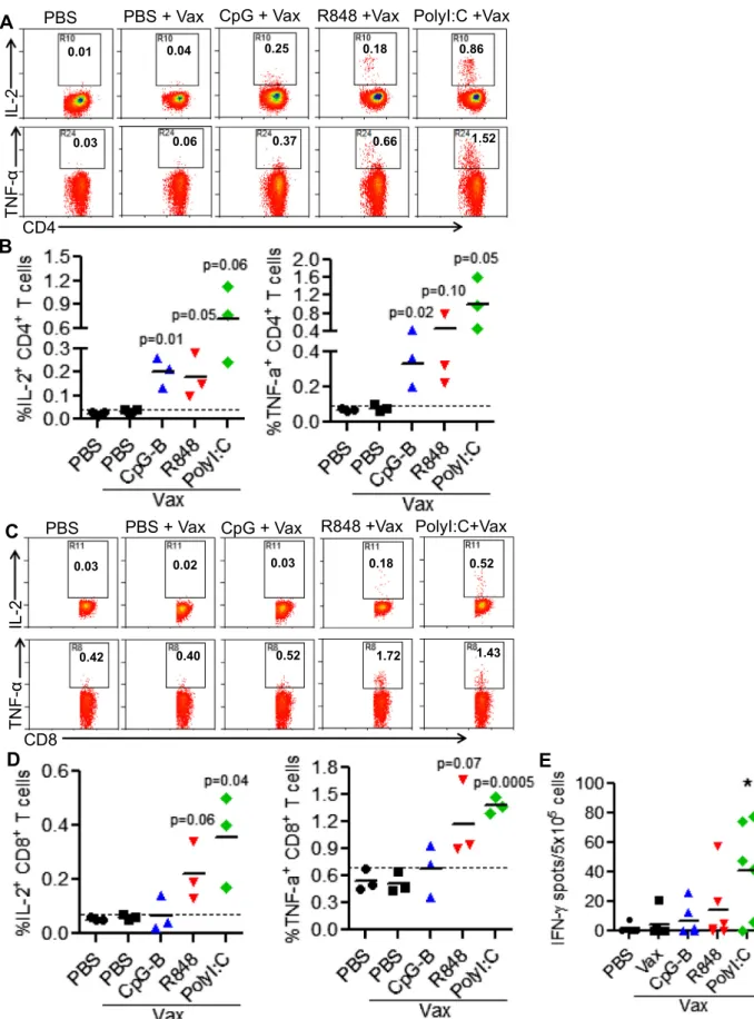
3.6. Poly I:C, R848 and CpG-B have different efficacy for enhancing antigen-specific T cell response
Recombinant antigen-based vaccines are often poorly immuno-genic and need adjuvants to induce efficient protective immunity, especially the CTL response[5]. Since TLR-Ls CpG-B, R848, and Poly I:C are better than other TLR-Ls tested here to activated human innate immune system in vivo, we further tested their ability to enhance antigen-specific T cell response to a CD40-targeting HIV vaccine, which is based on a recombinant human CD40 anti-body fused via the heavy (H) chain C-terminus to a string of the 5 HIV peptides (
a
CD40-HIV5pep) [34]. Humanized BLT (CD34+HSC, fetal liver, and thymus) mice were immunized with
a
CD40-HIV5pep with or without individual TLR-L as adjuvant. Antigen-specific T cell responses were evaluated one week after the boost vaccination. As shown inFig. 5A and B, after ex vivo stimulation with antigen-specific peptides, CD4+ cells from vaccinated micewith all three TLR-Ls as adjuvant, produced significantly higher levels of intracellular IL-2 and TNF-
a
than non-vaccinated control mice or vaccinated mice without adjuvant. In contrast, antigen-specific CD8+ T cell responses were only detected in vaccinated mice with Poly I:C and R848, but not CpG-B, as adjuvant(Fig. 5C and D). PolyI:C as adjuvant also enhanced the ability of antigen-specific T cells to produce IFN-
c
after stimulation with peptides ex vivo (Fig. 5E).3.7. The different ability of Poly I:C, R848 and CpG-B to enhance antigen-specific CTL response correlates with their ability to activate mDC
To induce antigen-specific CTL responses, exogenous protein vaccines need to be processed and cross-presented with MHC class I molecules by specific dendritic cells [10]. It is reported that, in vitro, both mDCs and pDCs from human lymphoid organs can cross-present soluble antigens [40,41]. Our data indicated that, in vivo, Poly I:C and R848 but not CpG-B as adjuvant enhanced antigen-specific CTL response to a protein based vaccine (Fig. 5). Of these three adjuvants, Poly I:C only activated human mDCs, R848 activated both human mDCs and pDCs, while CpG-B prefer-entially activated human pDCs in different lymphoid organs in vivo (Fig. 4). These results suggest that, in vivo, activation of human mDCs but not pDCs is important for inducing antigen-specific CTL response to protein based vaccination.
CpG-B has been reported to activate and promote the cross-presentation ability of mouse pre-CD8
a
+ mDC [42] and mousebone marrow-derived mDCs[43]. It also induces a sustained high level of IL-12 expression in mouse[44]. Thus, as adjuvant, CpG-B can significantly enhance mouse CTL responses to protein or pep-tide vaccine in mice[45,46] which is different from our data in humanized mice here. Again, our results suggest that the adjuvant activity of TLR-Ls in mice is distinct from that in humans.
Taken together, our data indicate that, consistent with their dif-ferent abilities for activating human mDCs, Poly I:C and R848 but not CpG-B have the ability to induce human CTL response to antigen-based vaccine. Thus, humanized mice provide a unique and highly relevant in vivo model to test human innate response, which can define intrinsic adjuvant activity and resulting enhance-ment of vaccine antigenicity prior to human testing.
4. Discussion
TLR-Ls, which can activate the innate immune system through specific receptors, are widely recognized as potential vaccine adju-vants[10]. Therefore, understanding the immunological effect of TLR-Ls, in vivo in humans, is essential for applying them in the clinic. Mouse models have served as the most widely used tools for mechanistic study and preclinical evaluation of TLR-Ls as adju-vants. However, since human and mouse diverged between 65 and 75 million years ago, fundamental differences exist between the two species, in particular in their innate immune systems[13]. In the present study, we took advantage of the well-developed humanized mouse model to study how distinct TLR-Ls stimulated the human immune system and as adjuvants in vivo. We report the Table 1
Cytokines induced by TLR-Ls in humanized mice in vivo resemble that in Rhesus macaque but different from that in C57BL/6 mice.
Cytokines induced Stimulation conditions
Humanized mousein vivo Rhesus macaquein vivo(32) C57BL/6 mousein vivo(25)
CpG-B R848 Poly I:C MPLA CpG-B R848 Poly I:C MPLA CpG-B R848 Poly I:C LPS
IFN-a ++ ++++ ++ – ++ ++++ NA – +/ ++ ++++ –
IL-12p70 + + +++ +/ NA NA NA NA – – – –
IL-6 + +++ +++ +/ + +++ NA +/ +++ +++ +++ ++
IFN-c + +++ ++ +/ + +++ NA – ++++ ++ ++ +
following novel findings: (1) we proved that leukocytes from spleens of humanized mice respond similarly to 6 different TLR agonists as human PBMCs in vitro. (2) We found that TLR-Ls stim-ulated distinct human cytokine production profiles in humanized mice in vivo, compared with that in vitro. (3) Humanized mice responded to TLR-Ls stimulation differently from C57BL/6 mice, but similarly to that reported in nonhuman primates. (4) We demonstrated that, consistent with their different abilities to acti-vate human mDCs, Poly I:C and R848 but not CpG-B were able to enhance antigen-specific CTL responses to a protein based vaccine in humanized mice.
We report here that TLR-Ls activated the human immune response differently from that in mice. It is reported that CpG-B treatment in mice induced high levels of pro-inflammatory cytoki-nes IL-6, TNF-
a
, IL-12 and IFN-c
, comparable to that induced by R848 or Poly I:C[25]. However, CpG-B induced much lower levels of human IL-6, TNF-a
, IL-12 and IFN-c
in humanized mice in com-parison to R848 and Poly I:C (Fig. 3). The distinct expression pat-terns of TLR-9 in mouse and human probably contribute to the different immunological effect induced by CpG-B[13]. Our results explained why CpG-B elicits TNF-a
-dependent toxicity in mice but not in humans [39]. Thus, knowledge about the immunological Fig. 4.Distinct TLR-Ls differentially activate human monocytes, mDC, and pDCs in different lymphoid organs. Humanized mice were injected intraperitoneally with CpG-B, R848, Poly I:C or PBS. Leukocytes from peripheral blood, spleen, and LNs were isolated for flow cytometry analysis 24 h after treatment. The MFI for CD40, CD86 and HLA-DR on CD14+monocytes (A), CD11 c+
mDCs (B) and CD303+
Fig. 5.CpG-B, R848 and Poly I:C differentially enhance antigen-specific CD4+
and CD8+
T cell responses to vaccination. Humanized BLT mice (with both human HSC and thymus co-transplant) were vaccinated withaCD40-HIV5pep alone or with indicated TLR-Ls twice at 3 weeks interval. Mice were euthanized 10 days after boost. Splenocytes from vaccinated humanized mice were stimulated ex vivo with the 5 specific HIV long peptides plusa-CD28 mAb. IL-2 and TNF-aexpression by CD4+
and CD8+
T cells were detected by intracellular staining. IFN-cproduction was detected by ELISPOT assay. (A and B) Representative plots and summarized data show percentages of IL-2+
and
TNF-a+CD4+T cells. (C-D) Representative plots and summarized data show percentages of IL-2+and TNF-a+CD8+T cells. Each dot represents one individual mouse with 3 mice
effect of TLR-Ls in humanized mice will be critical to evaluate human innate immune activation and adjuvant activity relevant to humans.
We also compared our data with reported work in nonhuman primates [32]. We found that TLR-Ls stimulated similar innate immune response in humanized mice as in Rhesus macaque, which has closer genetic relationship to human. Herein, we conclude that the humanized mouse serves as a highly relevant small animal model to test human immunological activity of TLR-Ls, in vivo.
In this study, we also demonstrated that TLR-Ls stimulated the human innate immune response differently in vivo compared to that in vitro. Three major classes of stimulatory CpG oligodeoxynu-cleotides (CpG ODN) have been identified based on their structural characteristics and activity on human pDCs and B cells[35,47]. Type I IFNs play important roles in stimulating dendritic cell mat-uration and migration, and enhancing humoral immunity as well as modulating effector and memory T cell response[48]. Therefore, CpG-A, which can induce robust type I IFN response in vitro, may have potential benefits as vaccine adjuvant. However, therapeutic potential of CpG-A remained largely unexplored. Here we showed that CpG-A, which can stimulate high levels of human IFN-
a
pro-duction in vitro, did not induce significant levels of IFN-a
in vivo in humanized mice (Figs. 1 and 2). CpG-A can form uncontrollable aggregation that complicates its manufacture [49,50]. Thus, one possible reason that CpG-A behaves poorly in vivo activity could be its highly aggregated structure, which may restrict its access to spleen, lymph node or bone marrow[51,52]. In contrast,CpG-B induced high levels of IFN-
a
in humanized mice in vivo(Fig. 2). In addition to stimulating IFN-
a
production, CpG-B also induced other cytokines and chemokines such as IL-6, IL-12,IFN-c
, and IP-10 in vivo in humanized mice. These results further sup-port the development of CpG-B as vaccine adjuvant in clinical trials.In comparison to CpG-A, the TLR7/8-L R848 was poor in induc-ing IFN-
a
, in vitro, both from human PBMCs and spleen cells from humanized mice. However, it stimulated fast, robust, and durable IFN-a
production, in vivo, in humanized mice (Fig. 3A). In addition to CpG-B and R848, we report here that Poly I:C is also a potent human innate immune stimulator, in vivo, in humanized mice. Poly I:C treatment induced significant levels of IFN-a
, in vivo, but not in vitro (Figs. 1–3). Most importantly, it stimulated much higher and more durable levels of IL-12 than CpG-B and R848, in vivo (Fig. 3B). Study has indicated that IL-12 plays an essential role for the development of antigen-specific CD8+T cell immuneresponses to vaccination in humans with cancer[53].
It will be of interest to explore the mechanisms by which TLR-Ls stimulate different cytokine production profiles, in vivo, compared to that in vitro. Lots of variables such as; physiological conditions, cell-cell interactions and dynamics, and accessibility of the drugs to the cells, might contribute to the differences observed here. Thus, the immunological effect of TLR-Ls, in vitro, cannot simply predict their activity in vivo. Using the humanized mouse model, we will further investigate how distinct TLR-Ls differentially stim-ulate the human innate immune response in vivo.
From a vaccine development state point, induction of robust, potent and durable CTL responses using a non-viral vector is chal-lenging. We hypothesize that targeting antigens through a DC recep-tor combined with DC stimularecep-tors may be required to optimize T cell responses. We show here that Poly I:C and R848, which can signifi-cantly stimulate human mDCs, induced antigen-specific CTL response to a DC-targeting HIV vaccine. In contrast, CpG-B, which activated human pDCs but not mDCs, enhanced antigen-specific human CD4+helper T cell but not CTL response in humanized mice
(Fig. 5). The results indicate that human pDCs activated by CpG-B did not cross-present antigen to CD8+T cells efficiently, although several reports showed that in vitro pDCs can cross-present soluble
antigen[40,41]. Taken together, our results suggest that activation of human mDCs but not pDCs in vivo is important for inducing antigen-specific CTL response to protein-based vaccines. Our data is consistent with the result from human clinical trials, which show that CpG-B preferentially enhances antibody responses and CD4+T
cell responses to the NY-ESO-1 protein[54]. Although CD8+T cell
responses are also detected in some vaccinated people at later time points during vaccination, it is believed that the cross-presentation of antigen is predominantly mediated by vaccine-induced antibody
[54].
The expression profile of several TLRs in mouse immune cells is different from that in human immune cells. It may lead to different adjuvant activity of Ls in mice and humans. For example, TLR-9 is widely expressed in nearly all myeloid cells in mice but it pref-erentially expressed in pDCs and B cells in humans. That explains why CpG-B has been used as a potent adjuvant to enhance CTL response in mice[45,46], which is different from our observation here in humanized mice. Humanized mice thus will provide a crit-ical model to test human adjuvant activity of TLR-Ls and other novel adjuvants, in vivo, prior to clinical trials.
In conclusion, we demonstrate that different TLR-Ls distinc-tively activate human innate immune responses and enhance antigen-specific CD4+or CD8+T cell responses in humanized mice.
The results will greatly help the development of novel human adjuvants in human clinical settings.
Acknowledgments
This work was supported in part by grants from the US National Institutes of Health (AI109784 and DK095962 to LS). This work was also supported by a grant from the Vaccine Research Institute, Paris, France. The funders had no role in study design, data collec-tion and analysis, decision to publish, or preparacollec-tion of the manuscript.
We thank the members of Su laboratory for discussions, and UNC DLAM, FACS cores for support, and the Vaccine Research Insti-tute (VRI), Paris France.
Conflict of interest statement
G.Z., S.Z., and Y.L., are named inventors on CD40-targeting vaccine patents and patent filings held jointly by INSERM and the Baylor Research Institute.
Appendix A. Supplementary material
Supplementary data associated with this article can be found, in the online version, at https://doi.org/10.1016/j.vaccine.2017.09. 052.
References
[1] Yellow fever vaccine. WHO position paper. Releve epidemiologique hebdomadaire/Section d’hygiene du Secretariat de la Societe des Nations = Weekly epidemiological record/Health Section of the Secretariat of the League of Nations. 2003;78:349–359.
[2]Crotty S, Felgner P, Davies H, Glidewell J, Villarreal L, Ahmed R. Cutting edge: long-term B cell memory in humans after smallpox vaccination. J Immunol. 2003;171:4969–73.
[3]Hammarlund E, Lewis MW, Hansen SG, Strelow LI, Nelson JA, Sexton GJ, et al. Duration of antiviral immunity after smallpox vaccination. NatureMed 2003;9:1131–7.
[4]Querec T, Bennouna S, Alkan S, Laouar Y, Gorden K, Flavell R, et al. Yellow fever vaccine YF-17D activates multiple dendritic cell subsets via TLR2, 7, 8, and 9 to stimulate polyvalent immunity. J Exp Med 2006;203:413–24.
[5]Pulendran B, Ahmed R. Immunological mechanisms of vaccination. Nat Immunol 2011;12:509–17.
[7]Kawai T, Akira S. The role of pattern-recognition receptors in innate immunity: update on Toll-like receptors. Nat Immunol 2010;11:373–84.
[8]O’Neill LA, Bowie AG. Sensing and signaling in antiviral innate immunity. Curr Biol: CB. 2010;20:R328–33.
[9]Iwasaki A, Medzhitov R. Regulation of adaptive immunity by the innate immune system. Science 2010;327:291–5.
[10]Coffman RL, Sher A, Seder RA. Vaccine adjuvants: putting innate immunity to work. Immunity 2010;33:492–503.
[11]Mbow ML, De Gregorio E, Valiante NM, Rappuoli R. New adjuvants for human vaccines. Curr Opin Immunol 2010;22:411–6.
[12]Duthie MS, Windish HP, Fox CB, Reed SG. Use of defined TLR ligands as adjuvants within human vaccines. Immunol Rev 2011;239:178–96. [13]Mestas J, Hughes CC. Of mice and not men: differences between mouse and
human immunology. J Immunol 2004;172:2731–8.
[14]Rongvaux A, Takizawa H, Strowig T, Willinger T, Eynon EE, Flavell RA, et al. Human hemato-lymphoid system mice: current use and future potential for medicine. Annu Rev Immunol 2013;31:635–74.
[15]McKenzie R, Fried MW, Sallie R, Conjeevaram H, Di Bisceglie AM, Park Y, et al. Hepatic failure and lactic acidosis due to fialuridine (FIAU), an investigational nucleoside analogue for chronic hepatitis B. N Engl J Med 1995;333:1099–105. [16]Lee EW, Lai Y, Zhang H, Unadkat JD. Identification of the mitochondrial targeting signal of the human equilibrative nucleoside transporter 1 (hENT1): implications for interspecies differences in mitochondrial toxicity of fialuridine. J Biol Chem 2006;281:16700–6.
[17]Xu D, Nishimura T, Nishimura S, Zhang H, Zheng M, Guo YY, et al. Fialuridine induces acute liver failure in chimeric TK-NOG mice: a model for detecting hepatic drug toxicity prior to human testing. PLoS Med 2014;11:e1001628. [18]Pabst R, Gehrke I. Is the bronchus-associated lymphoid tissue (BALT) an
integral structure of the lung in normal mammals, including humans? Am J Respir Cell Mol Biol 1990;3:131–5.
[19]Edwards AD, Diebold SS, Slack EM, Tomizawa H, Hemmi H, Kaisho T, et al. Toll-like receptor expression in murine DC subsets: lack of TLR7 expression by CD8 alpha+ DC correlates with unresponsiveness to imidazoquinolines. Eur J Immunol 2003;33:827–33.
[20]Suzuki K, Suda T, Naito T, Ide K, Chida K, Nakamura H. Impaired toll-like receptor 9 expression in alveolar macrophages with no sensitivity to CpG DNA. Am J Respir Crit Care Med 2005;171:707–13.
[21]Kadowaki N, Ho S, Antonenko S, Malefyt RW, Kastelein RA, Bazan F, et al. Subsets of human dendritic cell precursors express different toll-like receptors and respond to different microbial antigens. J Exp Med 2001;194:863–9. [22]Hornung V, Rothenfusser S, Britsch S, Krug A, Jahrsdorfer B, Giese T, et al.
Quantitative expression of toll-like receptor 1–10 mRNA in cellular subsets of human peripheral blood mononuclear cells and sensitivity to CpG oligodeoxynucleotides. J Immunol 2002;168:4531–7.
[23]Heil F, Hemmi H, Hochrein H, Ampenberger F, Kirschning C, Akira S, et al. Species-specific recognition of single-stranded RNA via toll-like receptor 7 and 8. Science 2004;303:1526–9.
[24]Hasan U, Chaffois C, Gaillard C, Saulnier V, Merck E, Tancredi S, et al. Human TLR10 is a functional receptor, expressed by B cells and plasmacytoid dendritic cells, which activates gene transcription through MyD88. J Immunol 2005;174:2942–50.
[25]Longhi MP, Trumpfheller C, Idoyaga J, Caskey M, Matos I, Kluger C, et al. Dendritic cells require a systemic type I interferon response to mature and induce CD4+ Th1 immunity with poly IC as adjuvant. J Exp Med 2009;206:1589–602.
[26]Tanaka S, Saito Y, Kunisawa J, Kurashima Y, Wake T, Suzuki N, et al. Development of mature and functional human myeloid subsets in hematopoietic stem cell-engrafted NOD/SCID/IL2rgammaKO mice. J Immunol 2012;188:6145–55.
[27]Pearson T, Shultz LD, Miller D, King M, Laning J, Fodor W, et al. Non-obese diabetic-recombination activating gene-1 (NOD-Rag1 null) interleukin (IL)-2 receptor common gamma chain (IL2r gamma null) null mice: a radioresistant model for human lymphohaematopoietic engraftment. Clin Exp Immunol 2008;154:270–84.
[28]Traggiai E, Chicha L, Mazzucchelli L, Bronz L, Piffaretti JC, Lanzavecchia A, et al. Development of a human adaptive immune system in cord blood cell-transplanted mice. Science 2004;304:104–7.
[29]Jiang Q, Zhang L, Wang R, Jeffrey J, Washburn ML, Brouwer D, et al. FoxP3+CD4 + regulatory T cells play an important role in acute HIV-1 infection in humanized Rag2-/-gammaC-/- mice in vivo. Blood 2008;112:2858–68. [30]Zhang L, Jiang Q, Li G, Jeffrey J, Kovalev GI, Su L. Efficient infection, activation,
and impairment of pDCs in the BM and peripheral lymphoid organs during early HIV-1 infection in humanized rag2(-)/(-)gamma C(-)/(-) mice in vivo. Blood 2011;117:6184–92.
[31]Meixlsperger S, Leung CS, Ramer PC, Pack M, Vanoaica LD, Breton G, et al. CD141+ dendritic cells produce prominent amounts of IFN-alpha after dsRNA
recognition and can be targeted via DEC-205 in humanized mice. Blood 2013;121:5034–44.
[32]Kwissa M, Nakaya HI, Oluoch H, Pulendran B. Distinct TLR adjuvants differentially stimulate systemic and local innate immune responses in nonhuman primates. Blood 2012;119:2044–55.
[33]Melkus MW, Estes JD, Padgett-Thomas A, Gatlin J, Denton PW, Othieno FA, et al. Humanized mice mount specific adaptive and innate immune responses to EBV and TSST-1. Nat Med 2006;12:1316–22.
[34]Flamar AL, Xue Y, Zurawski SM, Montes M, King B, Sloan L, et al. Targeting concatenated HIV antigens to human CD40 expands a broad repertoire of multifunctional CD4+ and CD8+ T cells. AIDS 2013;27:2041–51.
[35]Krieg AM. CpG motifs in bacterial DNA and their immune effects. Annu Rev Immunol 2002;20:709–60.
[36]Vollmer J, Weeratna R, Payette P, Jurk M, Schetter C, Laucht M, et al. Characterization of three CpG oligodeoxynucleotide classes with distinct immunostimulatory activities. Eur J Immunol 2004;34:251–62.
[37]Jurk M, Heil F, Vollmer J, Schetter C, Krieg AM, Wagner H, et al. Human TLR7 or TLR8 independently confer responsiveness to the antiviral compound R-848. Nat Immunol 2002;3:499.
[38]Casella CR, Mitchell TC. Putting endotoxin to work for us: monophosphoryl lipid A as a safe and effective vaccine adjuvant. Cell Mol Life Sci: CMLS 2008;65:3231–40.
[39]Campbell JD, Cho Y, Foster ML, Kanzler H, Kachura MA, Lum JA, et al. CpG-containing immunostimulatory DNA sequences elicit TNF-alpha-dependent toxicity in rodents but not in humans. J Clin Invest 2009;119:2564–76. [40] Segura E, Durand M, Amigorena S. Similar antigen cross-presentation capacity
and phagocytic functions in all freshly isolated human lymphoid organ-resident dendritic cells. J Exp Med 2013;210:1035–47.
[41]Hoeffel G, Ripoche AC, Matheoud D, Nascimbeni M, Escriou N, Lebon P, et al. Antigen crosspresentation by human plasmacytoid dendritic cells. Immunity 2007;27:481–92.
[42]de Brito C, Tomkowiak M, Ghittoni R, Caux C, Leverrier Y, Marvel J. CpG promotes cross-presentation of dead cell-associated antigens by pre-CD8alpha + dendritic cells [corrected]. J Immunol 2011;186:1503–11.
[43]Datta SK, Redecke V, Prilliman KR, Takabayashi K, Corr M, Tallant T, et al. A subset of Toll-like receptor ligands induces cross-presentation by bone marrow-derived dendritic cells. J Immunol 2003;170:4102–10.
[44]Krieg AM, Love-Homan L, Yi AK, Harty JT. CpG DNA induces sustained IL-12 expression in vivo and resistance to Listeria monocytogenes challenge. J Immunol 1998;161:2428–34.
[45]Davila E, Celis E. Repeated administration of cytosine-phosphorothiolated guanine-containing oligonucleotides together with peptide/protein immunization results in enhanced CTL responses with anti-tumor activity. J Immunol 2000;165:539–47.
[46]Miconnet I, Koenig S, Speiser D, Krieg A, Guillaume P, Cerottini JC, et al. CpG are efficient adjuvants for specific CTL induction against tumor antigen-derived peptide. J Immunol 2002;168:1212–8.
[47]Hartmann G, Battiany J, Poeck H, Wagner M, Kerkmann M, Lubenow N, et al. Rational design of new CpG oligonucleotides that combine B cell activation with high IFN-alpha induction in plasmacytoid dendritic cells. Eur J Immunol 2003;33:1633–41.
[48]Gonzalez-Navajas JM, Lee J, David M, Raz E. Immunomodulatory functions of type I interferons. Nat Rev Immunol 2012;12:125–35.
[49]Puig M, Grajkowski A, Boczkowska M, Ausin C, Beaucage SL, Verthelyi D. Use of thermolytic protective groups to prevent G-tetrad formation in CpG ODN type D: structural studies and immunomodulatory activity in primates. Nucl Acids Res 2006;34:6488–95.
[50] Gungor B, Yagci FC, Tincer G, Bayyurt B, Alpdundar E, Yildiz S, et al. CpG ODN Nanorings Induce IFNalpha from Plasmacytoid Dendritic Cells and Demonstrate Potent Vaccine Adjuvant Activity. Sci Transl Med 2014;6:235ra61.
[51]Kerkmann M, Costa LT, Richter C, Rothenfusser S, Battiany J, Hornung V, et al. Spontaneous formation of nucleic acid-based nanoparticles is responsible for high interferon-alpha induction by CpG-A in plasmacytoid dendritic cells. J Biol Chem 2005;280:8086–93.
[52]Marshall JD, Fearon KL, Higgins D, Hessel EM, Kanzler H, Abbate C, et al. Superior activity of the type C class of ISS in vitro and in vivo across multiple species. DNA Cell Biol 2005;24:63–72.
[53]Carreno BM, Becker-Hapak M, Huang A, Chan M, Alyasiry A, Lie WR, et al. IL-12p70-producing patient DC vaccine elicits Tc1-polarized immunity. J Clin Invest 2013;123:3383–94.