MicroRNAs: New Insights Into the Pathogenesis of
Endodontic Periapical Disease
Linda Chan
A thesis submitted to the faculty of the University of North Carolina at Chapel Hill in partial fulfillment of the requirements for the degree of Master of Science in the Department of
Endodontics, School of Dentistry
Chapel Hill 2013
Approved by:
Asma Khan
Salvador Nares
Ricardo Padilla
ii ABSTRACT
LINDA CHAN: MicroRNAs: New Insights Into the Pathogenesis of Endodontic Periapical Disease
(Under the direction of Asma Khan, Salvador Nares, Ricardo Padilla and Eric Rivera)
Apical periodontitis is an inflammatory disease of the periradicular tissues caused by the
host’s immune response to infection of the root canal system. MicroRNAs (miRNA) have
been shown to play an important role in the regulation of inflammation and the immune
response; however, their role in the pathogenesis of endodontic periapical disease has not
been explored. The purpose of this study was to examine the differential expression of
miRNAs in diseased periapical tissues as compared to healthy periodontal ligament and pulp
tissues. miRNA profiles were assessed using microarray technology and expression levels of
selected miRNAs linked to inflammation and the immune response were confirmed by
quantitative RT-PCR. Of the 381 miRNAs identified using microarray, 24 miRNAs were
down-regulated in diseased periapical tissues compared to controls (n=13) (P<0.003).
Down-regulation of 7 of 9 selected miRNAs was confirmed by qRT-PCR in a separate set of
diseased and healthy tissues (n=19) (P<0.05). Target genes of these miRNAs include key
mediators in the immune and inflammatory response such as of IL-6, MMP-9 and TGF-β.
These findings offer new insight into the pathogenesis of endodontic disease and have the
potential to impact the development of new methods for prevention, diagnosis, and treatment
iii
Acknowledgements
This thesis would not have been possible without the guidance and limitless help of my thesis
committee. Special thanks to Dr. Asma Khan for leading the research project and her
tremendous support throughout the study. I truly appreciate the guidance that Dr. Nares has
iv
TABLE OF CONTENTS
Chapter 1: Introduction
1.1 Pathogenesis of endodontic disease……… ………...2
1.2 Pathogenesis of apical periodontitis……….………...3
1.3 microRNAs in inflammation and the immune response………...……… 5
Chapter 2: Manuscript Introduction……….……….……..……...8
Materials and Methods………....………..…..…..9
Results……….…………...13
Discussion………..…….…………..…...14
Figures & Tables……….19
Chapter 3: Conclusion………...….………..………..……….….22
2
CHAPTER 1: INTRODUCTION
1.1 Pathogenesis of endodontic disease
Endodontic disease is characterized by inflammation of the pulp and the periapical tissues. In
endodontic disease, the tooth pulp first becomes infected with oral microflora either via
caries, fractures, cracks, coronal microleakage or operative dental procedures, which then
leads to necrosis of the pulp tissues. This involves a mixed, predominantly gram-negative,
anaerobic bacterial flora that elicits an initial immune response in the dental pulp (1-3). These microorganisms accumulate and become organized in biofilms where the cells are
embedded in a polysaccharide complex that the host defenses are incapable of eliminating
(4). The microbial invaders progress towards the apex and eventually their byproducts such as lipopolysaccharides (LPS) are able to move through the apical foramen, reaching the
periapex (5). Their egress from the apical foramen stimulates a secondary immune response in the periapical region that involves an initial innate immune response followed by the
activation of the adaptive immune response. This process can be viewed as the body’s
defense response to the infected root canal space in its attempt to localize the infection and
prevent its spreading and systemization (6, 7). First, the innate immune response is activated, which involves phagocytic leukocyte migration and proinflammatory cytokine production.
Next the adaptive immune response becomes activated, which involves T and B cells. Host
derived factors such as cytokines, arachidonic acid metabolites and neuropeptides are
3 1.2 Pathogenesis of apical periodontitis
The inflammatory disease of the periradicular tissues, apical periodontitis, has great
structural variation, especially in chronic lesions (8). However, initial tissue changes are characterized by hyperemia, vascular congestion, edema of the periodontal ligament and
extravasation of neutrophils (9). High concentrations of neutrophils and some macrophages are found in the acute phase of apical periodontitis while lymphocytes, macrophages and
plasma cells rise in number during the chronic phase of the disease process (10, 11). Neutrophils are drawn to the area of tissue injury by chemotaxis induced by LPS and
complement factor C5a. Neutrophils then release prostaglandins and leukotrienes such as
leukotriene B4 (LTB4) which attracts more neutrophils as well as macrophages to the
periapex. Prostaglandins are derived from arachidonic acid when it is metabolized through
the cyclooxygenase pathway. The prostaglandins, prostaglandin E2 (PGE2) and
prostaglandin I2 (PGI2) activate osteoclasts resulting in bone resorption (12). Neutrophils, which are the predominant cell type in the acute phase of inflammation, are an important
source of PGE2. Leukotrienes are formed from the oxidization of arachidonic acid via the
lipoxygenase pathway. LTB4 is a strong chemotactic agent for neutrophils and facilitates the
adhesion of PMNs to endothelial cell walls. PMNs contain cytoplasmic granules with many
enzymes capable of degrading cell structures and extracellular matrices, including several
matrix metalloproteinases. Although the primary role of PMNs is to destroy
microorganisms, host tissues inevitably become damaged in the process as neutrophils die in
large numbers at the site of inflammation, releasing their degradative enzymes, which do not
4
Macrophages enter the periapex later in the acute stage of the inflammatory response
and produce several key mediators such as proinflammatory cytokines, IL-1, IL-6, and TNF,
as well as chemotactic cytokine, IL-8. These cytokines increase the local vascular response,
bone resorption and degradation of the extracellular matrices. They also work with IL-6 to
up-regulate hematopoietic colony stimulating factor production, which mobilizes neutrophils
and promacrophages from the bone marrow. Antigen-antibody complexes are formed in the
later stages of the acute response as well (13). Proinflammatory and chemotactic cytokines involved in the pathogenesis of apical periodontitis include IL-1, IL-6, IL-8, and TNF (14). IL-1β is the main form expressed in human periapical lesions and their exudates (15, 16). This cytokine enhances the adhesion of leukocytes to endothelial walls, stimulates
lymphocytes, potentiates neutrophils, activates prostaglandin and proteolytic enzyme
synthesis, increases bone resorption and inhibits bone formation (12, 17). IL-6 down-regulates the production of IL-1. IL-8 is a chemotactic cytokine produced by monocytes and
fibroblasts under the influence of Il-1β and TNF-α and is involved in regulating PMN and
monocyte infiltration (18). TNF-α from macrophages and TNF-β from T-lymphocytes have many local effects similar to IL-1 (12).
In the chronic phase of apical periodontitis, the cellular composition shifts from a
neutrophil dominated lesion to a macrophage, lymphocyte and plasma cell dominated lesion
enclosed in a collagenous connective tissue periphery. Macrophage derived proinflammatory
cytokines stimulate lymphocytes differentiation. T-cells outnumber B-cells and there are
more CD4+ cells than CD8+ (11, 19-21). T-cells produce many cytokines that down-regulate the production of proinflammatory cytokines, thus suppressing osteoclastic activity, while
5
activation of macrophages, proliferation of fibroblasts, synthesis of connective tissue,
angiogenesis and down-regulation of many T-lymphocyte functions, thus helping to counter
some of the damaging effects of the inflammatory response (22, 23).
A periapical lesion is formed as a result of this dynamic interaction between the host
defense response and the bacteria contained in the root canal system. This inflammatory
disease of the periradicular tissues, referred to as apical periodontitis, is of central importance
in the practice of endodontics. In fact, the ultimate biological aim of root canal treatment
itself is to prevent or cure apical periodontitis (24).
The apical inflammatory response to infection of the root canal system involves the
induction of several hundreds of genes. It is a process that must be carefully regulated in
order to achieve pathogen clearance and at the same time prevent the consequence of
unregulated tissue damage. Despite advancements in the understanding of the pathogenesis
of endodontic disease, there is still much to discover about the genetic regulation of this
process.
1.3 microRNAs in inflammation and the immune response
The discovery of microRNAs (miRNAs) is one of the most significant scientific
breakthroughs in recent years and has dramatically changed the previous view of a linear
relationship between gene and protein expression (25). The first miRNAs were characterized in 1993; however, they were not recognized as a distinct class of regulators until 2001 when
the term “microRNA” was introduced (26, 27). miRNAs are short, non-coding, single stranded RNA molecules (18-25 nucleotides long) that mediate RNA-interference through
6
complementary sequences of their respective target messenger RNAs (mRNAs) and either
inhibiting their translation into proteins or initiating cleavage of the mRNA leading to its
degradation (28). Each miRNA can target many genes and it is thought that each gene is regulated by multiple miRNAs.
miRNAs play a fundamental role in mediating biological events and are involved in
virtually all physiologic processes such as proliferation, differentiation, apoptosis, cell fate
determination, signal transduction and organ development (25). They have also been implicated in a multitude of pathologic states such as cancer, developmental abnormalities,
cardiovascular diseases, neurodegenerative disorders and inflammatory diseases (29-33). They are emerging as novel biomarkers of disease, prognostic indicators, and targets for drug
therapy. The high sequence conservation of across species and tissue specificity are just
some of the properties that make miRNAs ideal biomarkers (34). Stable miRNAs have recently been identified in many body fluids including saliva and plasma, which allows for a
non-invasive means to easily measure miRNA profiles (35, 36). Functional miRNAs have also been discovered in exosomes, which presents a novel strategy to deliver RNA
therapeutic agents (37). miRNA-based technology is currently being implemented in a wide range of applications such as cancer diagnosis and prognosis, predicting risk of transplant
rejection, determining the quality of stored blood, and prenatal diagnostics (38).
miRNAs have been demonstrated to play an important role in the regulation of
inflammation and the immune response and their role in dentistry is just beginning to be
explored. Altered miRNA expression levels have been demonstrated in periodontal disease
7
healthy and diseased human dental pulps (42). miRNAs from the miR-181 family were among those identified, whose targets include IL-6 – one of the most important mediators of
the acute phase of the inflammatory response and a key player in the stimulation of
osteoclastic activity. Although some insight has been gained on the role of miRNAs in
endodontic pulpal disease, its role in endodontic periapical pathogenesis has not been
explored. The purpose of this study is to determine the differential expression of miRNAs in
diseased periapical tissues by comparing the miRNA profiles of diseased periapical tissues
8 CHAPTER 2
MicroRNAs: New Insights Into the Pathogenesis of Endodontic Periapical Disease
Introduction:
The discovery of microRNAs (miRNAs) is one of the major scientific breakthroughs
in recent years and has dramatically changed the view of a linear relationship between gene
and protein expression (25). miRNAs are short, non-coding, single stranded RNA molecules (18-25 nucleotides long) that mediate RNA-interference through post-transcriptional
modulation of gene expression. miRNAs silence genes by binding to complementary
sequences of their respective target messenger RNAs (mRNAs) either inhibiting their
translation into proteins or initiating cleavage of the mRNA leading to its degradation (28). Each miRNA can target many genes and it is thought that each gene is regulated by multiple
miRNAs.
miRNAs play a fundamental role in mediating biological events and are involved in
virtually all physiologic processes (25). They have also been implicated in a multitude of pathologic states such as cancer, developmental abnormalities, cardiovascular diseases,
neurodegenerative disorders and inflammatory diseases (29-33). miRNAs are emerging as novel biomarkers of disease, prognostic indicators, and targets for drug therapy. Their high
sequence conservation across species and tissue specificity are just some of their properties
9
measure miRNA profiles (35, 36). Functional miRNAs have also been discovered in exosomes, which presents a novel strategy to deliver RNA therapeutic agents (37). miRNA-based technology is currently being implemented in a wide range of applications such as
cancer diagnosis and prognosis, predicting risk of transplant rejection, determining the
quality of stored blood, and prenatal diagnostics (38).
miRNAs have been demonstrated to play an important role in the regulation of
inflammation and the immune response and their role in dentistry is just beginning to be
explored. Altered miRNA expression levels have been demonstrated in periodontal disease
by comparing healthy and inflamed gingival tissues (39-41). The first miRNA study in the field of endodontics found significant differential expression of several miRNAs between
healthy and diseased human dental pulps (42). miRNAs from the miR-181 family were among those identified, whose targets include IL-6 – one of the most important mediators of
the acute phase of the inflammatory response and a key player in the stimulation of
osteoclastic activity. Although some insight has been gained on the role of miRNAs in
endodontic pulpal disease, its role in endodontic periapical pathogenesis has not been
explored. The purpose of this study is to determine the differential expression of miRNAs in
diseased periapical tissues by comparing the miRNA profiles of diseased periapical tissues
and healthy control tissues.
Materials and Methods:
Study participants and sample collection: This study was approved by the Institutional
Review Board, at the University of North Carolina at Chapel Hill. The inclusion criteria
10
Patients who were immune compromised or currently taking antibiotics or other
medications known to influence the immune response were excluded from the study.
Written informed consent was obtained from all study participants.
Diseased periapical tissues were collected from teeth undergoing surgical
endodontic treatment (apicoectomy). These teeth had previous non-surgical endodontic
treatment and were associated with a non-healing periapical lesion. During the apicoectomy
procedure, granulation tissue from the periapical lesion was curetted from the bony cavity
prior to root end resection. Two different types of tissues were used as controls: normal
periodontal ligament and pulp tissues. These were collected from extracted non-carious
third molars or premolars. Healthy periodontal ligament was collected immediately
following extraction using a scaling instrument to separate the tissues from the surface of
the root. Healthy pulp tissue was extirpated using sterilized barbed broaches immediately
after extraction. Tissues samples were placed in a sterile eppendorf tube with 0.5ml
RNAsafer Stabilizer Reagent (VWR, Bridgeport, NJ) and stored at -80o C until processing.
13 samples (eight diseased periapical tissues and five healthy pulps) were used for the
microarray experiment and 19 samples (eight diseased periapical tissues, eight periodontal
ligaments and three healthy pulps) were used for qRT- PCR.
RNA isolation and miRNA microarray: Samples were thawed on ice and centrifuged at 4oC
for 2 minutes at 12,000 rpm to remove the stabilizer reagent. Total RNA was extracted
using the miRNeasy Mini kit (Qiagen, Valencia, CA) according to manufacturer’s
instructions. The RNA was quantitated using the NanoDrop (Thermo Scientific,
11
City, CA). The miRNA expression profiles were interrogated using Human miRNA
Microarrays (V3) and the miRNA Complete Labeling and Hyb Kit (both from Agilent
Technologies, Santa Clara, CA). The microarrays consist of glass slides containing 8
identical 15K oligonucleotide microarrays incorporating probes for 866 human miRNAs
represented from the Sanger miRBase 12.0. The procedure was performed as described
previously (42). Slides were scanned using the Agilent Microarray Scanner and the Agilent Feature Extraction Software version 10.5.1.1 (both from Agilent, Foster City, CA).
Bioinformatics miRNA analysis and target selection: Potential mRNA target genes for
differentially expressed miRNAs in diseased periapical tissues were identified using
miRWalk (http://www.rna.uni-heidelberg.de/apps/zmf/mirwalk/index_html). miRWalk is a
comprehensive database that provides information on human and murine miRNAs on their
predicted and validated targets associated with genes, pathways, diseases, organs, cell lines
and transcription factors. It is based on a comparison of computed mRNA 3’ UTRs
miRNA binding sites with 8 miRNA-target prediction programs. Candidate mRNAs were
selected if they were identified as validated miRNA targets in at least 5 out of 8 databases
and were linked to immunity, inflammation and pain by GO Biological Process
(www.geneontology.org). Results from miRWalk and PUBMED search were integrated to
reach our final results.
Quantitative RT-PCR: 9 miRNAs that demonstrated significant differential expression in the
microarray analysis and were known to be linked to inflammation and the immune response
12
Kit were purchased from Qiagen (Germantown, MD, USA). 450 ng total RNA was reverse
transcribed using cDNA synthesis kit according to manufacturer’s instructions. The PCR
reactions were run using miRNA specific primers and the miScript universal primer (Qiagen,
Catalogue #: MS00006692, MS00006699, MS00008841, MS00031500, MS00007588,
MS00009744, MS00031878, MS00010906, MS00003570, MS00007518). 20 μl reaction
mixes were prepared using 2X EvaGreen Master Mix (Biotium, Hayward, CA, USA), 2 μl of
1:10 diluted cDNA, and 10 pmoles of each forward and reverse primer. The real-time PCR
was carried out in the StepOne 7500 thermocycler (Applied Biosystems, Carlsbad CA,
USA). SNORD 44 served as an internal control and all reactions were run in triplicates.
Statistical analysis: For microarray data analysis, any expression value that was lower than
the reported error for that particular gene (which includes negative expression values) was set
to be equal to the estimated error rate. Quantile normalization was applied to the expression
data. To identify genes that were differentially expressed in each group, we applied a
permutation test to test the null hypothesis that the mean expression of each gene was the
same in both groups. An exact hypothesis test was used since the sample size was small. We
used the resulting p-values to estimate the false discovery rate q-value when the differential
expression of each miRNA is called "significant". For each resulting p-value, we computed
the q-value, which is defined to be the false discovery rate when all tests with a p-value less
than or equal to the given p-value are called “significant.”
For qRT-PCR analysis, the relative expression of miRNA as compared SNORD44
13
Differences were considered significant when the probability value was less than 5%
(P<0.05).
Results:
No significant differences were noted in gender distribution between experimental and
control groups. However, there was a significant difference in age between subjects from
which periapical tissue were collected and subjects from which healthy PDL and pulp were
collected (P < 0.05). The mean age of the periapical group was 53 yrs. (±15), while that of
the PDL controls was 28 yrs. (±16), and that of the pulp group was 18 yrs. (±3). Two
subjects from which periapical lesion tissue was collected for qRT-PCR analysis experienced
pain symptoms associated with the tooth prior to sample collection, while the remaining were
asymptomatic.
Microarray results
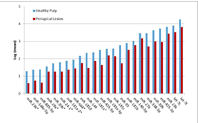
Of the 381 human miRNAs identified in periapical tissues, 24 miRNAs were significantly
down-regulated (P < .003, q < .08) in periapical tissues compared to healthy pulp control
tissues. Of these 24 down-regulated miRNAs, 15 showed a 2-5 fold change and six had more
than a five-fold change (Fig. 1). Of the 24 down-regulated miRNAs identified, nine miRNAs
that are linked to inflammation and immunity were selected for further analysis with
14 qRT-PCR results
The comparison between healthy periodontal ligament samples and diseased periapical tissue
samples showed a significant down-regulation of seven out of nine miRNAs tested (P < .05)
(Fig. 2). The fold change for this comparison varied from 1.2-fold to 21.2-fold. miR-95 was
down-regulated 1.2 fold while the remaining six miRNAs were down-regulated more than
4-fold. These same seven miRNAs were also significantly down-regulated when comparing
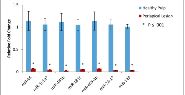
healthy pulp tissues to periapical lesion tissues (P ≤ .001) (Fig. 3). The fold regulation for
this comparison varied from 16-fold to 45-fold. These results were similar to those obtained
with microarray as both showed significant down-regulation of the seven miRNAs, however
the magnitude of the fold change was greater with qRT-PCR (16 to 45 fold change)
compared to microarray (2.4 to 10.8 fold change). The remaining two miRNAs, miR-455-5p
and miR-181d, were not detected in the samples using qRT-PCR.
Bioinformatics data
The potential targets of the differentially expressed miRNAs identified include key cytokines
involved in inflammation (IL-6, IL-10), chemokines (CCL8), pathogen recognition receptors
(TLR-4), growth factors (TGF-β1, VEGF-α) and proteins involved in macrophage
differentiation and the immune response to LPS (MMP-9) (Table 1).
Discussion:
In this study, multiple miRNAs from the miR-181 family (miR-181a*, miR-181b and
15
tissues compared to healthy control tissues. Down-regulation of miRNAs, which are
negative regulators themselves, results in an increase in their respective target messenger
RNAs (mRNA). The targets of miR-181a* include toll-like receptor-4 (TLR-4), which plays
a key role in pathogen recognition and activation of the innate immune response, and IL-6,
which stimulates neutrophil production and supports B-cell maturation. miR-181b also
targets IL-6 as well as CCL-8, MMP-9 and TGF-B1, which are involved in a wide range of
inflammatory pathways. For example, CCL-8 is chemotactic for and activates several
immune cells including monocytes, T cells and NK cells. MMP-9 is closely associated with
macrophage differentiation and TGF-β1 increases collagen biosynthesis and fibroblast
proliferation. The targets of miR-181c include SOCS1, which is involved in the LPS
response, and IL-2, which plays an essential role in the immune response to antigenic stimuli
and is important for the proliferation of T and B lymphocytes. The miR-181 family is also
notable for altering T-cell receptor signaling and increasing IFN-γ/IL-17 production by
Th1/17 cells.
Increasing evidence supports the role of the miR-181 family in inflammatory
pathologies. Circulating levels of miR-181a, miR-181a-2* and miR181c in whole blood were
significantly lower in patients diagnosed with complex regional pain syndrome, a disorder in
which neurogenic inflammation plays a key role (44). Blood plasma levels of miR-181b was found to be lower in patients with sepsis (a whole body inflammatory condition due to
infection) and in animal models of sepsis (45). Data from our previous study show that members of the miR-181 family are down-regulated in inflamed human pulps as compared to
16
pathologies could possibly be due to differences in the type of infection or the host tissue or
the sampling point after initiation of inflammation.
In addition to the miR-181 family, miR-24-1*, miR-95, miR-149 and miR-455-3p
were significantly down-regulated in diseased periapical tissues. These miRNAs also have a
variety of targets that are implicated in the immune and inflammatory response. For
example, miR-149 targets VEGF-α, which acts on endothelial cells to mediate increased
vascular permeability and promote cell migration to the site of inflammation. miR-455-3p
targets TLR-4 as well as IL-10, a cytokine produced primarily from monocytes that serve to
enhance B cell survival, proliferation and antibody production.
A limited number of studies have examined miRNA expression in inflammation
related to endodontic infection. Our previous study examined the differential expression of
miRNAs in inflamed and healthy pulps using microarray techniques (42). When comparing our microarray data to that of our previous study, we see that there is significant differential
expression of 13 of the same miRNAs in both periapical and pulp tissues. These include:
miR-29a*, miR-30b*, miR-181a-2*, miR-181d, miR-455-5p, miR199-5p and miR-664. All
seven of the significantly down-regulated miRNAs identified in diseased periapical tissues
were also shown to be down-regulated in inflamed pulp tissue compared to healthy pulp
tissue. The six of which showed significant down-regulation in both tissues include:
miR-24-1*, miR-95, miR-181a*, miR-181b, miR-181c and miR-455-3p. The extent of this cross-over
suggests some common miRNA regulatory network in pulpal and periapical disease
pathogenesis. This could be due to similar inflammatory processes occurring in both tissues.
However, the tissue specificity of miRNAs is apparent in the difference between the types of
17
exists, it is possible that the other miRNAs identified in this study could be unique to the
periapical disease process.
The limitations of this study include the small sample size and age matched controls
were not used. Both PDL and pulp were used as control tissues. In the absence of periapical
disease the tissues present in the periapex consist of PDL, pulp and bone tissue. Bone tissue
was not included as a control since its removal is not recommended in apicoectomy
procedures as this could delay healing. Bone tissue is also relatively acellular and its cellular
components are difficult to isolate from the hard tissue. After the microarray experiments, an
additional cohort of tissue samples were collected for qRT-PCR analysis. Despite using new
tissue samples for the qRT-PCR, the results confirmed the down-regulation of the same
miRNAs identified using microarray. Also, the same miRNAs were consistently
down-regulated in diseased periapical tissues when comparing to both pulp and PDL control
tissues. This correlation strengthens the findings and supports the inclusion of the selected
control tissues. The magnitude of down-regulation of the seven miRNAs was greater when
comparing diseased periapical tissues to pulp controls than when comparing to PDL controls.
This may be due to higher baseline expression levels of the miRNAs in the pulp since it is
more cellular in nature than PDL.
A significant difference in mean age between groups from which tissue samples were
collected was observed. Participants in the PDL and pulp control groups consisted primarily
of younger individuals undergoing extraction of third molars or premolars for orthodontic
purposes. Participants in the periapical group were older as these teeth had previous
18
Future studies that include a larger number of subjects are necessary to identify any
miRNAs that might correlate with pain symptoms. Studies that implement in situ
hybridization techniques could be used to determine the cellular sources of the identified
miRNAs. Confirmation of the altered target gene expression could then be investigated with
miRNA overexpression or knockdown studies.
This study explores the role of miRNAs in endodontic disease and provides new
insight into the genetic regulation of endodontic periapical pathogenesis. Seven specific
miRNAs that target key mediators in the inflammatory and immune response were
determined to be significantly down-regulated in diseased periapical tissues. This study
offers potential candidates for further investigation of miRNAs in endodontic disease. These
findings could facilitate the development of potential biomarkers and possible therapeutic
targets for the treatment of endodontic disease.
Acknowledgements:
19 Figures & Tables:
20
Figure 2. Relative fold changes of seven miRNAs in diseased periapical and healthy periodontal ligament tissues analyzed by qRT-PCR. Data was normalized to expression of endogenous control, SNORD44. Relative expression computed using the 2(-ΔΔCt) method. * P ≤ .001, ** P ≤ 0.05
Figure 3. Relative fold changes of seven miRNAs in diseased periapical and healthy pulp tissues analyzed by qRT-PCR. Data was normalized to expression of endogenous control, SNORD44. Relative expression computed using the 2(-ΔΔCt) method. * P ≤ .001
** * * * * * * 0 0.5 1 1.5 R e lativ e Fo ld C h an ge
Healthy Periodontal Ligament
Periapical Lesion
* P ≤ .001 ** P ≤ 0.05
* * * * * * * 0 0.5 1 1.5 R e lativ e Fo ld C h an ge Healthy Pulp Periapical Lesion
21
Table 1. Potential miRNA target genes were identified using PUBMED and miRWalk (http://www.rna.uni-heidelberg.de/apps/zmf/mirwalk/index_html).
miRNA Target gene Gene product function GO Term (Accession, Ontology)
miR-181a TLR-4 Toll-like receptor-4 (TLR) plays a fundamental role in pathogen recognition and activation of innate immunity GO:0035662, Molecular Function
IL-6 Acute and chronic inflammation and the maturation of B cells GO:0019981, Molecular Function
miR-181b IL-6
Negative regulation of cytokine secretion; negative regulation of collagen biosynthetic process; positive regulation of acute inflammatory response; response to cold, heat and
mechanical stimuli
GO:0005138, Molecular Function
CCL8 Immune response, inflammatory response, chemokine
activity, phospholipase activator
MMP9
Cell response to IL-1, LPS; macrophage differentiation; response to heat and mechanical stimuli; positive regulation of apoptosis and angiogenesis
GO:0004229, Molecular Function
TGFB1
Adaptive immune response; positive regulation of collagen biosynthesis, chemotaxis, fibroblasts migration, and ondontogenesis
GO:0034713, Molecular Function
miR-181c SOCS1 Cytokine mediated signaling pathway; negative regulator of
JAK-STAT pathway; LPS response
IL-2 Cytokine produced by T-cells in response to antigen or
mitogen stimulation GO:0005134, Molecular Function
miR-24
MAPK14
MAP kinase activated by various environmental stresses and proinflammatory cytokines, act as an integration point for multiple
biochemical signals GO:0051403, Biological Process
miR-95 EIF2C2
Eukaryotic translation initiation factor interacted with dicer1 in short-interfering-RNA-mediated gene silencing
miR-149 VEGFA
Increased vascular permeability, angiogenesis, vasculogenesis and endothelial cell growth, promoting cell migration, and inhibiting
apoptosis GO:0035924, Biological Process
miR-455
IL-10
Pleiotropic effects in immunoregulation and inflammation, B cell survival, proliferation, and antibody production, block
NF-kappa B activity GO:0019969, Molecular Function
22
CHAPTER 3: CONCLUSION
In this study, 24 miRNAs were found to be significantly down-regulated in periapical
tissues compared to healthy control tissues using microarray techniques. Of these 24
miRNAs, 9 which were linked to immunity and inflammation were then subjected to further
validation using qRT-PCR. Additional tissue samples were collected for qRT-PCR and
results showed significant down-regulation of 7 of the 9 miRNAs tested when comparing
diseased periapical tissue to both healthy pulp and periodontal ligament tissues.
Multiple miRNAs from the miR-181 family (miR-181a*, miR-181b and miR-181c)
were among those identified to be significantly down-regulated in diseased periapical tissues.
Down-regulation of miRNAs, which are negative regulators themselves, results in an
increase in their respective target messenger RNAs (mRNA). The targets of miR-181a*
include toll-like receptor-4 (TLR-4), which plays a key role in pathogen recognition and
activation of the innate immune response, and IL-6, which stimulates neutrophil production
and supports B-cell maturation. miR-181b also targets IL-6 as well as CCL-8, MMP-9 and
TGF-B1, which are involved in a wide range of inflammatory pathways. For example,
CCL-8 is chemotactic for and activates several immune cells including monocytes, T cells and NK
cells. MMP-9 is closely associated with macrophage differentiation and TGF-β1 increases
collagen biosynthesis and fibroblast proliferation. Studies using animal models have also
implicated miR-181b in the regulation of NF-kB-mediated endothelial cell activation and
23
implemented in anti-inflammatory therapy (45). The targets of miR-181c include SOCS1, which is involved in the LPS response, and IL-2, which plays an essential role in the immune
response to antigenic stimuli and is important for the proliferation of T and B lymphocytes.
Many miRNAs from the miR-181 family have been implicated in various
inflammatory pathologies. For example, circulating levels of miR-181a, miR-181a-2* and
miR181c are significantly lower in patients diagnosed with complex regional pain syndrome,
a disorder in which neurogenic inflammation plays a key role (44). In addition, circulating levels of miR-181b are lower in patients with sepsis (a whole body inflammatory condition
due to infection) and in animal models of sepsis (45). Data from our previous study show that members of the miR-181 family are down-regulated in inflamed human pulps as compared to
normal pulps (p≤0.001) (42). Conversely, miR-181c is upregulated in inflamed gingival tissues (40). These differences in expression of miR-181c in distinct inflammatory pathologies could possibly be due to differences in the type of infection or the host tissue or
the sampling point after initiation of inflammation.
In addition to the miR-181 family, miR-24-1*, miR-95, miR-149 and miR-455-3p
were significantly down-regulated in diseased periapical tissues. These miRNAs also have a
variety of targets that are implicated in the immune and inflammatory response. For
example, miR-149 targets VEGF-α, which acts on endothelial cells to mediate increased
vascular permeability and promote cell migration to the site of inflammation. miR-455-3p
targets TLR-4 as well as IL-10, a cytokine produced primarily from monocytes that serve to
enhance B cell survival, proliferation and antibody production.
A limited number of studies have examined miRNA expression in inflammation
24
miRNAs in inflamed and healthy pulps using microarray techniques (42). When comparing our microarray data to that of our previous study, we see that there is significant differential
expression of 13 of the same miRNAs in both periapical and pulp tissues. These include:
miR-29a*, miR-30b*, miR-181a-2*, miR-181d, miR-455-5p, miR199-5p and miR-664. Of
the seven significantly down-regulated miRNAs identified in diseased periapical tissues, six
miRNAs have also been shown to be down-regulated in inflamed pulp tissue compared to
healthy pulp tissue. These include miR-24-1*, miR-95, miR-181a*, miR-181b, miR-181c
and miR-455-3p. The extent of this cross-over suggests a common miRNA regulatory
network in pulpal and periapical disease pathogenesis.
Future studies that include a larger number of subjects are necessary to identify any
miRNAs that might correlate with pain symptoms. In this study, only two subjects
presented with pain symptoms prior to periapical tissue collection. A larger scale study
could include more symptomatic patients which would allow for statistical analysis to
determine if any specific miRNAs show differential expression between patients
experiencing pain and asymptomatic patients. Future studies could also investigate the
cellular sources of the identified differentially expressed miRNAs. Formalin fixed
periapical lesion tissues collected from apicoectomy procedures could be used with in situ
hybridization techniques to determine which cell types each of the identified miRNAs is
expressed in. In the present study, samples were determined to be periapical granulomas
rather than cysts based on only clinical impression. However, in situ hybridization would
allow for confirmation of the type of lesion while also providing a qualitative determination
25
expressed miRNAs could also shed light on whether the observed changes in their expression
levels are solely due to a change in the cell profile of the lesion or not.
In addition, the down-stream effects of altered miRNA expression could be explored.
Gene-specific experimental validation of miRNA targets can be done to identify individual
miRNA:mRNA interactions. Downstream effects of differential miRNA expression can be
observed at the protein level by western blot and at the mRNA level by qRT-PCR.
Confirmation of the altered target gene expression could then be investigated with miRNA
overexpression or knockdown studies. Over-expression can be achieved using transient
transfection of a synthetic miRNA precursor or by stable introduction of a miRNA
expression construct such as a lentiviral vector (46). miRNAs can be inhibited by expressing modified antisense oligonucleotides that bind to mature miNRAs thus blocking their activity.
Animal studies could also be used to sample lesions throughout the healing process in order
to determine whether there are changes in the miRNA profiles at different stages of healing.
This study is the first to explore the role of miRNAs in periapical disease, providing
new insight into the genetic regulation of endodontic disease. Seven specific miRNAs that
target key mediators in the inflammatory and immune response were determined to be
significantly down-regulated in diseased periapical tissues. This study begins to elucidate the
complex miRNA regulatory network in endodontic disease and offers potential candidates
for further investigation. The identified miRNAs have potential to serve as possible
biomarkers or therapeutic targets for the treatment of endodontic disease. These findings
could facilitate the development of improved methods of diagnosis and management of
26 REFERENCES
1. Ricucci D, Siqueira Jr. JF, Bate AL, Pitt Ford TR. Histologic investigation of root canal– treated teeth with apical periodontitis: A retrospective study from twenty-four patients. J Endod. 2009 4;35(4):493-502.
2. Sundqvist G. Taxonomy, ecology and pathogenicity of the root canal flora. Oral Surg Oral Med oral Pathol. 1994;78:522-30.
3. Craig Baumgartner J, Falkler WA. Bacteria in the apical 5 mm of infected root canals. J Endod. 1991 8;17(8):380-3.
4. Siqueira J, Rocas I, Lopes H. Patterns of microbial colonizationin primary root canal infections. Oral Surg Oral Med Oral Pathol Oral Radiol Endod. 2002;93:174-8.
5. Winstock D. Apical disease: An analysis of diagnosis and management with special reference to root lesion resection and pathology. Ann Royal Col Surg Engl. 1980;62:171.
6. Stashenko P. Role of immune cytokines in the pathogenesis of periapical lesions. Endodontics & Dental Traumatology. 1990;6:89-96.
7. Hou L, Sasaki H, Stashenko P. B-cell deficiency predisposes mice to disseminating anaerobic infections: Protectio by passive antibody transfer. Infec Immun. 2000;68:5645-5651.
8. Naidorf IJ. Immunoglobulins in periapical granulomas:A preliminary report. Journal of Endodontics. 1975.;1(15):1975.
9. Okiji T, Kawashima N, Kosaka T, Kobayashi C, Suda H. Distribution of la antigen-expressing nonlymphoid cells in various stages of induced periapical lesions in rat molars. J Endod. 1994 1;20(1):27-31.
10. Cohen S, Hargreaves KM, editors. Pathways of the pulp. 9th ed. Canada: Elsevier; 2006.
11. Kawashima N, Okiji T, Kosaka T, Suda H. Kinetics of macrophages and lymphoid cells during the development of experimentally induced periapical lesions in rat molars: A quantitative immunohistochemical study. J Endod. 1996 6;22(6):311-6.
12. Wang C, Stashenko P. Characterization of bone-resorbing activity in human periapical lesions. J Endod. 1993 3;19(3):107-11.
27
14. Nobuhara W, Del Rio C. Incidence of periradicular pathoses in endodontic treatment failures. Journal of Endodontics. 1993(19):315.
15. Ataoglu T, Ungor M, Serpek B, Haliloglu S, Ataoglu H, Ari H. Interleukin 1-B and tumour necrosis factor-a levels in periapical exudates. Int Endodon J. 2002;35:181.
16. Barkhordar RA, Hussain MZ, Hayashi C. Detection of interleukin-1 beta in human periapical lesions. Oral Surg Oral Med Oral Pathol. 1992;73:334.
17. Seckinger P, Klein-Nulend J, Alander C, Thompson R, dayer J, Raisz L. Natural and recombinant human il-1 receptor antagonists block the effects of IL-1 on bone resorption and prostaglandin production. J Immunol. 1990;145:4181-4184.
18. Jiang Y, Russell TR, Schilder H, Graves DT. Endodontic pathogens stimulate monocyte chemoattractant protein-1 and interleukin-8 in mononuclear cells. J Endod. 1998 2;24(2):86-90.
19. Love RM, McMillan MD, Jenkinson HF. Invasion of dentinal tubules by oral
streptococci is associated with collagen regeneration mediated by the antigen I/II family of polypeptides. Infec Immun. 1997;65(5157).
20. Cotti E, Toraginejad M. Detection of leukotriene C4 in human periradicular lesions. Int Endodon J. 1994;27(82).
21. Nilsen R, Johannessen A, Skaug N, Matre R. In situ characterization of mononuclear cells in human dental periapical inflammatory lesions using monoclonal antibodies. Oral Surg Oral Med oral Pathol. 1984;58(2):160-5.
22. Kulkarni A, Huh C, Becker D. Transformin growth factor B null mutation in mice causes excessive inflammatory response and early death. Proc Natl Acad Sci USA. 1993;90:770-774.
23. Diebold R, Eis M, Yin M. Early onset multifocal inflammation in the transforming growth factor B-null mouse is lymphocyte mediated. Proc Natl Acad Sci USA.
1995;92:12215-12219.
24. Orstavik D, Ford TP. Apical periodontitis: Microbial infection and host responses. In: Orstavik D, Ford TP, editors. Essential Endodontology. 2nd ed. Ames, IA: Wiley-Blackwell; 2007. p. 18.
25. Sonkoly E, Stahle M, Pivarcsi A. MicroRNAs and immunity: Novel players in the regulation of normal immune function and inflammation. Semin Cancer Biol. 2008;18:131-40.
28
27. Rubkun G. Molecular biology: Glimpses of a tiny RNA world. Science. 2001;294:797-9.
28. Ambros V. The functions of animal microRNAs. Nature. 2004;431:350-5.
29. Ryan B, Robles A, Harris C. Genetic variation in microRNA networks: The implications for cancer research. Nat Rev Cancer. 2010;10(6):389-402.
30. Bartel DP. MicroRNAs: Genomics, biogenesis, mechanism, and function. Cell. 2004 1/23;116(2):281-97.
31. Thum T, Catalucci D, Bauersachs J. MicroRNAs: Novel regulators in cardiac development and disease. Cardiovascular Research. 2008;79(4):562-70.
32. Zovoilis A, Agbemenyah HY, Agis-Balboa R, Stilling RM, Edbauer D, Rao P, et al. microRNA-34c is a novel target to treat dementias. EMBO J. 2011;30(20):4299-4308.
33. Jones SW, Watkins G, Le Good N, Roberts S, Murphy CL, Brockbank SMV, et al. The identification of differentially expressed microRNA in osteoarthritic tissue that modulate the production of TNF-α and MMP13. Osteoarthritis and Cartilage. 2009 4;17(4):464-72.
34. Etheridge A, Lee I, Hood L, Galas D, Wang K. Extracellular microRNA: A new source of biomarkers. Mutation Research. 2011;717:85-90.
35. Mitchell PS, Parkin RK, Kroh EM, Fritz BR, Wyman SK, Pogosova-Agadjanyan EL, et al. Circulating microRNAs as stable blood-based markers for cancer detection. Proc. Natl. Acad. Sci. USA. 2008;105:10513–10518.
36. Weber JA, Baxter DH, Zhang S, Huang DY, Huang KH, Lee MJ, et al. The microRNA spectrum in 12 body fluids. Clin. Chem. 2010;56:1733–1741.
37. Valadi H, Ekstrom K, Bossios A, Sjostrand M, Lee JJ, Lotvall JO. Exosome-mediated transfer of mRNAs and microRNAs is a novel mechanism of genetic exchange between cells. Nat Cell Biol. 2007;9:654-659.
38. Ladomery MR, Maddocks DG, Wilson ID. MicroRNAs: Their discovery, biogenesis, function and potential use as biomarkers in non-invasive prenatal diagnostics. Int. J. Mol. Epidemiol. Genet. 2011;2:253-260.
39. Stoecklin-Wasmer C, Guarnieri P, Celenti R, Demmer R, Kebschull M, Papapanou P. MicroRNAs and their target genes in gingival tissues. Journal of Dental Research.
2012;91(10):934-40.
29
41. Perri R, Nares S, Zhang S, Barros SP, Offenbacher S. MicroRNA modulation in obesity and periodontitis. Journal of Dental Research. 2012;91(1):33-8.
42. Zhong S, Zhang S, Bair E, Nares S, Khan A. Differential expression of MicroRNAs in normal and inflamed human pulps. Journal of Endodontics. 2012;38(6):746-52.
43. Livak K, Schmittgen T. Analysis of relative gene expression data using real-time quantitative PCR and the 2(-delta delta C(T)) method. Methods. 2001;25:402-408.
44. Orlova IA, Alexander GM, Qureshi RA, Sacan A, Graziano A, Barrett JE, et al. MicroRNA modulation in complex regional pain syndrome. J Transl Med. 2011 Nov 10;9:195,5876-9-195.
45. Sun X, Icli B, Wara AK, Belkin N, He S, Kobzik L, et al. MicroRNA-181b regulates NF-kappaB-mediated vascular inflammation. J Clin Invest. 2012 Jun 1;122(6):1973-90.