PLEKHG3 enhances polarized cell migration by
activating actin filaments at the cell front
Trang Thi Thu Nguyena,1, Wei Sun Parkb,1, Byung Ouk Parkc, Cha Yeon Kimd, Yohan Ohe, Jin Man Kimf, Hana Choia, Taeyoon Kyunga,c, Cheol-Hee Kimb, Gabsang Leee, Klaus M. Hahng, Tobias Meyerh, and Won Do Heoa,c,i,2
aDepartment of Biological Sciences, Korea Advanced Institute of Science and Technology, Daejeon 34141, Republic of Korea;bDepartment of Biology, Chungnam National University, Daejeon 34134, Republic of Korea;cCenter for Cognition and Sociality, Institute for Basic Science, Daejeon 34141, Republic of Korea;dGraduate School of Nanoscience and Technology, Korea Advanced Institute of Science and Technology, Daejeon 34141, Republic of Korea; eInstitute for Cell Engineering, Department of Neurology and Neuroscience, Johns Hopkins University School of Medicine, Baltimore, MD 21205;fGraduate School of Medical Science and Engineering, Korea Advanced Institute of Science and Technology, Daejeon 34141, Republic of Korea;gDepartment of Pharmacology, University of North Carolina at Chapel Hill, Chapel Hill, NC 27599;hDepartment of Chemical and System Biology, Stanford University School of Medicine, Stanford, CA 94305; andiKorea Advanced Institute of Science and Technology Institute for the BioCentury, Korea Advanced Institute of Science and Technology, Daejeon 34141, Republic of Korea
Edited by Thomas D. Pollard, Yale University, New Haven, CT, and approved July 15, 2016 (received for review March 24, 2016)
Cells migrate by directing Ras-related C3 botulinum toxin sub-strate 1 (Rac1) and cell division control protein 42 (Cdc42) activities and by polymerizing actin toward the leading edge of the cell. Previous studies have proposed that this polarization process re-quires a local positive feedback in the leading edge involving Rac small GTPase and actin polymerization with PI3K likely playing a coordinating role. Here, we show that the pleckstrin homology and RhoGEF domain containing G3 (PLEKHG3) is a PI3K-regulated Rho guanine nucleotide exchange factor (RhoGEF) for Rac1 and Cdc42 that selectively binds to newly polymerized actin at the leading edge of migrating fibroblasts. Optogenetic inactivation of PLEKHG3 showed that PLEKHG3 is indispensable both for inducing and for maintaining cell polarity. By selectively binding to newly polymer-ized actin, PLEKHG3 promotes local Rac1/Cdc42 activation to induce more local actin polymerization, which in turn promotes the recruit-ment of more PLEKHG3 to induce and maintain cell front. Thus, autocatalytic reinforcement of PLEKHG3 localization to the leading edge of the cell provides a molecular basis for the proposed posi-tive feedback loop that is required for cell polarization and directed migration.
PLEKHG3
|
cell polarity|
F-actin binding|
positive feedback|
PI3KC
ell polarity is essential for many cellular processes: It allows neurons to form dendrites and axons, enables dividing cells to produce daughter cells, engenders fibroblasts with wound-healing activity, and gives leukocytes the ability to crawl to infection sites (1, 2). Cell polarity is modulated by signaling cascades that center around the action of phosphoinositide kinases, the activation of Rho small GTPases by phosphatidylinositol-3,4,5-Tris phosphate (PIP3, a lipid product of PI3Ks), and the activity of F-actin (3, 4).Several studies have shown that F-actin generates positive feed-back for the activation of PI3K, Ras-related C3 botulinum toxin (Rac), and/or cell division control protein 42 (Cdc42) (4–6). The feedback activation of PI3K is, in turn, regulated by multiple Rho family small GTPases via coupled positive feedback loops (7) in which the existing filaments generate new free barbed ends, and new F-actin filaments grow on the free ends (8). In fibroblasts, T cells, and macrophages, cell polarity and chemotaxis are ab-rogated when Rac or Cdc42 is inhibited (9, 10). Rac and Cdc42 regulate cell migration through the precise spatial and temporal coordination of dynamic actin-based structures found at the leading edge of migrating cells (11, 12).
GTPase activity, which controls various cellular functions, is regulated by guanine-nucleotide exchange factors (GEFs) and GTPase activation proteins (GAPs). GEFs activate signaling by catalyzing the exchange of G protein-bound GDP to GTP, whereas GAPs terminate signaling by inducing the hydrolysis of GTP (13). Sixty-nine identified GEFs belong to the Dbl family and are re-sponsible for accelerating the intrinsic nucleotide exchange activity
of Rho-family small GTPases (14, 15). In addition to possessing a Dbl homology (DH)–pleckstrin homology (PH) module, most GEFs contain additional functional domains, such as the Src ho-mology 2 (SH2), Src hoho-mology 3 (SH3), Ras-GEF, and Ser/Thr or Tyr kinase domains. These regions are critical for GEFs’ability to interact with other regulatory proteins and to activate them in re-sponse to cellular signals (16).
Here, we report that the pleckstrin homology and RhoGEF domain containing G3 (PLEKHG3) is a PI3K-regulated RhoGEF for Rac1 and Cdc42 that selectively binds to newly polymerized actin at the leading edge of migrating fibroblasts. We discovered the existence of a positive feedback loop from actin filaments to PLEKHG3 from the observation that PLEKHG3 accumulates at the site of newly polymerized actin at the leading edge when the cell moves forward. PLEKHG3 was recruited to the newly formed protrusion area when a photoactivatable Rac1 (PA-Rac1) was continuously activated. Thus, PLEKHG3 is indispensable for both inducing and maintaining cell polarity and directional motility. These findings reveal that PLEKHG3 generates a positive feed-back loop that controls cell polarity and directional motility, explaining how Rac1 and actin polymerization are coupled by a positive feedback loop to ensure the stability of polarity.
Significance
Polarized cell migration plays a pivotal role in the development and repair of tissues. PI3K, Rho GTPases, and actin filaments are known to be involved in a positive feedback loop that induces and maintains cell polarity. Here, we show that the pleckstrin homology and RhoGEF domain containing G3 (PLEKHG3) se-lectively binds to newly polymerized actin and that this in-teraction exerts a positive regulatory effect on PLEKHG3 activity that enhances and sustains the cell front. A lack of PLEKHG3 ablates cell polarity, resulting in a decrease in cell migration. These findings provide the missing link that explains how Ras-related C3 botulinum toxin substrate 1 (Rac1) and actin polymerization are coupled by a positive feedback loop to ensure the stability of cell polarity.
Author contributions: T.T.T.N., T.M., and W.D.H. designed research; T.T.T.N., W.S.P., B.O.P., C.Y.K., Y.O., J.M.K., H.C., and T.K. performed research; T.T.T.N., W.S.P., C.-H.K., G.L., and K.M.H. contributed new reagents/analytic tools; T.T.T.N., B.O.P., C.Y.K., Y.O., J.M.K., T.K., and W.D.H. analyzed data; and T.T.T.N., T.M., and W.D.H. wrote the paper. The authors declare no conflict of interest.
This article is a PNAS Direct Submission.
Freely available online through the PNAS open access option.
1T.T.T.N. and W.S.P. contributed equally to this work.
2To whom correspondence should be addressed. Email: [email protected].
This article contains supporting information online atwww.pnas.org/lookup/suppl/doi:10.
1073/pnas.1604720113/-/DCSupplemental.
CELL
Results
PLEKHG3 Localizes to the Leading Edge and Controls Cell Migration.
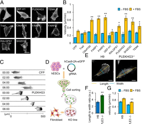
To identify new GEFs that control cytoskeletal dynamics during cell migration, we generated a library consisting of 63 human GEFs and analyzed their subcellular localizations in NIH 3T3 cells. The GEFs were classified into six groups based on their distinct subcellular localizations: one GEF was localized in the nucleus, one GEF was localized in microtubules, two GEFs were localized in actin filaments, six GEFs were localized in the plasma membrane (PM), six GEFs were distributed throughout the whole cell, and 47 GEFs were localized in the cytoplasm (Fig. S1andTable S1). We selected nine cytoskeleton-related GEFs that localized at the PM, actin filaments, or microtubules and examined their involvement in controlling cell migration (Fig. 1A). Cells expressing seven of the nine selected GEFs migrated more rapidly in the presence of FBS than did control cells (Fig. 1B). Most notably, PLEKHG3 demonstrated a polarized sub-cellular localization with enrichment at the leading edge (Fig. 1C
and Movie S1). To test whether the unique localization of
PLEKHG3 at the leading edge was a general feature of cell lines other than NIH 3T3, PLEKHG3 was expressed in human um-bilical vein endothelial cells (HUVECs) and MDA-MB-231 cells. Indeed, we observed the polarized subcellular localization of PLEKHG3 and the increased migration among HUVECs and MDA-MB-231 cells overexpressing this protein (Fig. S2 A–C). We confirmed the colocalization of PLEKHG3 and F-actin filaments by cytochalasin D treatment. A disruption pattern
visualized in F-actin was observed in PLEKHG3 (Fig. S2D) but not in Ras-specific guanine nucleotide-releasing factor 1 (RALGPS1), a PM GEF (17).
To confirm the apparent involvement of PLEKHG3 in con-trolling cell migration, a fibroblast cell line was differentiated from the PLEKHG3-knockout human ES cells (hESCs) based on the CRISPR/Cas9 method (Fig. 1D andFig. S2E andF). The knockout cell line showed an ablation of the PLEKHG3 expres-sion at the leading edge (Fig. 1E), a deviated morphology (Fig. 1F), and cell motility substantially decreased from that of control cells (Fig. 1G). The change in cell shape and cell motility was also recorded in cells of several cell lines treated with PLEKHG3 siRNA (Fig. S3).
PLEKHG3 Binds Directly to F-Actin Through an Actin-Binding Domain.
To elucidate the region of PLEKHG3 that is responsible for the colocalization with F-actin, we generated several truncated forms of PLEKHG3 and assessed their subcellular localizations in NIH 3T3 cells. Human PLEKHG3 [also known as ARHGEF43; National Center for Biotechnology Information (NCBI) no. BC129953] en-codes a 1,219-amino acid protein with a predicted mass of 134 kDa. It contains a tandem DH–PH domain catalytic cassette in the N-terminal sequence and does not harbor any other known domain or motif (Fig. S4A). Our results revealed that the region encompassing residues 910–940 of PLEKHG3 exhibited a sub-cellular localization similar to that of F-actin and that smaller frag-ments of this region were unable to colocalize to actin filafrag-ments
A
B
CFP GEF-H1 PLEKHG3
Trad C9orf100 FARP1 FARP2
RALGPS1 SH2D3C
TEM4 ̶ FBS + FBS
** **
** ** **
* *
hCas9-2A-eGFP
gRNA hESCs
Cell sorting
KO line
**
̶ FBS + FBS
0 0.4 0.8 1.2
H9
PLE3 -/
-v
e
lo
ci
ty
(μm/ml
n)
F
E
0 2 4 6
H9
PLE3 -/
-C
G
PLEKHG3
-/-H9
Length Width
**
00:00
02:00
PLEKHG3 04:00
06:00
00:00
02:00
04:00
06:00
CFP
(μm)
Fibroblast
D
0 500
Le
ngth to
w
idth
ratio
(a.u.)
0 0.2 0.4 0.6 0.8 1
Velocit
y
(
μ
m
/m
in)
(Fig. S4BandC). Furthermore, we found that both the DH–PH domain and actin-binding domain (ABD) of PLEKHG3 are re-quired for the polarized subcellular localization of PLEKHG3 and increased cell migration (Fig. S5 A–C). The ability of PLEKHG3 (amino acids 1–950) to induce cell polarity was also observed in the PLEKHG3−/−cells (Fig. S5DandE).
To determine whether the colocalization of PLEKHG3 and F-actin reflected a direct interaction, we used a high-speed actin cosedimentation assay to evaluate the binding ability of purified F-actin with purified recombinant GST-PLEKHG3(amino acids 890–950). Indeed, recombinant GST-PLEKHG3 (amino acids 890–950) was found predominantly in the F-actin–containing pellet (P) (Fig. S4D), demonstrating that PLEKHG3 binds directly to F-actin. Amino acids 910–940 of PLEKHG3 do not exhibit sequence similarity to any known ABD, such as Rho guanine nucleotide exchange factor 11 (PDZ-RhoGEF) (18), tumor en-dothelial marker 4 (TEM4) (19), FLJ00018 (20), or FYVE, RhoGEF, and PH domain-containing protein 4 (Frabin) (Fig. S4E) (21).
PLEKHG3 Enhances Cell Polarity by Activating Rac1 and Cdc42.Based on our observation of PLEKHG3 localization during cell migration, we found that PLEKHG3 localizes at the leading edge of the cell where most of the actin filaments are found. We hypothesized that the cell directionality depends on the accumulation of PLEKHG3 to newly synthesized F-actin at the leading edge. We therefore attempted to change the localization of PLEKHG3 from a sub-cellular region with high PLEKHG3 localization to a new region with lower PLEKHG3 localization and to observe any changes in cell directionality using the light-mediated dimerizer system cryp-tochrome 2 (CRY2)–CRY-interacting bHLH 1 (CIB1) (Fig. S6A). We used Lifeact (which is known to bind both G-actin and F-actin biochemically) for targeting PLEKHG3 to F-actin because Lifeact is used as a marker to visualize F-actin in living cells and is known
not to interfere with the actin dynamics in cells (22, 23). The illuminated regions showed the induction of lamellipodia, and the direction of the cells eventually changed because of the ac-cumulation of PLEKHG3 (Fig. S6BandDandMovie S2). To confirm that exogenous PLEKHG3 controls cell polarity and directionality during migration, we used an optogenetic method called “light-activated reversible inhibition by assembled trap” (LARIAT) to inhibit the function of exogenous PLEKHG3 (24). Upon light stimulation, the PLEKHG3-GFP proteins rapidly formed clusters. The cells shrank and lost polarity (Fig. S6Fand G). When the blue light was removed, the clusters disassembled, the cells returned to their original size, and cell polarity was regained
(Fig. S6H). To exclude the effect of endogenous PLEKHG3,
PLEKHG3−/−cells were transfected with exogenous PLEKHG3. We observed a similar phenomenon in PLEKHG3−/−cells when exogenous PLEKHG3 was inhibited by light stimulation (Fig. S6 I–K). To test the ability of PLEKHG3 to induce new polarity following disruption, the cells were locally illuminated at the leading edge. The illuminated region retracted rapidly and then reprotruded when the light was removed, changing the direction of cell migration (Fig. S6CandEandMovie S3). Collectively, these data indicate that PLEKHG3 controls cell polarity.
We examined the localization of 63 human GEFs and found two, PLEKHG3 and TEM4, which both localized to actin fila-ments but differed in their localization during cell migration. Assessment of migrating cells coexpressing exogenous TEM4 and PLEKHG3 confirmed that TEM4 is highly expressed at the trailing edge, whereas PLEKHG3 is highly expressed at the leading edge (Fig. S7 AandB). We speculated that this differ-ence could reflect differdiffer-ences in the ABDs of PLEKHG3 and TEM4. However, the ABDs of PLEKHG3 and TEM4 both showed localization patterns similar to that of F-actin (Fig. S7C), suggesting that other domains in PLEKHG3 and TEM4 may confer the ability to bind different F-actin filaments. It is known
F-actin Newly polymerized
actin filaments PLEKHG3
PLEKHG
3
F-actin Merge
GFP
F-a
c
ti
n
P
LEKHG
3
̶ Light + Light
R1 R2
PLEKHG3
Rac1
Actin Filaments
(+) f
e
e
d
b
a
c
k
PA-Rac1
R1 R2
0.2
30 15
0 0.7 1.2
Flourescence intensity
+ Light
R1 R2
PLEKHG3 PLEKHG3 F-actin F-actin
R1 R2
0 s
PLEKHG
3
F-actin
30 s
M
e
rg
e
60 s 90 s
N l l i d
0 3 6
Normaliz
e
d
fl
uorescence prof
ile
** ̶ Light + Light
(min)
A
C
B
F
E
G
D
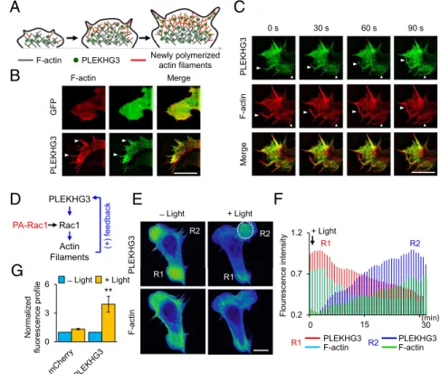
Fig. 2. PLEKHG3 enhances polarized cell migration via a positive feedback loop at the leading edge of the cell. (A) A model of how polymerized actin fila-ments increase the accumulation of PLEKHG3 at the leading edge. Gray indicates existing F-actin, red indicates newly polymerized actin filaments, and green circles indicate PLEKHG3. (SeeFigs. S6–S8and
Movies S2andS3.) (B) The localization of PLEKHG3-GFP and mCherry-Lifeact at the leading edge was observed via SIM microscopy with a 100×objective lens. Arrowheads show the colocalization of PLEKHG3 and F-actin at the tip of the leading edge of the cell. (C) PLEKHG3 accumulates near newly formed actin filaments. Arrowheads show the newly formed actin filaments. Time is shown in seconds. (Scale bar, 10μm.) (SeeMovie S4.) (D) A model for the positive feedback loop that connects actin filaments to PLEKHG3, as assessed using PA-Rac1. (E) Cells were cotransfected with PA-Rac1, mCherry-PLEKHG3, and iRFP670-Lifeact and were illuminated by blue light. PLEKHG3 is recruited to the newly formed protrusion where Rac1 is repeatedly activated by light (R2). mCherry-PLEKHG3 and infrared RFP (iRFP)-Lifeact are shown in a pseudo-colored intensity image. The white dotted circle indi-cates the position that was stimulated by light. (See
Movie S5.) (F) Fluorescence intensity profiles repre-senting the expression levels of PLEKHG3 and F-actin over time in region 1 (R1) and region 2 (R2), as assessed from the images presented inE. After light stimulation, the fluorescence intensity decreases in R1 but increases in R2. In R1, red indicates PLEKHG3, and cyan indicates F-actin. In R2, blue indicates PLEKHG3, and green
in-dicates F-actin. (G) Fluorescence intensity profiles representing the expression levels of mCherry-C1 (control) and mCherry-PLEKHG3 before and after light stim-ulation (n>50). The data represent the mean±SEM; *P<0.1; **P<0.01. (Scale bars, 20μm.)
CELL
that the Dbl family of most GEFs is responsible for accelerating the intrinsic nucleotide exchange activity of Rho-family small GTPases (14, 15). TEM4 has been reported to regulate the ability of Rho subfamily members (RhoA, B, and C) to promote the formation of actin stress fibers (19). To test which small GTPases are regulated by PLEKHG3, chemically induced het-erodimerization of FK506 binding protein (FKBP) and FKBP-rapamycin-binding-domain (FRB) (25) was used to translocate PLEKHG3 (DH–PH) from the cytosol to the PM. We observed that PLEKHG3 (DH–PH) induced the formation of lamellipodia and filopodia upon its recruitment to the PM following rapamycin treatment (Fig. S7D). This finding suggests that PLEKHG3 might regulate Rac or Cdc42, two small GTPases that are known to control dynamic actin filaments at the leading edge (26). To confirm this finding, we performed FRET imaging using Ras and interacting protein chimeric unit (Raichu)-Rac1 and Cdc42 FRET biosensors (Fig. S7E). A significant increase was observed in the FRET/CFP signal intensity ratio, indicating the activation of a small GTPases (Rac1 and Cdc42) at the PM (Fig. S7 F–H). In addition, to monitor the activation of the small GTPases Rac1 and Cdc42, we performed a pull-down assay with GST-PAK1-PDB on lysates. We detected higher levels of the active forms of Rac1 and Cdc42 in cells expressing PLEKHG3 (Fig. S7I). Together, these findings indicate that PLEKHG3 activates Rac1 and Cdc42.
PLEKHG3 Enhances Polarized Cell Migration via a Positive Feedback Loop at the Cell Leading Edge.We observed similar oscillations in the localization of PLEKHG3 and F-actin in migrating cells (Fig. S8A). Closer observation of the leading edge via super-resolution microscopy (SIM) showed that F-actin and PLEKHG3 are strongly colocalized at the filopodia of the leading edge (Fig. 2A
andBandFig. S8BandD). Moreover, PLEKHG3 accumulated
near newly formed actin filaments on a characteristic time scale
of∼30 s (Fig. 2CandMovie S4). Based on these observations, we hypothesized that there could be a positive feedback loop from polymerized actin to PLEKHG3. To test the involvement of PLEKHG3 in this positive feedback loop, we used PA-Rac1 to perform specific local activation at the leading edge (Fig. 2D). Upon light stimulation, Rac1 was activated, lamellipodia were formed, and PLEKHG3 was detected in the area of the newly formed protrusion (Fig. 2E–GandMovie S5). Haugh’s group observed the relocalization of PI3K signaling at the protru-sion upon photoactivation of PA-Rac1 (27). To eliminate the involvement of PI3K, cells were treated with the PI3K inhibitor LY294002 (LY29). Upon light stimulation, the accumulation of PLEKHG3 in the protrusion area was observed with treatment with LY29 (Fig. S8CandE). These findings are consistent with previous reports that the photoactivation of Rac at the leading edge of the cell can rescue the protrusion defects induced by PI3K inhibition (27, 28). PI3K is not required for the protrusion. Furthermore, to test the hypothesis that actin induces a positive feedback signal to PLEKHG3, cells were treated with cytocha-lasin D. PA-Rac1 failed to induce protrusion, and PLEKHG3 and F-actin were disrupted (Fig. S8 Cand E). Based on these findings, we propose that a positive feedback loop connects actin filaments and PLEKHG3.
PI3K Controls PLEKHG3 to Guide Directed Cell Migration.Because PI3K is known to regulate cell polarization, migration, and chemotaxis (29), we asked whether PI3K regulates the ability of PLEKHG3 to induce cell polarity during cell motility (Fig. 3A). To address this question, we treated cells with PDGF, an activator of PI3K. Under serum starvation, PLEKHG3 failed to induce cell polarity. Upon PDGF treatment, PLEKHG3 was translocated to the leading and trailing edges, and cell polarity was induced (Fig. S9 Aand D). Conversely, treatment with LY29 caused PLEKHG3-expressing
C
B
PLEKHG
3
Y
F-iSH2
̶ Rap + Rap
* ̶ Rap + Rap
0 0.5 1 1.5
YF YF-iSH2
N
o
rmal
iz
ed
fl
uorescence prof
ile
A
PLEKHG3
Actin Filaments
Polarized cells PI3K
Rac1
H
F
YF-iSH2
F-actin
D
+ Li
g
h
t
60 m 120 m 0 m
E
G
̶ Rap + Rap
̶
Li
g
h
t
+ Li
g
ht
Time (120 min)
R1
R2
Cell direction Before light
stimulation
After light stimulation
I
R1
R2
0 2.5 5
H9
PLE3-/-Norm
aliz
ed
fl
uorescence prof
ile
̶ Rap + Rap
*
0 1.25 2.5
- Light + Light
Normal
iz
ed
fl
uorescence prof
ile
Before After
* **
̶
Lig
h
t
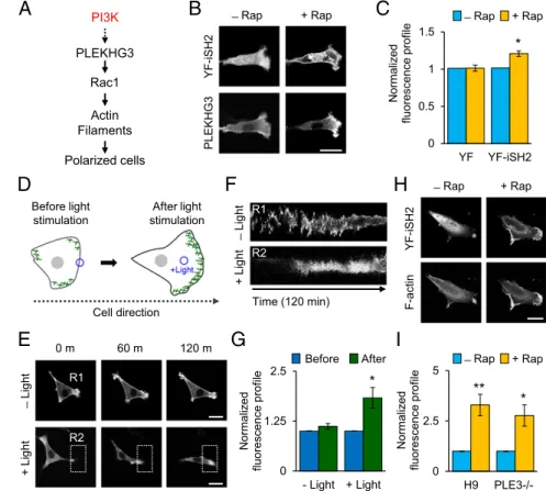
Fig. 3. PI3K controls PLEKHG3 to guide directed cell migration. (A) Schematic of how PLEKHG3 is regu-lated by PI3K. If PI3K regulates PLEKHG3, then the activation of PI3K triggers cell polarization as a result of PLEKHG3 relocalization. (B) Cells were cotransfected with Lyn-FRB, YFP-FKBP-iSH2 (YF-iSH2), and CFP-PLEKHG3 and were serum starved. CFP-PLEKHG3 activity was monitored following the activation of PI3K. Upon rapamycin treatment, PLEKHG3 localization was relo-cated and was detected mostly at the leading edge of migrating cells. (See Fig. S9.) (C) Intensity profiles representing the expression levels of CFP-PLEKHG3 at the leading edge before and after rapamycin treat-ment. YFP-FKBP-C1 (YF) is the negative control. (D) A model of PLEKHG3 accumulating at the leading edge and guiding cell migration upon light stimulation. The green line indicates PLEKHG3 expression. (E) Cells coexpressing mCherry-PLEKHG3, mCitrine-PHR-iSH2, and Lyn-CIBN-mCerrulean were serum starved and were illuminated with blue light within 120 min. The cells consistently migrated toward the illuminated area with PLEKHG3 accumulating at the leading edge, whereas the cells in the field without light exposure moved in random directions. The white dotted rect-angle indicates the position that was stimulated by light. (SeeMovie S6.) (F) The kymograph shows the PLEKHG3 intensity without light (R1) and with light (R2) stimulation over 120 min. (G) Fluorescence in-tensity profiles representing the expression levels of mCherry-PLEKHG3 in response to PI3K activation for 120 min of migration. (H) PLEKHG3−/− cells were transfected with Lyn-FRB, YFP-FKBP-iSH2, and CFP-Lifeact to test the ability of cells to trigger cell polari-zation when PIP3is produced. The cells were able to
cells to lose their polarity (Fig. S9BandE). To test whether PLEKHG3 is regulated via PI3K, we induced the activation of PI3K by recruiting the inter-SH2 (iSH2) domain from the cytosol to the PM and examined the distribution of PLEKHG3 localiza-tion. When rapamycin was added, PLEKHG3 was translocated to the leading edge of the cell (Fig. 3BandC). To test whether the recruitment of PLEKHG3 to the leading edge was caused by direct activation of PI3K or indirect activation through Rac-induced actin polymerization, cells were treated with cytocha-lasin D. In the presence of cytochacytocha-lasin D, endogenous PI3K failed to induce cell polarity; however, redistribution of PLEKHG3 localization was observed (Fig. S9 C and F). To confirm that PI3K controls PLEKHG3 to guide directed cell migration, we applied light-inducible PI3K (Fig. 3D) (30). We observed consistent migration toward the illuminated area, ac-companied by the accumulation of PLEKHG3 upon light stimu-lation of PI3K (Fig. 3E–Gand Movie S6). Collectively, these results indicate that PLEKHG3 guides directed cell migration via PI3K activation.
Several studies have reported that PI3K, Rac, and polymerized actin are the three core components of a local positive feedback loop that triggers cell polarization and migration (3, 4, 31). To test the ability of cells to induce polarity in the absence of PLEKHG3, we activated endogenous PI3K activity in PLEKHG3−/−cells. The PLEKHG3−/−cells are able to induce cell polarity when PI3K is activated (Fig. 3HandI); therefore PLEKHG3 is not the only RhoGEF that is activated by PI3K to induce cell polarity. In ad-dition to PLEKHG3, PI3K might regulate other GEFs that can also activate Rac1, such as guanine nucleotide exchange factor VAV2 (32) or T-lymphoma invasion and metastasis 1 (TIAM1) (3, 33, 34), to induce cell polarity and migration.
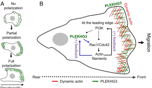
Based on the findings described above, we propose a tentative model for how PLEKHG3 pathways enhance cell polarity and cell migration. PLEKHG3 is a protein that binds directly to actin filaments. During cell migration, PLEKHG3 first redistributes to the leading and trailing edges (partial polarization); then, when the cell moves forward, it becomes localized at the leading edge (full polarization) (Fig. 4A). PLEKHG3 activity could be activated by PI3K-dependent or PI3K-independent pathways through Rac-induced actin polymerization (Fig. 4B). The activation of actin filaments generates a positive feedback loop involving PLEKHG3
and accounts for the localization of PLEKHG3 at the leading edge of the migrating cell. In conclusion, we show here that PLEKHG3 plays a role in controlling cell polarity and cell motility by selec-tively binding newly polymerized actin and activating Rac1 and Cdc42 to enhance local actin polymerization and subsequently further promote the recruitment of PLEKHG3 to induce and maintain the leading edge of the cell.
Discussion
We show here that the RhoGEF member PLEKHG3 contributes to the regulation of cell polarity and cell motility through its ability to bind F-actin filaments and to modulate the activities of Rac1 and Cdc42. When PLEKHG3 was recruited to different subcellular locations, new protrusions were induced, and the cell direction was changed upon light stimulation. PLEKHG3 binds directly to dynamic actin at the leading edge through an ABD that spans amino acids 910–940. Sequence comparison of the ABD of PLEKHG3 with those of other proteins known to bind to the actin cytoskeleton failed to identify any sequence simi-larity within known ABD proteins. The activity of PLEKHG3 depends on the presence of both the DH–PH domain and the ABD; deletion of either of these domains disrupts the localiza-tion of PLEKHG3 to the leading edge and abrogates cell po-larity. In analyzing PLEKHG3 localization during cell migration, we found that PLEKHG3 relocates mostly to the leading edge of the migrating cell to help the cell move forward. Knockout PLEKHG3 cells showed a substantial decrease in cell motility compared with control cells. These findings suggested that PLEKHG3 has an im-portant role in the regulation of cell migration. This effect was similar to that of cells lacking Coronin 1B, an F-actin–binding pro-tein that is localized to the leading edge of migrating fibroblasts (35). Cells that lacked PLEKHG3 had a longer shape, similar to that observed in cells lacking other well-known proteins, such as Cofilin (36) or WISp39 (37), that are localized to the leading edge. There was no significant difference in the actin structures of the H9 and PLEKHG3−/−cells. The elongated cell shape in the PLEKHG3−/− cells might result from the enrichment of myosin II at the trailing edge of cells, which leads to antagonistic protrusion activity (38, 39), or from an asymmetric localization of the Arp2/3 complex to the leading edge of the elongated cell (36).
Partial polarization
Full polarization
Directionality No polarization
Dynamic actin
Rear Front
PLEKHG3
Migration
PLEKHG3
Actin filaments Rac1/Cdc42 At the leading edge
(+)
f
e
edback
PI3K
(+) f
e
edback
A
B
Fig. 4. A tentative model for how PLEKHG3 pathways enhance cell polarity and cell migration. (A) PLEKHG3 does not induce polarity in nonmigratory cells. In contrast, in migratory cells, it first localizes to the leading and trailing edges to induce cell polarity (partial polarization) and then, when the cell moves forward, localizes mostly to the leading edge (full polarization). (B) PLEKHG3 activity could be activated by PI3K-dependent or PI3K-independent pathways through Rac-induced actin polymerization. The activation of actin filaments generates a positive feedback loop involving PLEKHG3 that accounts for the localization of PLEKHG3 at the leading edge of the migrating cell.
CELL
Here, we observed that PA-Rac1 led to the formation of lamellipodia, and PLEKHG3 was detected in the newly formed protrusion area on a characteristic time scale of∼30 s. Closer observation showed the recruitment of PLEKHG3 at the newly formed actin filaments at the leading edge. Our finding is similar to those of previous studies involving the PI3K signaling pathway. PI3K recruitment and its lipid products accumulate within 1 min after lamellipodium induction (40). The accumulation of PLEKHG3 in the protrusion area was observed in the presence of LY29 treatment. This finding provides evidence that a positive feedback loop con-sisting of PLEKHG3, Rac1/Cdc42, and actin filaments, is involved in regulating the cell migration machinery at the leading edge of the cell. It also explains why PLEKHG3 is recruited rapidly to the leading edge. These findings suggest that PLEKHG3 is involved in two positive feedback loops, one that involves PI3K, Rac1, and actin filaments (4, 6, 31, 41), and another involving Rac1 and actin filaments. Recruitment of PLEKHG3 to the leading edge by PI3K activation during cell migration triggers the local activation of Rac1 and Cdc42, thereby inducing further actin nucleation and branching. This effect appears to explain why PLEKHG3 is specifically localized at the leading edge, whereas other well-known actin-binding GEFs, such as Frabin and FLJ00018, were not detected at that location (20, 21). Some
reports have indicated that several actin-binding RhoGEFs, such as PDZ-RhoGEF (42) and FLJ000018 (20), have negative regulatory effects on GEF activity. Here, in contrast, we found that the binding of actin filaments to PLEKHG3 has a positive regulatory effect on GEF activity.
Our findings suggest that the interaction of PLEKHG3 with actin filaments at the leading edge of the cell is an important aspect of cell polarity and cell motility. It explains how Rac1 and actin polymerization are coupled by positive feedback, thereby stabilizing cell polarity.
Materials and Methods
The NIH 3T3 cells were transfected using a Neon transfection system (Invi-trogen). The PLEKHG3−/− cells were transfected using Lipofectamine LTX with Plus Reagent (Invitrogen). Live-cell imaging was conducted using a Nikon A1R confocal microscopy. Detailed experimental procedures are provided inSI Materials and Methods.
ACKNOWLEDGMENTS.We thank the members of the W.D.H. laboratory and Jason M. Haugh and Takanari Inoue for helpful comments and suggestions. This work was supported by Institute for Basic Science Grant IBS-R001-G1; in part by the Korea Advanced Institute of Science and Technology Institute for the BioCentury; and Grant P01-GM103723 (to K.M.H.) from the National Institute of General Medicine Science (NIGMS) at the NIH.
1. Van Keymeulen A, et al. (2006) To stabilize neutrophil polarity, PIP3 and Cdc42 augment
RhoA activity at the back as well as signals at the front.J Cell Biol174(3):437–445.
2. Muthuswamy SK, Xue B (2012) Cell polarity as a regulator of cancer cell behavior
plasticity.Annu Rev Cell Dev Biol28:599–625.
3. Inoue T, Meyer T (2008) Synthetic activation of endogenous PI3K and Rac identifies an
AND-gate switch for cell polarization and migration.PLoS One3(8):e3068.
4. Srinivasan S, et al. (2003) Rac and Cdc42 play distinct roles in regulating PI(3,4,5)P3
and polarity during neutrophil chemotaxis.J Cell Biol160(3):375–385.
5. Kölsch V, Charest PG, Firtel RA (2008) The regulation of cell motility and chemotaxis
by phospholipid signaling.J Cell Sci121(Pt 5):551–559.
6. Weiner OD, et al. (2002) A PtdInsP(3)- and Rho GTPase-mediated positive feedback
loop regulates neutrophil polarity.Nat Cell Biol4(7):509–513.
7. Yang HW, et al. (2012) Cooperative activation of PI3K by Ras and Rho family small
GTPases.Mol Cell47(2):281–290.
8. Sun CX, Magalhães MA, Glogauer M (2007) Rac1 and Rac2 differentially regulate actin
free barbed end formation downstream of the fMLP receptor.J Cell Biol179(2):239–245.
9. Kroschewski R, Hall A, Mellman I (1999) Cdc42 controls secretory and endocytic
transport to the basolateral plasma membrane of MDCK cells.Nat Cell Biol1(1):8–13.
10. Nobes CD, Hall A (1999) Rho GTPases control polarity, protrusion, and adhesion
during cell movement.J Cell Biol144(6):1235–1244.
11. Nalbant P, Chang YC, Birkenfeld J, Chang ZF, Bokoch GM (2009) Guanine nucleotide exchange factor-H1 regulates cell migration via localized activation of RhoA at the
leading edge.Mol Biol Cell20(18):4070–4082.
12. Ridley AJ, Paterson HF, Johnston CL, Diekmann D, Hall A (1992) The small GTP-binding
protein rac regulates growth factor-induced membrane ruffling.Cell70(3):401–410.
13. Bos JL, Rehmann H, Wittinghofer A (2007) GEFs and GAPs: Critical elements in the
control of small G proteins.Cell129(5):865–877.
14. Schmidt A, Hall A (2002) Guanine nucleotide exchange factors for Rho GTPases:
Turning on the switch.Genes Dev16(13):1587–1609.
15. Meller N, Merlot S, Guda C (2005) CZH proteins: A new family of Rho-GEFs.J Cell Sci
118(Pt 21):4937–4946.
16. Zheng Y (2001) Dbl family guanine nucleotide exchange factors.Trends Biochem Sci
26(12):724–732.
17. Rebhun JF, Chen H, Quilliam LA (2000) Identification and characterization of a new
family of guanine nucleotide exchange factors for the ras-related GTPase Ral.J Biol
Chem275(18):13406–13410.
18. Banerjee J, Wedegaertner PB (2004) Identification of a novel sequence in PDZ-RhoGEF
that mediates interaction with the actin cytoskeleton.Mol Biol Cell15(4):1760–1775.
19. Mitin N, Rossman KL, Der CJ (2012) Identification of a novel actin-binding domain
within the Rho guanine nucleotide exchange factor TEM4.PloS one7:e41876.
20. Sato K, et al. (2013) Identification of a Rho family specific guanine nucleotide
ex-change factor, FLJ00018, as a novel actin-binding protein.Cell Signal25(1):41–49.
21. Obaishi H, et al. (1998) Frabin, a novel FGD1-related actin filament-binding protein
capable of changing cell shape and activating c-Jun N-terminal kinase.J Biol Chem
273(30):18697–18700.
22. Riedl J, et al. (2008) Lifeact: A versatile marker to visualize F-actin.Nat Methods5(7):
605–607.
23. Riedl J, et al. (2010) Lifeact mice for studying F-actin dynamics.Nat Methods7(3):168–169.
24. Lee S, et al. (2014) Reversible protein inactivation by optogenetic trapping in cells.
Nat Methods11(6):633–636.
25. Banaszynski LA, Liu CW, Wandless TJ (2005) Characterization of the FKBP.rapamycin.
FRB ternary complex.J Am Chem Soc127(13):4715–4721.
26. Le Clainche C, Carlier MF (2008) Regulation of actin assembly associated with
pro-trusion and adhesion in cell migration.Physiol Rev88(2):489–513.
27. Welf ES, Ahmed S, Johnson HE, Melvin AT, Haugh JM (2012) Migrating fibroblasts
reorient directionality by a metastable, PI3K-dependent mechanism.J Cell Biol197(1):
105–114.
28. Yoo SK, et al. (2010) Differential regulation of protrusion and polarity by PI3K during
neutrophil motility in live zebrafish.Dev Cell18(2):226–236.
29. Servant G, et al. (2000) Polarization of chemoattractant receptor signaling during
neutrophil chemotaxis.Science287(5455):1037–1040.
30. Idevall-Hagren O, Dickson EJ, Hille B, Toomre DK, De Camilli P (2012) Optogenetic
control of phosphoinositide metabolism.Proc Natl Acad Sci USA109(35):E2316–E2323.
31. Wang F, et al. (2002) Lipid products of PI(3)Ks maintain persistent cell polarity and
directed motility in neutrophils.Nat Cell Biol4(7):513–518.
32. Booden MA, Campbell SL, Der CJ (2002) Critical but distinct roles for the pleckstrin homology and cysteine-rich domains as positive modulators of Vav2 signaling and
transformation.Mol Cell Biol22(8):2487–2497.
33. Zhu G, et al. (2015) An EGFR/PI3K/AKT axis promotes accumulation of the Rac1-GEF
Tiam1 that is critical in EGFR-driven tumorigenesis.Oncogene34(49):5971–5982.
34. Wang S, et al. (2012) Tiam1 interaction with the PAR complex promotes
talin-medi-ated Rac1 activation during polarized cell migration.J Cell Biol199(2):331–345.
35. Cai L, Marshall TW, Uetrecht AC, Schafer DA, Bear JE (2007) Coronin 1B coordinates
Arp2/3 complex and cofilin activities at the leading edge.Cell128(5):915–929.
36. Sidani M, et al. (2007) Cofilin determines the migration behavior and turning
fre-quency of metastatic cancer cells.J Cell Biol179(4):777–791.
37. Howell M, et al. (2015) WISp39 binds phosphorylated Coronin 1B to regulate Arp2/3
localization and Cofilin-dependent motility.J Cell Biol208(7):961–974.
38. Zhang H, et al. (2002) Phosphorylation of the myosin regulatory light chain plays a
role in motility and polarity during Dictyostelium chemotaxis.J Cell Sci115(Pt 8):
1733–1747.
39. Welf ES, Haugh JM (2011) Signaling pathways that control cell migration: Models and
analysis.Wiley Interdiscip Rev Syst Biol Med3(2):231–240.
40. Schneider IC, Haugh JM (2004) Spatial analysis of 3′phosphoinositide signaling in
living fibroblasts: II. Parameter estimates for individual cells from experiments.
Biophys J86(1 Pt 1):599–608.
41. Weiner OD (2002) Regulation of cell polarity during eukaryotic chemotaxis: The
chemotactic compass.Curr Opin Cell Biol14(2):196–202.
42. Banerjee J, Fischer CC, Wedegaertner PB (2009) The amino acid motif L/IIxxFE defines
a novel actin-binding sequence in PDZ-RhoGEF.Biochemistry48(33):8032–8043.
43. Komatsu N, et al. (2011) Development of an optimized backbone of FRET biosensors
for kinases and GTPases.Mol Biol Cell22(23):4647–4656.
44. Lee G, et al. (2012) Large-scale screening using familial dysautonomia induced
pluripo-tent stem cells identifies compounds that rescue IKBKAP expression.Nat Biotechnol