ADAPTATIONS IN IN VIVO CATECHOLAMINE SIGNALING IN MODELS OF STRESS AND ADDICTION
Megan Elizabeth Fox
A dissertation submitted to the faculty at the University of North Carolina at Chapel Hill in partial fulfillment of the requirements for the degree of Doctor of Philosophy in the
Department of Chemistry.
Chapel Hill 2016
Approved by: R. Mark Wightman Eric Brustad
ii © 2016
iii ABSTRACT
Megan Elizabeth Fox: Adaptations in in vivo catecholamine signaling in models of stress and addiction
(Under the direction of R. Mark Wightman)
Catecholamine neurotransmission plays a key role in regulating a variety of behavioral and physiological processes, and its dysregulation is implicated in both neurodegenerative and neuropsychiatric disorders. Understanding how catecholamine signaling is regulated in vivo may provide insight into its role in disease states ranging from anxiety and drug addiction to Parkinson’s disease. This work combines rapid, selective, and spatially resolved voltammetric measurements with pharmacology and behavior. We used this approach in divergent animal models to investigate the dynamics of in vivo norepinephrine and dopamine signaling. Our initial investigations focused on norepinephrine release in the ventral bed nucleus of the stria terminalis (vBNST), where we found differential regulation in models of anxiety and depression. When animals were challenged with social-isolation stress and drug-dependence, adaptations in vBNST norepinephrine regulation varied with respect to both stressor and baseline stress-reactivity. We hypothesized that certain stressors elicited
iv
animals. To understand how release could produce plasticity in catecholamine regulation mechanisms after drug dependence, we focused on opiate exposure and withdrawal. We found opposing responses from dopamine and norepinephrine: whereas dopamine fluctuations in the nucleus accumbens (NAc) increased during morphine intoxication, they decreased during precipitated withdrawal. Conversely, increased norepinephrine overflow in the vBNST was found only during withdrawal, and was time locked to somatic withdrawal behaviors. While probing real-time catecholamine
overflow, we also discovered hemispheric synchrony of NAc dopamine fluctuations, and revealed previously unappreciated cross-hemispheric projections in both the
v
ACKNOWLEDGEMENTS
The work in this dissertation reflects the combined efforts of many individuals. First, I would like to acknowledge my advisor, Dr. Mark Wightman. My experience working with you has made me a better independent scientist. You let me ask
interesting questions, even when you didn’t see why anyone would want to ask them. Your confidence in me helped me grow as a problem solver, and when results were confusing, we worked together to better communicate them. Second, I would like to acknowledge Dr. Zoe McElligott, under whose tutelage I learned how to make
norepinephrine measurements. Although some of the data we collected together verged on unpleasant (quantitative fecal boli, e.g.), I greatly appreciate the mentorship you provided during the first two years of my time here. Our work together is described in Chapter 2, and without this jumping off point, none of the subsequent work would have been possible. Third, I would like to acknowledge Dr. Elizabeth Bucher who taught me how to make measurements in freely moving animals, which was crucial for the work in Chapters 4 and 5. We worked together on many projects, and one of our joint efforts is described in Chapter 6. The work described in Chapter 5 was made possible with contributions from Masha Mikhailova and Dr. Evgeny Budygin at Wake Forrest, and Dr. Caroline Bass at SUNY Buffalo. It is exciting to uncover new properties of
vi
I also had extensive help from several talented undergraduate students. Isaac Studebaker and Nathaniel Swofford contributed to the work in Chapter 3, and amazed me with their ability to manage their own experiments. In this way, we were able to “divide and conquer” and for that I am especially grateful. I also received help from Daniel Seebold and Nicholas Boustead with electrode fabrication, and Hannah Yoo with HPLC. I would also like to acknowledge several current and former members of the Wightman Lab. Lindsay Walton helped with HPLC troubleshooting. Nathan Rodeberg contributed to some of the dopamine measurements in Chapter 4, and performed a great deal of the principal component analysis. Dr. Elyse Dankoski provided editorial assistance on nearly every chapter, in addition to helpful discussions, both in and outside of lab. Dr. Nina Owesson-White helped me order drugs, change cages, and her friendship and support during difficult times was invaluable. Lastly, to all past and
present members of the Wightman Lab who have lent a listening ear, a critical eye, or a cold beer, I am grateful.
I would also like to thank Dr. Jose Barreto and Patricia Barreto at Florida Gulf Coast University. You provided me with my first bench space and set of pipettes, and your feedback and support helped me to develop top-notch troubleshooting skills. Without this experience, I doubt I would have been able to push through my graduate work in the face of numerous obstacles. Finally, I would like to thank Jessica Finn for being my first science critic. Not only were you my greatest ally, both professionally and personally, but we can always talk about where our research is taking us despite
vii
TABLE OF CONTENTS
LIST OF TABLES ... xii
LIST OF FIGURES ... xiv
LIST OF ABBREVIATIONS ... xviii
CHAPTER 1: CONTRASTING REGULATION OF CATECHOLAMINE NEUROTRANSMISSION IN THE BEHAVING BRAIN ... 1
Introduction ... 1
1. Building the foundation for in vivo recordings ... 2
Fast-scan cyclic voltammetry ... 2
Regulation of extracellular catecholamines in brain slices ... 3
Catecholaminergic plasticity ... 5
Other modulators of catecholamine release... 6
2. In vivo recordings in anesthetized animals ... 7
Differential release of catecholamines in anesthetized animals ... 8
Adaptations in catecholamine function ... 11
3. Catecholamines function in awake animals ... 16
ICSS ... 17
Natural rewards and aversion ... 19
viii
4. Clinical Implications ... 26
5. Summary ... 29
CHAPTER 2: NORADRENERGIC SYNAPTIC FUNCTION IN THE BED NUCLEUS OF THE STRIA TERMINALIS VARIES IN ANIMAL MODELS OF ANXIETY AND ADDICTION ... 33
Introduction ... 33
Materials and Methods ... 36
Animal care ... 36
Evoked norepinephrine release ... 36
Determination of norepinephrine and dopamine content in tissue slices ... 38
Autoradiography ... 39
Acute morphine dependence ... 40
Elevated plus maze ... 40
Chemicals and drugs ... 40
Statistics... 41
Results ... 41
L rats had greater norepinephrine tissue content ... 41
L rats had altered noradrenergic neurotransmission compared with SD rats ... 42
Morphine-dependent SD rats had increased anxiety-like behavior ... 44
Norepinephrine dynamics were altered in morphine-dependent SD rats but not L rats ...45
Discussion ... 47
ix
CHAPTER 3: STRESS AND DRUG DEPENDENCE DIFFERENTIALLY MODULATE NOREPINEPHRINE SIGNALING IN ANIMALS WITH VARIED HPA AXIS FUNCTION
... 60
Introduction ... 60
Materials and Methods ... 62
Animal Care ... 62
Chemicals and Drugs ... 63
Measurement of Norepinephrine Release ... 63
Elevated Plus Maze ... 64
Morphine Dependence ... 65
DSP-4 Lesioning ... 65
Statistics... 66
Results ... 66
Norepinephrine dynamics were differentially altered in morphine-dependent SD and WKY rats...67
WKY and SD rats exhibited increased anxiety-like behavior following morphine dependence...69
Social-isolation altered norepinephrine signaling in SD, but not WKY rats ... 69
Social isolation induced anxiety-like behavior in SD rats ... 70
Coerulean lesion induced noradrenergic plasticity in SD, but not WKY rats ... 71
Coerulean lesion increased anxiety-like behavior in SD rats ... 72
Discussion ... 72
Support ... 78
x
Introduction ... 88
Materials and Methods ... 91
Animal care ... 91
Sterotaxic surgery ... 91
Voltammetric catecholamine measurements ... 92
vBNST pharmacology ... 93
Somatic withdrawal wigns ... 93
Statistics... 94
Results ... 94
Morphine-intoxication increases dopaminergic transmission in the nucleus accumbens ... 94
Naloxone-precipitated withdrawal decreases dopaminergic transmission in the nucleus accumbens...95
Morphine-withdrawal, but not intoxication elicits norepinephrine in the ventral bed nucleus of the stria terminalis...96
Catecholamine signaling differs during expression of withdrawal-related behaviors .. 97
Discussion ... 98
Support: ... 102
CHAPTER 5: CROSS HEMISPHERIC DOPAMINE PROJECTIONS HAVE FUNCTIONAL SIGNIFICANCE ... 111
Introduction ... 111
Materials and Methods ... 113
Animal care ... 113
xi
Spontaneous dopamine measurements-hemispheric synchrony ... 115
Lidocaine infusions ... 115
Stereotaxic virus infusion ... 115
Optogenetic stimulations ... 116
Immunohistochemistry ... 116
Mapping contralateral dopamine release ... 117
6-hydroxydopamine lesions ... 118
Pharmacology ... 119
Histology ... 119
Statistics... 120
Results ... 120
Spontaneous dopamine transients synchronize in the NAc. ... 120
Stimulation of dopamine neurons elicits release in the contralateral hemisphere. ... 121
Contralateral dopamine release is differentially regulated ... 123
Contralateral release is not solely compensatory ... 123
Discussion ... 125
Support ... 131
CHAPTER 6: CROSS-HEMISPHERIC NOREPINEPHRINE RELEASE IN THE VBNST ARISES FROM THE NUCLEUS OF THE SOLITARY TRACT ... 145
Introduction ... 145
Materials and Methods ... 147
Volammetric Norepinephrine Measurements ... 147
xii
Tissue Content Analysis ... 148
6-hydroxydopamine lesions ... 149
Knife-cut experiments ... 149
FluoroGold Tracing ... 150
Ibotenic Acid Infusion ... 151
Results ... 151
Stimulation of noradrenergic axons elicits release in the contralateral vBNST ... 150
DSP-4 treatment does not attenuate vBNST norepinephrine release ... 151
Physical, but not 6-OHDA LC lesions attenuate vBNST norepinephrine release ... 152
A2, but not LC neurons project bilaterally to the vBNST ... 153
DNB stimulations produce vBNST norepinephrine indirectly ... 153
Discussion ... 154
Support. ... 158
CHAPTER 7: CONCLUSION ... 165
Adaptations in in vivo norepinephrine regulation ... 165
Real-time catecholamine overflow during drug-intoxication and withdrawal ... 166
Unexpected cross-hemispheric catecholamine projections ... 167
Future Directions ... 168
xiii
LIST OF TABLES
Table 1.1: Opposing catecholamine responses...30
Table 2.1: Catecholamine tissue content analysis in the nucleus accumbens and ventral bed Nucleus of the stria terminalis of control and morphine-dependent Rats by HPLC...59
Table 3.1. Anxiety-like behavior following morphine withdrawal in Wistar-Kyoto
rats...86
Table 3.2: Anxiety-like behavior following differing stressors...87
xiv
LIST OF FIGURES
Figure 1.1: Fast-scan cyclic voltammetry for the detection of catecholamines ... 31
Figure 1.2: Spatially resolved measurements combined with pharmacology ensure selective dopamine or norepinephrine measurements ... 32
Figure 2.1: Fast-Scan Cyclic Voltammetry of norepinephrine in the bed nucleus of the stria terminalis in Sprague-Dawley and Lewis rats ... 54
Figure 2.2: Comparison of norepinephrine dynamics in naïve Lewis and
Sprague-Dawley rats ... 54
Figure 2.3: Effect of idazoxan and morphine on norepinephrine release in the ventral bed nucleus of the stria terminalis ... 55
Figure 2.4: Autoradiography for α2 adrenergic receptors and the norepinephrine
transporter in naïve, morphine-dependent, and control animals. ... 56
Figure 2.5: Morphine dependence in Sprague-Dawley and Lewis rats ... 57
Figure 2.6: Clearance half-life and change in response following idazoxan
administration in morphine/naloxone animals vs saline controls ... 58
Figure 3.1: Fast-scan cyclic voltammetry of norepinephrine in the ventral bed nucleus of the stria terminalis in Wistar-Kyoto rats ... 79
Figure 3.2: Norepinephrine release as a percent of baseline after BRL-44408 and JP-1302 in Sprague-Dawley and Wistar-Kyoto rats ... 80
xv
Figure 3.4: The effects of morphine dependence in Sprague-Dawley and Wistar-Kyoto rats ... 82
Figure 3.5: Norepinephrine response as a percent of pre-drug baseline to guanfacine in Sprague-Dawley and Wistar-Kyoto rats ... 83
Figure 3.6: The effects of social isolation in Sprague-Dawley and Wistar-Kyoto rats .... 84
Figure 3.7: The effects of coerulean lesioning in Sprague-Dawley and Wistar-Kyoto rats ... 85
Figure 4.1: Dopamine efflux in the nucleus accumbens varies with morphine intoxication and withdrawal ... 103
Figure 4.2: Naloxone-precipitated withdrawal produces norepinephrine overflow in the ventral bed nucleus of the stria terminalis. ... 104
Figure 4.3: Clorgyline suppresses oxidative current accompanying norepinephrine
release ... 105
Figure 4.4: Somatic withdrawal signs produce variable catecholamine release in the nucleus accumbens and ventral bed nucleus of the stria terminalis ... 106
Figure 4.5: Catecholamine signaling coincides with specific withdrawal behaviors ... 107
Figure 4.6: Idazoxan enhances naloxone-precipitated withdrawal signs ... 109
Figure 4.7: Mixed catecholamine recording locations respond to dopamine and
xvi
Figure 5.1: Spontaneous dopamine transients synchronize bilaterally in the nucleus accumbens ... 132
Figure 5.2: Stimulation of dopamine neurons produces localized release in the
contralateral striatum ... 134
Figure 5.3: Stimulation of contralateral dopamine neurons produces variable dopamine release in the striatum ... 135
Figure 5.4: Stimulation of the peduncolopontine tegmental nucleus elicits release in the contralateral dorsomedial striatum ... 136
Figure 5.5: Mapping contralaterally evoked dopamine release in the dorsolateral
striatum ... 137
Figure 5.6: Contralateral dopamine release is not a result of electrical spread between hemispheres ... 138
Figure 5.7: Channelrhodopsin-2 expression in the contralateral striatum... 139
Figure 5.8: Channelrhodopsin-2 expression is restricted to one hemisphere in the
substantia nigra ... 140
Figure 5.9: Contralateral dopamine release is differentially regulated in the dorsomedial striatum and nucleus accumbens ... 141
Figure 5.10: Effect of stimulation frequency on contralateral dopamine release ... 142
xvii
Figure 5.12: Tyrosine hydroxylase immunoreactivity is reduced in the substantia nigra after unilateral infusion with 6-hydroxydopamine ... 144
Figure 6.1. Stimulation of noradrenergic axon bundles produces hemispherically
equivalent norepinephrine release in the ventral bed nucleus of the stria terminalis ... 160
Figure 6.2. Chemical lesions of the locus coeruleus do not impact norepinephrine
release in the ventral bed nucleus of the stria terminalis ... 162
Figure 6.3. Unilateral fluorogold tracing in the ventral bed nucleus of the stria terminalis ... 163
Figure 6.4. Ibotenic acid infusions in the dorsal noradrenergic bundle attenuate norepinephrine release in the ventral bed nucleus of the stria terminalis, but not
anteroventral thalamus ... 164
xviii
LIST OF ABBREVIATIONS
4V fourth ventricle
6-OHDA 6-hydroxydopamine
[X]con contralaterally evoked analyte X
[X]con-max maximal contralaterally evoked analyte X
[X]ips ipsilaterally evoked analyte X
[X]max maximum analyte X concentration
µA microamperes
µg/g microgram/gram
µL microliters
µM micromolar
µm microns
AAV adeno associated virus
ac anterior commissure
aCSF artificial cerebral spinal fluid
ADHD attention deficit hyperactivity disorder Ag/AgCl silver/silver chloride
xix ANOVA analysis of variance
AP anterior-posterior AR adrenergic receptor
AV anteroventral thalamus
BNST bed nucleus of the stria terminalis
BRL BRL-44408
CaCl2 calcium chloride
cc central canal
ChR2 channelrhodopsin 2
CO2 carbon dioxide
COMT catechol-o-methyl transferase contra contralateral
CRF corticotropin releasing factor
CV cyclic voltammogram
DA Dopamine
DAB diaminobenzadine
DAT dopamine transporter
xx DBS deep brain stimulation
DIA diarrhea
dlBNST dorsolateral bed nucleus of the stria terminalis
dmBNST dorsomedial bed nucleus of the stria terminalis DMS dorsomedial striatum
DNB dorsal noradrenergic bundle
DS dorsal Striatum
DSP-4 N-(2-chloroethyl)-N-ethyl-2-bromobenzylamine
DV dorsal-ventral
Eapp applied potential
EB excessive eye-blinks
EDTA ethylenediaminetetraacetic acid
EEP spontaneous erection, ejaculation, penile-grooming EPM elevated plus maze
FG fluorogold
FITC fluorescein isothiocyanate FSCV fast-scan cyclic voltammetry
xxi GABAA gamma-aminobutyric acid A
GBR GBR-12909
GFC guanfacine
GFP green fluorescent protein GRK G-protein receptor kinase
HClO4 perchloric acid
HDCV high definition cyclic voltammetry
HEPES 2-[4-(2-hydroxyethyl)piperazin-1-yl]ethanesulfonic acid HPA hypothalamic-pituitary-adrenal
HPLC high performance liquid chromatography
HSD honestly significant difference i.p. intraperitoneal
i.v. intravenous IBA ibotenic acid
ICSS intra-cranial self-stimulation
IDA idazoxan
IgG immunoglobulin G
xxii
JP JP-1302
KCl potassium chloride Km Michaelis constant
L Lewis
LC locus coeruleus
L-DOPA L-3,4-dihydroxyphenylalanine
LIDO lidocaine
LV lateral ventricle
MAO-A monoamine oxidase A MFB medial forebrain bundle
mg/kg milligram/kilogram MgCl2 magnesium chloride
ML medial-lateral
mL milliliters
mm millimeters
mM millimolar
M-N morphine-naloxone
xxiii mPFC medial prefrontal cortex
MRP morphine
ms millisecond
nA nanoamps
NAc nucleus accumbens
NAcc nucleus accumbens core NaCl sodium chloride
NAcsh nucleus accumbens shell NaH2PO4 monosodium phosphate
NaHCO3 sodium bicarbonate
NAL naloxone
NE norepinephrine
NET norepinephrine transporter NGS normal goat serum
nM nanomolar
NMDAR N-methyl D-aspartate receptor
NO nitrous oxide
xxiv
O2 molecular oxygen
OCT organic cation transporter OT olfactory tubercle
P pair housed
PAG periaqueductal gray
PBS phosphate buffered saline
PBST phosphate buffered saline-tween 20
PCA principal component analysis PFC prefrontal cortex
PGi nucleus paragigantocellularis
pM picomolar
PPtg penducolopontine tegmental nucleus
PT ptosis
PTSD post-traumatic stress disorder r2 coefficient of determination
Rac raclopride
RM-ANOVA repeated measures analysis of variance
xxv
RT room temperature
S single housed
s.c. subcutaneous
SD Sprague-Dawley
SEM standard error of the mean
SM swallowing-movements
SN substantia nigra
S-N saline-naloxone
SNc substantia nigra pars compacta SNr substantia nigra pars reticulata
t1/2 clearance half-life
TC teeth-chattering
TH tyrosine hydroxylase TrisHCl trizma hydrochloride
UBE3A ubiquitin protein ligase E3A
V volts
vBNST ventral bed nucleus of the stria terminalis
xxvi Vmax maximum velocity of uptake VNB ventral noradrenergic bundle VP ventral pallidum
VTA ventral tegmental area
WDS wet dog shakes
WKY Wistar-Kyoto
1
CHAPTER 1: CONTRASTING REGULATION OF CATECHOLAMINE NEUROTRANSMISSION IN THE BEHAVING BRAIN
Introduction
The catecholamine neurotransmitters dopamine and norepinephrine modulate a variety of behavioral and physiological processes through their actions on post-synaptic receptors, and their dysregulation underlies the pathophysiology of many disease states. Deficits in dopamine and norepinephrine are a hallmark of neurodegenerative disorders such as Alzheimer’s (Weinshenker, 2008) and Parkinson’s disease (Schapira, 2009), and maladaptive catecholamine signaling promotes the development of
psychiatric disorders such as depression and drug addiction (Chaudhury et al, 2015; Koob and Volkow, 2010). Despite making up a relatively small proportion of synapses within the brain (Brady, 2005), catecholamine neurons can signal through volume transmission (Cragg et al, 2001; Garris et al, 1994a) to affect a wide variety of
downstream targets and influence communication throughout the entire brain. Over 40 years ago, Ralph Adams envisioned using electrochemistry in vivo to measure
oxidizable neurotransmitters (Kissinger et al, 1973). This technique has enabled researchers to measure neurotransmitter release and uptake in brain slices,
2 measurements.
It is impossible to cover the last four decades of in vivo electrochemistry in a single review. Therefore this introduction has three goals. First, we will provide a
general overview of fast-scan cyclic voltammetry and the pioneering work in brain slices that led to measurements in intact animals. Second, we will focus on the use of
anesthetized animals combined with pharmacology to reveal differences in regulation of dopamine and norepinephrine. Third, we will highlight studies in freely moving animals that reveal contrasting roles for these catecholamines in modulating behavior.
1. Building the foundation for in vivo recordings
Fast-scan cyclic voltammetry
In fast-scan cyclic voltammetry (FSCV), a potential sweep is applied to an electrode at a rapid scan rate (100-1000V/s) to oxidize and reduce electroactive species. The resultant current from this potential sweep can be converted into
concentration estimates for the species of interest through the use of in vitro calibration factors and multivariate analysis techniques (Keithley et al, 2009; Rodeberg et al, 2015). FSCV measurements are typically conducted at glass-encased, carbon-fiber
3
Regulation of extracellular catecholamines in brain slices
In brain slices, or “ex vivo” recordings, a carbon-fiber electrode is positioned ~100 µm into the tissue near a stimulating electrode. The release and subsequent uptake of neurotransmitters is measured while directly depolarizing nearby terminals. Early studies focused on dopamine, and FSCV in brain slices allowed researchers to determine that extracellular concentrations were a balance of release and uptake, with metabolism operating on a slower time scale (Near et al, 1988). In contrast to striatal dopamine, norepinephrine release in slices containing the bed nucleus of the stria terminalis (BNST) requires delivery of longer pulse trains and follows different release kinetics (Miles et al, 2002) which may be due to the differences in vesicular release rate between norepinephrine and dopamine (Chiti and Teschemacher, 2007) or differential dependence on N-time calcium channel activity (Mitchell and Adams, 1993).
Additionally, maximal norepinephrine efflux only weakly depends on stimulation current above 250 µA, unlike striatal dopamine, which does not approach saturation even with stimulation currents of 450 µA (Kennedy et al, 1992). Despite evidence that both catecholamines are released from similar sized vesicles (Bergquist and Ludwig, 2008; Papke et al, 2012), the mechanism underlying differential release kinetics is unknown.
sub-4
regions of the striatum (Salinas et al, 2016; Trout and Kruk, 1992) which might be attributed differences in striosome and matrix compartments (Salinas et al, 2016). The primary clearance mechanism for norepinephrine is the norepinephrine transporter (NET), but non-NET mechanisms may play a larger role in norepinephrine clearance, as the lifetime of vBNST clearance is only prolonged six-fold in NET knockout mice (Miles et al, 2002; Xu et al, 2000). DAT is also expressed in the vBNST, and there is some evidence that DAT may help clear norepinephrine from the extracellular space in mice lacking NET. Although DAT has a higher affinity for dopamine over norepinephrine, catecholamine transporters are notoriously promiscuous (Daws, 2009), and mice lacking DAT exhibit a slight reduction in norepinephrine clearance rate (Miles et al, 2002). However, DAT is not likely a major contributor to norepinephrine clearance in animals with normal transporter function, because pharmacological blockade of DAT in rats does not affect norepinephrine clearance rate (Palij and Stamford, 1992). However, in cases of prolonged signaling, DAT may serve as an additional mechanism for
norepinephrine clearance.
5
persistent elevation of extracellular dopamine concentrations (Jones et al, 1999). The principle autoreceptor controlling norepinephrine efflux is the α2, and it operates on a
similar time course as D2 receptors (Palij and Stamford, 1993; Trendelenburg et al, 2001). Despite having similar mechanisms in place to control extracellular
concentrations, the fundamental differences in dopamine and norepinephrine release and uptake position them to influence neuronal communication in diverse ways. Catecholaminergic Plasticity
A number of studies have used brain slice preparations to investigate
6
Dopamine regulation can also become dysregulated in rats reared in social isolation. These animals exhibit enhanced dopamine release and uptake as compared with group-reared rats. Interestingly, social isolation amplifies psychostimulant, but not cocaine inhibition of DAT (Yorgason et al, 2016). Because methamphetamine and cocaine both inhibit dopamine uptake, these findings suggest other adaptations outside of DAT play a role in the plasticity. The stress of social-isolation likely facilitates release of corticosterone, which has a profound impact on other transport mechanisms, such as organic cation transporters (Gasser et al, 2006).
Although fewer studies have used slice voltammetry to assay changes in noradrenergic function, work from the Stamford lab demonstrated adaptations in
somatodendritic norepinephrine release in the locus coeruleus (LC). Bath application of the analgesic tramadol reduces norepinephrine clearance (Halfpenny et al, 1999), but chronic treatment does not affect uptake mechanisms. Instead, chronic tramadol appears to sensitize α2 function (Hopwood et al, 2001). In mice lacking the metabolic
enzyme monoamine oxidase A (MAO-A), presynaptic control mechanisms become dysregulated. Peak LC norepinephrine efflux is higher, and accompanied by decreased clearance rates and α2 control over release (Owesson et al, 2002; Owesson et al,
2003). Similar adaptations to vBNST norepinephrine were found in anesthetized rats following stressors as discussed below in section 2.
Other modulators of catecholamine release
7
striatum, insulin also activates cholinergic interneurons to enhance dopamine release (Stouffer et al, 2015), however in the ventral tegmental area (VTA), it suppresses somatodendritic release and enhances dopamine reuptake (Mebel et al, 2012). Nitric oxide (NO) donors also modulate dopamine in a manner dependent on cholinergic activity. Under reduced acetylcholine, NO modulates dopamine release in a frequency-independent manner and acts directly on dopamine terminals, in contrast to its
frequency-dependent modulations in the presence of nicotinic receptor activity (Hartung et al, 2011). We could find no reports detailing how norepinephrine release is influenced by other signaling molecules in slices. Since infusion of NO donors into the BNST
produces anxiety (Faria et al, 2016), and norepinephrine in the BNST regulates the stress axis (Forray and Gysling, 2004), NO donors may potentiate norepinephrine release in the BNST to produce elevated stress/anxiety, but this has not yet been
shown. Additionally, a host of neuropeptides are known to regulate BNST activity (Kash et al, 2015) and may, in turn, regulate norepinephrine release. By combining slice voltammetry with the diverse tool-box of genetic manipulations, specific cell-type activation, and selective pharmacology, new modulators of catecholamine release will be identified that will contribute to a better understanding of how these
neurotransmitters are regulated, and aid in the development of more effective pharmacotherapies.
2. In vivo recordings in anesthetized animals
8
stimulating neurons or their axon bundles directly. Early studies used direct electrical stimulation, although recent optogenetic strategies provide an opportunity to excite or inhibit discrete cell-types (McCutcheon et al, 2014; Witten et al, 2011). Dopamine and norepinephrine differ structurally by only one hydroxyl group and their voltammograms in vivo are indistinguishable (Park et al, 2009). Thus, we have turned to a multi-step approach to validate the origin of the signal at the electrode. First, we limit our measurements to regions containing predominately dopamine or norepinephrine. Tissue homogenate studies have confirmed that norepinephrine is the primary catecholamine in the ventral bed nucleus of the stria terminalis (vBNST) and the anteroventral thalamus (AV), thus our first in vivo norepinephrine measurements were restricted to those regions (Park et al, 2009). Many dopamine rich regions lie adjacent to the vBNST and without the visual confirmation of electrode placement afforded by a slice preparation, we turned to a pharmacological approach to rule out contributions by dopamine (Figure 1.2). Voltammetric signals are only considered noradrenergic if they respond to norepinephrine drugs (e.g., α2 antagonist idazoxan) but not dopamine drugs
(e.g., D2 antagonist raclopride). Finally, a constant current is applied to the carbon-fiber electrode to make a lesion in the brain for subsequent histological validation of
electrode placement in the target region.
Differential release of catecholamines in anesthetized animals
9
The excellent spatial selectivity afforded by microelectrodes enables the
characterization of regulation mechanisms in discrete structures or microdomains of a given region. Early studies used voltammetry in anesthetized rats to reveal an apparent heterogeneity of release and uptake in compartments of the striatum and basolateral amygdala (Garris and Wightman, 1994b; May and Wightman, 1989). Dopamine regulation also differs between the NAc and the olfactory tubercle (OT). Dopamine release reaches smaller concentrations in the OT as compared with the NAc, and DAT inhibition produces smaller increases in OT dopamine compared with NAc
(Wakabayashi et al, 2016). Recent work in our lab has used this classical mapping approach combined with multiple electrodes and pharmacology to reveal an unexpected population of dopamine neurons that release dopamine into the contralateral striatum. Whereas NAc dopamine release is ~20x higher as elicited by ipsilateral vs contralateral VTA stimulations, dopamine release in the contralateral dorsomedial striatum is
hemispherically equivalent with SN or pendunculopontine tegmental nucleus (PPTg) stimulations. The contralaterally projecting dopamine neurons originating from the VTA are also differentially regulated by D2 receptors, as they are more sensitive to the D2 antagonist raclopride than ipsilateral VTA projections (Fox et al, 2016a). The high spatial resolution afforded by microelectrodes combined with pharmacological
approaches will continue to enable functional characterization of dopaminergic circuits in intact animals.
10
and norepinephrine release simultaneously in the NAc and vBNST with a stimulation that targeted both noradrenergic axons and the VTA/SN. As previously demonstrated in slices, release and uptake of norepinephrine in the vBNST is slower as compared with dopamine in the NAc, even with identical stimulation location. The two catecholamines are also differentially regulated. Basal levels of dopamine increase when D2 receptors are antagonized and uptake is blocked with amphetamine (Park et al, 2011). In the vBNST, α2 autoreceptor antagonism with amphetamine does not elevate basal
norepinephrine (Park et al, 2011). Additionally, tyrosine hydroxylase inhibition depletes dopamine release faster than norepinephrine (Park et al, 2011). This contrasting regulation is further corroborated in studies using more selective uptake inhibition. When dopamine regulating mechanisms are blocked in anesthetized animals (i.e., D2 and DAT), dopamine concentrations fluctuate spontaneously in the striatum (Venton and Wightman, 2007). However, similar blockade of noradrenergic regulation (i.e., α2
and NET) does not elicit spontaneous norepinephrine fluctuations in the vBNST (Park et al, 2015). Although norepinephrine and dopamine have similar regulation mechanisms, they signal in distinct ways when control mechanisms are blocked. These findings hint at uncovered mechanisms controlling norepinephrine release. Moreover, the larger stimulations required to elicit norepinephrine suggest that norepinephrine is only released endogenously under extreme physiological conditions.
11
(Garris et al, 1993). This finding was confirmed in a recent report; however, D2 receptor antagonism paradoxically attenuates dopamine release in this region (Shnitko and Robinson, 2014b). Pharmacology in cortical regions must thus be selected carefully for signal validation, particularly because NET takes up dopamine in the PFC (Moron et al, 2002). Both the dorsal BNST and the NAc shell receive some norepinephrine
innervation in addition to dopamine. Selective pharmacology indicates norepinephrine in the NAc is restricted to the more caudal portion of the shell, (Park et al, 2010) and norepinephrine in the dorsal BNST is contained to the medial portion. One study found dmBNST norepinephrine is ~50% of vBNST release and is accompanied by slower clearance and reduced α2 and NET function compared to vBNST regulation (Herr et al,
2012). These studies highlight that voltammetry, combined with anatomical and
pharmacological validation enables characterization of release and uptake mechanisms in an intact brain. Moreover, this basic understanding provides a basis for comparison following manipulations such as stress or drug exposure.
Adaptations in catecholamine function
12
despite unchanged levels of tyrosine hydroxylase or DAT (Simpson et al, 2014). Without the intact circuitry afforded by an anesthetized preparation, these differences in release may not have been discovered. Recent work in our lab has examined how baseline genetic differences impact catecholamine regulation mechanisms. Norepinephrine in the vBNST of Sprague-Dawley (SD) rats was compared with Lewis and Wistar-Kyoto (WKY) rats. Whereas release and uptake were similar between SD and WKY rats (Fox et al, 2015), there were marked differences in release, uptake, and autoreceptor control in Lewis rats, without differential α2 or NET expression (McElligott et al, 2013).
Additionally, depletion of LC norepinephrine with DSP-4 produced adaptations to α2
receptors and uptake in SD, but not WKY rats without changing norepinephrine release magnitude (Fox et al, 2015). These findings underscore the importance of voltammetric catecholamine measurements in intact systems, because differential release and uptake likely contribute to the phenotypic variations observed in genetically diverse animal models.
13
injection of either cocaine or speedball in animals with self-administration history (Pattison et al, 2012). In another study, one day pre-treatment with cocaine does not alter dopamine response to a subsequent cocaine challenge. However, 7 days of
cocaine exposure potentiates the effect of a cocaine challenge on dopamine signaling in the NAc and is accompanied by an increase in apparent Km of DAT (Addy et al, 2010).
There is some evidence that kappa opioid receptors mediate cocaine-potentiated dopamine release in the NAc. The endogenous ligand for kappa opioid receptors is dynorphin, which is released during stress and promotes dysphoria (Chavkin, 2013). Kappa activation alone inhibits evoked dopamine in the NAc, and on a short-time scale, pre-treatment with a kappa agonist attenuates dopamine response to cocaine in the NAc. However, on a longer time-scale, pre-treatment with a kappa agonist increases the cocaine-induced increase in NAc dopamine (Ehrich et al, 2014).This is an interesting finding in the context of stress-induced cocaine use. It is possible that dynorphin released by stress promotes a dysphoric state that drives drug administration. On a short time-scale, this stress decreases cocaine’s actions on mesolimbic dopamine, resulting in escalation of drug-intake to compensate for cocaine’s attenuated effect. However, on a longer time-scale, kappa activation potentiates cocaine’s effects on dopamine, further driving its reinforcing properties. Although cocaine clearly increases extracellular dopamine concentrations, there are other signaling mechanisms outside of elevated dopamine that drive persistent drug use.
14
methamphetamine is administered in the presence of reward-associated cues, but can be attenuated with cannabinoid receptor antagonism (Loewinger et al, 2012). This disruption in normal dopamine function likely drives increased methamphetamine use. Similar to methamphetamine in the striatum, a high dose of ethanol decreases evoked dopamine and slows clearance in the mPFC. However, when the same dose is given to adult animals with a history of intermittent alcohol exposure in adolescence, dopamine clearance is slowed, but ethanol does not suppress dopamine release (Shnitko et al, 2014a; Shnitko et al, 2015b). In the NAc, adolescent alcohol use reduces tonic levels of dopamine in adulthood. However PPTg, but not MFB evoked dopamine release is increased in these animals. The administration of an allosteric GABAA agonist
attenuates increased dopamine release and decreases risk-taking behavior of animals with adolescent alcohol exposure (Schindler et al, 2016). These findings suggest that there is increased inhibitory tone after alcohol exposure that drives changes in
dopaminergic signaling through a disinhibitory mechanism. Dopamine modulated learning signals are disrupted in humans with a long history of alcohol-intake (Deserno et al, 2015), which may increase craving and contribute to the increased risk-taking behavior found in both humans and animal models. Although ethanol does not exert direct actions on dopamine neurons, its disruption of normal dopamine signaling contributes to alcohol abuse.
15
rats, both α2 and uptake are blunted, whereas only α2 function is attenuated in WKY.
The treatment did not impact regulation mechanisms in Lewis rats, highlighting the importance of genetic factors in susceptibility to catecholamine dysregulation after drug exposure (Fox et al, 2015; McElligott et al, 2013). Furthermore, when SD and WKY rats were exposed to 2-weeks of social isolation, SD rat noradrenergic function resembles that of a morphine-addict, whereas WKY norepinephrine is unchanged (Fox et al, 2015). Similar to drug exposure, Lewis rats also did not alter noradrenergic synaptic function following social isolation, suggesting their regulation mechanisms are maximally
disrupted (Fox and Wightman, unpublished observations). Researchers have also used anesthetized animals to investigate the mechanisms of non-abused drugs on
catecholamine regulation. For example, the dopamine precursor, L-DOPA, enhances dopamine release in both dorsal and ventral striatum, but causes a delayed inhibition of dopamine release in the dorsal striatum. L-DOPA also reduces uptake rates by
decreasing Vmax of DAT (Harun et al, 2015). Since dopamine is the metabolic precursor
to norepinephrine, L-DOPA may also affect norepinephrine concentrations, but this area is currently unexplored. Noradrenergic deficits are also a key component in the
development of Alzheimer’s disease, and the way pharmacotherapies alter
16 3. Catecholamines function in awake animals
The first awake-animal FSCV dopamine measurements were conducted nearly 20 years ago and the signals researchers found were closely associated with a novel stimulus or environment (Garris et al, 1997; Rebec et al, 1997; Robinson et al, 2001). However, dopamine concentrations were also found to fluctuate in the absence of any external stimuli in awake animals at rest (Robinson et al, 2002). Spontaneous
dopamine fluctuations, or “transients” have been measured in the dorsal striatum, nucleus accumbens, and olfactory tubercle (Robinson et al, 2002), and they display heterogeneity in their frequency, amplitude, and duration in subregions of the NAc (Wightman et al, 2007). Dopamine transients in the NAc originate from cell-firing in the VTA (Sombers et al, 2009), are increased by cannabinoid receptor activation (Cheer et al, 2004), and are the main source of average extracellular NAc dopamine levels
(Owesson-White et al, 2012). DAT blockade with nomifensine increases spontaneous and stimuli-related dopamine transients (Robinson and Wightman, 2004), and acute phenylalanine/tyrosine depletion reduces the frequency, but not amplitude of
17
fluctuate spontaneously in the vBNST of animals at rest, further illustrating the differences in endogenous catecholamine signaling.
ICSS
Intracranial self-stimulation (ICSS) was first described by Olds and Milner (Olds and Milner, 1954), and through extensive mapping studies, it was determined that sites that supported the best self-stimulation were centered around the medial forebrain bundle, implicating catecholamine signaling as a principle mediator of the behavior. In this paradigm, an animal is trained to respond instrumentally (e.g. lever-press) to deliver an electrical stimulation to its brain. The presentation of the lever is traditionally
preceded by a cue that predicts reward availability. Dopamine is released following direct electrical stimulation of the VTA/SN or MFB, but as animals become well trained, the NAc dopamine release moves to the cue in a time-locked fashion and is
accompanied by decreases in stimulation-evoked dopamine (Owesson-White et al, 2008). To this end, the dopamine response elicited by a reward-predicting cue, but not the reward, provides strong support for dopamine’s involvement in reward prediction error (Schultz, 2013). Disruption of endocannabinoid signaling reduces cue-evoked dopamine, whereas increases in cannabinoid receptor 1 signaling increase cue-evoked dopamine and reward seeking (Oleson et al, 2012). Recent work employing a
18
multimodal sensor enable localized drug delivery to chemotype nearby cells (Belle et al, 2013); i.e., unit activity is selectively altered by either D1 or D2 drug delivery and the functional receptor activity of individual neurons is identified in real time. In this recent report, rats were trained to press a lever for electrical stimulation of the VTA. As before, dopamine is elicited by the lever-predicting cue, but cue-associated dopamine acts directly on identified D2-containing neurons. Dopamine responses associated with the lever press are instead mediated by both D1 and D2-containing neurons (Owesson-White et al, 2016). In agreement with this, intra-NAC shell infusions of D1 antagonists block lever-press behavior and alter lever-press associated firing (Cheer et al, 2007a). The coupling of iontophoresis with electrochemical and electrophysiological recordings provide a technical advantage over previous drug delivery techniques, since large volumes of drugs can disrupt animal behavior. Future work employing this discrete drug delivery technique will allow for receptor-level mechanisms to be uncovered in real time, without necessitating the use of transgenic animals. Furthermore, this technique may be extended into regions containing mixed catecholamines for signal validation prior to making measurements during behavior.
Typical ICSS stimulations also target noradrenergic axons, and in one study, norepinephrine and dopamine responses were recorded in subterritories of the BNST during ICSS. Both norepinephrine and dopamine are evoked by the electrical
19
norepinephrine and dopamine overflow switch. Whereas norepinephrine is released in the vBNST at the time of stimulation anticipation, dopamine concentrations in both NAc and dlBNST decrease (Park et al, 2013). It is tempting to speculate that vBNST
norepinephrine is acting to suppress dopamine release through its actions on VTA projecting neurons in the BNST. Norepinephrine is not elicited by cues predicting ICSS, but its release during extinction may serve as a signal to guide action selection. When effort no longer results in reward or positive outcome, norepinephrine may shape
dopamine’s response in order to facilitate learning about the negative outcome. Indeed, animals lacking LC norepinephrine lever-press longer and with more vigor during ICSS extinction (Mason and Iversen, 1979), and LC lesions impair attention (Selden et al, 1990). Future work should address the involvement of norepinephrine in extinction from other reward paradigms, which may provide insight into how action selection is shaped in the context of negative or unanticipated outcomes.
Natural Rewards and Aversion
20
the effects of high calorie food and insulin on dopamine signaling is an important area of investigation (Stouffer et al, 2015). Stressful life experience dysregulates dopaminergic signaling (Yorgason et al, 2016), and may contribute to the development of eating disorders through its actions on the dopamine system.
In contrast to dopamine, little is known regarding norepinephrine signaling during caloric rewards or changes to diet, which is of particular interest given norepinephrine’s involvement in anorexia (Janhunen et al, 2013; Nedelescu et al, 2016). One study showed unexpected sucrose delivery does not elicit release in the vBNST (Park et al, 2012), however norepinephrine may be released during prolonged omission of food as it is during omission of ICSS reward. Moreover, norepinephrine influences the HPA axis (Forray et al, 2004) and by engaging the brain’s stress centers, it may suppress feeding behavior and promote anorexia. Recording norepinephrine overflow in awake animals after delivery/omission of high-calorie foods may reveal its modulation of dopamine signaling and contribute to dysregulated patterns of food consumption.
Food-predictive cues also elicit dopamine in the NAc (Roitman et al, 2004), and sucrose-predictive cues evoke greater dopamine release than those that predict saccharin (McCutcheon et al, 2012). Inactivation of the basolateral amygdala with baclofen/muscimol attenuates NAc dopamine evoked by sucrose paired cues without blunting VTA-stimulated release in the NAc (Jones et al, 2010). Dopamine release can also time-lock to sucrose-predictive cues in the dorsolateral, but not dorsomedial striatum (Shnitko and Robinson, 2015a). NAc dopamine selectively modulates
21
and duration in the shell as compared with the core (Cacciapaglia et al, 2012;
Cacciapaglia et al, 2011). When rats must press one lever to extend a second lever for sucrose delivery, dopamine responses vary between the NAc core and shell. In the core, dopamine release is greatest after presentation of the first lever, or the “seeking lever”, and less for subsequent presentation of the “taking lever” and reward delivery. In the shell, dopamine release is robust to both levers as well as reward delivery (Saddoris et al, 2015a). When animals are asked to choose between immediate and delayed reward, dopamine responses in the shell scale with the interval between cue and reward delivery (Day et al, 2010), and with an animals’ preferred reward (Day et al, 2010; Sugam et al, 2012). Enhancing dopamine release in the NAc during cues with optogenetics alters the decisions rats make regarding which lever to press to get which magnitude of reward, and they alter choice based on delay, but not magnitude of reward (Saddoris et al, 2015b). Norepinephrine may be released in the vBNST during
presentation of a lesser-magnitude reward than anticipated by the animal, but no
studies have investigated norepinephrine signaling in paradigms involving different food reward magnitude.
Dopamine is also elicited during delivery of a non-caloric reward. When animals are sodium-depleted, NAc dopamine signaling increases when animals are given a salt solution, and over time, dopamine responses move to the salt-predictive cue in sodium-depleted rats (Cone et al, 2016). Delivery of aversive stimuli suppress dopamine
22
suppression of NAc dopamine signaling after the aversive tastant quinine depends on corticotropin releasing factor-VTA interactions (Graf et al, 2013; Twining et al, 2015). During delivery of an aversive tastant, decreased dopamine in the NAc is accompanied by increased vBNST norepinephrine (Park et al, 2012; Roitman et al, 2008). The
reciprocal signaling of dopamine and norepinephrine during rewarding and aversive stimuli is corroborated in studies investigating the response of these catecholamines during delivery of a noxious stimulus (Park et al, 2015). Dopamine responses vary after cessation of a tail pinch, although the predominant effect is suppression of dopamine overflow during the noxious stimulus. A tail pinch increases norepinephrine in the
vBNST, and although these studies were conducted in anesthetized animals, one might suspect that norepinephrine in the vBNST would also increase in awake animals during a noxious stimulation, such as a foot-shock. Indeed, markers of noradrenergic activity increase following foot-shock (Passerin et al, 2000; Rassnick et al, 1998). How
norepinephrine is released during a variety of noxious stimuli should be investigated, as there may be an important role of norepinephrine to suppress dopamine responses to support learning about aversive or negative outcomes.
Social interactions also elicit phasic dopamine responses, and presentation of a conspecific increases the frequency of dopamine transients, although habituation decreases this response (Robinson et al, 2002). Dopamine is also released in the NAc in response to prosocial ultrasonic vocalizations, and like conspecific presentation, decline rapidly upon subsequent playbacks (Willuhn et al, 2014). NAc dopamine is also modulated by reward delivery to a conspecific, as stronger dopamine release is
pro-23
social interactions, this response attenuates, and even becomes reversed in repeated trials, with reductions in dopamine during conspecific reward delivery (Kashtelyan et al, 2014). Interaction with an aggressive, and unfamiliar rat also elicits dopamine release in the accumbens (Anstrom et al, 2009); however, how unconditioned aggression interacts with norepinephrine signaling has not been explored. Interestingly, aggressive Lewis rats have higher norepinephrine content in the amygdala (Patki et al, 2015), and social defeat stress increases norepinephrine synthesis and NET expression in the LC (Chen et al, 2012; Fan et al, 2013). Given that norepinephrine influences the HPA axis, it is tempting to hypothesize norepinephrine is released during an animal’s decision to fight or flee during aggressive social interactions, and this should be a topic of future
investigation. Drugs of Abuse
Drugs of abuse have variable actions on catecholamine neurons, and those that interfere with reuptake (e.g. cocaine, psychostimulants) have been recently reviewed (Covey et al, 2014). Emerging evidence supports cannabinoid modulation of dopamine signaling and drug-reward (Cheer et al, 2007b; Hernandez et al, 2014) and has been reviewed in detail elsewhere (Covey et al, 2015). Despite reports that cannabinoids increase LC norepinephrine activity (Oropeza et al, 2005; Page et al, 2007), how cannabinoid receptor activation influences phasic norepinephrine release has not been investigated.
24
2013). However, there is disparity in alcohol’s effects on neurochemistry. Microdialysis data support a biphasic dopaminergic response, where low doses produce increases in NAc dopamine (Yoshimoto et al, 1992) and decreases at higher doses (Blanchard et al, 1993). As measured by FSCV, acute ethanol dose-dependently decreases evoked dopamine in the dorsal striatum (Budygin et al, 2001), but there is an apparent
heterogeneity of dopamine response in the NAc. In some recording locations, ethanol increases dopamine transient frequency, whereas in some the frequency is decreased or even unaffected (Robinson et al, 2009). Interestingly, cues that predict ethanol reward are time locked to dopamine release in the dorsolateral striatum and NAc, but not in the dorsomedial striatum (Shnitko et al, 2015a). This apparent heterogeneity in dopamine response to ethanol should be addressed, because alcohol exposure has circuit specific effects on dopaminergic transmission (Schindler et al, 2016).
Furthermore, information regarding the effect of phasic norepinephrine signaling after alcohol is lacking, despite microdialysis work demonstrating a sensitization to
norepinephrine response to alcohol after early life stress (Karkhanis et al, 2014).
Polymorphisms in vesicular monoamine transporter 2 are associated with increased risk for alcoholism (Fehr et al, 2013), and how alcohol impacts catecholaminergic signaling in these individuals would be an interesting area of exploration.
In addition to alcohol, recent work has examined the effect of an acute
25
dopamine transmission (Vander Weele et al, 2014). In our laboratory, we extended this work to determine the impact of drug withdrawal in addition to drug exposure on
dopamine release. In contrast to the previous report, subcutaneous administration of morphine produces a persistent (>1 hr) increase in dopamine transient frequency, which may reflect the differential time course of drug delivery between the two studies. When animals underwent naloxone-precipitated withdrawal, dopaminergic transmission decreased in the NAc. Although naloxone decreased the frequency of dopamine transients regardless of prior drug-exposure, only in animals undergoing withdrawal were dopamine transient concentrations suppressed. We also measured
norepinephrine in the vBNST, and found that morphine-exposure did not elicit
norepinephrine responses, but robust norepinephrine release events occurred during precipitated withdrawal that coincided with specific somatic withdrawal behaviors (Fox et al, 2016b). Similar the opposing responses highlighted above, contrasting dopamine and norepinephrine signaling under drug exposure and withdrawal confirm their
reciprocal signaling during appetitive and aversive stimuli. These findings are interesting in the context of the allostasis model, which suggests that during the development of addiction, positively reinforced behaviors dominate early on, with negatively reinforced behaviors emerging later in the addiction cycle (Koob et al, 2010). It is tempting to speculate that the noradrenergic response during withdrawal acts to suppress transient dopamine concentrations through its actions on VTA projecting vBNST neurons. Since glutamatergic inputs from the vBNST exert an excitatory influence over VTA dopamine neurons (Georges and Aston-Jones, 2002), and norepinephrine’s actions through α2A
26
norepinephrine release during withdrawal/aversion may act through α2 receptors in the
vBNST to suppress VTA activity and decrease dopaminergic output as a consequence. This hypothesis is corroborated by the suppression of NAc dopamine concentrations after α2 receptor activation with clonidine (Murai et al, 1998). Furthermore,
norepinephrine causes GABAA inhibition of VTA-projecting BNST neurons (Dumont and
Williams, 2004), which may serve as an additional source of reduced dopaminergic output via VTA inhibition. Thus noradrenergic suppression of dopamine may attenuate drug-potentiated dopamine signaling, causing escalation of intake per the allostasis model. Indeed noradrenergic signaling is exacerbated in Lewis rats, a model of increased drug consumption (McElligott et al, 2013), which may contribute to their increased drug self-administration (Picetti et al, 2012). Future work should address how this progresses longitudinally, as well as how exposure and withdrawal from other drugs of abuse might produce opposing responses from catecholamines to contribute to the addiction cycle.
4. Clinical Implications
Voltammetric catecholamine measurements in models of human disease have provided new insights into their pathogenesis. To model Parkinson’s disease,
researchers have turned to animals that express the mutant proteins found in human patients to uncover how these mutations lead to catecholaminergic deficits. For
27
In another study, expression of mutant α-synuclein in tyrosine-hydroxylase neurons produces differential catecholamine deficits that are regionally specific: evoked dopamine is reduced in the dorsal striatum, but not in the NAc, and vBNST
norepinephrine release is unchanged (Taylor et al, 2014). These genetic manipulations afford ways to test early circuit function, and might become a useful preclinical model for testing Parkinson’s therapeutics. Researchers have also used voltammetry to
investigate aberrant dopamine signaling in a model of the neurodevelopmental disorder, Angelman’s syndrome. Mice lacking maternal ubiquitin ligase E3A (UBE3A) exhibit increased mesolimbic dopamine and attenuated nigrostriatal dopamine in the absence of adaptations to the number of dopaminergic cells (Riday et al, 2012). Although dopamine replacement therapies such as L-DOPA may have some therapeutic
potential, due to the pathway-specific adaptations in these mice, such global elevation of extracellular dopamine may instead lead to more dysfunction. In apparent contrast to this, a recent report found optogenetically evoked NAc dopamine in brain slices did not differ between mice lacking UBE3A and their controls (Berrios et al, 2016). It is
28
apparently in a slice preparation with selective terminal depolarization. Moving forward, we need to consider the evidence from both preparations to integrate how circuit
function becomes disrupted in disease models.
Recent advances have enabled researchers to also conduct electrochemical measurements in humans in parallel with deep brain stimulation (DBS) probes. Human recordings typically employ a fused-silica microelectrode assembly that house reference and recording electrodes and are positioned within or nearby DBS probes. In the first human recordings, striatal dopamine fluctuations were measured during a decision-making task (Kishida et al, 2011). Separate work at the Mayo clinic has made voltammetric measurements of adenosine during implantation of DBS probes for
treatment of essential tremor using a wireless system (Chang et al, 2012a; Kasasbeh et al, 2013). More recent work asked how dopamine encodes reward prediction errors in Parkinson’s patients (Kishida et al, 2016). Although this study found dopamine
integrates reward prediction error with counterfactual prediction error, it is difficult to determine if this is a normal property of dopamine neurons, given the relatively low dopamine concentrations and possible adaptations following pharmacological
intervention in Parkinson’s patients. Human measurements are further complicated by the calibration methodology often employed. Although standardized training sets for principle component analysis may predict the identity of a given analyte (e.g.
29 5. Summary
Over the last four decades, electrochemical methods for measuring
30
Table 1.1: Opposing Catecholamine Responses
Stimulus NAc Dopamine vBNST Norepinephrine
At rest -
ICSS
Stimulation
ICSS-predictive cue -
ICSS-extinction
Food reward
Unexpected Food -
Food-predictive cue ?
Food-omission ?
Drugs of abuse
Drug exposure -, ?
Drug Withdrawal , ?
Noxious/Aversive
Quinine
Fear cues ?
31
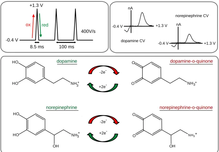
Figure 1.1: Fast-scan cyclic voltammetry for the detection of catecholamines. The most commonly used waveform sweeps from -0.4 to +1.3 V at a scan rate of 400 V/s. The positive-going scan oxidizes dopamine and norepinephrine to their ortho-quinone form, and the negative going scan reduces them back to dopamine or norepinephrine.
Plotting the resultant current vs potential results in identical characteristic cyclic voltammograms (CVs) for both dopamine and norepinephrine.
NH3+ HO
HO
NH3+ O
O -2e
-+2e
--0.4 V +1.3 V
nA O OH NH3 O HO OH NH3 HO dopamine dopamine-o-quinone norepinephrine norepinephrine-o-quinone -0.4 V +1.3 V
8.5 ms 100 ms
400V/s
ox red
dopamine CV
-0.4 V +1.3 V
norepinephrine CV nA
-2e
-32
Figure 1.2: Spatially resolved measurements combined with pharmacology ensure selective dopamine or norepinephrine measurements. Brain slice, adapted from the atlas of Paxinos and Watson showing norepinephrine terminals (red) surrounded by dopamine terminals (yellow), highlighting the need for a small sensor. Boxes show mock electrically stimulated response types to different drugs. Red bar denotes stimulation. In the green box, a pure dopamine signal increases with D2 antagonism, and remains elevated with α2 antagonism; a pure norepinephrine signal does not
increase following D2 antagonism and only responds to α2 antagonism. In the red box, a
mixed dopamine / norepinephrine signal responds to both D2 and α2 antagonists.
33
CHAPTER 2: NORADRENERGIC SYNAPTIC FUNCTION IN THE BED NUCLEUS OF THE STRIA TERMINALIS VARIES IN ANIMAL MODELS OF ANXIETY AND
ADDICTION1
Introduction
In drug-dependent individuals, researchers have highlighted the development of a persistent ‘negative emotional state’ when access to drugs is terminated (Koob, 2009). Substance abuse is often co-morbid with anxiety disorders and impacted by stressful life experiences (Hyman et al, 2009; Sinha, 2008). The risk for developing an addiction, however, varies considerably between individuals, and different people can have very different responses to drug or stress exposure. One means for addressing this issue is examining animals with divergent behavioral phenotypes in appropriate neuronal structures. Akin to human addicts, inbred Lewis (L) rats self-administer opiates at high levels (George and Goldberg, 1989), and show escalation of drug-taking
behavior (Picetti et al, 2012). Additionally, L rats display several anxiety-like phenotypes as compared with outbred Sprague-Dawley (SD) rats, as well as hypocortisolemia; thus, they have been suggested to be a good model of post-traumatic stress disorder (PTSD)
1 This chapter originally appeared as an article in Neuropsychopharmacology. The original
citation is as follows : McElligott ZA, Fox ME, Walsh PL, Urban DJ, Ferrel MS, Roth BL, Wightman RM (2013). “Noradrenergic Synaptic Function in the Bed Nucleus of the Stria
Terminalis Varies in Animal Models of Anxiety and Addiction.” Neuropsychopharm 38(9):
34
35
and amygdalar inputs, and stress and reward centers (Drolet, 2009). The BNST is densely innervated by noradrenergic fibers arising from the A1, A2 (via the ventral noradrenergic bundle or VNB), and A6 (via the dorsal noradrenergic bundle) cell bodies (Forray et al, 2004). Norepinephrine signaling within the BNST modulates anxiety-like behavior and influences induction of the hypothalamic–pituitary–adrenal (HPA) axis (Cecchi et al, 2002), affects expression of learned and physical opiate withdrawal behaviors (Delfs et al, 2000) and contributes to stress-induced reinstatement of drug seeking (Erb et al, 2000; Leri et al, 2002; Wang et al, 2001). Furthermore,
norepinephrine impacts both excitatory (McElligott and Winder, 2009) and inhibitory synaptic transmission (Dumont et al, 2004), induces synaptic plasticity (McElligott and Winder, 2008), and releases corticotropin-releasing factor (CRF) in the BNST
(McElligott et al, 2010; Nobis et al, 2011). The Morilak lab found that extracellular norepinephrine in the BNST was the same in non-manipulated L and SD rats; however, when restrained for 30 min, extracellular norepinephrine was elevated in L rats
compared with SD rats (Pardon et al, 2002) . Furthermore, L rats exhibit fear generalization and anxiety-like behaviors that are BNST dependent (Duvarci et al, 2009).
36
mechanisms underlying differences in norepinephrine overflow in L rats as compared with SD rats, and to test the hypothesis that morphine dependence alters regulatory mechanisms at noradrenergic neurons. We found that non-manipulated L rats had a reduced rate of clearance and decreased sensitivity at their α2-adrenergic receptors
(ARs) as compared with SD rats. Furthermore, using high-performance liquid chromatography (HPLC), we found that non-manipulated L rats had higher
norepinephrine tissue content. When SD rats were made physically dependent on morphine (Schulteis et al, 1999), modeling the condition of a human addict, there was a significant reduction in their norepinephrine uptake rate and in their response to a challenge with an α2-AR antagonist as compared with controls. In contrast, neither
clearance rate nor α2-AR sensitivity were different in morphine-dependent L rats as
compared with control or non-manipulated animals. Correlated to these alterations at norepinephrine synapses in the BNST, morphine-dependent SD rats, but not L rats, showed heightened anxiety-like behavior as compared with controls. The changes that occurred in morphine-dependent SD rats were profound: they exhibited
indistinguishable norepinephrine uptake rates and similar responses to an α2-AR
37 Materials and Methods
Animal care
All experiments were performed in accordance with the University of North Carolina at Chapel Hill (UNC) Institutional Animal Care and Use Committee’s
guidelines. SD and L rats (males; 350–450 g; L rats total n=70, SD total n=85; Charles River, Wilmington, MA) were housed within UNC animal facilities and given food and water ad libitum. Electrochemical, biochemical, and behavioral experiments were performed as follows: voltammetry, SD n=34, L n=26; HPLC, SD n=22, L n=15; autoradiography, SD n=14, L n=13; elevated plus maze, SD n=15, L n=16.
Evoked norepinephrine release
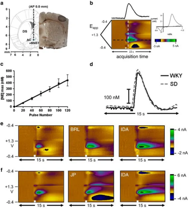
Norepinephrine release in the vBNST was evoked from stimulation of the VNB as previously described (Park et al, 2009). Briefly, animals were anesthetized with
urethane (1.5 g/kg i.p.) and placed in a stereotaxic frame (Kopf, Tujunga, CA). Holes were drilled in the skull for the carbon-fiber and stimulating electrodes at coordinates from a rat brain atlas(Paxinos and Watson, 2007). A Ag/AgCl reference electrode was placed in the contralateral hemisphere. A carbon-fiber microelectrode was placed in the vBNST (+1.2 mm ML, 0 mm AP, 7.2–7.8 mm DV). An untwisted bipolar stimulating electrode (Plastics One, Roanoke, VA) was placed in the VNB (+1.0 mm ML, −5.2 mm A-P, 8.0–8.5 mm DV from bregma). Both the carbon-fiber and stimulating electrodes were adjusted in the dorsal-ventral axis to obtain optimum norepinephrine release.