THE HEART OF THE MATTER:
CARDIOVASCULAR VULNERABILITY OF BREAST CANCER SURVIVORS IMPLICATIONS FOR AEROBIC CAPACITY AND CARDIOVASCULAR RISK
Jordan Talmadge Lee
A dissertation defense submitted to the faculty of the University of North Carolina at Chapel Hill in fulfillment of the requirements for the degree of Doctor of Philosophy in the School of
Medicine (Interdisciplinary Program in Human Movement Science).
Chapel Hill 2020
ABSTRACT
Jordan Talmadge Lee: The Heart of the Matter: Cardiovascular Vulnerability of Breast Cancer Survivors. Implications for Aerobic Capacity and Cardiovascular Risk
(Under the direction of Claudio L. Battaglini)
TABLE OF CONTENTS
LIST OF FIGURES AND TABLES... viii
CHAPTER ONE: INTRODUCTION ... 1
Background ... 1
Statement of Purpose... 5
Definition of Terms and Abbreviations ... 6
Research Aims and Hypotheses ... 7
Assumptions ... 9
Limitations ... 9
Delimitations... 10
Significance ... 10
CHAPTER TWO: LITERATURE REVIEW ... 12
Overview ... 12
Section I: Breast Cancer ... 12
Side Effects of Breast Cancer Treatment ... 13
Section II: Cardiovascular Toxicity of Breast Cancer Therapy... 14
Surgery ... 15
Chemotherapy ... 16
Radiation ... 24
Hormonal Therapy ... 26
Summary ... 28
Section III: Cardiorespiratory Fitness: Oxygen Cascade and Hemodynamics ... 28
The Oxygen Cascade ... 28
Cardiovascular Hemodynamics: Central Aspects ... 31
Cardiovascular Hemodynamics: Peripheral Aspects ... 34
Section IV: Exercise for Cardiorespiratory Health and Fitness... 36
Mechanisms of Exercise-induced Changes ... 37
Response Heterogeneity and Power of Predictors ... 41
CHAPTER THREE: METHODOLOGY... 43
Subjects ... 43
Recruitment ... 43
Instrumentation ... 44
Research Design Overview ... 45
General Procedures ... 46
Assessments ... 47
Sample Size Estimate ... 52
Statistical Analysis ... 52
CHAPTER FOUR: MANUSCRIPT ONE ... 56
CHAPTER FIVE: MANUSCRIPT TWO ... 78
Major Findings ... 96
Exploratory Findings ... 99
Strengths and Limitations ... 100
Future Research ... 101
LIST OF FIGURES AND TABLES
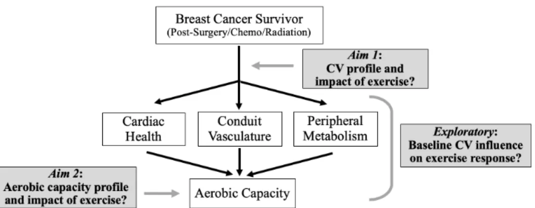
Figure 1. Conceptual model leading to research aims……….…8
Table 1. Study assessment and intervention overview………..45
Table 2. Progression of aerobic training exercise prescription………..50
CHAPTER ONE: INTRODUCTION
Background
Research has demonstrated that lifestyle risk factors at diagnosis from physical inactivity and/or poor dietary habits in concert with toxic cancer therapies is a particularly dangerous combination for long-term cardiovascular health in the breast cancer population (L. W. Jones, Haykowsky, Swartz, Douglas, & Mackey, 2007). The loss of cardiovascular health/function related to cancer and treatments in BCS is also characterized by exercise intolerance and an attenuated maximal capacity to uptake and utilize oxygen (“aerobic capacity”) (L. W. Jones, Courneya, et al., 2012). Aerobic capacity is the gold standard measure for cardiopulmonary fitness, and reflects an individual’s integration of cardiac and vascular systems/structures in the body. Loss of aerobic capacity can have a direct impact on patient quality and quantity of life, and impairments warrant specific attention for individual well-being (L. W. Jones, Haykowsky, Pituskin, et al., 2007a; Kaminsky et al., 2013). Lower aerobic capacities have been associated with increased mortality, both cancer and non-cancer specific, increased morbidity, and less favorable patient outcomes (J. B. Peel et al., 2009). Associations between low aerobic capacity and increased mortality following surgery have been observed in specific cancer populations. Unfortunately, aerobic capacities also diminish throughout the cancer continuum, especially during adjuvant therapy, but exercise can provide powerful leverage to improve aerobic capacity in the adjuvant setting (L. W. Jones et al., 2016; Lakoski et al., 2013; Lakoski, Jones, Krone, Stein, & Scott, 2015; J. B. Peel et al., 2009; J M Scott et al., 2018).
damage has not received the same attention. However, due to the integration and dependency between cardiac, vascular, and muscular structures of the body, a combination of insults likely contributes to the overall decreased aerobic capacity and increased cardiovascular disease risk observed in BCS. Chemotherapies and targeted therapies, such as anthracyclines and trastuzumab, have known detrimental effects on the cardiac system and developing evidence supports the vascular system may be insulted as well (Jain, Russell, Schwartz, Panjrath, & Aronow, 2017; Mulrooney, Blaes, & Duprez, 2012). It is also possible that a “domino effect” exists: observed cardiac damage in BCS may be downstream from cancer therapy-induced vascular damage, and/or compromised conduit vascular function may impair peripheral microvascular function. The overall effect may be represented by a decreased systemic aerobic capacity and increased susceptibility to future cardiovascular events. However, more work is needed to elucidate the cardiovascular profiles of BCS following treatment and the relationship of these individual systems to overall aerobic capacity. Clarifying the specific cardiovascular components potentially affected by cancer and cancer therapies, how they relate to overall aerobic capacity, and the potential ability to protect or recover their health and function with targeted interventions would provide important advances for the field of cardio-oncology.
Statement of Purpose
Cardiovascular disease mortality is a global burden and a particular threat to BCS due to potential cardiac and vascular specific damage from cytotoxic cancer therapies. There is a critical need to evaluate cancer therapy-induced cardiovascular damage in BCS which may be an
important contributor to the observed increased cardiovascular risk and decreased aerobic capacity in this population. Furthermore, determining interventions that may improve or protect vulnerable cardiovascular organs/systems will provide patients with means to enhance long-term well-being.
Therefore, the purpose of this study was to compare the cardiovascular and aerobic capacity profiles of BCS who have recently completed primary cancer treatments with otherwise healthy, non-cancer controls (CTL) before and after a 16-week, community-based exercise program. Exploratory analyses investigated the utility of baseline cardiovascular measures as predictors of exercise response (quantified by deltaVO2peak), and also investigated the
relationship between baseline p16INK4a, a marker of molecular aging, and important measures of clinically and functionally measures of patient outcomes.
Definition of Terms and Abbreviations
Aerobic capacity: The ability of the body to uptake, distribute, and utilize oxygen in the body and is quantified in this study as VO2peak (mL O2 /kg/min).
Breast Cancer Survivors (BCS): Woman with a confirmed breast cancer diagnosis and includes all phases of the cancer continuum from diagnosis to treatment to decades following treatment completion living with a history of cancer. In this study, BCS were early stage (0-III) survivors who were within one year of completing their primary breast cancer treatments (surgery, chemotherapy, radiation).
Otherwise healthy, non-cancer controls (CTL): Women ≥21 years old without any cancer diagnosis/history or other overt, known health concerns and who exercise two or fewer days per week at enrollment.
VO2peak: peak quantity (mL O2/kg/min) of oxygen consumed during a cardiopulmonary exercise test (CPET) on a cycle ergometer. VO2peak was calculated as the average of the three highest, five-second averages of recorded oxygen consumption measures during the last minute of a CPET.
Oxygen Cascade: The stepwise flow/movement of oxygen in the body from
Cardiovascular profile/components: Measures of cardiac and vascular health outcomes including arterial stiffness (quantified by pulse wave velocity (PWV)), augmentation index (AIx), and Buckberg Index obtained via non-invasive tonometry methods (PWV, AIx, Buckberg).
Community-based exercise program: Refers to the University of North Carolina Get REAL & Heel (GRH) Breast Cancer exercise rehabilitation program for cancer survivors. This is a 3-day per week, supervised group exercise program specifically designed for breast cancer patients and includes both aerobic and strength training customized to each patient’s ability.
p16INK4a: Tumor suppressor protein that has been evaluated as a biomarker of molecular aging in peripheral T-cells collected from standard venipuncture
Research Aims and Hypotheses
Figure 1. Conceptual model leading to research aims.
Primary and Secondary Aims
Aim 1:To determine the cardiovascular profile of breast cancer survivors compared to women
without a cancer history before and after a 16-week, community-based exercise program.
Null Hypothesis 1a: PWV will not differ between BCS and CTL at baseline.
Null Hypothesis 1b: Changes in PWV will not differ between BCS and CTL following participation in community based, exercise training.
Aim 2: To determine cardiorespiratory fitness of breast cancer survivors compared to women
without a cancer history before and after a 16-week, community-based exercise program.
Null Hypothesis 2a: VO2peak will not differ between BCS and CTL at baseline. Null Hypothesis 2b: Changes in VO2peak will differ between BCS and CTL following participation in community based, exercise training.
Exploratory Analyses
In addition to the previously stated Research Aims, three exploratory analyses were also conducted. The first exploratory analysis evaluated the relationship between baseline
baseline cardiovascular variables (PWV, AIx, Buckberg Index) with training response
(responders (DVO2peak ≥ 2.5ml/kg/min) vs. non-responders (DVO2peak < 2.5ml/kg/min)) in BCS and CTL. A third exploratory analysis evaluated the relationship between baseline p16INK4a biomarker of aging and clinical/functional patient outcomes including but not limited to DVO2peak, arterial stiffness, 6MWT, and lean body mass.
Assumptions
1. All of the participants followed the pre-test and post-test guidelines.
2. All of the participants were honest in answering questions related to medical history and physical activity.
3. All participants honestly engaged in exercise at the community-based facility to the best of their abilities.
4. All participants maintained current/normal lifestyles (nutrition, other physical activity) throughout the 16-week study period with the exception of the exercise intervention prescribed by researchers.
5. All participants remained under routine cancer care and long-term therapy (endocrine treatment), as appropriate, and immediately communicate any pertinent health related changes (especially oncologic or cardiovascular related changes) with the research team.
Limitations
2. Exercise intervention intensity was primarily evaluated using subjective, patient reported exertion (using Borg rating of perceived exertion (RPE) scale) which may be less specific than heart rates or oxygen uptake but is a necessary design for realistic and practical implementation in a community-based, non-randomized controlled design.
3. Negative impacts (both objective and subjective) of long-term cancer related medications (ex: endocrine therapy) may become enhanced over the study duration, however this is standard of cancer care and were accommodated as best as possible during the study.
Delimitations
1. A relatively select sample size of locally-residing women due to need to attend exercise sessions at Get REAL & Heel 3 days per week for 16 weeks.
2. Only women who had completed their major breast cancer anti-cancer treatments within the past year were eligible to participate in the study.
3. Only women who were not currently participating in regular exercise defined as physically active no more than 2 days per week.
Significance
CHAPTER TWO: LITERATURE REVIEW
Overview
For the purpose of organization, this review of literature is divided into four main sections. The first section provides an overview of breast cancer and associated side effects of treatment. The second section reviews specific cardiovascular toxicities of breast cancer therapies. The third section will discuss cardiorespiratory fitness based on the oxygen cascade and cardiovascular hemodynamics. The fourth section will focuses on the potential benefit of exercise training in the oncology setting for improving cardiovascular physiology, function, and performance.
Section I: Breast Cancer
improved disease-free survival (DFS) and overall survival (OS) for breast cancer patients also place healthy cells at risk for damage.
Side Effects of Breast Cancer Treatment
While tumor cells are the primary target for cancer therapies, chemotherapy, for example, is toxic to rapidly dividing cells - a defining feature of cancer (Hanahan & Weinberg, 2000). However, other non-cancerous cells in the body, like those of the gastro-intestinal tract, hair, and skin, are also rapidly dividing. Chemotherapy-induced damage to these otherwise healthy cells can result in undesired and potentially debilitating side effects like nausea, vomiting, neutropenia, thrombocytopenia, fatigue, and paresthesia, among others. If side effects become too severe, cancer therapy can be delayed, require dose reductions, or even be discontinued based on patient recovery response. While a treatment hiatus provides opportunity for patient rebound from negative side effects, a discontinuous regimen may compromise the efficacy of the established benefits of therapy (Hershman et al., 2011). Side effects, both long and short term, can significantly impact a patient’s quality and quantity of life (Basch et al., 2017; K.A. Nyrop et al., 2018).
dying from CVD than breast cancer, likely due to the ‘multiple hit’ of poor lifestyle habits, cardiotoxic cancer therapies, and overall autonomic dysfunction (Gernaat et al., 2017; L. W. Jones, Haykowsky, Pituskin, et al., 2007b; Lakoski et al., 2015; Patnaik et al., 2011; J M Scott et al., 2018; Shiovitz & Korde, 2015). Additionally, damage to the cardiovascular system can compromise independence and impair functional capacities potentially altering quality of life (K S Courneya et al., 2003; Paterson, Cunningham, Koval, & St Croix, 1999). Especially as the numbers of BCS grow, efforts to prevent, attenuate, and reverse cardiotoxic consequences of cancer treatments are warranted and necessary (Armenian et al., 2017; Schmitz, Prosnitz, Schwartz, & Carver, 2012).
Section II: Cardiovascular Toxicity of Breast Cancer Therapy
specific damage than vascular specific damage but natural and physical linkage between the cardiac and vascular system, the nature of cancer therapy administration via intravenous infusion (chemotherapy) and/or central vessel exposure during radiation suggests that the vascular system may be particularly vulnerable to toxic effects of cancer therapies. Evidence suggesting prevalence of vascular injury is increasing and vascular damage from cancer therapy may be a unique contributor to cardiovascular disease risk, may develop independently from cardiac-specific damage, and/or may enhance cardiac specific dysfunction in breast cancer populations (Blaes et al., 2017; L M Jones, Stoner, Brown, Baldi, & McLaren, 2013; Koelwyn et al., 2016; J M Scott, Adams, Koelwyn, & Jones, 2016). Therefore, more attention to understand the impact of primary cancer therapies on both cardiac and vascular structures and their ability to support oxygen utilization in the body is extremely warranted.
Surgery
accompanied by lymph node dissection, either sentinel or axillary, that help oncologists determine the likelihood that the tumor has spread to other parts of the body. Sentinel nodes are located close to the breast and are the most likely nodes to contain rogue cancer cells. If evidence of cancer cells exists in the sentinel nodes, a surgeon may complete an axillary lymph node dissection. These additional lymph nodes located peripheral to the breast are removed and help indicate the likelihood of metastasis to the body (Galimberti et al., 2013).
In terms of cardiovascular risks, breast cancer surgery alone has not been associated with long-term cardiovascular disease development.
Chemotherapy
Perhaps the most critical yet challenging goal of cancer treatment is preventing tumor progression and minimizing metastatic potential. Chemotherapy, targeted therapy, and hormonal therapies circulate the whole body to target rogue cancer cells that may have metastatic potential. Chemotherapy, given intravenously or by pill, has proven tremendously successful in preventing recurrence and promoting cancer-specific survival, and is a cornerstone treatment in our arsenal of cancer therapies (Mansour et al., 1989, 1998; Peto et al., 2012). Targeted therapy and hormonal therapy are systemic treatments with use dependent on tumor cell membrane receptor expression characteristics (D. Slamon et al., 2011).
(WHO) (“19th WHO Model List of Essential Medicines (April 2015),” 2015). These foundational drugs are the most widely used chemotherapies for systemic treatment of breast cancer, used in over one-third of breast cancer patients (Giordano, Lin, Kuo, Hortobagyi, & Goodwin, 2012; Mulrooney et al., 2012). Mechanistically, anthracycline agents intercalate between base pairs of DNA and RNA, interfere with critical DNA replication enzymes, and induce substantial oxidative damage resulting in tumor cell death (Jain et al., 2017). Common chemotherapy routines including anthracyclines involve four to six cycles of therapy occurring in a two or three-week frequency with drugs given individually or in combination, depending on the regimen selected by the treating oncologist (Gradishar et al., 2018).
Cardiotoxicity of Anthracycline Chemotherapy
Chemotherapy-induced cardiotoxicity from anthracycline therapy is well documented in the literature (Floyd et al., 2005; Jain et al., 2017). First identified in the 1970’s, anthracycline-induced cardiotoxicity is dose-dependent with incidence ranging from 3%-48%, based on the cumulative dose received. This discovery led to the development of guidelines limiting a patient’s cumulative lifetime dose of anthracyclines to approximately 500mg/m2 (Von Hoff et al., 1979). However, cardiotoxicity can occur at doses below this threshold and nevertheless, constrains treatment efficacy (Swain, Whaley, & Ewer, 2003; Von Hoff et al., 1979).
heart failure (CHF), or may become clinically detectable only after permanent damage has occurred (Doyle, Neugut, Jacobson, Grann, & Hershman, 2005; Lakoski et al., 2015; Von Hoff et al., 1979). With a hazard ratio of 3.46 compared to , this specific heart failure imparts an especially poor prognosis for breast cancer patients (Felker et al., 2000). In the absence of standard long-term cardiac-specific follow up practices for cancer patients, effective monitoring of potential cardiotoxicity is logistically challenging (Schmitz et al., 2012). In attempt to capture and treat potential cardiotoxicity as early as possible, a recent review suggested that suspicion for cardiotoxicity in cancer survivors should remain high and standards for evaluation be low (Armenian et al., 2017). Other groups have suggested cardiovascular-specific evaluations be completed in all patients starting adjuvant treatment, regardless the specific therapy (Koelwyn, Khouri, Mackey, Douglas, & Jones, 2012). Needless to say, developing improved strategies to identify patients at high cardiovascular disease risk and implementing cardio-protective approaches to cancer care is highly warranted.
for the growing number of survivors may outpace the medical providers available to serve (Bluethmann et al., 2016). Efforts to clarify the mechanisms responsible for cardiovascular damage and toxicity will enhance the ability to design and implement cardioprotective strategies.
No single mechanism has been identified as the predominant contributor to anthracycline-induced cardiotoxicity, and the process is indeed complex. It has also been suggested that the mechanisms responsible for tumor cell death are distinct from those that result in cardiac cell death, and/or that cardiomyopathy results when drug-induced damage is combined with particularly harmful lifestyle choices (Menna, Salvatorelli, & Minotti, 2008). Current evidence supports oxidative stress due to mitochondrial dysfunction, ion dysregulation, energy depletion, and disrupted cardiac signaling pathways all contribute to anthracycline-induced cardiomyopathy. As evidenced through some of these speculated pathways, anthracycline toxicity may actually amplify in the body as the drug is metabolized (Mordente, Meucci, Silvestrini, Martorana, & Giardina, 2012).
properly metabolize fuel substrates for energy which can lead to mitochondrial death. In terms of tumor fighting capacity, this effect has clear benefit. However, the same impact cannot be said for cardiomyocytes. Furthermore, it has been demonstrated that anthracyclines directly damage mitochondrial DNA (mtDNA) of cardiac muscle, but less so skeletal muscle, likely due to intercalation into strands or via production of reactive oxygen species (Adachi et al., 1993; Ashley & Poulton, 2009; Dirk Lebrecht, Kokkori, Ketelsen, Setzer, & Walker, 2005). Unfortunately, mtDNA damage, electron transport chain dysfunction, and reactive oxygen species production can compound and accumulate over time. This latency effect is speculated as the primary reason for development of late-onset cardiomyopathy and irreversible damage (D Lebrecht et al., 2007).
Vascular Toxicity of Chemotherapy
Targeted Therapy
Unlike the blanket-style approach of systemic chemotherapy, targeted therapies are specific to blocking the growth of a cancer cell via a precise receptor or vulnerable characteristic of that tumor. Trastuzumab (HerceptinÒ) and pertuzumab (PerjetaÒ) are two targeted therapies used for treatment of primary and metastatic HER2+ breast cancer tumors, respectively. Both are humanized monoclonal antibodies that act on specific and different sites of the human epidermal growth receptor located on the tumor cell membrane. These drugs prevent dimerization of HER2 receptors, successfully interfering with cell signaling pathways critical to growth and proliferation, which can consequently prevent tumor cell survival (Albini et al., 2011). Trastuzumab has been shown to improve disease free survival by 51% and overall survival by 37% when administered following anthracycline therapy, providing substantial benefit for HER2+ breast cancer patients (Romond et al., 2005; D. J. Slamon et al., 2001; Zeglinski, Ludke, Jassal, & Singal, 2011).
Cardiotoxicity of Trastuzumab Targeted Therapy
et al., 2011; Zeglinski et al., 2011). While the precision of directed therapies like trastuzumab has contributed substantially to the arsenal of tools available for HER2+ cancer therapy, mechanistically the drug targets a ubiquitous receptor, rendering non-tumor cells susceptible to injury (Sandoo, Kitas, & Carmichael, 2015; Zeglinski et al., 2011). Cardiac tissue expresses human epidermal growth factor receptors, although the degree of expression is debated, resulting in vulnerability to the effects of trastuzumab (Albini et al., 2011). The resulting cardiotoxicity usually presents more quickly than anthracycline-induced damage but manifests similarly as a decline in LVEF. Also dissimilar to anthracycline cardiotoxicity, trastuzumab-induced cardiac decline has been attributed to disruption in Neuregulin1 and ErbB receptor signaling pathways, mitochondrial function, and cellular energetics rather than structural damage of cardiomyocytes (Brero et al., 2010; Lee et al., 1995; Rochais & Fischmeister, 2010; Spallarossa et al., 2010). Permanent damage to the cardiac tissue from trastuzumab is less common than anthracycline-induced damage and recovery of cardiac function is possible following cessation of trastuzumab therapy. Interestingly, some patients are also able to restart anti-HER2 therapy without subsequent cardiac symptoms. However, the therapy is a modern intervention, thus, long term side effects like cardiotoxicity profile are not yet well-known (L. W. Jones, Haykowsky, Swartz, et al., 2007).
Vascular Toxicity of Trastuzumab Targeted Therapy
Extremely limited research is available evaluating the impact of trastuzumab on vascular specific outcomes in breast cancer survivors (Grover et al., 2015). Two published studies
treated with anthracyclines so delineating the specific impact of trastuzumab alone is complicated. Regardless, pulse wave velocities increased by over 2 m/s in only 4 months
following therapy which doubles the clinically meaningful change usually seen in approximately 10 years of aging (C.-Y. Liu et al., 2012; Yersal et al., 2018). More work is needed to determine the specific impact of targeted therapies on vascular health.
Radiation
Cardiotoxicity of Radiation
Due to the location of vital organs in proximity to breast tissue that may be targeted, incidental exposure of the heart, lungs, and thoracic vessels to radiation can result in substantial organ and structure damage (M. Clarke et al., 2005; Cuzick et al., 1994; Marks et al., 2005). A staggering 62% increase in cardiac deaths has been observed in women who received radiation therapy for their breast cancer (Yusuf, Sami, & Daher, 2011). Research supports that radiation can damage proper physical contraction of the muscular ventricular wall, leading to perfusion deficits in the heart; especially as more heart volume is included in the radiation field in addition to damage of the cardiac vasculature (Gyenes, Fornander, Carlens, Glas, & Rutqvist, 1996; Marks et al., 2005). Valvular heart disease is recognized especially in women who receive radiation doses in excess of 30 Gray (Hull, Morris, Pepine, & Mendenhall, 2003). However, radiation-induced damage can result independent of detectable changes in left ventricular ejection fraction (LVEF), which is the most widely used cardiac surveillance technique for many cancer patients. The silent development of heart disease in this population further reiterates the need for consecutive cardiac surveillance techniques.
Vascular Toxicity of Radiation
albeit likely related to older techniques not commonly used today (Darby et al., 2013). Improvements in administration techniques, like respiratory gating/breath holding, since the 1970’s have resulted in decreased risk of cardiovascular disease in patients treated with radiation (Hooning et al., 2007). While currently speculative without long term prospective data, radiotherapy-induced cardiac perfusion deficits, wall motion abnormalities, and local arterial stiffening may collectively or individually contribute to long term cardiovascular disease development in cancer patients.
Hormonal Therapy
Hormonal therapy is a third example of commonly used systemic therapies for women with HR+ tumors. Tamoxifen (NolvadexÒ) is a selective estrogen receptor modulator (SERM) given to block estrogen from reaching estrogen receptors in ER+ and/or PR+ breast cancer cells. Five years of Tamoxifen therapy provides a significant reduction in risk of same-side recurrence, ipsilateral recurrence, and death from breast cancer for ER+ tumors (Early Breast Cancer
specific events and remains a concern (Foglietta et al., 2017; L. W. Jones, Haykowsky, Swartz, et al., 2007). Tamoxifen, on the other hand, appears to elicit cardioprotective effects related to blood lipid profiles due to its actions as an estrogen agonist. Overall, it has not yet been
determined whether the cardiovascular disease risk profile differences between the two hormonal therapies are a result of protective effects of Tamoxifen or harmful effects of AI’s, but these changes may predispose the survivor to unwanted comorbidities (Foglietta et al., 2017; Thurlimann et al., 2005).
Cardiotoxicity of Aromatase Inhibitors
Estrogen is thought to play a protective role in terms of cardiovascular health based on the relatively low incidence of cardiovascular disease in premenopausal compared to
Vascular Toxicity of Aromatase Inhibitors
Aromatase inhibitors are thought to elicit negative effects on the vascular endothelium disrupting vasodilation, coagulation and antioxidant regulation, potentially predisposing survivors to atherosclerosis and/or hypercholesterolemia (Mendelsohn, 2002; Nathan et al., 2001). Women receiving aromatase therapy have demonstrated increased arterial stiffness and less favorable lipid profiles but a distinguished, vascular-specific toxicity due to AI damage versus Tamoxifen protection has not yet been observed (Foglietta et al., 2017; Yersal et al., 2018).
Summary
Primary breast cancer therapies have the potential to significantly disrupt cardiovascular health, with the impact of some treatments more toxic, evident, and/or well-studied than others. Further work is needed to clarify each therapy’s damage-inducing mechanism and the organs and tissues that may be most vulnerable. Further, the ability to feasibly and accurately monitor and detect early deterioration in the cardiac and vascular system would likely provide opportunity for earlier intervention and potentially curb the incidence of permanent cardiovascular disease. Additionally, the potential role of complementary therapies such as exercise to protect or promote cardiac and vascular-specific health of BCS in the adjuvant setting warrants investigation.
Section III: Cardiorespiratory Fitness: Oxygen Cascade and Hemodynamics
The Oxygen Cascade
measured directly through maximal exercise testing and is quantified in terms of maximal aerobic capacity or VO2max. This maximal oxygen consumption is also described mathematically in the Fick equation (VO2max = CO * a-vO2diff) as a product of cardiac output (stroke volume * heart rate) times the arterial venous oxygen difference (a-vO2diff). An individual’s aerobic capacity is therefore a product of central and peripheral components; the former dominated by the heart and conduit vasculature, and the latter by microvasculature, skeletal muscle, and mitochondria (Hoppeler & Weibel, 1998). In series, these components reflect links in the oxygen cascade – a model describing the flow of oxygen delivery, transport, and consumption. Mathematically, it can be demonstrated that impairments, individual or in combination, in this cascade will result in an overall impaired aerobic capacity (VO2max). Interestingly, decreased aerobic capacity is a well-established phenomenon observed in breast cancer patients and is critical due to the well-established relationship between depressed maximal aerobic capacities with increased morbidity and mortality (L. W. Jones et al., 2010; Terence Kavanagh et al., 2003). From diagnosis to years beyond, breast cancer patients have demonstrated aerobic capacities up to 30% lower than age-matched, non-cancer controls (L. W. Jones, Courneya, et al., 2012). This loss of aerobic capacity has been observed despite preserved stroke volume, and survivors demonstrate impairments in contractility and an inability to meet functional demand (Koelwyn et al., 2016). Therefore, determinants of oxygen transport other than central cardiac factors must be compromised, but have not yet been well studied in this population.
(Jessica M Scott et al., 2018). Only a handful of studies have concurrently evaluated overall aerobic capacity with potential determinants in BCS, and have observed central performance impairments with implication of peripheral deficiencies, especially at higher workloads (Beaudry et al., 2019; L. W. Jones, Haykowsky, Pituskin, et al., 2007b; Koelwyn et al., 2016). Improving understanding of vulnerable cardiovascular components in potential need of surveillance, will allow researchers the opportunity to propose and design interventions specific to protecting or repairing vulnerable links as a way to preserve systemic oxygen consumption.
Cardiovascular Hemodynamics: Central Aspects
(i.e. in the area of assessment: radial, brachial, carotid-femoral), and AIx changes based on systemic arterial stiffness influenced by heart rate, whole-body reflected wave amplitudes, and PWV (Kim & Braam, 2013). Nonetheless, greater arterial stiffness (i.e. indicated by faster PWV and higher AIx) indicates less elastic, more collagenous vasculature and a less favorable cardiovascular profile (Sutton-Tyrrell et al., 2005; Weber et al., 2004). Clinical conditions such as diabetes and peripheral artery disease can result in compromised vascular health as exhibited by increased PWV (Benetos, Laurent, Hoeks, Boutouyrie, & Safar, 1993; Boutouyrie et al., 1992; O’Rourke, Staessen, Vlachopoulos, Duprez, & Plante, 2002). While significantly less studied compared to diabetes and peripheral artery disease, cancer survivors also exhibit increased arterial stiffness as measured by PWV, likely due to varying vascular-related toxicities from cancer therapies (Mozos et al., 2017; Souza et al., 2018). This unfavorable vascular profile may be a contributor to the increased incidence of cardiovascular mortality observed in BCS.
compromised (for example due to anthracycline-induced cardiotoxicity), the heart must increase contraction rate (heart rate) to maintain cardiac output. This is a natural response of the cardiac system observed under stress or during exercise but may become less transient if the heart/vasculature becomes chronically pathologic. Over time, if stroke volume cannot be maintained and heart rate increases to conciliate, perfusion of the heart may be compromised which increases risk of cardiac ischemia (Hoffman & Buckberg, 2014). An index using diastolic pressure, systolic pressure, and time integrals over a cardiac cycle has been developed using characteristics derived from a validated central pressure waveform (Hwang et al., 2014). This value is termed the Buckberg Index and reflects cardiac subendocardial viability; a parameter of diastolic function. While index values are not specifically intended to diagnose ischemia, serial measures may capture changes that illustrate functional deterioration and help identify those at risk of diminishing cardiac performance (Budinskaya et al., 2017; Hoffman & Buckberg, 2014). This measure, derived from the waveform of validated and automated tonometry systems used for pulse wave analysis, has been minimally studied and never reported in the breast cancer literature, to the best of our knowledge (Butlin et al., 2013). The Buckberg index, in addition to PWV and AIx, could be an important, non-invasive, hemodynamic measure to obtain serially when monitoring patients like BCS who have particular cardiovascular concerns.
Non-Invasive Measurement of Central Cardiovascular Health
2013). Central aortic pressures and characteristics are superior to peripheral pressures in describing cardiac stress, however, these technologies are not generally utilized in standard oncology clinics (Wassertheurer et al., 2010). Failing to evaluate central pressures, AIx, and Buckberg indices may compromise more detailed understanding of patient cardiovascular health status. In terms of BCS who are at higher risk of cardiovascular and coronary artery disease due to potentially cardiotoxic therapies, serial cardiovascular surveillance with the previously described non-invasive technologies may offer unique supplemental diagnostic and prognostic value for identifying important, centrally-located cardiovascular vulnerabilities (Harris et al., 2006; L M Jones et al., 2013). However, it should be recognized that values such as the Buckberg Index obtained from pulse wave analysis are derivatives, not direct measurements, and should therefore be interpreted with caution until further direct validations exist.
Cardiovascular Hemodynamics: Peripheral Aspects
feasible and validated measure when used appropriately (Lucero et al., 2018; Mancini et al., 1994). NIRS has been extensively used in clinical populations such as peripheral artery disease and diabetes mellitus, but has been minimally implemented in cancer patients, and never in a group of all breast cancer survivors to the best of our knowledge (Barroco, Sperandio, Reis, Almeida, & Neder, 2017; Lanfranconi et al., 2014; Layec et al., 2016).
Section IV: Exercise for Cardiorespiratory Health and Fitness
Well designed and implemented exercise can have an important role for improving cardiorespiratory health, fitness, fatigue, and quality of life among other factors in the breast cancer population (Battaglini et al., 2014b; A A Kirkham et al., 2016; McNeely et al., 2006; Schmitz et al., 2010). Recent publications support exercise as an effective adjuvant therapy for increasing cardiorespiratory fitness in BCS and other clinically relevant outcomes (A A Kirkham et al., 2016; J M Scott et al., 2018). Interestingly, while exercise can positively impact multiple organ systems, enhance overall cardiovascular reserve capacity, and is an established therapy to improve exertional intolerance for patients with heart failure (Pandey et al., 2015), the same utility has been less consistently appreciated in the breast cancer population (L. W. Jones, Haykowsky, Swartz, et al., 2007; Lakoski et al., 2012; Mora, Cook, Buring, Ridker, & Lee, 2007). However, it has been shown that increasing levels of exercise is associated with a decrease in incidence of cardiovascular events in women with non-metastatic breast cancer (L. W. Jones et al., 2016). All-cause and cancer-specific mortality have been reduced by 13% and 15%, respectively, in non-cancer populations following an increase in cardiorespiratory fitness of approximately 3.5mL/kg/min (1 MET) (Barlow et al., 2012; Kodama et al., 2009; Myers et al., 2002).
especially as they relate to overall cardiorespiratory fitness. Cardiovascular outcomes other than cardiorespiratory fitness are rarely reported but warrant attention as they may be important and unique indicators of heart, macro, and microvascular health and may be more feasibly obtained and monitored in clinic settings than a CPET (Jessica M Scott et al., 2018). Determinants of aerobic capacity (i.e. those representative of the components in the Fick equation) have also been minimally studied in concurrence with systemic aerobic capacity measurements (Beaudry et al., 2019; L. W. Jones, Haykowsky, Pituskin, et al., 2007a) Systemic cardiorespiratory fitness can be improved with properly designed and implemented exercise, which suggests cardiac and vascular determinants supporting the overall oxygen consumption are improving either independently or as a system. This phenomena is well-described in non-cancer populations (M. G. Wilson, Ellison, & Cable, 2016) and supporting and speculated mechanisms will be explained below. However, the impact of exercise training on individual components, especially those related to vascular health and peripheral oxygen metabolism, have not been well studied or reported in BCS (Beaudry et al., 2019; J M Scott et al., 2018). Evaluating these more intricate relationships may provide an enhanced understanding of specific vulnerable components of a BCS’s cardiovascular system following cancer therapy, and the unique contribution of components to systemic oxygen consumption. Long term, these components may develop as new targets for consecutive surveillance or intervention throughout a BCS’s journey.
Mechanisms of Exercise-induced Changes
general, greater amounts of exercise appear to maximize cardiovascular health (T Kavanagh, 1983; M. G. Wilson et al., 2016). Athletes commonly exhibit superior fitness, enlarged heart structures, and lower resting blood pressures when compared to sedentary controls as a result of continuous engagement in structured physical activity(M. G. Wilson et al., 2016). These changes promote cardiovascular efficiency and enhance exercise capacity due to improvements in ability to uptake, distribute, and utilize oxygen within the body. Therefore, exercise training can be used, alone or in concert with pharmacologic interventions, to target vulnerable systems and improve outcomes for at-risk populations.
Cardiac Benefits of Exercise Training
increasing force-producing potential (Mihl et al., 2008). However, increases in cardiac contractile strength due to strength training has shown minimal impact on overall aerobic capacity in BCS, unlike the adaptations of endurance training, but provide other beneficial systemic effects pertinent to BCS (A A Kirkham et al., 2016; Sasso et al., 2015; Schmitz et al., 2010).
Vascular Benefits of Exercise Training
populations like BCS with demonstrated autonomic dysfunction and potential endothelial damage, the effects may be dampened or absent (Lakoski et al., 2015; J M Scott et al., 2014). Two recently published studies observed attenuated blood flow response in cancer survivors, which negatively impacts proper utilization of oxygen at working tissue, but mechanisms
responsible for these decrements are not yet understood (Didier et al., 2017; Ederer et al., 2016). Furthermore, it is relatively unknown the impact or potential benefit of chronic exercise training on blood flow in cancer populations.
Oxidative Stress Benefits of Exercise Training
Exercise naturally produces free-radicals in mitochondria of working cells but chronic exercise training reduces oxidative damage, improves antioxidant enzymes, and reduces
Response Heterogeneity and Power of Predictors
T-cells (Sanoff et al., 2014). Interestingly, the 10-14 years of molecular aging observed via increased p16 INK4a expression in the previously mentioned study approximates the same aging phenomenon related to aerobic capacity observed in cancer patients following therapy (L. W. Jones, Courneya, et al., 2012). The relationship between p16 INK4a and VO2, and other functional or clinical outcomes, has yet to be evaluated but warrants further investigation (Sanoff et al., 2014).
CHAPTER THREE: METHODOLOGY Subjects
Two cohorts of women were recruited for the proposed study. One group was early stage, non-metastatic BCS who were within one year of completing primary cancer therapy (surgery, chemotherapy, radiation), and were otherwise free from overt cardiovascular, metabolic, orthopaedic complications. The second group was an age and physical activity matched group of women who did not have a cancer history and were otherwise healthy. Both groups required physician clearance to participate and were cleared by a cardiologist before completion of any maximal exercise testing. Both groups completed study-related testing (described later) on 2 days at baseline, and then completed 16-weeks of progressive, supervised aerobic and strength training exercise at the UNC GRH exercise facility, followed by 2 days of post testing mirroring pre-testing.
Recruitment
research team members to discuss the study and allow survivors time to ask any questions. Those interested in participating in the GRH study were asked to sign a UNC IRB-approved informed consent form before completing any study related assessments.
Instrumentation
A Physical Activity Readiness Questionnaire (PAR-Q) and a medical history
questionnaire were used to determine whether or not approval by physician would be required to take part in the study prior to officially signing consent to participate. The short version of the International Physical Activity Questionnaire (IPAQ) was used to confirm current physical activity participation in both groups. Literature supports breast cancer survivors report substantial levels of physical inactivity throughout the cancer care continuum, so exercise participation was not used as an exclusion in our breast cancer survivors (Kwan et al., 2012). To best mirror this population, recruited control participants did not complete more than two days of structured exercise at baseline.
analysis using ParvoMedics metabolic system. The UNC GRH exercise program facility was used for the 16-week exercise training intervention.
Research Design Overview
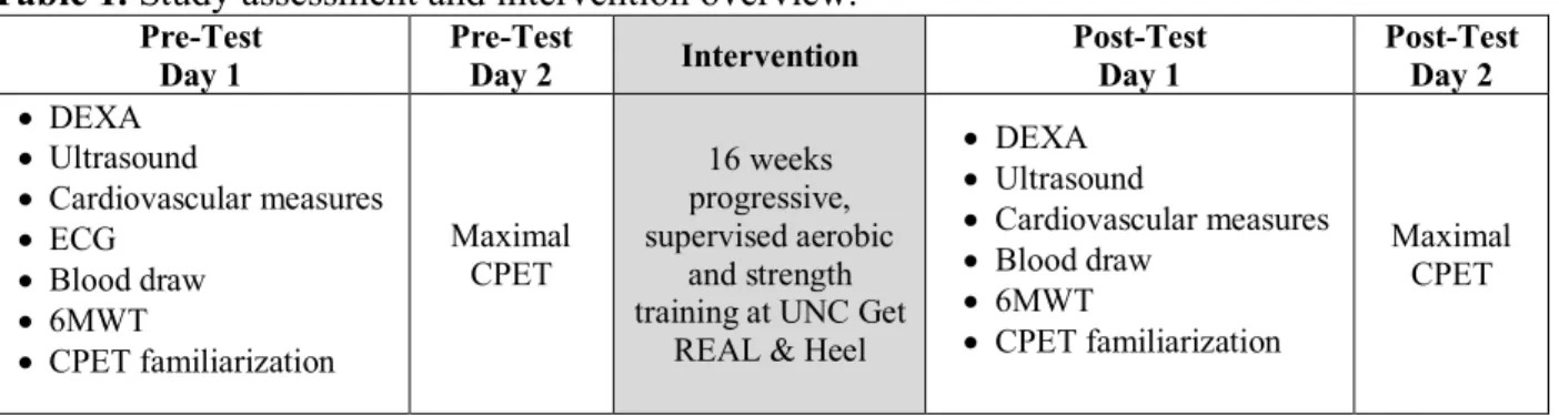
In this non-randomized study, all subjects completed two laboratory visits prior to (pre-test) and immediately following (post-(pre-test) a 16-week supervised exercise intervention as illustrated in Table 1. Both laboratory visits were conducted in the Exercise Oncology Research Laboratory (EORL) in the department of Exercise and Sport Science at UNC-Chapel Hill. Pre and post testing were completed within a two-week window. The first day of testing included: fasted body composition, 12-lead ECG evaluation, fasted and resting cardiovascular measures, and familiarization to the exercise test on the cycle ergometer. The second day of testing included the true maximal exercise test on the cycle ergometer with a 3-minute post completion lactate measurement. The 16-week exercise intervention included a combination of progressive aerobic and strength training for 3 small group sessions per week at approximately 1 hour per session at the UNC GRH exercise facility. Post-testing followed the exact same format as pre-testing (minus ECG) and occured over a two-week timeframe immediately following conclusion of the 16-weeks of exercise training.
Table 1. Study assessment and intervention overview. Pre-Test
Day 1
Pre-Test
Day 2 Intervention
Post-Test Day 1
Post-Test Day 2 • DEXA
• Ultrasound
• Cardiovascular measures • ECG
• Blood draw • 6MWT
• CPET familiarization
Maximal CPET 16 weeks progressive, supervised aerobic and strength training at UNC Get
REAL & Heel
• DEXA • Ultrasound
• Cardiovascular measures • Blood draw
• 6MWT
• CPET familiarization
General Procedures
After subjects demonstrated interest to participate in this study and met baseline inclusion criteria (clear medical history, ≤2 days/week exercise, physician approval if needed), subjects were informed of pre-assessment guidelines to follow in the day(s) prior to physical fitness pre and post-testing and were scheduled for pretesting in the EORL. This included being fasted for most of the day one assessments and no caffeine for either testing day, but participants were encouraged to stay hydrated with water. On the first day of pre-testing, subjects read and signed an informed consent form and were assigned a 4-digit identification code. The primary
investigator verbally insured the subject adhered to pre-assessment guidelines. The primary investigator collected demographic information of each subject including age, race, sex, height and weight. Cancer-specific information was collected from electronic medical records for survivors who agreed to participate and release related information.
Following demographic data collection, each subject completed in the following order: one DEXA assessment, one ultrasound scan of the vastus lateralis, resting cardiovascular measurements (AIx, Buckberg Index, PWV) taken in duplicate and averaged for analysis, one resting 12-lead ECG, one blood draw for complete blood count and p16INK4a, one 6-minute walk test (6MWT), two timed up-and-go tests with the fastest time recorded for analysis, and a familiarization session on the bike including wearing a mask and completing submaximal exercise up to 75% of heart rate reserve. Participants were required to be fasted (overnight) for the first five assessments on day one but were encouraged to bring and consume a light snack before the walking test and bike familiarization.
Following the first day, research team members obtained cardiologist review and
The second day included a maximal exercise test on the cycle ergometer. Participants were fitted with appropriately sized metabolic mask for gas exchange analysis. Participants completed warm up then an incremental (15 watt/min) ramp maximal exercise test on the cycle ergometer.
Following completion of testing days, participants were enrolled in the 16-week exercise intervention. Following completion of the intervention, participants were scheduled to return to the EORL for post-testing and completed the same assessments in the same order as pre-testing. The blood draw was not completed at post testing.
Assessments
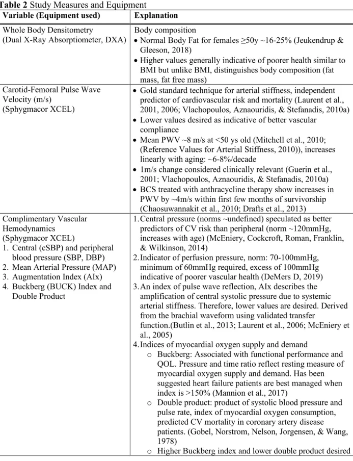
Height and Body Mass
Subject’s height was measured without shoes, standing with their back against the stadiometer and looking straight ahead. Body mass was measured without shoes and in minimal clothing using an electronic scale.
Body Composition
Total body weight-mass (BW) and compositional aspects of lean body mass (LBM), fat tissue mass (FM), and percentage body fat (% BF) was examined for patient demographics using standard DEXA screening procedures.
Non-invasive cardiovascular measurements
Gold standard measures of arterial stiffness including PWV and AIx, in addition to other hemodynamic variables, were collected using the usingthe SphygmorCor XCEL device (AtCor Medical, Sydney, Australia) following standard manufacturer guidelines and protocols
Contralateral placement of the cuff has been shown to yield congruent readings (Hwang et al., 2014). The cuff as electronically inflated/deflated and central and peripheral blood pressures, AIx, and Buckberg Index were measured by the device and software. Measures were taken in duplicate and averaged for analysis. For Pulse Wave Velocity, participants lied in a supine position with a blood pressure cuff on upper thigh of one side of the body to detect femoral pulse, and a handheld tonometer at the ipsilateral carotid artery. Segmental measurements between the cuff and the tonometer were made and were necessary for the software to estimate the linear distance the pressure wave travels in the conduit vessel of interest. The tonometer was placed on the carotid artery and the leg cuff inflated electronically. Once a sufficient pulse wave was detected, the carotid-femoral PWV reading was captured over 10 seconds. Measures were repeated twice or until there was no more than a 10% difference between measures. PWV was automatically calculated as the distance between the detected carotid artery pulse and the detected femoral artery pulse divided by pulse transit time.
Blood Draw
cells with purity exceeding 90% were used for RNA extraction. TaqMan RT-PCR were performed in duplicates and 18S were used as controls to measure the expression of p16INK4a. Additionally, TaqMan includes control samples of known p16INK4a and were run as additional methodological control.
6-Minute Walk Test
The 6-minute walk test is a clinically feasible test used to assess aerobic endurance and will be completed following resting cardiovascular measures on the first day of testing.
Participants were encouraged to walk as quickly as possible around a 50-yd rectangular course for a total of 6 minutes. Participants had to complete the testing at a walk; jogging or running was not be permitted. Participants were verbally encouraged for maximal possible effort throughout the testing. A member of the research team tallied the number of laps completed to the nearest 5yd. Final distances were converted to meters upon completion.
Cardiopulmonary Exercise Test (CPET)
Participants were required to familiarize with the electronically braked cycle ergometer and mask used for the CPET at the end of day one. Participants completed an identical protocol to the maximal test, including the strength progression and use of the Borg RPE scale, but were stopped at ~75% of their heart rate reserve (HRR) on day one. The maximal CPET was completed on day two and is described in detail below.
continuously by 15watts/min until test termination. Termination of the test was determined by subject reaching volitional exhaustion and signaling to stop the test, VO2 plateau or decrease with increase in exercise intensity, or an abnormal subject response to the test was observed and therefore the research team terminated the test (however, did not occur). This protocol has been previously successfully implemented in other clinical populations (Wilkerson et al., 2011). Heart rate and RPE were continually monitored and recorded throughout the testing. Blood lactate was collected by fingertip puncture 3-minutes after the completion of the maximal CPET.
Get REAL & Heel Exercise Intervention
Following testing, participants completed exercise training 3 days/week for 16 weeks at the UNC GRH Exercise Facility. Exercise was a combination of both strength and strength exercise and was designed to progressively increase to a moderate-high workload but adapted to each participant as necessary. Aerobic exercise included a variety of options including walking, cycling, elliptical, etc. For the first 2 weeks of exercise training, exercise volume and intensities began at 10-15 minutes at low intensity of 50-60% of the participant’s heart rate reserve (HRR) with a corresponding RPE between 8-11. For participants who were deconditioned, exercise duration may have been reduced by 5-10 minutes. The goal was for participants to achieve 30 minutes of moderate intensity aerobic exercise by mid to end of the 16-week program using 65-75% HRR with a respective RPE between 12-14 (Table 2).
Table 2. Progression of aerobic training exercise prescription.
AEROBIC EXERCISE WEEK1-2 WEEK3-7 WEEK8-16
• Treadmill • Stationary bike • Elliptical • Stepper
Duration (min) 10-15 10-30 30
Intensity Low Moderate
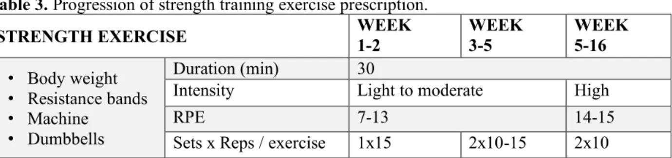
Strength exercises targeted large muscle groups in the upper body, lower body, and core to improve strength, balance, and functionality. Each session of strength training lasted approximately 30 minutes. Two sets of each exercise were performed for 10-15 repetitions with intensity progressing in weeks 1-5 from light to moderate (RPE 7-13). After week 5, attempts were made to increase the intensity from moderate to high (RPE 14-15) for 2 sets of 10 repetitions of each exercise. Exercises targeting major muscles groups included: lateral raises wall push-ups, rows, squats, bridge, plank, reverse sit up, shrugs, tandem stance, and lateral pulldown. Modifications to these exercises were made for any existing injuries, orthopedic limitations, or sequela from anti-cancer treatments (Table 3).
Table 3. Progression of strength training exercise prescription.
STRENGTH EXERCISE WEEK1-2 WEEK3-5 WEEK5-16
• Body weight • Resistance bands • Machine
• Dumbbells
Duration (min) 30
Intensity Light to moderate High
RPE 7-13 14-15
Sets x Reps / exercise 1x15 2x10-15 2x10
Training progression was accomplished slowly, starting with 1 set of strength exercises using no weight or very light weight in the first two weeks of training with the number of repetitions achieving 15, then increased based on the participant’s ability and limitations up to 2 sets during weeks 3-5 of training. As participants progressed to tolerate increased resistance, a balance component was carefully added to the training regimen. This included performing strength exercises with a level of complexity that challenged postural control.
all participants were continuously monitored and supervised during all phases of the exercise sessions at GRH.
Participant adherence and compliance were quantified using exercise logs to determine the exercise dose completed over 16-weeks. Adherence reflects attendance (ATT), simply the number of days participants came to the GRH facility out of the total days possible in 16 weeks (48 days total). Aerobic compliance (aCOMP) was calculated as the number of days (out of 48) where participants completed ≥80% of the prescribed duration at the prescribed intensity. Strength compliance (sCOMP) was calculated as the number of days (out of 48) where participants completed ≥80% of the prescribed volume (sets x reps) at the prescribed intensity.
Sample Size Estimate
Sample size estimates were calculated a priori and were based on the primary outcome of aerobic capacity. Arterial stiffness analysis was evaluated for power post hoc. Published
systematic reviews and meta analyses support exercise therapy to increase aerobic capacity in breast cancer survivors by approximately 2.3 - 2.8 mL/kg/min (Battaglini et al., 2014b; J M Scott et al., 2018). We opted for a change of 2.5mL/kg/min. The sample size required was 26 total participants using magnitude-based inferences, but oversampling was performed to account for potential dropouts and missing data.
Statistical Analysis
Statistical analyses were performed using Statistical Package for Social Sciences (SPSS) version 25.0 (IBM, Armonk, NY) and jamovi open source computer software (The jamovi project, version 1.2.5) . Baseline descriptive statistics (means (SD); percentages for categorical variables) were computed to summarize participant demographics and breast cancer
compliance with the exercise intervention. Independent t-tests were used to compare groups at baseline, and to compare exercise attendance and compliance following training. Cohen’s d was calculated for exercise attendance and compliance measures, using the difference between the outcome means for the BCS and CTL groups divided by the pooled SD. For interpretation of effect sizes, Cohen’s “rules of thumb” were used: small=0.20, medium=0.50, and large=0.80 (Cohen, 1977).
Because the purpose of this study was also to evaluate the impact of community-based exercise training on arterial stiffness and aerobic capacity, univariate linear regression models were used to evaluate associations of pre-post change in PWV (deltaPWV, adjusted for MAP) and pre-post change in VO2peak (delta VO2peak, adjusted for age) with days since end of treatment (EOT), days of exercise attendance (ATT), days of aerobic exercise compliance (aCOMP), and days of strength exercise compliance (sCOMP) in the BCS group. For exploratory purposes, univariate analyses were then repeated using the pooled sample (BCS plus CTL). Due to sample size limitations, multivariable analyses were not conducted.
Aim 1:To determine the cardiovascular profile of breast cancer survivors compared to women
without a cancer history before and after a 16-week, community-based exercise program.
Null Hypothesis 1a: PWV will not differ between BCS and CTL at baseline.
Null Hypothesis 1b: Changes in PWV of BCS and CTL will not differ between following participation in community based, exercise training.
priori for all statistical procedures at < 0.05. If time-by-group interactions were not significant, the final models estimated the main effects of group and time.
Aim 2: To determine the aerobic capacity of breast cancer survivors compared to women
without a cancer history before and after a 16-week, community-based exercise program.
Null Hypothesis 2a: VO2peak will not differ in BCS than CTL at baseline.
Null Hypothesis 2b: Changes in VO2peak of BCS and CTL will not differ following participation in community based, exercise training.
For Hypothesis 2a, a linear mixed model was used to evaluate the effects of time (pre vs. post) and group (BCS vs. CTL) on VO2peak. Models used fixed effects of time and group and a random effect of subject with adjustment for age. The α-level was set a priori for all statistical procedures at < 0.05. If time-by-group interactions were not significant, the final models estimated the main effects of group and time.
Exploratory Aim 1: To determine the relationship between baseline cardiovascular variables
(PWV, AIx, Buckberg Index) with changes in aerobic capacity (DVO2peak) in breast cancer
survivors and women without a cancer history.
Exploratory Aim 2: To determine the relationship between baseline cardiovascular variables
(PWV, AIx, Buckberg Index) with training response (responders (DVO2peak ≥ 2.5ml/kg/min) vs.
non-responders (DVO2peak < 2.5ml/kg/min)) in breast cancer survivors and women without a
Exploratory Aim 3: To determine the relationship between baseline p16INK4a and
clinical/functional patient outcomes including but not limited to DVO2peak, arterial stiffness,
6MWT, and lean body mass.
CHAPTER FOUR: MANUSCRIPT ONE
Background
With ever-increasing survival rates among women with early breast cancer (DeSantis et al., 2019), the risk of dying from cardiovascular disease (CVD) exceeds that of dying from breast cancer (Armenian et al., 2017; Patnaik et al., 2011; Sturgeon et al., 2019). Cardiac-specific damage is well recognized and congestive heart failure is the most concerning cardiovascular risk for BCS, especially those treated with anthracycline chemotherapy (Henriksen, 2018; Jain et al., 2017). Heart failure can be assessed using echocardiograms to detect changes in resting left ventricular ejection fraction (LVEF) (Felker et al., 2000) and while not a standardized practice, is typically monitored during treatment or when survivors become symptomatic, which can be years beyond diagnosis (Armenian et al., 2017; Jain et al., 2017; Schmitz et al., 2012). Therefore, cardiac monitoring and evaluation may be logistically challenging to coordinate. There is also concern that ventricular dysfunction may not be detected at rest (standard testing procedure) but, instead, only when the cardiovascular system is taxed with exercise (Beaudry et al., 2019, 2018; Foulkes et al., 2019; Koelwyn et al., 2016, 2016). Deterioration in cardiac performance may therefore be unintentionally missed until overt enough to induce noticeable changes at rest, which is likely indicative of permanent damage.
(Chaosuwannakit et al., 2010; Didier et al., 2017; Drafts et al., 2013; Ederer et al., 2016; Grover et al., 2015; Jain et al., 2017; Mulrooney et al., 2012). Precisely how the heart is damaged by cancer therapies is a topic of on-going research but vascular-specific changes may occur upstream of cardiac-specific damage (Khouri et al., 2012; A. F. Yu & Jones, 2016; Zagar, Cardinale, & Marks, 2016). Cardiac tissue is perfused by the aorta and coronary arteries;
therefore, one hypothesis is that cancer treatments may cause acute stiffening of central vascular structures. This, in turn, may increase afterload on the heart and damage cardiac tissue, which may lead to ventricular dysfunction, heart failure, and/or overall CV fitness decline (L. W. Jones, Courneya, et al., 2012; Laurent et al., 2006). Therefore, vascular health decline may be an early clinical sign of impending CV damage or increased CV risk, but there is a paucity of data related to vascular health profiles of BCS and more work is needed (Armenian et al., 2017; Erbel et al., 2014; Mehta et al., 2018; Mozos et al., 2017).
Brown, Baldi, & McLaren, 2019; Vlachopoulos, Aznaouridis, O’Rourke, et al., 2010; Vlachopoulos, Aznaouridis, & Stefanadis, 2010a).
Of the limited existing data in oncology patients, treatment with anthracycline
chemotherapy and radiation have been associated with increased arterial stiffness (Mozos et al., 2017). Fortunately, two studies suggest aortic stiffness may normalize or recover years after cancer chemotherapy, but further research is needed to evaluate factors that may contribute to this recovery process or prevent initial decline in cancer survivors. (Grover et al., 2015;
Koelwyn et al., 2016). Exercise is a known promoter of CV health in the general population, has shown promise to improve arterial stiffness in patients with coronary artery disease, and may be beneficial to BCS vascular health following chemotherapy (D J Green et al., 2011, 2012; Daniel J Green & Smith, 2018; Oliveira, Ribeiro, Alves, Campos, & Oliveira, 2014). In one study evaluating cardiac and vascular health in long-term BCS, no difference was found between survivor and control groups; however, both groups self-reported substantial exercise engagement (about 55 minutes/day) (Koelwyn et al., 2016). To our knowledge, only one study has evaluated the impact of exercise training on arterial stiffness in BCS and found significant improvement in aortic PWV following 12 weeks of circuit training (Lynnette M Jones, Stoner, Baldi, &
McLaren, 2020). However, in the two previously mentioned studies, survivors were
approximately 7 years post treatment completion (Lynnette M Jones et al., 2020; Koelwyn et al., 2016). The impact of exercise training on vascular health acutely following treatment completion (within ~1-2 years) has yet to be explored.
Research evaluating the vascular health profile and the potential benefit of interventions such as exercise on vascular outcomes in women with breast cancer would further our
(Lynnette M Jones et al., 2019; J M Scott et al., 2016). Advances in this area of research may also generate improved CV monitoring and management strategies clinicians can use in the future to optimize patient-centered care. The current study aimed to profile the cardiovascular system of women with early breast cancer who have recently completed primary cancer
treatment, and to evaluate the impact of a 16-week community-based exercise training program on arterial stiffness compared to an age-matched control group of women who are cancer-free. Methods
Study Overview
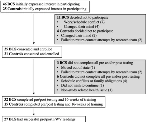
Figure 1 Recruitment and retention
Study Participants
Breast cancer survivors had been diagnosed with early-stage (0-III) breast cancer and were within one year of completing primary therapy (chemotherapy, radiation, surgery). Participants in the CTL group were age-matched, did not have a history of cancer, and self-reported they were physically active no more than 2 days per week. Both groups were free from overt cardiovascular, metabolic, or orthopedic limitations as reported by medical history
including Raleigh, Durham, Chapel Hill and surrounding areas recruited via electronic and paper fliers and word of mouth.
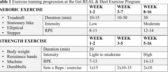
Intervention
For both groups, the 16-week intervention entailed supervised, progressive, aerobic and strength exercise training three days a week for approximately one hour per day (Table 1). Table 1 Exercise training progression at the Get REAL & Heel Exercise Program
AEROBIC EXERCISE WEEK1-2 WEEK3-7 WEEK8-16
• Treadmill • Stationary bike • Elliptical • Stepper
Duration (min) 10-15 10-30 30
Intensity Low Moderate
RPE 8-11 12-14
STRENGTH EXERCISE WEEK1-2 WEEK3-5 WEEK5-16
• Body weight • Resistance bands • Machine
• Dumbbells
Duration (min) 30
Intensity Light to moderate High
RPE 7-13 14-15
Sets x Reps / exercise 1x15 2x10-15 2x10
The supervised training took place at the UNC Get REAL & Heel (GRH) Exercise Program for cancer survivors, an off-campus facility at a convenient location in the community. Participants were asked to maintain their current lifestyle habits outside of GRH training in order to best evaluate the program-specific effects. A variety of equipment for both aerobic and
resistance/strength training has been used at GRH to adapt to individual participant fitness and mobility needs which allows trainers the ability to maximize patient safety and exercise
engagement. For example, participants could choose treadmills, stationary bikes or ellipticals for aerobic work and dumbbells, resistance bands, or machine weights for strength training,