of five riboswitches and one ribozyme
ZHICHAO MIAO,
1RYSZARD W. ADAMIAK,
2,3MACIEJ ANTCZAK,
3ROBERT T. BATEY,
4ALEXANDER J. BECKA,
5MARCIN BIESIADA,
2MICHA
Ł
J. BONIECKI,
6JANUSZ M. BUJNICKI,
6,7SHI-JIE CHEN,
8CLARENCE YU CHENG,
5FANG-CHIEH CHOU,
5ADRIAN R. FERRÉ-D
’
AMARÉ,
9RHIJU DAS,
5WAYNE K. DAWSON,
6FENG DING,
10NIKOLAY V. DOKHOLYAN,
11STANIS
Ł
AW DUNIN-HORKAWICZ,
6CALEB GENIESSE,
5KALLI KAPPEL,
5WIPAPAT KLADWANG,
5ANDREY KROKHOTIN,
11GRZEGORZ E.
Ł
ACH,
6FRANÇOIS MAJOR,
12THOMAS H. MANN,
5MARCIN MAGNUS,
5,6KATARZYNA PACHULSKA-WIECZOREK,
2DINSHAW J. PATEL,
13JOSEPH A. PICCIRILLI,
14,15MARIUSZ POPENDA,
2KATARZYNA J. PURZYCKA,
2AIMING REN,
13,16GREGGORY M. RICE,
17JOHN SANTALUCIA JR.,
18,19JOANNA SARZYNSKA,
2MARTA SZACHNIUK,
2,3ARPIT TANDON,
11JEREMIAH J. TRAUSCH,
4SIQI TIAN,
5JIAN WANG,
20KEVIN M. WEEKS,
17BENFEARD WILLIAMS II,
11YI XIAO,
20XIAOJUN XU,
8DONG ZHANG,
8TOMASZ ZOK,
3and ERIC WESTHOF
11–20[Author affiliations appear at end of paper.]
ABSTRACT
RNA-Puzzles is a collective experiment in blind 3D RNA structure prediction. We report here a third round of RNA-Puzzles. Five puzzles, 4, 8, 12, 13, 14, all structures of riboswitch aptamers and puzzle 7, a ribozyme structure, are included in this round of the experiment. The riboswitch structures include biological binding sites for small molecules (S-adenosyl methionine, cyclic diadenosine monophosphate, 5-amino 4-imidazole carboxamide riboside 5′-triphosphate, glutamine) and proteins (YbxF), and one set describes large conformational changes between ligand-free and ligand-bound states. The Varkud satellite ribozyme is the most recently solved structure of a known large ribozyme. All puzzles have established biological functions and require structural understanding to appreciate their molecular mechanisms. Through the use of fast-track experimental data, including multidimensional chemical mapping, and accurate prediction of RNA secondary structure, a large portion of the contacts in 3D have been predicted correctly leading to similar topologies for the top ranking predictions. Template-based and homology-derived predictions could predict structures to particularly high accuracies. However, achieving biological insights from de novo prediction of RNA 3D structures still depends on the size and complexity of the RNA. Blind computational predictions of RNA structures already appear to provide useful structural information in many cases. Similar to the previous RNA-Puzzles Round II experiment, the prediction of non-Watson–Crick interactions and the observed high atomic clash scores reveal a notable need for an algorithm of improvement. All prediction models and assessment results are available at http://ahsoka.u-strasbg.fr/rnapuzzles/.
Keywords: 3D prediction; bioinformatics; force fields; X-ray structures; models; structure quality
INTRODUCTION
Our growing knowledge of the biological functions of RNA
demands an increased rate of modeling the structures of
RNA. Riboswitches are mRNA segments, mostly located in
5
′UTRs that carry out regulatory functions. A riboswitch
un-dergoes conformational changes upon ligand binding and
functions as a switch in transcriptional or translational levels.
Aptamers are regions of RNA that selectively bind small
molecules, whereas riboswitches are natural RNA aptamers
embedded in leader sequences of genes. Since riboswitches
are functional and may include conformational changes,
the 3D structures of riboswitches are of vital importance
for understanding the molecular mechanisms of their
regu-latory functions. One of the aims of computational
predic-tions of 3D RNA structure is to help in the understanding
of the binding of small RNA molecules, the conformational
changes induced and
in fine
to contribute to the unraveling
of the molecular mechanisms of riboswitches.
RNA-Puzzles is a CASP-like (Moult et al. 2016) collective
blind experiment for three-dimensional (3D) RNA structure
Corresponding authors: [email protected], e.westhof@ ibmc-cnrs.unistra.fr
Article is online at http://www.rnajournal.org/cgi/doi/10.1261/rna. 060368.116. Freely available online through theRNAOpen Access option.
prediction evaluation. The aims are to identify the capacities
and bottlenecks in the RNA prediction problem. The
struc-tures to be predicted are unknown to the databases and to
the modelers and, thus, biases due to prior knowledge are
avoided. The prediction methods result from various
ap-proaches, but often combine fragment assemblies from
known structures present in databases and energy
minimiza-tions through various types of force fields adapted to the
granularity of the models and the stages of the modeling
(for a recent review on the commonly applied algorithms,
see Dawson and Bujnicki 2016). Experimental data, newly
collected (Cordero et al. 2014), can be also used as constraints
during the prediction process. Until now, 16 puzzles have
been set up and assessments of six puzzles were previously
published (Cruz et al. 2012; Miao et al. 2015). In the recent
and ongoing stages of RNA-Puzzles, we have strongly
en-couraged the development of novel, automatic, and efficient
RNA structure prediction algorithms to help the community
in understanding real-world RNA structure
–
function
rela-tionships, as well as to promote the development of
automat-ed and user-friendly web servers. Since the inauguration of
RNA-Puzzles, the field has progressed first and foremost
through the numerous discussions and exchanges between
the various modeling groups. This has led to agreed protocols
for delivery of models, descriptions of computations, and
assessments. At the same time, the automatization of the
modeling processes has steadily progressed. At this stage, it
is probably still too early to offer a comparative analysis of
the prediction and modeling methods.
Here we report a third round of RNA-Puzzles and we focus
on the prediction of RNA riboswitches and ribozymes,
evaluated on the basis of six RNA structures: the SAM-I
riboswitch aptamer, the SAM-I/IV riboswitch, the
ydaO
riboswitch, the ZTP riboswitch, the L-glutamine riboswitch,
and the Varkud satellite ribozyme. These molecules are
func-tionally significant as they can bind ligands, may include
conformational changes, or can catalyze chemical reactions.
Contributing to the stringency of this round, all six molecules
included regions without homology to previously solved
structures, and in most cases the problem required modeling
the entire structure de novo. According to the prediction
re-sults, we discuss several critical aspects of RNA 3D structure
predictions: (i) the prediction of RNA noncanonical
con-tacts, (ii) the prediction of structural topology, and (iii) the
understanding of small molecule binding and the induced
conformational changes.
We find that RNA 3D structure prediction has already
achieved a high level of accuracy for template-based and
homology-based structure predictions and, thus, can
al-ready contribute significantly to our understanding of the
underlying molecular mechanisms in some cases. The
prediction of ligand binding and the resulting
conforma-tional changes are also possible but cannot be guaranteed.
For a large de novo structure, the prediction is still a
diffi-cult endeavor.
RESULTS AND DISCUSSION
The five RNA-Puzzles on riboswitches
Puzzle 4: SAM-I riboswitch aptamerThis SAM-I riboswitch problem is an aptamer where the P3
helix is engineered as an extended helix (Baird et al. 2012). It
binds an
S-adenosyl methionine (SAM) molecule in its
cen-ter and can bind L7Ae-like proteins (YbxF and YlxQ) at the
K-turn module. The 126-nucleotide (nt)-long sequence is
the following:
5
′-GGCUUAUCAAGAGAGGUGGAGGGACUGGCCCGA
UGAAACCCGGCAACCACUAGUCUAGCGUCAGCUU
CGGCUGACGCUAGGCUAGUGGUGCCAAUUCCUG
CAGCGGAAACGUUGAAAGAUGAGCCA-3
′After the prediction deadline, the 2.8 Å diffraction
resolu-tion structure was deposited in PDB with ID 3V7E. Before the
experiment, homologous structures identical to this
particu-lar riboswitch were already available (Stoddard et al. 2010).
The SAM binding position is identical to the prior structure
(see
Supplemental Fig. S1
), and the ligand-free state (3IQP)
also adopts the same topology. The L7Ae/YbxF
protein-bind-ing region is easily detected as a K-turn module. The RMSD
of aligned parts between previously available 3IQR and the
new protein-bound coordinates 3V7E is <1 Å. The difference
lies on the engineered P3 helix and the minor deviations
of the P4 helix. Therefore, this puzzle is a template-based
prediction of high-resolution modeling.
Puzzle 8: SAM-I/IV riboswitch
The SAM-I/IV riboswitch aptamer is a structure with
homol-ogies to both SAM-I and SAM-IV families (Trausch et al.
2014). It binds SAM in a region similar to the SAM-I
ribos-witch but may originate from a different ancestor. The
sequence is 96-nt long:
5
′-GGAUCACGAGGGGGAGACCCCGGCAACCUGGGAC
GGACACCCAAGGUGCUCACACCGGAGACGGUGGA
UCCGGCCCGAGAGGGCAACGAAGUCCGU-3
′Puzzle 12:ydaO riboswitch
Two cylclic diadenosine monophosphate (c-di-AMP)
mole-cules bind the
ydaO
riboswitch, which is involved in
sporula-tion, osmotic stress responses, and cell wall metabolism, in
two pseudo-symmetry-related pockets (Ren and Patel 2014).
The sequence of the 108-nt
ydaO
riboswitch is as follows:
5
′-AUCGCUGAACGCGGGGGACCCAGGGGGCGAAUCU
CUUCCGAAAGGAAGAGUAGGGUUACUCCUUCGAC
CCGAGCCCGUCAGCUAACCUCGCAAGCGUCCGAA
GGAGAA-3
′The structures of the complex were solved at resolutions
2.65 Å (binding with c-di-dAMP, PDB 4QLN) and 2.72 Å
(binding with AMP, PDB 4QLM). Although a
c-di-GMP bound riboswitch was previously solved, the
ydaO
riboswitch is a new structure topology and is difficult to
pre-dict. When predicting the two c-di-AMP binding pockets,
non-Watson
–
Crick edges of specific aptamer nucleotides
directly contacting the ligands were unknown.
Puzzle 13: ZTP riboswitch
A ZTP (5-amino 4-imidazole carboxamide riboside 5
′-tri-phosphate) riboswitch can up-regulate de novo purine
syn-thesis in response to increased intracellular levels of ZTP or
ZMP (Trausch et al. 2015). The sequence of the ZTP
ribos-witch structure is of 60 nt:
5
′-GGGUCGUGACUGGCGAACAGGUGGGAAACCACCG
GGGAGCGACCCGCCGCCCGCCUGGGC-3
′Two PDB structures were solved at resolutions 2.5 Å (PDB
4XW7) and 1.8 Å (PDB 4XWF), respectively. Despite the lack
of a homologous structure, the secondary structure of the
riboswitch is relatively simple and the size of the structure
is small, which facilitated the prediction.
Puzzle 14: L-glutamine riboswitch
The L-glutamine riboswitch goes through dramatic
confor-mational changes in the P3 helix upon glutamine binding
(Ren et al. 2015). The length of the structure is 61 nt. Two
sequences, corresponding to constructs used to crystallize
li-gand-bound and ligand-free versions of the aptamer, were as
follows:
5
′-CGUUGACCCAGGAAACUGGGCGGAAGUAAGGCCC
AUUGCACUCCGGGCCUGAAGCAACGCG-3
′(Bound)
5
′-CGUUGGCCCAGGAAACUGGGUGGAAGUAAGGCCC
AUUGCACUCCGGGCCUG AAGCAACGCU-3
′(Free)
The ligand-free state structure was solved at resolution
3.1 Å and deposited in PDB with ID 5DDO, while three
L-glutamine bound structures were solved: 5DDP at 2.3 Å,
5DDQ at 2.4 Å, and 5DDR at 2.61 Å resolution. Although
5DDQ and 5DDR were solved in Mn
2+-soaked and Cs
+-soaked conditions, their structural differences from 5DDP
are subtle. The structural modules GAAA tetra-loop and
U1A-protein-binding loop, engineered to replace Loops
L2 and L3, were interesting in prediction. For
conforma-tional changes, the nucleotides G22 and G23, disordered
in the free state, form critical long-range interactions in the
ligand-bound state. The correct prediction of these
interac-tions was expected to influence overall prediction accuracy.
The RNA-Puzzle on a ribozyme
Puzzle 7: the Varkud satellite (VS) ribozymeBesides riboswitches, we also report in this round of
experi-ments the prediction of a self-cleaving ribozyme. The Varkud
satellite ribozyme (Suslov et al. 2015), as part of VS RNA, is
the largest known small nucleolytic ribozyme. The 185-nt
se-quence covers residues 601
–
785 of VS RNA:
5
′-GCGCUGUGUCGCAAUCUGCGAAGGGCGUCGUCG
GCCCGAGCGGUAGUAAGCAGGGAACUCACCUCCA
AUGAAACACAUUGUCGUAGCAGUUGACUACUGUU
AUGUGAUUGGUAGAGGCUAAGUGACGGUAUUGG
CGUAAGCCAAUACCGCGGCACAGCACAAGCCCGCU
UGCGAGAUUACAGCGC-3
′Two single site mutant structures of the VS ribozyme were
solved to the same resolution of 3.07 Å, PDB 4R4P and 4R4V.
The structure deviations between the two structures are slight.
However, the structure is large and no homologous structure
was available before the experiment, making it a difficult
problem. Furthermore, the RNA crystallized as a dimer, and
modelers were challenged with predicting a complex with a
total size of 370 nt, the largest RNA-Puzzle problem to date.
Experimental data description
The Das group provided
“
fast-track
”
experimental data to all
the modelers for puzzles 7, 8, 12, 13, and 14.
One-dimension-al chemicOne-dimension-al mapping using SHAPE, CMCT, DMS, and
hy-droxyl radical footprinting and multidimensional chemical
mapping measurements based on mutate-and-map (M
2)
and multiplexed
•
OH cleavage analysis (MOHCA-seq, for
targets 12
–
14) were acquired as described in Cordero et al.
(2014), Kladwang et al. (2014), and Cheng et al. (2015a).
Data were distributed via the private RNA-Puzzles website
or via entries with anonymized sequences in the RNA
Mapping Database (Cordero et al. 2012); deposition IDs
are summarized in
Supplemental Table S8
.
Overall comparison results
Assessment methods
with more than one crystal structure. When multiple
struc-tures were available, we assumed the RNA might populate
a diverse ensemble of structures and every structure is a
pos-sible native structure. Hence, when assessing the prediction
quality of a structure, comparisons were made with all
available crystal structures. Up to now, there is no single or
universal metric that can be considered as the major
determi-nant of the overall accuracy of a predicted model. Therefore,
we use a set of metrics to assess all the models. Because it
is the most commonly used and obvious metric, the root
mean square deviation (RMSD) between the predicted
mod-els and the crystal structure is used for ranking the modmod-els.
RMSD is a metric for global topology comparisons, but it
spreads the errors all over the structure. Indeed, we can
find some special cases where the RMSD shows a different
ranking from other metrics. Different metrics were used to
assess different aspects of the predictions: RMSD stands for
the global similarity of all the atoms; deformation index
(DI) and the complete deformation profile matrix (DP) stand
for prediction accuracy of the nucleotide interactions,
while the interaction network fidelity (INF) assesses the
in-teraction accuracies at different levels (Parisien et al. 2009);
the Clash score evaluated by MolProbity (Chen et al. 2010)
assesses the atomic harmony of the structure, and the mean
of circular quantities (MCQ) score (Zok et al. 2014) assesses
the structural similarity with the native structure in the
torsion angle space. Each of those metrics, because they
assess very different structural characteristics, has
advan-tages and drawbacks. Thus, DI, which stands for the local
pairwise superimpositions, does not show the differences
when all predicted structures are far away from the native
structure. INF defines the quality of a certain type of
predicted interaction but not all the elements. Clash score
only demonstrates the reasonability of the atomic distances
and is not discriminative. MCQ compares the dihedral
angles without considering bond lengths or bond angles.
We now add radar diagrams (
Supplemental Figs. S21
–
S27
)
to give a general idea and an overview of the scores related
to the first ranked and best RMSD models of the
parti-cipating groups. The advantage of using a set of metrics
assessing various molecular characteristics is that it shows
the qualities and deficiencies of the various algorithms as
a function of the size and type of RNA molecule being
predicted.
The five RNA-Puzzles on riboswitches
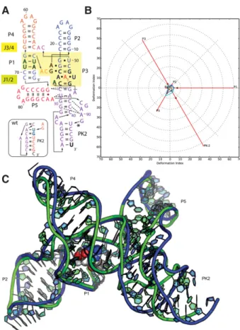
Puzzle 4: the SAM-I riboswitch aptamer (see Fig. 1;Supplemental Figs. S1,S2)
Because of the availability of homologous templates, such as
3IQP, the prediction accuracy of Puzzle 4 is extremely high.
As shown in
Supplemental Table S1
,
28
out of 30 total
predicted structures have a RMSD within 6 Å, while 10
prediction models from the Chen laboratory are within
3.5 Å. The Watson
–
Crick base pairs were perfectly predicted.
Generally, a better prediction always includes a better
identi-fication of non-Watson
–
Crick pairs and of stacking contacts.
Further, we find the prediction models from the Chen
labo-ratory are well optimized for atomic clashes. This indicates
that a very good level of high-resolution homology modeling
of RNA structures has been achieved (see Fig. 1). The Das
group provided predictions of SAM binding and the
ori-entation of the SAM that are very close to the native binding
position, shown in
Supplemental Figure S3
. In homology
modeling, the SAM binding region can also be inferred
from known templates. The contacts between SAM and the
riboswitch are compared in
Supplemental Figure S4
; most
of the contacts (mainly hydrogen bonds) predicted by Das
model 1 have been predicted in a correct manner. Several
groups also predicted the fold of the YbxF protein and its
binding of the SAM I K-turn. The availability of prior
tem-plates for K-turns bound to proteins such as L7Ae allowed
these groups to achieve near-atomic accuracy (Fig. 1). Most
of the groups predicted the protein at the right positions,
since the L7Ae-Kturn binding is well known.
Puzzle 8: the SAM-I/IV riboswitch (see Fig. 2;Supplemental Figs. S5,S6)
Although no high-resolution structural template exists for
this puzzle, structural clues about the SAM-I/IV riboswitch
can be derived from SAM-I riboswitch structures.
Potential-ly, similar distant-homology-based predictions could be an
important application of RNA structure prediction in
inter-preting molecular mechanism and biological functions.
Even if only four out of 42 predictions were predicted below
7 Å RMSD, the best one is still within 5 Å RMSD (
Supple-mental Table S2
). This model, from the Das group, correctly
predicts helix P5 stacked against the backbone of pseudoknot
PK2 in an unexpected manner (Fig. 2). Even at 11 Å RMSD,
many prediction models could potentially be helpful in
un-derstanding the structure. In the top ranking cases,
Wat-son
–
Crick pairs and stacking were predicted to a very high
level, but the prediction of the non-Watson
–
Crick pairs still
needs to be improved. Although no SAM binding was
pre-dicted in this puzzle, the SAM binding site can be inferred
from related structures since they maintain identical
contacts. A comparison between SAM-I riboswitch and
SAM-I/IV is shown in
Supplemental Figure S6
.
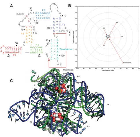
Puzzle 12: the ydaO riboswitch (see Fig. 3;Supplemental Figs. S7,S8)
The
ydaO
riboswitch can be defined as a
“
difficult case
”
because of its relatively large size, its lack of homology to
any previously solved RNA structures, and because of
bind-ing effects of the two ligands, which requires special
consid-eration of the availabilities of the non-Watson
–
Crick edges.
The RMSD of the 51 submitted predictions range from 10
to 36 Å with an average value of 16.6 Å, as shown in
Supplemental Table S3
. The P4 and P5 helical regions, longer
and thus more stable, are better predicted, while P2 and P3
are worse. The bubble between P2 and P3 helix was mostly
unresolved in the X-ray map, implying that this region
could be less stable in structure. Nevertheless, independent
crystallographic solutions of homologous c-di-AMP
struc-tures (Gao and Serganov 2014; Jones and Ferré-D
’
Amaré
2014) (also released after RNA-Puzzle 12 modeling) showed
strong agreement with all resolved parts of the crystal
struc-ture considered herein, suggesting that the overall fold is
well defined and a valid target for prediction. Many of the
predicted structures do not fully consider the binding of
the two c-di-AMP ligands, but the global topologies of
the top ranking models are still visually similar to the X-ray
structure (Ding group model in Fig. 3). The pseudoknot
and the bubble are difficult to predict in this puzzle,
and the superimposition of the best model is shown in
Supplemental Figure S8
. The pseudoknot is very well
pre-dicted while the bubble region is poor in all models, likely
due to an incorrect secondary structure used by most
model-ers. This may be largely related to the flexibility of the
struc-ture, as the pseudoknot includes many base pair interactions
and is stable. However, the bubble is too flexible so can
only be partly solved in crystal and the prediction is thus
more difficult.
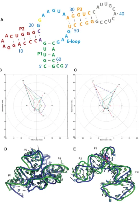
Puzzle 13: the ZTP riboswitch (see Fig. 4;Supplemental Figs. S9,S10)
Like Puzzle12, the ZTP riboswitch is a full de novo prediction,
but the size of the structure is relatively small and mainly
com-prised of Watson
–
Crick interactions. Thus, the top two
pre-dictions, from the Das group, achieved RMSD within 6 Å,
strikingly similar to the native topology, shown in
Supple-mental Table S4
. According to Figure 4, we find that the
pre-dicted structure adopts exactly the same fold as the native
structure, except that the curvature of the helix deviates
slightly. The predicted structure gives a structure model of
the single-stranded loop between P1 helix and P3 helix, which
is not solved by crystallography. This loop region may be too
dynamic for a unique conformation and the predicted
struc-ture is also disordered. For the ZMP binding, the coordinates
FIGURE 2. Puzzle 8: SAM-I/IV riboswitch. (A) Secondary structuregiven by the Das laboratory model 7 were quite close to the
na-tive binding region, but with an opposite orientation that is
less buried in the RNA structure. As shown in
Supplemental
Figure S10
, the native ZMP binding position is between Das
model 1 and model 7.
Puzzle 14: the L-glutamine riboswitch (see Fig. 5;Supplemental Figs. S11–S14)
The L-glutamine riboswitch is the first RNA molecule in
RNA-Puzzles for which a large conformational change upon
ligand binding has been experimentally captured. The
se-quences of molecules used to crystallize both the free and
bound states were released for the prediction experiment.
This puzzle points to the important question of how well
RNA conformational change can be predicted by
state-of-the-art methods. If a prediction can achieve reasonable
quality in both states, it increases the likelihood that de novo
RNA structure prediction might provide useful
hypo-theses of explanations for molecular mechanism in the near
future. Structure comparisons between predictions and native
structures are available in Figure 5, while 2D heat maps of
deformation profiles are demonstrated
in
Supplemental Figures S11
–
S13
.
Further, to begin quantifying the help
of the inclusion of additional
experimen-tal data, Puzzle 14 was divided into
“
pre-experiment prediction
”
and
“
post-exper-iment prediction.
”
Fifty-one predicted
structures of free state and 64 of bound
state are listed in
Supplemental Table S5
and
Supplemental Table S6
. The best
pre-diction of free state is from the
“
post-ex-periment
”
model 2 of the Das laboratory
that is 6.5 Å RMSD away from native
structure, while the best for bound state
is from
“
pre-experiment
”
model 2 of
the Bujnicki laboratory, which is 5.0 Å
RMSD away from the native structure.
Interestingly, these most accurate
predic-tions were made with Rosetta FARFAR
methods and are capable of explaining
the ligand-induced conformational
chan-ge, but the worst cases in prediction are
quite far away,
∼
20 Å RMSD. In the free
state prediction (
Supplemental Table
S5
), only two of the top 10 models are
pre-experiment ones: Das
pre-experi-ment models 1 and 9 ranking at second
and eighth. However, six out of the top
10 models are pre-experiment
predic-tions in the bound state prediction, while
the top three models are also
pre-experi-ment ones. Consequently, utility of
exper-imental data (or at least the additional
time allowed for modeling) on the free state prediction
appears clear; but such improvement is not detected in the
bound state modeling. The Bujnicki pre-experiment models
1
–
4 are the best models for bound state predictions, but after
the integration of experimental data, these models were not
recognized as the best ones and were not included in
post-experiment models, suggesting that either the post-experimental
data were misleading or were not helpful in late-stage
refine-ment of these models.
A Loop E motif, present in the Puzzle14 structure, is a
critical structural module in RNA structure and was
recog-nized previously based on sequence conservation (Ames
and Breaker 2011). The prediction of the Loop E structure
is compared in
Supplemental Figure S14
for both free and
bound states. In the free state, only Das group
post-experi-ment model 2 gave the right prediction. In the bound state,
Bujnicki group pre-experiment model 2 and Das group
pre-experiment model 6 are the best predicted ones. The
well-predicted loop E modules are always detected in the
top ranking predictions, which underscores the importance
of predicting correctly any well-known structural modules
for the final resulting model.
The RNA-Puzzle on a ribozyme
Puzzle 7: the Varkud satellite ribozyme (see Fig. 6;
Supplemental Figs. S15–S13)
As the only ribozyme in this round of RNA-Puzzles and the
largest of the small nucleolytic ribozymes, the VS ribozyme
was very difficult to predict de novo, although much
struc-tural data and many models were previously available
(Wilson and Lilley 2011). The RMSD of the predictions range
from 20 to 60 Å, with a mean value of 29 Å. Figure 6B
dem-onstrates that the best models from, e.g., the Das group
mod-el 1, accuratmod-ely predict the important modules. Neverthmod-eless,
the relative orientation between the different helical
struc-tures in 3D space is difficult to predict. In
Supplemental
Figure S16
, all of the 7 hairpins and internal stems (P1
–
P7)
are compared between native and prediction. Although the
predictions are each similar to the native, small deviations
of non-Watson
–
Crick interactions then led to global
topo-logical differences clearly detected in
Supplemental Figure
S16A,D,F
. In particular, the P2
–
P3
–
P6 junction was
incor-rectly assumed in prior modeling efforts and RNA-puzzle
modeling herein. Using
“
standard
”
rules (Lescoute and
Westhof 2006) and characterization of the junction in
isola-tion (Lafontaine et al. 2001), P3 and P6 were expected to be
coaxially stacked as there are no unpaired nucleotides
be-tween them; but the crystal structure revealed these helices
to be separated.
The native structure adopts a more expanded state than
the predicted structures. Still, we find that the prediction
of non-Watson
–
Crick interactions is poor, as shown in
Supplemental Table S7
, although the structure topology
could be softened by these noncanonical interactions.
However, such deviations in local structure may result in
topological change between different structural domains.
As a further challenge, the RNA crystallized as a dimer,
swapping the stem P1 holding the cleavage site between
the two partners. These reasons explain the poor global
RMSD that could be achieved for this structure. Therefore,
further efforts are still needed in predicting large multimeric
structures de novo.
General comments
Model ranking
For each Puzzle, predictors were allowed to submit up to
10 structural models that they ranked from the most
reli-able model down to the least relireli-able ones. This ranking
should constitute a direct measure of the quality of the
overall scoring function and especially of its correlation
with structural models as derived from crystallography.
As for automated structure prediction web servers, one
is likely to consider only the first few models. However
important, ranking the prediction models is a nontrivial
but practical step.
If we only take the first model into consideration, the
ac-curacies of the prediction worsen substantially. For example,
for Puzzle 12, a model of the Ding group gives the best RMSD
to the X-ray structure, but that model was ranked worst (12th
of 12 submissions) from that group during blind modeling.
The same group
’
s top-ranked model is only the 18th best
model and brings a 5 Å decrease in RMSD compared with
their most accurate model, as assessed post facto. The bound
state of Puzzle 14 is another case where models with accurate
global folds were submitted but were ranked low by the
mod-eling groups on their ranked lists. To give a better view on
re-lationships between highly scored models and their ranking
with respect to different metrics, we provide radar diagrams
illustrating the relative rankings built for the first models and
for the best RMSD models (
Supplemental Figs. S21
–
S27
).
They clearly show that the best RMSD model is not
necessarily the best in terms of all measures. In extreme cases,
highly scored models in one ranking can be significantly
worse than top-ranked models in the other ranking. Thus,
we should be aware of how important it is to consider various
evaluation measures when assessing the results of 3D
struc-ture prediction.
Crystal derived B-factor values and the quality
of the predictions
In
Supplemental Figures S2
,
S5
,
S7
,
S9
,
S11
–
S13
,
S15
,
the 2D heat maps of the deformation profiles are used to
demonstrate the prediction accuracy
locally in each part between the
pre-dicted and the crystal RNA structures.
The majority of the heat maps point
to high accuracies of the top ranking
models, except Puzzle 12 (RMSD 10 Å)
and Puzzle 7 (RMSD 20 Å). According
to
Supplemental Tables S1
–
S7
, there
is a good correlation between RMSD
and deformation profile (DI all).
Gen-erally, accurate prediction requires good
relative positions between local parts.
Further,
the
B-factors
plotted
are
aligned on the left of the heat maps as
histogram plots. We find many of the
badly predicted regions correspond to
higher B-factor values, which suggests
that these nucleotides or large RNA
seg-ments are highly mobile or that several
of the nucleotide atoms may not have
clear density in the electron density
map. In such cases, the X-ray data may
not be sufficient to fully determine
the coordinates of such nucleotides and
the derived coordinates may present
large errors. For example, the high
B-factor region of Puzzle 4 (three residues
in the apical loop of the P3 extension)
corresponds to those parts with the
highest deformation profile and thus
potentially badly modeled, either by the
crystallographers or the modelers, or
that this region does not assume a single
conformation, especially in solution.
Examination of the electron densities of
these three high B factor residues in
Supplemental Figure S17
reveals that
the coordinates of the crystal structure
do not fit the electron densities well.
Hence, it is difficult to assess the
accuracy of the predictions for those
nucleotides. Leaving out such residues,
the majority of the local structure parts
have been predicted quite accurately.
This suggests that RNA 3D structure
prediction may help build better
struc-tural models in crystallography or in
conjunction with other structural
deter-mination methods.
MATERIALS AND METHODS
In the following, we briefly introduce the methods used and focus on the new updates and special treatments in the prediction.
Adamiak group
In all the puzzles, a fully automated prediction method (Popenda et al. 2012) provided by RNAComposer (http://rnacomposer.ibch. poznan.pl and http://rnacomposer.cs.put.poznan.pl) was applied to conduct the RNA 3D structure prediction. RNAComposer is a knowledge-based method that employs automated fragment assembly, based on the secondary structure tree graph represen-tation and homology of structural elements. At present the RNAComposer dictionary contains as much as 490,000 3D struc-ture elements. Models delivered by RNAComposer are energy min-imized and refined.
The prediction fidelity of RNAComposer depends critically on the accuracy of the RNA secondary structure, used as an input (Popenda et al. 2012; Purzycka et al. 2015). Therefore, secondary structures predicted in silico using tools integrated in the RNAComposer system were adjusted according to experimental data, if available. The Adamiak group also applied Rfam (Gardner et al. 2009) to identify conserved base pairs (Puzzle 13). Information about pseudoknots was obtained by manual analysis of the experimental data (Puzzles 7, 8, and 13) and/or based on the literature review (Puzzles 12 and 13).Supplemental Table S9
presents input RNA secondary structure topologies applied to the considered puzzles. Information about pairing patterns within the pseudoknots was introduced into the RNAComposer dot-bracket annotated input using square brackets. The structure of each poten-tial pseudoknot was additionally refined using distance restraints derived from canonical A-RNA structure (Supplemental Table S9)
applying the described procedure (Huang et al. 2013). In some cases, the generated 3D structures did not correlate with long-dis-tance interactions or relative positioning of the helixes deduced from the provided ex-perimental data (i.e., Puzzle 8). This prompt-ed us to develop new functionality of the RNAComposer system (Antczak et al. 2016) that allows the user to introduce a particular, user-defined 3D element for the structure as-sembly. This functionality was subsequently utilized in solving Puzzles 13 and 14. Difficulties in predicting some interactions from the experimental data resulted in anoth-er improvement of the RNAComposanoth-er sys-tem. New algorithms are being developed that will increase the pool of promising 3D el-ements that can be applied for 3D structure assembly by allowing the user to explore RNA FRABASE (Popenda et al. 2008, 2012) using new wild card characters. Such charac-ters will be introduced in the definition of the query patterns.
All 3D models were evaluated using the criteria of total energy and right geometry to exclude knotted structures, and those that did not fulfilled experimental restraints cri-terion. Final 3D models were verified using RNApdbee server (Antczak et al. 2014), avail-able at http://rnapdbee.cs.put.poznan.pl, to confirm that input secondary structure topol-ogy is preserved. Our RNA 3D models are highly ranked according to measured INFWC.
The Adamiak group was able to generate hundreds of 3D struc-ture models very fast. The major limitation was a manual analysis of provided experimental data, and identification of these 3D mod-els that agreed best with the experimental data within the given time.
Puzzle 4
The Adamiak group focused on the prediction of the RNA–protein complex (Fig. 1). The high level of homology forB. subtilisYbxF protein (sequence: GSYDKVSQAKSIIIGTKQTVKALKRGSVKEV VVAKDADPILTSSVVSLAEDQGISVSMVESMKKLGKACGIEVGA AAVAIIL) allowed us to obtain a protein 3D model based on homol-ogous modeling with I-TASSER (Yang et al. 2015).
RNA secondary structure was extracted from the SAM-I ribos-witch ofB. subtilis(PDB id: 3NPB) and used to predict RNA 3D structure in the fully automated mode of RNAComposer. Three-di-mensional models of RNA/protein complexes were obtained using HADDOCK (Dominguez et al. 2003). The Adamiak group defined protein–RNA interfaces based on the structures of L7Ae bound to the K-turn motif in box C/D RNA (1RLG—A. fulgidus L7Ae, 1SDS—M. jannaschiiL7Ae, 3PLA—S. solfataricusL7Ae) as well as flexible regions (“semiflex”and “fullyflex”) in RNA and protein. From obtained 3D models of RNA/protein complexes, five were chosen that showed the highest score returned by HADDOCK and the number of well-defined, specific hydrogen bonds between RNA and protein. For model 3, the RMSD value (4.008 Å) of the predicted complex was nearly identical to the RNA component
itself (RMSD 4.091 Å). Outermost results were obtained when the “fullyflex”option was used upon docking.
Puzzle 8
After introduction of additional restraints on the residues constitut-ing the pseudoknot (Supplemental Table S9), obtained 3D models were inspected for the agreement with the provided Mutate-and-Map data. However, the predicted 3D models did not preserve the tertiary interactions deduced from the provided experimental data. Therefore, the Adamiak group decided to submit the best two 3D structure models based on the total energy calculated by RNAComposer. Those models displayed very good INFNWC
(0.612 and 0.548) as depicted onSupplemental Figure S23.
Puzzle 12
The RNA secondary structure topology was based on published data (Nelson et al. 2013). Topologies with 5 or 6 base pairs (bp) forming the pseudoknot were used as the RNAComposer input (Supplemental Table S9). Additional base pairs 27–101 and 32–37 were also introduced based on expert analysis of initially predicted 3D models.
Puzzle 13
Secondary structure was developed based on the data published by Kim et al. (2015). New functionality of RNAComposer was imple-mented (Antczak et al. 2016), and user-defined 3D structure elements for the internal loop (7-UGACUGGCGAACAG-20; 33-CGGGGAG-39 and the single strand: 7-UGACUGGCGAACAG-20) were introduced. Those structural elements were identified in RNA FRABASE (Popenda et al. 2008, 2010) using our own scripts. Thirty-three structurally diverse 3D elements displaying 25%–42% of sequence identity and nine with the best sequence identity were chosen for the internal loop and for the single strand element, re-spectively. The Adamiak group applied all combinations of those 3D elements in the automated model assembly by RNAComposer and obtained 297 highly different resultant 3D structures. Subsequently, the final 3D models were chosen according to highest agreement with the experimental data and acceptable total energy delivered by RNAComposer. The 3D structure model displaying the best compatibility with experimental data was submitted. This model shows the best INFWCscore out of all (55) ranked 3D
struc-tures (Supplemental Fig. S25).
Puzzle 14
RNA secondary structure generated using RNAfold (Zuker and Stiegler 1981) was analyzed and adjusted. The G5–C55 base pair was substituted with G23–C55. This change allows RNAComposer to automatically recognize the internal loop: 23-GAAGUAA-29; 50-UGAAGC-55, as E-loop motif. Such a motif was previously re-ported also for glutamine aptamers (Ames and Breaker 2011). RNAComposer generated ten 3D structures. Two structures with the extreme values of radius of gyration were selected. Analysis of provided MOHCAseq data resulted in identification of contacts be-tween regions nt 9–10 : 26–29 : 49–52 and nt 16–18 : 28–31. In order to force preservation of those contacts in the resultant 3D model with the lowest radius of gyration, the three-way junction 4-UGAC-7; 20-GCGG-23; 55-CA-56 was manually adjusted in
PyMol. This 3D motif was used as a user-defined element during 3D model assembly (Antczak et al. 2016). 3D models predicted by RNAComposer are characterized by very good local agreement with the reference structure in regard to the E-loop motif and 10 nt apical loop, and are therefore ranked within the top five 3D models within the MCQ category (Zok et al. 2014) and all INF-based categories (Supplemental Fig. S26).
Puzzle 7
The RNA secondary structure was adjusted based on provided ex-perimental data, and base-pairing between regions 27–35 and 90– 97 was anticipated from this analysis. Due to the difficulties in prediction of 3D structures preserving mentioned base-pairing in-teractions and appropriate total energy score, hundreds of various 3D models were generated. The five most diverse 3D models were finally submitted. Since interactions that occur between 27–34 and 90–97 are inter- rather than intramolecular (Suslov et al. 2015), ap-plied experimental data resulted in substantially decreased accuracy.
Bujnicki group
The Bujnicki group used a hybrid modeling strategy (Piatkowski et al. 2016) based on the approach tested in the previous editions of the RNA-Puzzles experiment (Cruz et al. 2012; Miao et al. 2015). If the target sequence exhibited detectable similarity to an RNA with known experimentally determined structure (as happened in the case of Puzzles 4 and 8), the Bujnicki group generated models of the whole molecule or its parts using a template-based (compar-ative) modeling method ModeRNA (Rother et al. 2011b) or its server version (Rother et al. 2011a). Remodeling of uncertain regions and modeling of RNA molecules that lacked suitable templates relied mostly on template-free folding using the coarse-grained method SimRNA (Boniecki et al. 2016). In the course of the competition, the Bujnicki group experimented with various versions of the SimRNA force field. For instance, in Problems 12, 13, and 14 they tested different variants of the force field, including ones that could be more easily extrapolated to the standard Turner energy rules (Xia et al. 1998), and introduced further concepts from polymer chemis-try (Flory 1969). This is an ongoing project wherein improvements are being continually added. In the course of the experiment the Bujnicki group has also introduced a completely automated version of the program available as the SimRNAweb web server (Magnus et al. 2016). Wherever available, template-free folding was aided by spatial restraints obtained from computational predictions (e.g., in-formation on the secondary structure or orientation of helices in junctions) and from data identified in the literature or made avail-able in the course of the RNA-Puzzles experiment by the Das group. For high-resolution refinement of models, the Bujnicki group used the QRNAS method (J Stasiewicz and JM Bujnicki, unpubl.) that ex-tends the AMBER force field with energy terms explicitly modeling hydrogen bonds, idealizes base pair planarity, and regularizes the backbone conformation.
from one to a few days per target (depending on the size of the sequence modeled in the template-free mode), and the final refinement was typically run overnight. In addition, the introduction of various restraints often required extensive time developing additional scripts to handle the unique problems inherent in each Puzzle.
The Bujnicki group also explored a completely new way of 3D ge-ometry prediction for RNA molecules. This project was started by M. Magnus during his internship at Stanford with R. Das and was inspired by a method to combine structure prediction runs across diverse homologs, as is carried out in protein structure predic-tion (Bonneau et al. 2001). Based on the observapredic-tion that sequences from the same RNA family fold into similar structures, the Bujnicki group explored the possibility that a similar process can be observed in computational modeling and could be used to detect the global helical arrangements for a given target sequence based on the ar-rangements within a subset of homologs. The proposed method explores the use of multiple sequence alignment information and parallel modeling of RNA homologs to improve 3D structure predic-tion over modeling of single RNA sequences. To build a structural model of the target sequence, a multistep modeling process is per-formed. First, for the target sequence, a subset of homologous sequences is selected using the Rfam database. Subsequently, inde-pendent folding simulations are carried out for these homologs; in this series of RNA-Puzzles experiments the Bujnicki group used ROSETTA/FARNA (Das and Baker 2007), but in principle any tem-plate-free folding method can be used. Structural fragments corre-sponding to the evolutionarily conserved regions (in particular helices)—determined from the alignment—are extracted from all obtained models and clustered to identify the most common struc-tural arrangement. The Bujnicki group also explored a way to con-strain the simulation by keeping the conserved residues identified by the alignment as being in close spatial proximity. The approach was used in modeling of Puzzles 13 and 14. In a blind prediction of Puzzle 13, a model obtained with this methodology was second, and in a Puzzle 14 (bound form), one model was best in terms of the RMSD to the reference structure. This approach is now under further systematic tests in preparation to make it automated and available for the community. The current version of the program, documentation, and input files to solve Puzzles 13 and 14 can be ac-cessed under a github repository at https://github.com/mmagnus/ EvoClustRNA.
Puzzle 4
For Puzzle 4, the Bujnicki group submitted the structure of the com-plete RNA–protein complex. The models of both RNA and protein components have been constructed using homology modeling, as suitable templates could be found in both cases. The RNA part was based on a homologous Thermoanaerobacter tengcongensis
SAM-I riboswitch structure (PDB ID: 3IQP) used as a template. The structure of the YbxF protein was predicted by fold-recognition using the GeneSilico metaserver (Kurowski and Bujnicki 2003), fol-lowed by template-based modeling using the L7Ae protein structure (PDB ID: 2FC3) as a template, with MODELLER (Sali and Blundell 1993). RNA–protein docking was performed using a procedure that in the meantime has been automated and now is available via the NPDock web server (Tuszynska et al. 2015). The models turned out to be quite accurate (the best one had 3.99 Å RMSD to the ref-erence structure).
Puzzle 7
This RNA structure was predicted in a template-free mode. The Bujnicki group knew from the problem description that the target sequence crystallized as a dimer. Following this clue, the Bujnicki group generated two types of dimers, all based on an initial model of a monomer, generated with restraints on secondary structure. Two types of dimers were proposed: one type obtained by manual docking of two copies of the monomeric structure, and another obtained by domain swapping. The domain-swapping hypothesis was later found to be incorrect. While the experimen-tally determined structure forms a dimer using intermolecular base pairs between residues 31–33 in one chain and 92–94 (number-ing accord(number-ing to the target sequence) in the other chain, the Bujnicki group assumed that these residues form intramolecular pairs within the monomer, leading to a distortion of the global ar-chitecture, despite otherwise correct predictions of other structural elements.
Puzzle 8
This RNA structure was predicted by template-based modeling, based on the structure of theS-adenosylmethionine riboswitch reg-ulatory mRNA element (PDB ID: 2GIS), followed by template-free refinement with restraints on secondary structure, including a pseu-doknot. The models turned out to be quite accurate (the best one had 6.71 Å RMSD to the reference structure).
Puzzle 12
The structure was modeled in a template-free mode, using restraints on secondary structure derived from a search of Rfam and other sources for homologous sequences with known structures. Simulations were run in two versions: with and without the predict-ed pseudoknot. The Bujnicki group also introducpredict-ed some mutate-and-map restraints based upon visual estimation of intensity and a very simple in-house program for reading the mutate-and-map data provided by the Das laboratory. Based on information provided for this problem, the Bujnicki group assumed there should be a binding pocket for cdiAMP and they filtered the models obtained based on the proximity of conserved residues. This turned out to be an error, as the structure turned out to have two binding pockets. Nonetheless, the Bujnicki group was able to obtain models with globally correct architecture, despite local inaccuracies in secondary structure and wrong topology of the chain in one of the regions (be-tween P2 and P3 helices). Other than that, the biggest challenge was to get the coaxial stacking of helices right. In this exercise the Bujnicki group ran out of time and did not subject the models to re-finement with QRNAS before the deadline, which led to the high steric clashes in the models.
Puzzle 13
on the experimental data gave better models (<6 Å RMSD to the reference structure).
Puzzle 14
This structure was modeled using a hybrid approach, by combining template-based modeling for individual structural fragments, fol-lowed by global folding with restraints. As templates, the Bujnicki group used the E-loop motif from the structure ofH. marismortui
ribosome (PDB ID: 1JJ2) and of the sarcin–ricin loop motif (PDB ID: 1Q9A). Models of these fragments were used as a source of dis-tance restraints for SimRNA, derived using an in-house-developed tool. The Bujnicki group also used restraints on the secondary structure of the whole molecule derived from an Rfam alignment (GlnA family). Further, they used the CARTAJ method (Lamiable et al. 2012) to predict the architecture of the junction and generated restraints to enforce coaxial stacking of helical regions. Further, re-straints on individual base pairs were added based on our interpre-tation of additional information available for the free and bound forms. A separate set of folding simulations was carried out with additional information from the mutate-and-map data and MOHCA data provided by the Das laboratory (to automatically generate the SimRNA restraints using this type of information, the Bujnicki group used our in-house tools developed by W. Dawson). The restraint on the G23–C60 base pair predicted for the bound form turned out to be correct and the Bujnicki group obtained very accurate models. The best prediction of our group was generated using M. Magnus’ “evolution-based modeling”and it was the best model among all groups (in terms of RMSD) (Magnus et al. 2016). However, due to mispredicting a G22–U61 base pair (which is not present in the free form), the Bujnicki group failed to predict the large conformational change between the bound and the free form, resulting in globally incorrect models of the free form.
Chen group
The Chen group used a hierarchical approach to predict RNA 3D structure from the sequence (Xu et al. 2014). For a given RNA se-quence, they first predict the secondary structure from the free en-ergy landscape using the Vfold2D model. Then they predict the all-atom 3D structures using the Vfold3D model, based on the predict-ed 2D structure. In general, the accuracy of the 3D structure prpredict-edic- predic-tion mainly relies on the accurate predicpredic-tion of the 2D structure, since the Vfold3D model uses the 2D motif to search for the tem-plates to build the all-atom 3D structures. A slight change in 2D structure, such as the closing or opening of a single loop-closing base pair, may lead to a different 3D template for the loop structure in the predicted 3D structures.
In order to further increase the accuracy of RNA 2D structure prediction, they also applied Rfam (Burge et al. 2013) to identify the possible conserved base pairs and used the most conserved base pair information as a constraint to the Vfold2D algorithm to predict 2D structures. They found that, for the cases of which Rfam results are available (Puzzles 8, 12, 13, and 14), the 2D struc-tures could be correctly determined by the hybrid method (com-bining the sequence analysis and the Vfold2D free energy-based model). For structures containing no cross-linked base pairs, such as Puzzles 8 and 14, the Vfold2D obtained the same/similar predictions as the results from the hybrid method. However, for
the cases with cross-linked base pairs, such as Puzzles 12 and 13, the Rfam data can indeed increase the accuracy for the 2D structure prediction. For Puzzle 7, which did not have Rfam data, the Vfold2D gave∼90% (54/60 bps) prediction of the native base pairs (see also the INF all/wc/nwc result in the result sum-mary tables).
For the 3D template-based structure prediction algorithms, such as Vfold3D, a critical limitation is that not all motifs have proper templates in the template database built from the known PDB data-base. They used the Rosetta (assembly) package to sample the all-atom motif 3D structures for the motifs without any templates. The top five cluster centers (centroid structures) were selected for further structure assembly in the Vfold3D to build 3D structures of the whole RNA. For example, the all-atom structures of one of the three-way junctions in Puzzle 12 were generated by the Rosetta package. They used the A-form helix to build the all-atom helix structures. The treatment of the A-form helices, which have small differences from the real (slightly distorted) all-atom 3D helix structures, could also result in notable structural differences in the global fold (Xu and Chen 2015).
In summary, the computation involved two steps: the prediction of the 2D structure and the construction of the 3D structure, fol-lowed by the AMBER minimization. They manually incorporated the constraints from the Rfam results into the Vfold2D model for 2D structure predictions. In general, the prediction can be complet-ed within 5 h. However, if the Rosetta package is uscomplet-ed to generate the centroid motif structures, it may take 2 d to sample 50,000 structures using 8 CPUs (Intel Core i7-2600 CPU at 3.40 GHz). Their predic-tions did not take into account the effect of the ligand molecules on the structure of RNA.
Das group
The Das group created 3D models through homology modeling and de novo modeling (fragment assembly of RNA with full-atom re-finement) methods in the Rosetta modeling package, as described in Cheng et al. (2015b). By the time of later targets (puzzles 12– 14), these runs became largely automated after identifying second-ary structure, possible templates, and experimentally guided con-straints, as noted below:
Puzzle 4
This protein/RNA modeling provided an opportunity to test high-resolution stepwise assembly of the RNA-contacting protein loop and is described in Das (2013). The helix extension P3ext was mod-eled in an over-compressed conformation due to use of an incorrect force field in Rosetta’s rna_helix.py; after the crystal structure was released, this problem was identified and then traced by Molprobity analysis. The repulsion between molecular hydrogens was too weak in Rosetta. The effect was corrected for later targets and in other Rosetta RNA applications (Chou et al. 2016). Independently, protein modeling work in Rosetta discovered a sim-ilar issue (Park et al. 2016).
Puzzle 7
case in the crystal structure. However, problems were apparent during modeling due to mismatches of which regions were modeled as buried compared to regions found to be protected in hydroxyl radical footprinting experiments (Supplemental Fig. S18). Furthermore, an apparent signal for a contact between A652 and A726 in mutate-and-map measurements (Supplemental Fig. S18), which turned out to be correct, could not be reconciled with our models. After release of the crystal structure, these inconsistencies with experiments were traced to using models of the P2–P3–P6 junction enforcing P3–P6 coaxial stacking (Lafontaine et al. 2001), which turned out to be incorrect; modeling with less human bias and more automated use of experimental data might have improved accuracy.
Puzzle 8
This RNA was modeled based on previously detected homology to SAM I in the core, secondary structure confirmed by mutate-and-map experiments, and de novo building of peripheral tertiary con-tacts, essentially as described in Cheng et al. (2015b).
Puzzle 12
Although mutate-and-map experiments gave accurate secondary structures, this RNA was modeled with an incorrect extra stem pair P4, based on prior literature modeling, which precluded agree-ment in the“bubble”region. The Rosetta modeling included mod-eling of the two cdiAMP ligands, with possible binding partners tested among conserved guanosines in the riboswitch, and ranking based on structural convergence of the RNA. MOHCA-seq, which discovers tertiary proximities, was applied too late in the modeling to influence the structure prediction, but post facto comparisons suggest inclusion of MOHCA-seq would have improved accuracy (Supplemental Fig. S19). See Tian and Das (2016) for further discus-sion of how more automated use of experiments would likely have improved accuracy.
Puzzle 13
The most accurate model came from use of a strong MOHCA-seq constraint connecting A28 to the molecule’s core (Supplemental Fig. S20). Rosetta modeling included ZMP ligands with binding partners posited to be conserved uracils in the riboswitch. This target also promoted the development of aclustix-Rosetta method to combine structure prediction runs across diverse homologs, as is carried out in protein structure prediction (Bonneau et al. 2001), in a collaborative effort with Magnus and Bujnicki, as de-scribed above (Magnus et al. 2016).
Puzzle 14
This modeling benefited from recognition of the loop E module, but otherwise involved standard FARFAR or stepwise modeling in Rosetta. Experimental data for the bound state, which turned out to be rather extended, did not reveal any proximity to aid modeling.
Ding group
RNA structure predictions by the Ding group try to incorporate various structural data (both secondary and tertiary structures)
available in the literature (e.g., bioinformatics, hydroxyl radical foot printing, and in-line probing) or provided by the organizer (e.g., SHAPE and proximity-mapping from the Das group). Briefly, for each given sequence, the database search with Rfam (Burge et al. 2013; Daub et al. 2015) is first performed to deter-mine the RNA molecule’s identity, function, homology, and mul-tiple sequence alignment (MSA). In the case of Puzzle 4, where structures of homologous RNA sequences are experimentally known (e.g., PDB ID: 2GIS), a homology modeling approach with multiscale discrete molecular dynamics (DMD) modeling (Ding et al. 2008a) is adopted. With the coarse-grained RNA mod-el, sequence variations such as mutations, insertions, and deletions are straightforwardly carried out. The evolutionarily conserved structural elements are constrained with respect to the corre-sponding experimental structures in the coarse-grained DMD simulations. The coarse-grained structural models with low free energies during the course of DMD simulations are collected and subjected to clustering analysis to select representative model structures (i.e., 10 models). For each coarse-grained structural model, all-atom reconstruction using the Medusa force field (Ding et al. 2008b) is performed as described in iFoldRNA (Sharma et al. 2008).
In other cases without known homology structures, a hierar-chical modeling approach is applied. At the coarse-grained modeling stage, non-pseudoknot secondary structures and then the pseudoknots are imposed stepwise using base pair constraints (Gherghe et al. 2009). The base pairs obtained from MSA are used if the RNA sequence is annotated in the Rfam database. Otherwise, base pairs derived from secondary structure predic-tions (with SHAPE if available) are used. Usually, 10 independent simulations are performed with different initial conditions. Next, the tertiary structural information including chemical probing by hydroxyl radical footprinting and in-line probing and proximity mapping are incorporated. The solvent accessibility information can be obtained from the chemical probing experiments (Ding et al. 2012). Since the chemical probing data from the literature are often nonquantitative, the nucleotides are simply categorized as exposed, buried, and intermediate based on visual inspection of published plots. For the proximity-mapping data (e.g., the MOHCA data), the Ding group develops a weak bias potential model to drive DMD simulations toward the experimentally consistent state. Attractive potentials are assigned between nucle-otide pairs with the MOHCA intensity,I, larger than the average value, <I>; and the corresponding attraction strength,ɛ=−KBTln
(I/〈I〉)/2, whereKBTis assumed 0.6 kcal/mol for the temperature T= 300 K, andKBdenotes the Boltzmann constant. A stepwise
Dokholyan and Weeks groups
The Dokholyan group participated in Puzzles 4, 7, 8, 12, and 13; Puzzle 12 was undertaken in collaboration with the Weeks group. Structure modeling was performed using discrete molecular dynam-ics (DMD) simulations (Dokholyan et al. 1998; Proctor et al. 2011). The Dokholyan group’s structure prediction pipeline comprises sequential steps. First, simulations are performed using coarse-grained RNA geometry and a simplified force field (Ding et al. 2008a). Each nucleotide is represented by three pseudoatoms corre-sponding to the phosphate group, sugar, and nucleobase. The con-nectivity and the local geometry of the RNA chain is supported by restraints on bond lengths, bond angles, and dihedral angles opti-mized using a set of high-resolution RNA structures. The model in-cludes potentials for base-pairing (A–U, G–C, and U–G), base-stacking, short-range phosphate–phosphate repulsion, and hydro-phobic interactions (Ding et al. 2008a). Additional constraints can account for putative secondary structure, tertiary contacts, or ligand binding pockets. Replica exchange simulations are used to enhance sampling of the RNA conformational space. Next, the lowest (10% of) energy structures are selected from the replica exchange simula-tions and clustered hierarchically to identify the dominant state among the lowest energy ensemble. Finally, the centroids of the most populated clusters are selected for all-atom reconstruc-tion. All-atom reconstruction is adapted from protein modeling (Shirvanyants et al. 2012). The DMD-direct modeling process is ful-ly automated (Krokhotin et al. 2015). For Puzzle 4, the Dokhoful-lyan group built the initial tertiary structure of the target RNA based on the structure of a homologous sequence (PDB ID: 3IQP). This structure was converted to a coarse-grained model, run through DMD simulations, and converted back to an all-atom model for refinement.
In all other puzzles, the Dokholyan group performed simulations starting from extended RNA molecules providing a secondary struc-ture as restraints. In Puzzles 7, 8, and 13, the secondary strucstruc-ture was predicted using Mfold (Zuker 2003). For Puzzle 12, the Dokholyan group first searched for the sequence and obtained a single hit from
T. tengcongensis, 5′of the OppA9 gene, annotated as an ABC-type protein with nickel transporting ability. SHAPE-MaP data were then obtained with and without the (incorrectly) presumed ligand Ni2+. Despite using the incorrect ligand, significant changes in
SHAPE reactivity were observed as a function of Ni2+. SHAPE-MaP-directed modeling (Siegfried et al. 2014) using ShapeKnots (Hajdin et al. 2013) resulted in a structure with a correctly predicted pseudoknot and with overall sensitivity and positive prediction val-ues of 82.3% and 69%, respectively. This minimum free energy sec-ondary structure was used as the basis for submissions 1 and 2. After the Das group released their MOHCA-seq (Cheng et al. 2015a) proximity data, the Dokholyan group selected an alternative second-ary structure (with improved 94.3% sensitivity and 80.5% positive predictive value). Despite the high accuracy of the secondary struc-ture model, the accuracy of the predicted tertiary strucstruc-ture was only moderate (submission 3, RMSD ∼19.8Å). Ultimately, they were challenged by their inability to correctly predict the ligand and by the short turnaround time for the puzzle.
Xiao group
In this round of RNA-Puzzles, the Xiao group participated in Puzzles 12 and 13. In each case, Mfold (Zuker et al. 1999) was
used to predict 2D structure and 3dRNA (Zhao et al. 2012) to pre-dict 3D structures of the given sequence, without using any experi-mental data. The 2D structures used were“…..((((..(((….)))(((((((. ((((….)))).(((..(((((((….))))))).((((….))))))).)))….))))))))…(((((… …..)).)))……” in Puzzle 12 and “…((((…..))))..[[[[[[[…… ((((((…..))))))……..(((.)))]]]].]]]” in Puzzle 13. It is noted that the 2D structures used by them are different from the native ones and so the predicted WC pairs are low in accuracy (group Xiao in
Supplemental Table S3). The Xiao group assembled a preliminary structure based on the 2D structure using our updated template li-brary of secondary structure elements (SSE), and then repeatedly re-placed the template of a randomly selected SSE to produce an ensemble of 1000 candidates. These candidates were clustered into 10 classes and scored by 3dRNAscore (Wang et al. 2015) to pick out the best scoring structure from each class. All the steps are fully automated and done quickly. It is noted that one of the 10 structures submitted for Puzzle 13 (number 1 of group Xiao inSupplemental Table S4) was optimized using molecular dynamics for 80 ns. 3dRNA could not yet consider the effect of ligands.
Discussion and conclusions
With this round of RNA-Puzzles, we started to test and evaluate sev-eral aspects of RNA 3D structure prediction aside from structural similarity compared to native crystal structures. We assessed the pre-dictions in template-based structures, homology-derived structures, and fully de novo structures. The targets were quite diverse and included single-ligand binding and double-ligand binding ribos-witches, a structure that showed notable ligand-induced conforma-tional changes, and a large ribozyme. We also began to assess more carefully the influence of fast-track chemical mapping data.
For template-based predictions, current modeling methods have achieved a consistently high level of accuracy: It is possible to model nearly all the structural details when a clear homolog can be identi-fied. For the SAM riboswitch aptamer bound to YbxF protein and the SAM I/IV riboswitch aptamer, it was possible to predict a struc-ture with known close or rather distant relatives to explore function-al characteristics in structure, including protein–RNA binding interfaces and previously unseen peripheral RNA tertiary domains. Particularly for the SAM I/IV RNA, structural clues were effectively derived for modeling despite quite distant homology, as illustrated by the accuracy of the final structures. In addition, the ligand bind-ing sites were readily inferred via homology.