THE INFLUENCE OF THE CERVICAL MUSCULATURE, VISUAL
PERFORMANCE, AND ANTICIPATION ON HEAD IMPACT SEVERITY IN HIGH SCHOOL AND COLLEGIATE FOOTBALL
Julianne Denice Schmidt
A dissertation submitted to the faculty of the University of North Carolina at Chapel Hill in partial fulfillment of the requirements for the degree of Doctor of Philosophy in Curriculum of Human Movement Science.
Chapel Hill 2013
Approved by:
Kevin M. Guskiewicz, PhD, ATC J. Troy Blackburn, PhD, ATC
© 2013
ABSTRACT
JULIANNE SCHMIDT: The Influence of the Cervical Musculature, Visual Performance, and Anticipation on Head Impact Severity in High School and Collegiate Football
(Under the direction of Kevin M. Guskiewicz)
Context: Athletes with weaker, smaller, and less stiff cervical musculature; diminished visual performance; and that do not anticipate an oncoming collision are thought to be more likely to experience rapid head acceleration during collision. Objective: To compare the odds of sustaining higher magnitude head impacts between athletes with higher and lower performance on cervical characteristic and visual
performance measures and to compare head impact magnitudes between anticipated and unanticipated collisions. Participants: Forty-nine high school and collegiate football players. Interventions: Participants completed the cervical testing protocol and visual performance assessment prior to the season. Video footage of on-field collisions was analyzed to determine each player’s level of anticipation at the time of head impact. Head impact biomechanics were captured at each practice and game. Main Outcome
Measures: Cervical muscle strength, size, and stiffness, visual performance measures, level of anticipation, and head impact biomechanical measures. Results: Football players with greater cervical stiffness had reduced odds of sustaining higher magnitude head impacts, rather than head impacts in the 1st quartile, compared to players with less
ACKNOWLEDGEMENT
This body of research could not have been completed without the love and support of my committee, my husband, my family, and my colleagues.
To Dr. Kevin Guskiewicz- I sincerely thank you for your careful guidance over the past six years. I admire your ability to foster the best in all people and, most of all, the best in me. Thank you letting me share in the successes of the Gfeller Center, for opening your home to me, letting me get to know your extraordinary family, and for always having my best interests in mind. As a colleague and a friend, I wish you the very best in your new role as dean.
To Dr. Troy Blackburn- Thank you for providing me an example of integrity and excellence in research. Thank you for teaching me to endure through the pitfalls and to hold true to the scientific process.
To my friend and advisor from day one, Dr. Jason Mihalik- It has been a joy to watch you transition from a thriving doctoral student to a successful junior faculty member. Your example will serve me well as I prepare to make the same transition. You, Johna, and Jenna will always have my love and friendship.
To Dr. Steven Marshall- I have appreciated your positive words more than you will ever know. You have always found a way of encouraging me when I need it the most. Your career is an inspiration.
To my constant source of energy and courage, Dave- Your unwavering support over the past six years has been the single biggest catalyst to my success. Thank you for enduring the dissertation and job search process with me and for being my biggest fan along the way. I cannot wait for the journey that is the rest of our lives. I am lucky to have you, “tonight and forever”.
To Dad, Mom, and Todd- Thank you for your truly unconditional love. Thank you for teaching me that family is everything. I count myself as one of the most fortunate people because no matter where I am in the world, I have three people who are always on my side cheering me on. To quote my dad: "Any job, big or small, do it right, or not at all." I hated hearing that as a kid, but that saying has fortunately become my mantra.
TABLE OF CONTENTS
LIST OF TABLES ... xiv
LIST OF FIGURES ... xvi
Chapter I. INTRODUCTION ... 1
Cervical Muscle Characteristics ... 2
Visual Performance ... 4
Collision Anticipation ... 5
Specific Aims ... 6
Variables ... 6
Independent Variables ... 6
Dependent Variables ... 8
Research Questions ... 9
Research Question 1: Cervical Muscle Characteristics ... 10
Research Question 2: Visual performance ... 11
Research Question 3: Level of Anticipation ... 12
Research Question 4: Predicting Head Impact Biomechanical Measures ... 12
Research Hypotheses ... 13
Research Hypotheses for Research Question 1: Cervical Characteristics ... 13
Research Hypothesis for Research Question 3: Level of Anticipation ... 15
Research Hypotheses for Research Question 4: Predicting Head Impact Severity ... 15
Operational Definitions ... 16
Assumptions ... 19
Limitations ... 19
Delimitations ... 20
Significance of the Study ... 20
II. REVIEW OF LITERATURE... 22
Introduction ... 22
Epidemiology ... 22
Pediatric Brain Injuries ... 23
Gender Comparisons ... 24
Injury Mechanisms... 24
Football Brain Injuries ... 25
Neurometabolic Cascade Following Concussion ... 26
The Adolescent Versus the Adult Brain ... 28
Negative Post-Concussion Outcomes ... 30
Second Impact Syndrome ... 30
Post-concussion Syndrome ... 30
Recurrent Concussion ... 32
Chronic Traumatic Encephalopathy ... 32
Cognitive Decline ... 33
Biomechanics of Mild Traumatic Brain Injury ... 35
Animal research ... 35
Model research ... 36
In Vivo Accelerometer-Based Research ... 38
Modifiable Factors ... 41
The Dynamic Cervical Response ... 42
Cervical Strength ... 43
Cross-Sectional Area ... 46
Cervical Stiffness ... 47
Muscle Activation ... 48
Anthropometrics & Cervical Posture ... 49
Visual performance ... 51
Anticipation... 53
Methodological Considerations ... 55
Rationale for Participant Population ... 55
Rationale for Measurements and Instrumentation ... 57
Summary of Rationale for the Study ... 60
III. METHODOLOGY ... 61
Study Participants ... 61
Study Design ... 61
Measurements & Instrumentation ... 62
Cervical Testing Protocol ... 63
Ultrasonographic Cross-Sectional Area ... 65
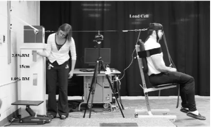
Cervical Perturbation ... 66
Visual Performance Assessment ... 70
Visual Clarity.. ... 70
Contrast Sensitivity ... 71
Depth perception ... 72
Near-Far Quickness ... 72
Target Capture ... 73
Perception Span ... 73
Eye-Hand Coordination ... 74
Go/No Go…… ... 74
Reaction Time. ... 75
Head Impact Biomechanics ... 76
Video Assessment of Level of Anticipation ... 77
Play Exposure ... 77
Data Reduction... 78
Cervical Characteristics ... 79
Isometric Strength ... 79
Ultrasonographic Cross-Sectional Area ... 80
Cervical Perturbation ... 80
Visual Performance ... 82
Head Impact Biomechanics ... 84
Play Exposure ... 87
Statistical Analyses ... 87
Research Question 1: Cervical Characteristics ... 87
Research Question 2: Visual performance ... 88
Research Question 3: Level of Anticipation ... 89
Research Question 4: Predicting Head Impact Severity ... 89
Manuscript Legend ... 93
IV. MANUSCRIPT I ... 94
Introduction ... 94
Methods... 98
Study Participants ... 98
Measurements & Instrumentation ... 99
Isometric Strength ... 100
Ultrasonographic Cross-Sectional Area ... 101
Cervical Perturbation ... 103
Head Impact Biomechanics ... 106
Data Reduction... 107
Isometric Strength ... 107
Ultrasonographic Cross-Sectional Area ... 108
Cervical Stiffness ... 108
Cervical Electromyographic Measurement ... 109
Head Impact Biomechanics ... 110
Results ... 113
Cervical Isometric Strength ... 114
Cervical Muscle Size ... 115
Cervical Perturbation ... 115
Discussion ... 127
Cervical Isometric Strength & Cervical Muscle Size ... 127
Cervical Perturbation ... 130
Conclusions ... 133
V. MANUSCRIPT II ... 134
Introduction ... 134
Methods... 137
Study Participants ... 137
Measurements & Instrumentation ... 138
Visual Performance Assessment ... 138
Head Impact Biomechanics ... 138
Procedures ... 139
Visual Performance Assessment ... 139
Head Impact Biomechanics ... 140
Data Reduction... 142
Statistical Analyses ... 143
Results ... 144
Discussion ... 148
VI. MANUSCRIPT III... 153
Introduction ... 153
Methods... 155
Study Participants ... 155
Procedures ... 156
Head Impact Biomechanics ... 156
Video Footage Capture ... 157
Data Reduction... 157
Head Impact Biomechanics ... 157
Video Assessment of Level of Anticipation ... 158
Statistical Analyses ... 159
Results ... 160
Discussion ... 162
Conclusions ... 164
VII. RESEARCH QUESTION FOUR OVERVIEW ... 166
APPENDIX I: PLAYER TO PLAYER FORM ... 170
APPENDIX II: PLAY EXPOSURE LOG ... 171
LIST OF TABLES
Table
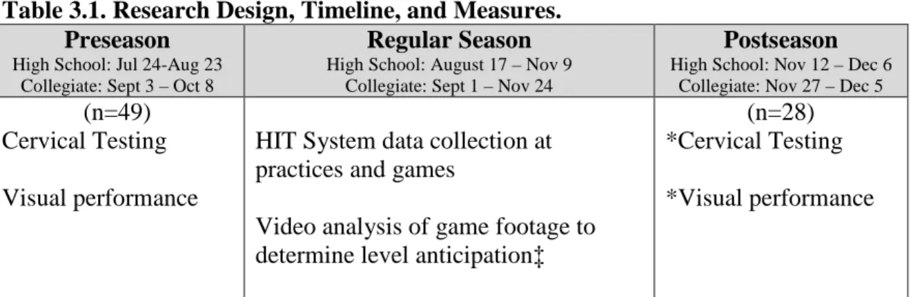
3.1. Research Design, Timeline, and Measures………...…...…….62 3.2 Data Summary Table for Research Questions 1-3..………..…………...91 3.3 Data Summary Table for Research Question 4.……...……...…..…………...92 4.1. Demographic information for both high school and collegiate
football players……….………..99
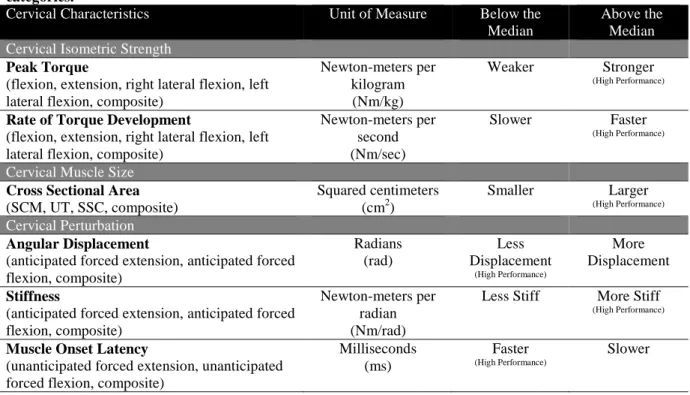
4.2. Head impact biomechanics categorization cutoffs and frequencies……...111 4.3. Cervical characteristic variable table indicating the unit of
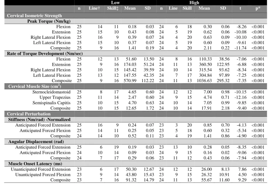
measure and high performance categories………...113 4.4. Descriptive statistics and between group comparisons for low and
high performers for each cervical characteristic………...…...………...117 4.5. Cervical isometric strength (Group overall): Odds ratios (OR) and
95% confidence intervals (CI) indicating the high performance group’s odds of sustaining higher magnitude head impacts, rather than 1st quartile head
impacts, compared to the low performance group………..……...118 4.6. Cervical isometric strength (Skill players only): Odds ratios (OR) and
95% confidence intervals (CI) indicating the high performance group’s odds of sustaining higher magnitude head impacts, rather than 1st quartile head
impacts, compared to the low performance group………...119 4.7. Cervical isometric strength (Linemen only): Odds ratios (OR) and
95% confidence intervals (CI) indicating the high performance group’s odds of sustaining higher magnitude head impacts, rather than 1st quartile head
impacts, compared to the low performance group………...120 4.8. Cervical muscle size (Group overall): Odds ratios (OR) and
95% confidence intervals (CI) indicating the high performance group’s odds of sustaining higher magnitude head impacts, rather than 1st quartile head
impacts, compared to the low performance group.………..121 4.9. Cervical muscle size (Skill players only): Odds ratios (OR) and
95% confidence intervals (CI) indicating the high performance group’s odds of sustaining higher magnitude head impacts, rather than 1st quartile head
4.10. Cervical muscle size (Linemen only): Odds ratios (OR) and
95% confidence intervals (CI) indicating the high performance group’s odds of sustaining higher magnitude head impacts, rather than 1st quartile head
impacts, compared to the low performance group…………..…………...…...123 4.11. Cervical perturbation (Group overall): Odds ratios (OR) and
95% confidence intervals (CI) indicating the high performance group’s odds of sustaining higher magnitude head impacts, rather than 1st quartile head
impacts, compared to the low performance group………..………...124 4.12. Cervical perturbation (Skill players only): Odds ratios (OR) and
95% confidence intervals (CI) indicating the high performance group’s odds of sustaining higher magnitude head impacts, rather than 1st quartile head
impacts, compared to the low performance group………..…...…...125 4.13. Cervical perturbation (Linemen only): Odds ratios (OR) and
95% confidence intervals (CI) indicating the high performance group’s odds of sustaining higher magnitude head impacts, rather than 1st quartile head
impacts, compared to the low performance group….………..126 5.1. Demographic Information…………....………...138 5.2. Nike SPARQ Sensory Station subtest protocol and outcome
measure description………….…...…..………..………141 5.3. Head impact biomechanics categorization cutoffs and frequencies...…...143 5.4. Visual performance variable table indicating the unit of measure
and high performance categories…………...………....………...144 5.5. Descriptive statistics and between group comparisons for low and
high performers for each visual performance variable………..…...…...…..…146 5.6. Visual Performance: Odds ratios (OR) and 95% confidence
intervals (CI) indicating the high performance group’s odds of sustaining higher magnitude head impacts, rather than 1st quartile head impacts,
LIST OF FIGURES
Figure
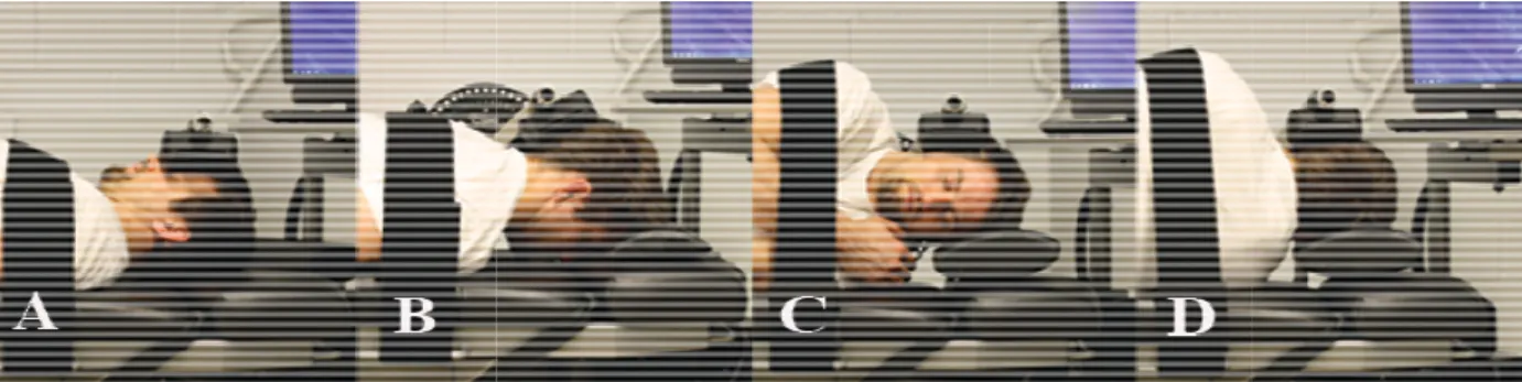
4.1. Participant positioning for cervical spine isometric (A) flexor, (B) extensor, (C) right lateral flexor, (D) and left lateral flexor
Chapter 1 INTRODUCTION
As many as 3.8 million sports-related traumatic brain injuries occur each year, not counting injuries that go unreported (Langlois, Rutland-Brown, & Wald, 2006; McCrea, Hammeke, Olsen, Leo, & Guskiewicz, 2004). Concussion is defined as “a complex
pathophysiological process affecting the brain, caused by traumatic biomechanical forces” (McCrory, et al., 2009). Acutely, concussed athletes experience diminished cognitive function, altered motor control, and symptoms such as headache, nausea, and dizziness (Guskiewicz, Ross, & Marshall, 2001; McCrea, et al., 2005; McCrea, et al., 2003). Sport-related concussion is of particular concern in youth athletes because younger athletes are more susceptible to sustaining concussions (Buzzini & Guskiewicz, 2006; Gessel, Fields, Collins, Dick, & Comstock, 2007; Guskiewicz, Weaver, Padua, & Garrett, 2000). In fact, concussion incidence rates among high school football players are higher than in any of the three collegiate divisions (Guskiewicz, et al., 2000). Concussions can have severe acute and long-term consequences for youth athletes because of the ongoing
neurocognitive development that occurs throughout adolescence (Patel & Greydanus, 2002).
depression, mild cognitive impairment, and early onset Alzheimer’s later in life (Dale, Leigh, Luthert, Anderton, & Roberts, 1991; Guskiewicz, et al., 2005; Guskiewicz,
Marshall, et al., 2007). Some speculate that these debilitating conditions could also result from the cumulative effects of the thousands of subconcussive (non-injurious) impacts to the head that athletes experience throughout their careers (Spiotta, Shin, Bartsch, & Benzel, 2011). For high school athletes who continue to play in college and then
professionally, exposure to a high number of cumulative head impacts may increase their risk of developing neurodegenerative disorders during late-life. Animal studies have demonstrated that higher magnitude impacts to the head or body cause the brain to accelerate and decelerate rapidly within the skull resulting in greater brain tissue strain (Ommaya & Gennarelli, 1974). Extrapolating these data to humans, it seems possible that reducing the magnitude of head impacts that athletes sustain during sport participation may reduce the risk of concussion, the severity of subconcussive head impacts, and, subsequently, the risk of developing late-life cognitive declines that some have
speculated are associated with concussions and repetitive brain trauma. However, little research is available addressing the modifiable factors that could help mitigate the severity of head impacts that result during sport, leaving sports medicine professionals with limited options for preventing concussion. Cervical muscle characteristics, visual performance, and the ability to anticipate impending collisions are three modifiable factors that could potentially be targeted to reduce head impact magnitude during sport.
Cervical Muscle Characteristics
cervical musculature response may be predisposed to concussion because they are less able to generate adequate internal preparatory and reactive forces to counter head acceleration (Viano, Casson, & Pellman, 2007). Contraction of the cervical musculature strong enough to make the cervical spine a rigid segment is believed to link the head, neck, and thorax as a single segment. If inadequate force is generated rapid acceleration of the head occurs. Force imparted to the athlete during a collision is theoretically dispersed over the effective mass of the head, neck, and thorax segments combined, thereby reducing head acceleration. When the cervical musculature is not fully contracted, such as when a player receives an unexpected hit, the impact force is imparted to the head rather than across the neck to the thorax. It seems possible that as cervical muscle activity increases players simultaneously experience a proportional decrease in the severity of head impact. Previous studies have manipulated neck tension in Hybrid III
anthropometric head models and have observed that increasing neck tension resulted in a 35% decline in concussion risk, as measured by the head injury criterion, based on laboratory measures (Viano, et al., 2007). The role of the cervical musculature in modifying head impact forces remains unclear in human models (Mihalik, et al., 2011; Tierney, et al., 2008; Tierney, et al., 2005). Possessing certain anatomical and dynamic cervical spine characteristics may enable an athlete to better increase his or her effective head-neck-thorax mass, making the player better prepared to limit rapid head acceleration. However, the role of cervical muscle strength, physiological cross-sectional area,
Visual Performance
The eyes supply sensory information to the brain, the brain then decodes and integrates the visual information while also considering vestibular and somatosensory information (Zimmerman, Lust, & Bullimore, 2011). The brain then sends out an
appropriate motor signal to the muscles based on the supplied sensory information. Many sports involve quick and unpredictable movement of an object, teammates, and
competitors. Athletes must be able to accurately perceive and identify both static and dynamic features, scan and interpret visual information at differing contrast levels, alternate between focusing on objects at varying distances, perform efficient eye movements, and respond quickly to visual stimuli (Henderson & Hollingworth, 2003; Zimmerman, et al., 2011; Zupan, Arata, Wile, & Parker, 2006). Numerous studies conclude that athletes demonstrate better visual abilities than non-athletes, and that elite athletes have visual abilities superior to novice athletes (Hitzeman & Beckerman, 1993; Laby, et al., 1996; Stine, Arterburn, & Stern, 1982; Uchida, Kudoh, Murakami, Honda, & Kitazawa, 2012). It seems possible that enhanced visual performance would allow an athlete to better anticipate impending collisions with other players allowing them to better mitigate head impact severity.
Tong, & Schor, 2012). Further research is needed to determine if visual performance influences head impact biomechanics.
Collision Anticipation
Anticipatory responses to impending head or body collisions may help mitigate acceleration of the head, thereby reducing the potential risk for sustaining a brain injury and reducing the magnitude of subconcussive impacts. An athlete that is able to foresee an impending impact will instinctively and cognitively react with anticipatory responses, such as leaning, using the arms to block the face, or recoiling the head by elevating the shoulders (Metoyer, Zordan, Hermens, Wu, & Soriano, 2008). During sport, athletes must maintain gaze fixation on a target area, such as a goal or ball, for accurate aiming. Gaze fixation may limit the athlete’s ability to foresee and prepare for impending impacts (van der Kamp, 2011). In youth ice hockey, unanticipated collisions tend to result in more severe head impact magnitudes than anticipated collisions (Mihalik, Blackburn, et al., 2010). In contact sports, the striking player prepares for impending collision by aligning the head, neck, and thorax to impart maximum force on an opponent by driving through the struck player. Previous studies, that have modeled helmet-to-helmet impacts, show that the struck players, on average, experienced greater linear and rotational head acceleration than the striking player (Viano, et al., 2007). However this study used a small sample of head impacts that were reconstructed in a laboratory setting and
anthropometric models that lack the ability to anticipate an impending collision. Because the striking player fully anticipates the impending collision he imparts much greater force on the struck player. Thus, further research is necessary to determine the effect of
Specific Aims
1. To evaluate the effect of cervical musculature characteristics measured during the preseason on head impact biomechanics sustained in-season among high school and collegiate football players.
2. To evaluate the effect of visual performance measured during the preseason on head impact biomechanics sustained in-season among high school football players. 3. To evaluate the effect of level of anticipation at the time of collision on head
impact biomechanics among high school football players.
4. To determine if preseason measures of cervical musculature characteristics and visual performance, and level of anticipation at the time of collision predict head impact biomechanics among high school and collegiate football players.
Variables
Independent Variables
1. RQ1: High and low performance on the following cervical musculature characteristics:
a. Composite peak torque
b. Composite rate of torque development c. Composite cross-sectional area
d. Composite stiffness
e. Composite angular displacement f. Composite muscle onset latency
a. Visual acuity b. Contrast sensitivity c. Depth perception d. Near-Far quickness e. Target capture f. Perception span
g. Eye-Hand coordination h. Go/No Go
i. Reaction Time 3. RQ3: Level of anticipation
a. Anticipated b. Unanticipated
4. RQ4a: Predicting Head Impact Biomechanics a. Composite peak torque
b. Composite rate of torque development c. Composite cross-sectional area
d. Composite stiffness
e. Composite muscle onset latency f. Visual acuity
k. Perception span
l. Eye-Hand coordination m. Go/No Go
n. Reaction Time o. Level of Anticipation
5. RQ4b: Predicting Head Impact Biomechanical Profiles a. Composite peak torque
b. Composite rate of torque development c. Composite cross-sectional area
d. Composite stiffness
e. Composite muscle onset latency f. Visual acuity
g. Contrast sensitivity h. Depth perception i. Near-Far quickness j. Target capture k. Perception span
l. Eye-Hand coordination m. Go/No Go
n. Reaction Time
Dependent Variables
a. Frequency of categorized head impact magnitude by linear acceleration (1st quartile, 2nd quartile, 3rd quartile, 4th quartile, 95th percentile, 99th percentile)
b. Frequency of categorized head impact magnitude by rotational acceleration (1st quartile, 2nd quartile, 3rd quartile, 4th quartile, 95th percentile, 99th percentile)
c. Frequency of categorized head impact magnitude by Head Impact Technology Severity Profile (HITsp) (1st quartile, 2nd quartile, 3rd quartile, 4th quartile, 95th percentile, 99th percentile)
2. RQ 3 & 4a: Game Head Impact Biomechanical Measures a. Peak linear acceleration
b. Peak rotational acceleration
c. Head Impact Technology Severity Profile (HITsp)
3. RQ 4b: Cumulative Game Head Impact Biomechanical Measures Per Play Exposure
a. Cumulative game linear acceleration per play exposure b. Cumulative game rotational acceleration per play exposure c. Cumulative game HITsp per play exposure
4. RQ 4b: Cumulative Game Head Impact Frequency Per Play Exposure
Research Questions
Research Question 1: Cervical Muscle Characteristics
We split football players into a group of high and a group of low performers for each cervical characteristic measure.
a. Do football players with high and low preseason composite cervical peak torque performance differ in odds of sustaining head impacts in 2nd quartile, 3rd quartile, 4th quartile, 95th percentile, or 99th percentile, rather than head impacts in the 1st quartile?
b. Do football players with high and low preseason composite cervical rate of torque development performance differ in odds of sustaining head impacts in 2nd quartile, 3rd quartile, 4th quartile, 95th percentile, or 99th percentile, rather than head impacts in the 1st quartile?
c. Do football players with high and low preseason composite cervical cross-sectional area performance differ in odds of sustaining head impacts in 2nd quartile, 3rd quartile, 4th quartile, 95th percentile, or 99th percentile, rather than head impacts in the 1st quartile?
d. Do football players with high and low preseason composite cervical stiffness performance differ in odds of sustaining head impacts in 2nd quartile, 3rd quartile, 4th quartile, 95th percentile, or 99th percentile, rather than head impacts in the 1st quartile?
f. Do football players with high and low preseason composite cervical muscle onset latency performance differ in odds of sustaining head impacts in 2nd quartile, 3rd quartile, 4th quartile, 95th percentile, or 99th percentile, rather than head impacts in the 1st quartile?
Research Question 2: Visual performance
We split high school football players into a group of high and a group of low performers for each visual performance measure.
a. Do football players with high and low preseason visual acuity performance differ in odds of sustaining head impacts in 2nd quartile, 3rd quartile, 4th quartile, 95th percentile, or 99th percentile, rather than head impacts in the 1st quartile?
b. Do football players with high and low preseason contrast sensitivity performance differ in odds of sustaining head impacts in 2nd quartile, 3rd quartile, 4th quartile, 95th percentile, or 99th percentile, rather than head impacts in the 1st quartile? c. Do football players with high and low preseason depth perception performance
differ in odds of sustaining head impacts in 2nd quartile, 3rd quartile, 4th quartile, 95th percentile, or 99th percentile, rather than head impacts in the 1st quartile? d. Do football players with high and low preseason near-far quickness performance
differ in odds of sustaining head impacts in 2nd quartile, 3rd quartile, 4th quartile, 95th percentile, or 99th percentile, rather than head impacts in the 1st quartile? e. Do football players with high and low preseason target capture performance
f. Do football players with high and low preseason perception span performance differ in odds of sustaining head impacts in 2nd quartile, 3rd quartile, 4th quartile, 95th percentile, or 99th percentile, rather than head impacts in the 1st quartile? g. Do football players with high and low preseason eye-hand coordination
performance differ in odds of sustaining head impacts in 2nd quartile, 3rd quartile, 4th quartile, 95th percentile, or 99th percentile, rather than head impacts in the 1st quartile?
h. Do football players with high and low preseason go/no go performance differ in odds of sustaining head impacts in 2nd quartile, 3rd quartile, 4th quartile, 95th percentile, or 99th percentile, rather than head impacts in the 1st quartile?
i. Do football players with high and low preseason reaction time performance differ in odds of sustaining head impacts in 2nd quartile, 3rd quartile, 4th quartile, 95th percentile, or 99th percentile, rather than head impacts in the 1st quartile?
Research Question 3: Level of Anticipation
a. Is there a significant difference in head impact biomechanical measures between anticipated and unanticipated collisions in high school football players?
Research Question 4: Predicting Head Impact Biomechanical Measures
b. Do preseason cervical characteristics and visual performance predict cumulative game head impact biomechanical measures per play exposure in high school and collegiate football players?
c. Do preseason cervical characteristics and visual performance predict cumulative game head impact frequency per play exposure in high school and collegiate football players?
Research Hypotheses
Research Hypotheses for Research Question 1: Cervical Characteristics
a. Football players that are high performers on composite cervical peak torque will have reduced odds of sustaining head impacts in 2nd quartile, 3rd quartile, 4th quartile, 95th percentile, and 99th percentile, rather than head impacts in the 1st quartile.
b. Football players that are high performers on composite cervical rate of torque development will have reduced odds of sustaining head impacts in 2nd quartile, 3rd quartile, 4th quartile, 95th percentile, and 99th percentile, rather than head impacts in the 1st quartile.
c. Football players that are high performers on composite cervical cross-sectional area will have reduced odds of sustaining head impacts in 2nd quartile, 3rd quartile, 4th quartile, 95th percentile, and 99th percentile, rather than head impacts in the 1st quartile.
quartile, 95th percentile, and 99th percentile, rather than head impacts in the 1st quartile.
e. Football players that are high performers on composite cervical angular displacement will have reduced odds of sustaining head impacts in 2nd quartile, 3rd quartile, 4th quartile, 95th percentile, and 99th percentile, rather than head impacts in the 1st quartile.
f. Football players that are high performers on composite cervical muscle onset latency will have a reduced odds of sustaining head impacts in 2nd quartile, 3rd quartile, 4th quartile, 95th percentile, and 99th percentile, rather than head impacts in the 1st quartile.
Research Hypotheses for Research Question 2: Visual performance
a. Football players that are high performers on visual acuity will have reduced odds of sustaining head impacts in 2nd quartile, 3rd quartile, 4th quartile, 95th percentile, and 99th percentile, rather than head impacts in the 1st quartile.
b. Football players that are high performers on contrast sensitivity will have reduced odds of sustaining head impacts in 2nd quartile, 3rd quartile, 4th quartile, 95th
percentile, and 99th percentile, rather than head impacts in the 1st quartile.
c. There will be no differences in odds of sustaining head impacts in 2nd quartile, 3rd quartile, 4th quartile, 95th percentile, and 99th percentile, rather than head impacts in the 1st quartile between high and low performers on depth perception.
d. Football players that are high performers on near far quickness will have reduced odds of sustaining head impacts in 2nd quartile, 3rd quartile, 4th quartile, 95th
e. There will be no differences in odds of sustaining head impacts in 2nd quartile, 3rd quartile, 4th quartile, 95th percentile, and 99th percentile, rather than head impacts in the 1st quartile between high and low performers on target capture.
f. Football players that are high performers on perception span will have reduced odds of sustaining head impacts in 2nd quartile, 3rd quartile, 4th quartile, 95th percentile, and 99th percentile, rather than head impacts in the 1st quartile. g. Football players that are high performers on eye-hand coordination will have
reduced odds of sustaining head impacts in 2nd quartile, 3rd quartile, 4th quartile, 95th percentile, and 99th percentile, rather than head impacts in the 1st quartile. h. There will be no differences in odds of sustaining head impacts in 2nd quartile, 3rd
quartile, 4th quartile, 95th percentile, and 99th percentile, rather than head impacts in the 1st quartile between high and low performers on go/no go.
i. Football players that are high performers on reaction time will have reduced odds of sustaining head impacts in 2nd quartile, 3rd quartile, 4th quartile, 95th percentile, and 99th percentile, rather than head impacts in the 1st quartile.
Research Hypothesis for Research Question 3: Level of Anticipation
a. Unanticipated collisions will result in significantly higher head impact biomechanical measures than anticipated collisions.
Research Hypotheses for Research Question 4: Predicting Head Impact Severity
biomechanical measures. Muscle onset latency, visual acuity, depth perception, target capture, eye-hand coordination, and reaction time will significant direct predictors of game head impact biomechanical measures. Depth perception and go/no go will not be significant predictors.
b. Composite cervical peak torque, rate of torque development, stiffness, cross-sectional area, and composite visual performance raw score will be significant inverse predictors of cumulative game head impact biomechanical measures while controlling for play exposure. Muscle onset latency will be significant direct predictors of mean game head impact biomechanical measures.
c. Composite visual performance raw score will be a significant inverse predictor of cumulative game head impact frequency while controlling for play exposure. Composite cervical peak torque, rate of torque development, stiffness, cross-sectional area, and muscle onset latency will not be significant predictors.
Operational Definitions
1. Head Impact Technology severity profile (HITsp): A weighted composite score including linear acceleration, rotational acceleration, impact duration, and impact location.
2. Cervical Characteristics:
b. Composite rate of torque development: A calculated sum of the rate of torque development of the cervical flexors, extensors, right lateral flexors, and left lateral flexors. Rate of torque development is defined as the maximal value of the slope of the force-time curve, calculated using a 50-millisecond sliding window from onset to peak force (Almosnino, Pelland, & Stevenson, 2010).
c. Composite cross-sectional area: A calculated sum of the cross-sectional area of the sternocleidomastoid, upper trapezius, and semispinalis capitis measured using ultrasonographic imaging.
d. Composite stiffness: A calculated sum of flexor and extensor stiffness. Stiffness is a measure of an elastic body’s resistance to deformation. Flexor stiffness was determined by measuring the flexor muscle group’s resistance to deformation during forced extension after an applied load. Extensor stiffness was determined by measuring the extensor muscle group’s resistance to deformation during forced flexion after an applied load.
e. Composite angular displacement: A calculated sum of peak angular displacement of the head relative to the thorax following perturbation into both flexion and extension.
f. Composite muscle onset latency: The sum of the duration of time between force application and the onsets of myoelectric activity in the
3. Visual performance: Visual performance measures in this study include: visual acuity, contrast sensitivity, depth perception, near-far quickness, target capture, perception span, eye-hand coordination, go/no go, and reaction time.
4. Level of anticipation: Level of anticipation was determined by evaluating video of each head impact sustained during games. The Player to Player form was used to grade the player’s relative body position at the time of impact (Mihalik,
Blackburn, et al., 2010; Ocwieja, et al., 2012).
a. Anticipated: An impact occurring while the athlete is looking in the direction of the impending collision, is in a general athletic readiness position (knee and trunk flexion with feet shoulder-width apart), and uses their legs to drive their shoulders through the collision.
b. Unanticipated: An impact occurring where the athlete is looking in the direction of the oncoming collision but is not in an athletic readiness position or an impact occurring while the athlete is not looking in the direction of the impending collision.
c. Unknown: Collisions where the investigator is unable to identify the direction of gaze or the positioning of the body.
5. Play exposure: The number of plays that each athlete participates in during all games throughout the entire season as recorded by the primary investigator. 6. Cumulative Game Head Impact Biomechanical Measures Per Play Exposure:
(g) from head impacts sustained in all games divided by the number of play exposures.
b. Cumulative Game Rotational Acceleration Per Play Exposure: The average rotational acceleration per play, computed as the sum of the rotational acceleration (rad/sec2) from head impacts sustained in all games divided by the number of play exposures.
c. Cumulative Game HITsp Per Play Exposure: The average HITsp per play, computed as the sum of the HITsp from head impacts sustained in all games divided by the number of play exposures.
7. Cumulative Head Impact Frequency: The average number of head impacts per play, computed as the sum of the frequency of head impacts sustained in all games divided by the number of play exposures.
Assumptions
1. Preseason measures of cervical characteristics and visual performance reflect the changes that occur over the course of the season.
2. Participants gave their best efforts during pre and post-season testing sessions 3. These lab measures accurately reflect cervical function in the athletic setting. 4. The head-neck segment moves about the thorax as a rigid body
5. Athletes did not alter their sport technique due to the presence of the instrumentation and investigators.
Limitations
2. Cervical isometric strength and stiffness were measured in just one plane at a time. 3. Head to thorax movement measured during cervical perturbation does not account
for movement of individual vertebrae or movement of the head relative to C1. 4. The Head Impact Telemetry system does not measure rotational acceleration
about the Z-axis.
5. Results from this study may not apply to athletes that participate at other levels of play or female athletes.
6. We were not able to determine if cervical characteristics, visual performance, and level of anticipation influence the odds of sustaining a concussion.
Delimitations
1. Data collection was limited to only practices and games during a single competitive season.
2. Athletes were recruited from a single high school and single collegiate institution. 3. This study did not examine impacts to the head that result in concussion.
4. Participants were all males.
Significance of the Study
sustaining high magnitude impacts to the head. This study provides guidance for
Chapter 2
REVIEW OF LITERATURE
Introduction
Sport-related mild traumatic brain injury is a major public health concern in the United States (Langlois, et al., 2006). Concussions result from rapid acceleration and deceleration of the brain caused by biomechanical forces transmitted from an impact to the head or indirectly through the body (McCrory, et al., 2009). Athletes with insufficient cervical musculature strength may be predisposed to more severe head impacts because they are less able to generate adequate internal preparatory and reactive force to counter head acceleration (Viano, et al., 2007). The purpose of this review is to discuss relevant literature regarding concussion epidemiology, neurometabolic cascades that follow traumatic brain injury, development and recovery of the adolescent brain, negative postconcussive outcomes, head impact biomechanics, important cervical characteristics, sport visual performance, and anticipation.
Epidemiology
Sniezek, & Thurman, 1996). By observing epidemiologic patterns in sport-related concussion, sports medicine professionals can guide targeted preventive measures.
As many as 3.8 million sport-related traumatic brain injuries occur annually in the United States (Langlois, et al., 2006), with evidence that many go unrecognized,
unreported, and untreated (Langlois, et al., 2006; McCrea, et al., 2004; Valovich McLeod, Schwartz, & Bay, 2007). Evaluation and treatment of sport-related concussion cost
approximately 60 billion dollars each year (Langlois, et al., 2006). Concussions represent 13.2% of all sport-related injuries reported in the high school setting (Marar, McIlvain, Fields, & Comstock, 2012). Earlier epidemiologic studies report slightly lower incidences of concussion ranging from 5.5-8.9% of all injuries, but this is likely because these
studies have not included contact sports like ice hockey and lacrosse (Gessel, et al., 2007; Powell & Barber-Foss, 1999; Schulz, et al., 2004). Overall, the concussion rate is
approximately 2.5 concussions per 10,000 athlete exposures (Gessel, et al., 2007; Marar, et al., 2012).
Pediatric Brain Injuries
Nearly half of all concussions among youth and adolescents result during participation in sport (Bakhos, Lockhart, Myers, & Linakis, 2010; Meehan & Mannix, 2010). High school athletes have a higher incidence of concussion compared to their collegiate counterparts (Gessel, et al., 2007; Guskiewicz, et al., 2000). Some researchers theorize that adolescent athletes have less protection for their developing nervous system because they have relatively decreased neuronal myelination, a greater head-to-body ratio, and thinner cranial bones (Buzzini & Guskiewicz, 2006). High school athletes that
full resolution of symptoms, take longer than three weeks to return to sport, and are more likely to be medically disqualified from sport (Castile, Collins, McIlvain, & Comstock, 2011). Athletes who sustain their first concussion at a young age and continue to
participate in sport on into high school and college have a longer window of time they are participating in sports which increases their exposure and therefore risk of re-injury (Guskiewicz & Valovich McLeod, 2011). Symptoms that persist following concussion in adolescent athletes are particularly concerning because these deficits can significantly affect academic performance and social function during a critical period of development (Blume, Lucas, & Bell, 2011).
Gender Comparisons
Among gender-comparable sports, females have a higher concussion rate than males (Castile, et al., 2011; Gessel, et al., 2007; Marar, et al., 2012). Likewise, females have higher rates of recurrent concussions than males (Castile, et al., 2011). Some
researchers and clinicians speculate that observed gender differences could be attributable to head and cervical biomechanical differences (Mansell, Tierney, Sitler, Swanik, & Stearne, 2005; Tierney, et al., 2008; Tierney, et al., 2005). However, concussion rates may differ between genders because female athletes are generally more honest about reporting injuries than male athletes, due to cultural norms (Dick, 2009).
Injury Mechanisms
concussion rates are greater during competition compared to practice (Gessel, et al., 2007; Marar, et al., 2012; Schulz, et al., 2004). This could be explained by evidence in football that collisions that take place after two players travel a longer closing distances and in ice hockey where collisions occur on the open ice result in higher magnitude head impacts (Mihalik, Blackburn, et al., 2010; Ocwieja, et al., 2012). Some studies suggest that football players sustain a greater number of impacts and more severe impacts during games compared to practices (Broglio, et al., 2009). However, a similar study in a college sample suggests that head impacts sustained during helmets-only and full-contact
practices are more severe than head impacts sustained during games (Mihalik, Bell, Marshall, & Guskiewicz, 2007).
Football Brain Injuries
Among high school sports, football accounts for nearly half of all reported
(McCrea, et al., 2004). Because athletes may be largely unaware of the signs and symptoms of concussion and the seriousness of premature return to play, prevalence of concussion in high school football is likely higher than previously published
epidemiology literature.
Neurometabolic Cascade Following Concussion
Post-concussion deficits occur in the absence of detectable structural pathology and typically resolve completely over time. Neuronal dysfunction following concussion result from ionic shifts, altered metabolic demand, impaired neuronal connectivity, and changes in neurotransmission (Giza & Hovda, 2001). Understanding the neurometabolic cascade that follows concussion is vital for understanding the underlying
pathophysiology.
Traumatic brain injury sets off a complex and interwoven sequence of ionic and metabolic events from which damaged cells may eventually recover, or in certain cases, degenerate and dies. Membrane disruption and axonal stretch caused by a direct or indirect impact to the head, results in opening of voltage-dependent potassium channels and a subsequent efflux of potassium from cells to the extracellular space. Potassium is released into the extracellular space by leaking through the mechanically stretched cell membrane and by passing through voltage-gated potassium channels (Katayama, Becker, Tamura, & Hovda, 1990; Takahashi, Manaka, & Sano, 1981). Non-specific
work overtime, but simultaneously consume increasing amounts of adenosine triphosphate (Mayevsky & Chance, 1974; Rosenthal, LaManna, Yamada, Younts, & Somjen, 1979). To meet elevated adenosine triphosphate requirements, there is a marked upregulation of cellular glycolysis, which occurs within minutes after brain injury
(Ackermann & Lear, 1989). Hypergycolysis results in lactate byproduct, which builds up within the neuron (Nilsson & Nordstrom, 1977; Nilsson & Ponten, 1977; Yang, DeWitt, Becker, & Hayes, 1985).
In addition to potassium efflux, NMDA receptor activation permits a rapid and sustained influx of calcium. Elevated intracellular calcium can be sequestered by the mitochondria, but will eventually lead to dysfunction of oxidative metabolism, which further increases the cell 's dependence on glycolysis-generated adenosine triphosphate (Giza & Hovda, 2001). Calcium accumulation eventually leads to cell dysfunction, damage, and sometimes death. Ionic shifts and acute alterations in cellular energy metabolism occur during a period when cerebral blood flow is reduced (Yamakami & McIntosh, 1989; Yuan, Prough, Smith, & Dewitt, 1988). Imbalance between glucose delivery and glucose consumption predisposes neurons to secondary injury and secondary cell death (Giza & Hovda, 2001). After the initial period of ionic disturbance and increase in glucose metabolism, the local cerebral metabolic rate for glucose and oxidative
The Adolescent Versus the Adult Brain
Cognitive and cortical growth generally occurs in cycles, with a series of sporadic spurts and drops. High school athletes are still developing in cognitive areas like
concentration, memory, reasoning, and problem solving (Hunt & Ferrara, 2009). The rate of progression through each phase of cognitive and cortical development differs between individuals, but most individuals go through a common developmental process (Fisher & Rose, 1998). Passing through these phases requires reorganization and simplification, which allows the individual to move through the four different tiers: reflex, action, concrete representation, and abstraction (Fisher & Rose, 1998). Once the infant moves beyond the reflexive tier, the action tier is identifiable as the infant begins building
complex sensorimotor actions, typically between three months and two years (e.g. names, emotions). Between ages two and 12, the child develops concrete representational
capacities and eventually understands his or her first abstractions (understanding mathematic calculations, literary meanings, concepts of law). Optimal abstraction capacities appear between 10 and 25 years of age and produce the capacity to build principles relating multiple abstractions (Fisher & Rose, 1998). Although most cognitive and cortical development is complete by the time an individual enters college,
development continues on into early adulthood (Luna, et al., 2001).
2001). Recent research using juvenile rats support this concept. Compared to adult rats, younger rats show longer periods of apneas, shorter periods of unconsciousness, present with post-percussion hypotension, and have higher mortality following traumatic brain injury (Prins, Lee, Cheng, Becker, & Hovda, 1996). Despite displaying more severe immediate response to brain injury, younger rats with mild and moderate traumatic brain injury continue to perform well on spatial learning tasks (Prins & Hovda, 1998).
However, when moderately concussed juvenile rats are reared in an enriched
environment, they fail to develop increased cortical thickness and enhanced cognitive performance seen in sham-injured rats raised in the same enriched environments
(Fineman, Giza, Nahed, Lee, & Hovda, 2000). Brain injury that occurs in the developing brain, even without early signs of damage, may lead to impaired plasticity.
Long-term deficiencies have been observed in human research, as well.
Symptom-free high school athletes with a history of two or more concussions perform similarly on neurocognitive testing to athletes who have just experienced a recent
Negative Post-Concussion Outcomes
Research regarding both the short- and long-term effects of concussion has raised considerable concern about brain function. Negative post-concussion outcomes include second impact syndrome, post-concussion syndrome, recurrent concussion, chronic traumatic encephalopathy, cognitive decline, and depression.
Second Impact Syndrome
Second impact syndrome is defined as occurring when ‘‘an athlete who has sustained an initial head injury, most often a concussion, sustains a second head injury before symptoms associated with the first have fully cleared’’ (Cantu, 1998). A second insult to the brain, sometimes occurring from a seemingly innocuous hit to the head or body, that occurs prior to brain recovery is thought to results in catastrophic brain swelling. Although second impact syndrome is undoubtedly the most severe negative outcome that could occur following concussion, evidence supporting the existence of second impact syndrome remains anecdotal (McCrory, 2001; McCrory, Davis, &
Makdissi, 2012; Randolph, 2011). Brain swelling is a common result from a head injury; however, it remains unknown whether a second concussive injury is a risk factor for this condition. Although it seems logical that returning an athlete to play before concussion related symptoms have resolved could increase the athletes vulnerability to negative postconcussive outcomes, the number of athletes that prematurely return to play without negative consequences is still unknown.
Post-concussion Syndrome
seven to ten days following injury (Guskiewicz, et al., 2001; McCrea, et al., 2003). However, a small, but clinically significant number of athletes experience
post-concussion syndrome, which consists of a complex mixture of cognitive, behavioral, and physical symptoms that persists for an extended period of time after the concussion (Jotwani & Harmon, 2010; Williams, Potter, & Ryland, 2010). Definitions of post-concussion syndrome differ across diagnostic criteria, resulting in widespread confusion about identifying and treating athletes with prolonged recoveries (Jotwani & Harmon, 2010). A reliable and consistent definition is necessary to further scientific research and provide clarity to clinical decisions regarding post-concussion syndrome.
Some authors speculate that persistent post-concussion symptoms are a
consequence of psychological illness rather than brain injury (Lishman, 1988; Williams, et al., 2010). Some literature suggests the stress triggered by brain injury results in depression and anxiety, which disrupts concentration and other mental operations. In a prospective longitudinal study, Yeates et al. (Yeates, et al., 2009) observed that severity of head injury predicted post-concussion symptoms in most but not all patients
history of previous concussion and those who have preexisting psychiatric issues could be at higher risk of developing post-concussion syndrome (Jotwani & Harmon, 2010).
Recurrent Concussion
Like many sports injuries, history of similar injury is the best predictor of recurrent injury. Epidemiologic studies have time and time again identified a history of previous concussions as a risk factor for suffering recurrent concussion (Gerberich, Priest, Boen, Straub, & Maxwell, 1983; Guskiewicz, et al., 2000; Schulz, et al., 2004). It is possible that the brain’s ability to respond to traumatic insults may be compromised in previously concussed athletes making them more susceptible to another concussion. The risk of recurrent concussion in the youth and adolescent athletes is currently unknown (Guskiewicz & Valovich McLeod, 2011). Although a few studies indicate that a previous history of concussion may increase an athlete’s risk of sustaining additional concussion, these trends could be attributable to the fact that these same athletes may continue to be exposed to more play-time, may exhibit risky biomechanics, or be exposed to more intense athletic activities.
Chronic Traumatic Encephalopathy
Chronic traumatic encephalopathy describes the presence of tau protein within the cerebral tissue that results in neurologic deterioration and is only observed among
undergone sustained axonal stretching and deformation that later triggers neurocognitive decline (Yuen, Browne, Iwata, & Smith, 2009). Sport type, level of competition, position, and playing career duration may all influence an athlete’s risk of developing chronic traumatic encephalopathy (Stern, et al., 2011). All diagnosed cases of chronic traumatic encephalopathy have a history of brain trauma exposure, but controversy exists over the risk of exposure to brain trauma because not all individuals with exposure to repetitive brain trauma develop chronic traumatic encephalopathy.
The clinical presentation of chronic traumatic encephalopathy is distinct from post-concussion syndrome because patients do not present with unrelenting symptoms immediately following a concussion. Rather, the symptoms of chronic traumatic encephalopathy result from a progressive, but gradual, decline in neuronal function (McKee, et al., 2009). Typically, chronic traumatic encephalopathy symptoms present in midlife as cognitive, emotional, and behavioral symptoms, usually decades after exposure to repetitive brain trauma. Behavioral symptoms are often the most concerning since they present as a depressed mood, apathy, emotional instability, suicidal tendencies and behaviors, and problems with impulse control (Stern, et al., 2011).
Cognitive Decline
2005). Although additional prospective research is necessary to determine how exposure to head trauma influences the onset of dementia-related syndromes in athletes, these studies present compelling evidence that mild cognitive impairment may be initiated by multiple concussions. More acutely, a history of multiple concussions has been
associated with reduced neurocognitive performance, increased symptom severity, and delayed resolution of concussion related symptoms (Collins, et al., 1999; Colvin, et al., 2009; Guskiewicz, et al., 2003; Guskiewicz, et al., 2000; Iverson, Gaetz, Lovell, & Collins, 2004). These short-term consequences of recurrent concussion support the findings regarding the more chronic consequences of years of playing football. Further research is necessary to further elucidate whether cognitive impairment, both short- and long-term, results from sport-related concussion.
Depression
Many patients that suffer a traumatic brain injury are at a high risk for developing subsequent major depression (Kreutzer, Seel, & Gourley, 2001). Although the prevalence of depression is especially high in individuals after suffering a severe traumatic brain injury (Jorge, et al., 1993), retired professional football players with a history of three or more mild traumatic brain injuries are at a threefold risk of being diagnosed with clinical depression compared with those with no prior history (Guskiewicz, Marshall, et al., 2007). Links between mild traumatic brain injury and major depression could possibly be due to neuronal changes that occur in areas of the brain that modulate mood. Neuroanatomical structures such as the hippocampus (Sheline, Sanghavi, Mintun, & Gado, 1999),
depression. The loss of neurons caused by recurrent concussion could put individuals at risk of depression, which results in further structural changes within regions of the brain that control mood. Many individuals also suffer from disruption of social relationships, disruptions in friendships and social support, lack opportunities to build new friendships, and often withdraw from leisurely activities (Morton & Wehman, 1995). Although the link between the pathophysiology of recurrent concussion and the lifetime risk of depression is unclear, it seems possible that recurrent mild traumatic brain injury may result in a similar structural and psychosocial impact that eventually leads to depressive disorders.
Biomechanics of Mild Traumatic Brain Injury
Research on biomechanical factors and their influence on outcomes after sport-related concussion remain inconclusive. Previous studies using animal and crash test dummy models provide preliminary evidence, but new advancements in real-time technologies may aid future research on this topic. All research regarding the
biomechanics of head injury operate under the same tenet that kinetic energy from an impact to the head is transmitted to the tissue of the brain.
Animal research
Early research primarily utilized primates and other larger mammalian animal models, but changes in ethics regulations animal research in this area has been limited to the rat and other small mammalians. In one of the earliest studies of head injury
These results were later replicated demonstrating that when rotation of the head is restricted, allowing only translation, cerebral concussion does not occur (Ommaya & Gennarelli, 1974). Translational mechanisms are thought to cause focal brain tissue strain, while rotational mechanisms are thought to cause more diffuse axonal injury. Rotational acceleration of the head is thought to cause the cerebrum to rotate about the relatively fixed brainstem (Ommaya & Gennarelli, 1974). Because the midbrain and upper
brainstem are responsible for alertness and responsiveness, the strain experienced during rotational mechanisms are more likely to result in loss of consciousness than linear mechanisms (Ommaya & Gennarelli, 1974). If this concept is applied to human models, contraction of the cervical musculature could limit rotational movements of the head, thereby, reducing diffuse axonal injury.
Model research
In the early 1970’s, the National Operating Committee on Standards for Athletic Equipment contracted Wayne State University Department of Neurosurgery to develop standards for football helmets established standards for the impact performance of football helmets (Gurdjian, Lissner, Hodgson, & Patrick, 1964). The Wayne State
University Concussion Tolerance Curve, computed from impact duration and magnitude, was used to propose a theoretical threshold of 90g of linear acceleration necessary to produce a mild traumatic brain injury. These experiments were conducted using cadavers and metal headforms, but were instrumental in developing standards for new and
As part of the same series of studies published in Neurosurgery, Viano et al. (Viano, et al., 2007; Viano & Pellman, 2005) evaluated the mechanics of both the struck and striking player for plays resulting in injury. Alarmingly, the authors found that the struck players, on average, experience 98g of linear head acceleration while the striking player only experienced 58.5g (Viano, et al., 2007; Viano & Pellman, 2005). Because the striking player fully anticipates the impending collision they are able to optimize their biomechanics to impart much greater force on the struck player. The striking player often delivers maximum force by lowering the head to align the head, neck, and torso. Linking the head, neck, and thorax was found to increase the effective mass of the striking athlete by up to 67% (Viano, et al., 2007). The struck player is most often the one affected by concussion because of the high inertial load imparted by the striking athlete.
In Vivo Accelerometer-Based Research
Real-time accelerometer data collection is a novel method available to researchers who are attempting to better understand the biomechanics of concussion. In one of the first studies to use accelerometry in vivo, Naunheim et al. (Naunheim, Standeven, Richter, & Lewis, 2000) measured head acceleration using a single triaxial accelerometer
imbedded in the helmet of high school hockey and football players during actual game play. The authors measured peak linear acceleration and computed the Gadd Severity Index and Head Injury Criterion scores for head impacts sustained during actual play periods in several games over four seasons. The authors also recorded acceleration of head impacts of soccer players while heading a soccer ball while wearing the
current research paradigms, the author estimated mean linear acceleration measured in the football and ice hockey players of 29.2g and 35.0g, respectively, just slightly higher than current estimates (Broglio, et al., 2009; Mihalik, et al., 2007).
The Head Impact Telemetry System was designed to allow clinicians and
researchers to measure real-time head impact biomechanics in helmeted athletes. Helmets are equipped with six spring-loaded single-axis accelerometers. When an impact occurs to the head data are collected, time-stamped, encoded, and relayed to a near-by sideline-controller antennae and laptop computer for storage (Beckwith, Chu, & Greenwald, 2007; Broglio, Eckner, Surma, & Kutcher, 2011; Broglio, et al., 2009; Brolinson, et al., 2006; Duma, et al., 2005; Eckner, Sabin, Kutcher, & Broglio, 2011; Greenwald, Gwin, Chu, & Crisco, 2008; Guskiewicz, Mihalik, et al., 2007; Mihalik, et al., 2007). Duma et al. (Duma, et al., 2005) and Brolinson et al. (Brolinson, et al., 2006) were first to publish important descriptive data regarding head impacts in collegiate football. These authors reported a mean linear acceleration of 32g. The primary finding of this study was that the accelerometry system proved effective at collecting thousands of head impact data and that the system provide useful information to both researchers and clinicians. The
invention of an in-helmet accelerometry system that allows for real-time analysis of head impact biomechanics has great potential to shed light on the biomechanical risk factors for concussion allowing for measurement of the severity, frequency, and location of impacts occurring at the head in football, hockey, and boxing (Beckwith, et al., 2007).
Since these early exploratory studies, further efforts have been made to examine the biomechanical characteristics of impacts to the head with the hopes of further
identifying a theoretical threshold of concussion (Guskiewicz & Mihalik, 2011). Despite advancements in technologies, questions regarding why some athletes withstand high magnitude impacts without sustaining a concussion, whereas others are injured by lower magnitude impacts remains unanswered. In contrast to previously published theoretical injury thresholds, Mihalik et al. (Mihalik, et al., 2007) reported that less than 0.35% of impacts that exceeded 80g of linear acceleration resulted in concussion. Guskiewicz et al. (Guskiewicz, Mihalik, et al., 2007) established that no relationship existed between biomechanical characteristics of head impacts that resulted in concussion and clinical neurocognitive, postural control, and symptom severity measures. In a similar study design utilizing high school athletes rather than collegiate athletes, Broglio et al. (Broglio, et al., 2011) observed that same results. Combined, these studies suggest that concussions occur from impacts in a wide range of magnitude and that post-concussion declines are independent of head impact biomechanics (Broglio, et al., 2011; Guskiewicz, Mihalik, et al., 2007). In an attempt to understand the dynamic nature of the injurious threshold, Eckner et al. (Eckner, Sabin, et al., 2011) evaluated the subconcussive impact profiles that preceded 20 concussive head impacts. Their data suggested that impact volume and intensity preceding a concussive event did not influence concussion threshold in high school football athletes. Although clinical outcomes measures seem to remain unaffected by head impact biomechanical measures, it seems possible that impacts occurring beyond the purposed 70 to 75g injury threshold may result in subtle neurocognitive and postural control deficits in the absence of a concussion diagnosis.
consequences of subconcussive head impacts (Spiotta, et al., 2011). McCaffrey et al. (McCaffrey, Mihalik, Crowell, Shields, & Guskiewicz, 2007) assessed these short-term clinical outcomes in asymptomatic collegiate football players following low and high magnitude impacts. Their findings suggested that sustaining an impact greater than 90g did not result in observable deficits in neurocognitive or postural control performance or in an increase in self-reported symptoms (McCaffrey, et al., 2007). Likewise, a similar study evaluating changes in neurocognitive, postural control, and symptom severity prior to and following a season of exposure to head impacts found that repetitive
subconcussive head impacts did not appear to result in short-term neurologic impairment (Gysland, et al., 2012). Since these studies, the sensitivity of the neurocognitive measures used to identify neurocognitive deficits has been brought into question (Coldren, Russell, Parish, Dretsch, & Kelly, 2012). Recent strides have been made in understanding head impacts characteristics, but more scientific research is necessary to better understand the causes of concussions in sport and how the brain is influenced by repetitive trauma.
Modifiable Factors
In an early initiative to prevent concussion, Dr. Robert Cantu suggested that
sport of football. Rule changes, such as eliminating tackling from youth football, may prevent some concussions by limiting exposure, but would likely encounter considerable social opposition and risk substantial change to the sport (Johnson, 2012). In contrast changes in conditioning provide an alternative to drastic rule changes. Improving the dynamic response of the cervical musculature through conditioning has promising potential for reducing head impact severity (Cantu, 1996).
The Dynamic Cervical Response
medicine professionals and strength and conditioning coaches when designing cervical training programs.
Cervical Strength
The osteoligamentous structures of the cervical spine contribute approximately 20% of the minimally needed mechanical stability of the cervical spine (Panjabi, et al., 1998). This leaves a remaining 80% of the mechanical load to be managed by the cervical musculature. During trauma, the contribution of the cervical musculature becomes even more important. Much like in the sports setting, technological advancements have improved fighter plane airframe materials, propulsion systems, and flight controls, allowing fighter pilots to fly farther, faster, higher (Seng, Lam, & Lee, 2003). A large number of studies addressing cervical strength have been focused on the fighter pilot population (Alricsson, Harms-Ringdahl, Larsson, Linder, & Werner, 2004; A. F. Burnett, Naumann, Price, & Sanders, 2005; Seng, et al., 2003). Much like the modern athlete, fighter pilots are at greater risk for injury associated with their profession. Although weak cervical musculature has been proposed as a potential risk factor for concussion,
strengthening programs are not emphasized in most sports.