i
P ÍRODOV DECKÁ FAKULTA
LEAF SENESCENCE
AS A LIGHT-DEPENDENT PROCESS
HABILITATION THESIS
MARTINA ŠPUNDOVÁ
i
Abstract
The habilitation thesis deals with leaf senescence, a fundamental process
that is essential for seed production and plant viability in the next generation or season, as allowing plants to recycle leaf nutrients. The understanding of senescence mechanisms is important not only with respect to scientific questions, but also in context of maintaining or increasing plant productivity and minimizing postharvest quality loss during transportation and storage. As a part of leaf development, senescence is primarily induced and controlled by endogenous factors, but it is pronouncedly modulated by environmental factors including light. During the past two decades there has been a significant progress in the understanding the senescence mechanisms and regulation, but they are still far from being elucidated.
In the first part of the thesis, recent knowledge of some aspects of leaf senescence (such as chloroplast and chlorophyll degradation, impairment of photosynthesis, and cytokinin action) is briefly summarized. The attention is also paid to a role of light in senescence processes. In the second part of the thesis, main results of our publications (chronologically ordered) dealing with leaf senescence are described. The relevant papers are included in the Appendix.
It has been shown that light pronouncedly affects rate of chlorophyll degradation and inhibition of photosynthetic processes, activity of xanthophyll cycle and antioxidative enzymes, and level of oxidative damage. Light also modifies the antisenescence effect of exogenous cytokinin and endogenous level of main cytokinin types and forms in detached leaves. It has been suggested that the delaying effect of light on senescence of detached Arabidopsis leaves may be related to the persisted biosynthesis of isopentenyladenine. It has been revealed that light can eliminate an acceleration of senescence-associated processes caused by the loss-of-function mutation in cytokinin receptors. Finally, it has been observed that chlorophyll b deficiency accelerates decrease in photosynthetic activity during dark-induced senescence of detached barley leaves and
that exogenous cytokinin is able to eliminate this effect, probably through the stabilization of photosynthetic reaction centres.
ii
Abstrakt
H V
k tvorb semen a podporuje
Po , ale i v rostlin a u I senescen ji B k v k V inhibici P procesech. V
(chronologicky P publikace jsou uvedeny
B uje rychlost degradace chlorofylu a inhibici
D modifikuje antise v P senescenci Arabidopsis B v . V b urychluje
sene aplikace cytokininu toto
stabilizace
iii
Abbreviations
A antheraxanthin
AHK Arabidopsis histidine kinase APX ascorbate peroxidase
ARR Arabidopsis response regulator
ATG(s) autophagy gene(s)
B (light) blue (light)
BAP N6-benzylaminopurine
CCV(s) CHLOROPLAST VESICULATION-containing vesicle(s) Chl chlorophyll
CK(s) cytokinin(s)
CKX cytokinin oxidase/dehydrogenase CLH chlorophyllase
CRY cryptochrome CWINV cell wall-invertase
cZ cis-zeatin
DEPS de-epoxidation state of xanthophyll cycle pigments DMSO dimethylsulfoxide
FR (light) far-red (light)
GR glutathione reductase
HPt histidine phosphotransfer proteins iP isopentenyladenine
iPR isopentenyladenine riboside IPT isopentenyl transferase LHC(s) light-harvesting complex(es)
LHCII light-harvesting complex of photosystem II LOOH(s) lipid hydroperoxide(s)
LOX lipoxygenase LP lipid peroxidation MDA malondialdehyde
mT meta-topolin
NPQ non-photochemical chlorophyll fluorescence quenching PAO pheophorbide a oxygenase
PG(s) plastoglobule(s) PHY(s) phytochrome(s) PL(s) phospholipase(s) PSI photosystem I PSII photosystem II
PUFA(s) polyunsaturated fatty acid(s)
qP photochemical quenching of chlorophyll fluorescence qN non-photochemical quenching of chlorophyll fluorescence R (light) red (light)
RC(s) reaction center(s)
RCB(s) RUBISCO-containing bodies RCI reaction centre of photosystem I RCII reaction centre of photosystem II ROS reactive oxygen species
RUBISCO ribulose-1,5-bisphosphate carboxylase/oxygenase
iv
SAV(s) senescence-associated vacuole(s) TBARs thiobarbituric acid-reactive substances TF(s) transcription factor(s)
tZ trans-zeatin
tZR trans-zeatin riboside
V violaxanthin
Z zeaxanthin
f,D quantum yield for constitutive non-regulatory dissipation processes NPQ quantum yield for regulatory non-photochemical quenching
P effective quantum yield of PSII photochemistry in light-adapted state PSII actual quantum yield of photosystem II photochemistry
v
Content
1 INTRODUCTION ... 1
1.1 Leaf senescence ... 2
1.1.1 Definition of leaf senescence ... 2
1.1.2 Senescence under stress conditions ... 3
1.1.3 Systems used in senescence studies ... 5
1.1.4 Leaf senescence and gene expression ... 5
1.2 Effect of light on leaf senescence... 7
1.2.1 Leaf senescence in dark ... 8
1.2.2 Leaf senescence under light ... 9
1.2.3 Effect of red and far-red light ... 10
1.2.4 Effect of blue light ... 11
1.2.5 Effect of excessive light ... 11
1.2.6 Effect of UV-radiation... 11
1.3 Leaf senescence and chloroplasts ... 12
1.3.1 Chloroplast degradation ... 12
1.3.2 Chloroplast movement ... 15
1.4 Senescence-associated pigment changes ... 16
1.4.1 Chlorophyll degradation ... 16
1.4.2 Carotenoids ... 17
1.4.3 Anthocyanins ... 19
1.5 Role of ROS in leaf senescence ... 20
1.6 Leaf senescence and cell membranes ... 21
1.7 Leaf senescence and photosynthesis ... 22
1.7.1 Senescence-associated inhibition of photosynthesis ... 22
1.7.2 Degradation of photosynthetic proteins ... 24
1.7.3 Chloroplast retrograde signalling during senescence ... 26
1.8 Cytokinin regulation of leaf senescence ... 28
1.8.1 Cytokinins ... 28
1.8.2 Cytokinin effects on photosynthesis in senescing leaves ... 29
1.8.3 Cytokinin derivatives ... 29
1.8.4 Cytokinin-mediated acceleration of leaf senescence ... 30
vi
2 AIMS OF THE THESIS ... 35
3 RESULTS ... 36
3.1 Ultra-structural and functional changes in the chloroplasts of detached barley leaves senescing under dark and light conditions ... 36
3.2 Xanthophyll cycle activity in detached barley leaves senescing under dark and light ... 37
3.3 Plant shading increases lipid peroxidation and intensifies senescence-induced changes in photosynthesis and activities of ascorbate peroxidase and glutathione reductase in wheat ... 38
3.4 Protective cytokinin action switches to damaging during senescence of detached wheat leaves in continuous light ... 39
3.5 The interplay between cytokinins and light during senescence in detached Arabidopsis leaves... 41
3.6 Exogenous application of cytokinin during dark senescence eliminates the acceleration of photosystem II impairment caused by chlorophyll b deficiency in barley ... 43
4 CONCLUSIONS AND FUTURE PERSPECTIVES ... 44
5 REFERENCES ... 45
1 You are like the yellow leaf. The messengers of death are at hand. You are to travel far away. What will you take with you? Buddha
1 INTRODUCTION
The habilitation thesis deals with leaf senescence, a fundamental process in plant kingdom. Efficient senescence is important for seed production and plant viability in the next generation or season. On the other hand, premature senescence can reduce yield and quality of crop plants. In addition, senescence pronouncedly contributes to postharvest loss in vegetable and ornamental plants. The understanding of senescence mechanisms is therefore important not only with respect to scientific questions, but also in context of maintaining or increasing plant productivity and minimizing the postharvest quality loss during transportation and storage. This issue has grown in importance in the context of global climate change and ensuring sufficient food for the population worldwide.
Senescence includes unique physiological, molecular and genetic mechanisms. As a part of plant or leaf development, it is primarily induced and controlled by endogenous factors, but it is also pronouncedly modulated by environmental factors including light. During the past two decades there has been a significant progress in the understanding the senescence mechanisms and regulation, but they are still far from being elucidated.
In the first part of the thesis, recent knowledge of some aspects of leaf senescence such as chloroplast deterioration, chlorophyll degradation, impairment of photosynthesis, and cytokinin action is summarized. The attention has also been paid to a role of light in senescence processes. In the second part of the thesis, main results of our publications (chronologically ordered) dealing with leaf senescence are briefly described. The relevant papers are included in the Appendix. Citations of these publications are written in bold blue in the text of the first part, citations of other publications I co-authored are written in blue.
2
1.1 Leaf senescence
1.1.1 Definition of leaf senescence
The word L senescere
In Merriam-Webster online dictionary, senescence is defined as process of becoming old or as the growth phase in a plant or plant part (such as a leaf) from
full maturity to death . The
its exact meaning can differ. In animals (and humans), senescence is usually synonymous with aging. The meaning of senescence in plants is described below.
In plants we can distinguish two basic types of senescence: mitotic and post-mitotic. Mitotic (or replicative or proliferative) senescence, demonstrated in the arrest of cell division, occurs in shoot apical meristem, in fruits and in leaves at very early stages of development (for a review, see e.g. Gan 2007). Post-mitotic senescence is typical for mature leaves and floral petals. It can be divided into three phases: initiation, re-organization, and termination (Fig. 1). The thesis deals with the post-mitotic leaf senescence.
Senescence is often an annual event. Due to its importance for agriculture, leaf senescence of annual crops (e.g. wheat, barley, and rice) has been most intensively studied. Additionally, as in other areas of plant research, Arabidopsis is used as an important (annual) model plant. These plants show monocarpic senescence which is associated with formation and maturation of seeds. In some cases, the removal of reproductive structures delays leaf and whole-plant senescence (e.g., Srivalli and Khanna-Chopra 2004). Whole-plant senescence in Arabidopsis is controlled by reproduction as well, but the onset of leaf senescence is determined mainly by age-related processes, not by the appearance of flowers and seeds (Hensel et al. 1993).
In contrast to the annuals (more precisely, to monocarpic plants), in perennial plants leaf senescence may not be directly associated with seed production. However, a re-translocation of nutrients (e.g., nitrogen, phosphorus, sulphur, metal ions, and carbon skeletons) from senescing plant parts to surviving ones occurs in both plant types. In the annual plants, nutrients are transported to the seeds, while in the perennial plants nutrients are stored in surviving organs such as roots, bulbs, or bark. Leaf senescence can be therefore primarily imagined as a process during which leaf cell metabolism switches from anabolism to catabolism and in which cellular components are actively degraded and remobilized. The final stage of leaf senescence is the leaf death, but it is actively hindered until all nutrients have been removed. This is clearly demonstrated by the reversibility of leaf senescence: a completely yellow leaf (when majority of its nutrients is mobilized) can be induced to re-green by various treatments (e.g., Zavaleta-Mancera et al. 1999). Such reversibility indicates that leaf senescence is controlled from the start to the finish.
P being critical for
plant fitness, survival and reproduction. Integration of multiple internal and external signals allows the plant to regulate timing, rate and nature of senescence processes. Leaf senescence as a part of normal leaf development (referred as developmental, natural or age-dependent senescence) is primarily induced and controlled by endogenous factors (such as age,
3
reproductive development, and phytohormone levels) (Fig. 1) and is considered to be a type of programmed cell death (PCD) controlled by a genetic program (Nam 1997). Cell death during natural senescence comes slowly than during other types of PCD to ensure the efficient remobilization of nutrients.
As senescence is obviously linked to previous development of plant, it should be seen
Leaf senescence is influenced not only by the contempo-rary internal and external conditions, but also by all previous events that co-determine properties and fitness of the plant. Thus, progress of senescence can vary pronouncedly between plants or leaves whose properties or fitness differ before start of senescence, even though the conditions during senescence are the same.
Senescence proceeds heterogeneously through the plant (usually starting from the plant base) and also through area and cross-section of an individual leaf. Within the senescing leaf, individual cells are usually at many different phases of senescence. The veinal tissue stays green and alive until the final stage of senescence whereas interveinal tissues undergo senescence-associated changes including de-greening (Gan and Amasino 1997, Niewiadomska et al. 2009). Obviously, this spatial heterogeneity of leaf senescence allows maximizing the transport of nutrients from the leaf. An unequal rate of senescence was found also in epidermal, guard, and mesophyll cells (Keech et al. 2007).
1.1.2 Senescence under stress conditions
The timing and progress of developmental senescence is pronouncedly modulated by environmental factors (Fig. 1). When plants suffer from abiotic or biotic stresses, premature leaf senescence is often induced (e.g., K ). Under such conditions, it can be beneficial for the plant to start premature senescence of the leaves that are not photosynthetically productive. However, the leaves formed represent a significant investment for the plant therefore the start of senescence has to be strictly controlled to avoid its induction under only temporarily undesirable conditions.
The environmental stress factors affect plant productivity and yield by altering plant cell homeostasis and modifying source-sink relations (for a review, see e.g. Albacete et al. 2014). Drought, excess or low light intensity, high and low temperatures, soil salinity and nutrient deficiency are major abiotic stress factors impairing plant productivity and food quality. The global climate change will probably generate further deterioration of environmental conditions resulting in more severe decrease in plant fitness and yield.
A decrease in both, source and sink, can occur during stress conditions. Premature and/or accelerated leaf senescence accompanied by a decrease in chlorophyll (Chl) content and by impairment of photosynthetic apparatus leads to a decrease in the source strength due to inhibition of leaf photosynthesis. On the other hand, the decreased sink strength caused by reduced growth of sink tissues under stress conditions leads to accumulation of assimilates in the source leaves, which can cause a feedback inhibition of photosynthesis. In addition, stress conditions can impair also transport processes from source to sink which further decreases the sink strength and may contribute to the limitation of growth and productivity.
4
The a
M -Bosch 2008, Sade et al. 2018). The annual plants usually accelerate their transition to a reproductive stage and increase allocation of nutrients into seeds (Albacette et al. 2014). The stress conditions induce leaf senescence at whole-plant level, starting in older leaves and continuing to younger ones, which is accompanied by the gradual remobilization of nutrients to seeds. Thus, in case of the annual plants, the stress-induced senescence can contribute to successful plant survival or reproduction.
Unlike the annual plants, the perennial ones allocate biomass and nutrients preferentially to vegetative tissues (for example, to roots), which allows surviving the stress conditions and subsequent plant recovering (e.g., Zwicke et al. 2015). Usually, senescence is
induce M -Bosch 2008, Sade et al. 2018).
A P -Ramos et al. 2013) as well as negative correlation (Zwicke et al. 2015) between leaf senescence and plant survival have been found in stressed perennial grasses. Thus, in some cases the maintenance of photosynthesis and source strength under stress conditions could be for survival of the perennial plants more important than leaf senescence.
5
1.1.3 Systems used in senescence studies
Different senescence are used in studies of leaf senescence (Fig. 2), having specific advantages and disadvantages. Developmental senescence of plants growing under natural field conditions represents the most natural system, but too many factors (known and unknown) can affect the senescence process and results can be unrepeatable and difficult to interpret. Results obtained on plants growing and undergoing developmental senescence under controlled conditions are better reproducible. In some senescence studies cotyledons are used, but their senescence may not be the same as in true leaves (e.g., Weaver and Amasino 2001) as some specific organ-dependent differences (including gene expression) can be expected (e.g., Brown and Hudson 2015).
When detached leaves are used instead of the intact ones, the situation is simplified due to abolishing an impact of the plant on the leaf and interruption of the nutrient transport from the leaf. However, as these effects can pronouncedly change the progress of senescence, results obtained with the detached leaves may not be representative for senescence of the intact ones. Finally, dark-induced senescence of whole plants or leaves (detached or intact ones, individually darkened) is often used (Fig. 2). The dark-treatment allows synchronizing and accelerating the senescence process (Weaver and Amasino 2001) and results are well reproducible. However, due to the absence of light and photosynthesis, processes induced by sugar starvation are prevailing, which may not fully correspond to natural senescence. Indeed, a comparative transcriptome analysis in Arabidopsis revealed significant differences in both gene expression and signalling pathways between developmental senescence and senescence induced by darkening of whole plants (Buchanan-Wollaston et al. 2005). Van der Graaff et al. (2006) showed pronounced differences in gene expression between individually darkened intact leaves and leaves during developmental senescence. A different response of the gene response has been found also between individually darkened intact leaves and leaves of whole darkened plants (Law et al. 2018). These findings indicate that senescence processes are not the same in the particular therefore results obtained with one system should not be generalized for the others. Interactions between the leaf and the rest of the plant as well as light conditions significantly influence the way leaf senescence will run.
1.1.4 Leaf senescence and gene expression
As a highly complex process, leaf senescence is strictly regulated at multiple levels, including chromatin-mediated regulation and regulation at transcriptional, post- transcriptional, translational and post-translational level (for a review, see e.g., Woo et al. 2013). The chromatin-mediated regulation of leaf senescence involves histone modification and changes in chromatin architecture (Ay et al. 2009, Chen et al. 2016). The transcriptional regulation consists in a pronounced alteration of gene expression (see below). The post-transcriptional regulation is mediated mainly by microRNAs and tasiRNAs (trans-acting small-interfering RNAs). Finally, phosphorylation and ubiquitylation are the main post-translational modifications regulating leaf senescence (Woo et al. 2013).
6
Fig. 2 Systems used in leaf senescence studies. a developmental senescence under normal (light) conditions; b detached leaves senescing under light conditions; c whole plants darkened; d individually darkened intact leaves (the rest of the plant is under normal light conditions); e detached leaves senescing in dark.
Dramatic re-programming of cellular metabolism and degradation of cellular structures during leaf senescence are connected with extensive changes of gene expression. Thousands of - SAGs) have been identified by transcriptomic analysis (e.g., Lin and Wu 2004, Breeze et al. 2011). Generally, genes involved in degradation of cellular components, transport of nutrients, and detoxification of oxidative metabolites are induced or up-regulated during leaf senescence, while genes related to photosynthesis are down-regulated (Gan and Amasino 1997, Lin and Wu 2004, Wojciechowska et al. 2018). SAG-encoded proteins include proteases, nucleases, lipid-, carbohydrate-, and nitrogen-metabolizing enzymes, stress-responsive proteins, and transcription regulators (Buchanan-Wollaston et al. 2003). Most of known autophagy genes (ATGs) are co-ordinately up-regulated at an early stage of develop-mental senescence (van der Graaff et al. 2006). Further, genes whose products are components of signal transduction pathways, such as the mitogen-activated protein kinase (MAPK) cascades are induced (Guo et al. 2004). It appears that senescence-promoting genes as well as anti-senescence regulators are up-regulated during leaf anti-senescence in order to control precisely its initiation and progression.
A L“D the senescence
regulation has been developed as a useful tool for the further study of molecular aspects of leaf senescence (Liu et al. 2011). Using LSD, Li et al. (2012) created gene networks and identified common regulators of leaf senescence in Arabidopsis.
Although many SAGs are induced during both developmental and dark-induced senescence, the transcriptome analysis revealed that the senescence program differs significantly during these two types of senescence (Park et al. 1998, Buchanan-Wollaston et al.
7
2005, van der Graaff et al. 2006, Breeze et al. 2011). Interestingly, gene expression profiles differed also between senescing flag and second leaves of rice (Lee et al. 2017) indicating the different senescence progress in the particular leaves coordinated on the plant level.
The expression of SAGs is controlled by an activation of transcription factors (TFs). In microarray expression analysis, more than 200 transcription factors have been found to regulate leaf senescence (Wollaston et al. 2003, Miao et al. 2004, Buchanan-Wollaston et al. 2005). Two TF families, NAC and WRKY, have been identified as the major TFs regulating senescence. More than 30 NAC genes were found to be significantly up-regulated during leaf senescence of Arabidopsis (for a review, see Woo et al. 2013), including ORE1
(positive regulator of senescence) and JUB1 (negative regulator of senescence). It seems that NAC TFs integrate different internal and external stimuli with the developmental age (Woo et al. 2013). Regarding WRKY TFs, WRKY53 (positive regulator of dark-induced senescence), WRKY54, and WRKY70 (both negative regulators of senescence) were found to regulate leaf senescence (Besseau et al. 2012). The interactions of WRKY TFs apparently contribute to the fine-tuning senescence regulation at the transcriptional level.
Recently, a time-course gene-expression profiling of Arabidopsis leaves has revealed that senescence is regulated by time-evolving networks based on the temporal transition of interactions among senescence regulators including TFs (Breeze et al. 2011, Woo et al. 2016, Kim et al. 2018). For example, NAC ANAC017, ANAC082, and ANAC090 TFs) has shifted from positive to negative at a pre-senescent stage (Kim et al. 2018).
Woo et al. (2016) reported that during leaf senescence of Arabidopsis, a transcriptional coordination of chloroplast and nuclear genes encoding photosynthetic proteins increases. The authors assume that the enhanced coordination of chloroplast and nuclear transcriptomes during senescence allows the optimal transition of chloroplasts from source of energy and assimilates to source of nutrients for recycling. Existence of a system specialized for transcri-ptional coordination between the nucleus and chloroplasts has been proposed (Woo et al. 2016).
The findings suggest that the regulation of senescence is extremely complex, variable in time and dependent on the conditions of senescence. It can be assumed that the senescence regulation is different in various types of plants (for example, in annuals and perennials) and can differ also among species or cultivars. Importantly, properties and fitness of the plant, co-determined by the environmental conditions in which it evolves, can significantly influence the progress of senescence. However, these questions are not yet taken into account in senescence studies.
1.2 Effect of light on leaf senescence
Light plays an essential role in plant life. It is indispensable for photosynthesis and acts as a signal for plant and leaf development. Photoreceptors (namely phytochromes, phototropins and cryptochromes) as well as chloroplasts serve as sensors in light sensing-mediated pathways regulating gene expression. It is known that leaf senescence is influenced by light, but this knowledge is based mainly on the finding that in darkened or shaded leaves senescence
8
is accelerated. The role of light in regulation of senescence under natural conditions is not yet elucidated. The effect of light on senescence processes has been viewed mainly in terms of providing the energy for photosynthesis (thereby life sustaining), but now it is clear that light influences senescence by several other routes such as generation of reactive oxygen species (ROS) (e.g., Causin and Barneix 2007), signalling via light receptors and interactions with phytohormones (e.g., Zdarska et al. 2015). Both light quantity (intensity and photoperiod) and quality (i.e., spectral composition) are important N “ , Marchetti et al. 2018).
The question remains, what is the main cause of accelerated senescence in the absence or deficiency of light. Is it the lack of assimilates and energy due to not working or inhibited photosynthesis or the absence of light signals suppressing senescence? According to Liebsch and
K
in darkened or shaded leaves, but the phytochrome-mediated light signalling could be superior to the starvation in the inhibition of senescence. It is not excluded that both the starvation and light-signalling absence are required for the senescence induction and that also other factors can play a role (Liebsch and Keech 2016).
The light regulation of leaf senescence is largely or leaf and can differ, for instance, between sun- and shade-acclimated plants. Responses to the light treatment can differ also between intact and detached leaves because of diverse source-sink relations and interactions with the rest of plant. Both negative effect (senescence-delaying) and positive effect (senescence-accelerating) of light have been shown. The senescence promoting effect is usually observed when the light intensity and/or dose are increased (e.g.,
P W Velez-Ramirez et al. 2017). However, as mentioned,
senescence can be induced and/or accelerated also by darkening, shading or shortening of photoperiod.
1.2.1 Leaf senescence in dark
Dark is known to accelerate pronouncedly the rate of leaf senescence compared to
(growth) light conditions. This acceleration has been found in both individually darkened intact leaves and detached leaves (e.g., Weaver and Amasino 2001, Causin et al. 2006, Keech et al. 2007, Ja ). However, darkening of whole Arabidopsis plants for several days does not induce the senescence process (Weaver and Amasino 2001, Keech et al. 2007, Law et al. 2018). In the leaves of whole darkened plants, both chloroplasts and mitochondria were largely preserved, Chl content and photosynthetic capacity remained high, while rate of mitochondrial respiration decreased (Keech et al. 2007).
On the contrary, in the individually darkened intact leaves the photosynthetic function decreased rapidly as well as the Chl content and chloroplast number and size, but the rate of mitochondrial respiration was maintained. The faster decline in photosynthetic function was accompanied by a more pronounced down-regulation of genes encoding photosynthetic proteins (Law et al. 2018). The authors suggest that in the leaves of whole darkened plants,
-
9
induced, cellular components (including the photosynthetic apparatus) are degraded, all in order to translocate nutrients efficiently from the darkened leaves to the other parts of the plant (Keech et al. 2007, Law et al. 2018).
1.2.2 Leaf senescence under light
Compared to darkened system , the light treatment usually slows down the rate of senescence-associated changes (e.g., Hidema et al. 1992, Kar et al. 1993, Chang and Kao 1998,
Š ,J ). In the last years, the slowing down effect of light
(continuous light of low intensity or light pulses) on senescence is practically used to delay post-harvest senescence of green vegetables, for example fresh basil (Costa et al. 2013) or broccoli (Favre et al. 2018).
It is assumed that the delay of senescence by light is not only due to functional photosynthesis providing assimilates and energy, but also due to light signals inhibiting the onset of senescence (see part 1.2.3). In some cases, light treatment does not delay senescence of detached leaves compared with dark (e.g., Š , V ), and when high light doses are used, senescence may be even accelerated. These different effects of light can be related to a balance between supply and demand of assimilates in the detached leaf as their transport from the leaf is eliminated. When the supply and demand of assimilates are roughly balanced, which can be expected in case of relatively low light intensity, the detached leaf can remain alive for a relatively long time. However, when the supply of assimilates outweighs their demand in the leaf (in case of higher light intensity or dose), a feed-back inhibition of photosynthesis may lead to over-excitation of photosynthetic apparatus, ROS accumulation and oxidative damage, thereby senescence will be accelerated.
In case of whole plants and intact leaves, a decrease in light intensity or length of photoperiod is known to start or promote senescence (e.g., Weaver and Amasino 2001,
Š b, Fracheboud et al. 2009, Brouwer et al. 2012). It is assumed that the decrease in light intensity below the compensation point (resulting in a negative carbon balance) induces changes in gene expression related to senescence (Brouwer et al. 2012). Law et al. (2018) reported that transcriptional response to shading is similar to a response in darkened leaves (at least during the first days) indicating similar signalling mechanisms and metabolic strategies. A reduced antioxidative protection (Š b, Causin et al. 2015) and decreased cytokinin content (Marchetti et al. 2018) can be also involved in the accelerated progress of senescence in the shaded leaves.
On the other hand, the application of supplemental light has begun to be used in practice to reduce senescence of shaded leaves in commercial canopies of leafy vegetables. For instance, supplemental lighting has been shown to retard senescence of outer leaves
in lettuce grown in so- and proposed to be the
efficient way to improve yield and profitability of lettuce (Zhang et al. 2015).
In perennial deciduous trees, the shortening of photoperiod is thought to be one of the main factors starting leaf senescence (Lee et al. 2003, Fracheboud et al. 2009). However, the strict photoperiodic control of the onset of autumn senescence is not a rule as other factors might have interactive effects on senescence timing such as low temperatures, shortage
10
of water or nutrients, and pathogen infection (Fracheboud et al. 2009). In Arabidopsis, the length of photoperiod also affects leaf senescence but by other way than in the deciduous trees. A delay of leaf senescence under the short photoperiod and earlier senescence under the long photoperiod are typical for Arabidopsis. However, it has been proposed that in this case
N al. 1996).
1.2.3 Effect of red and far-red light
The light environment of individual leaves on the plant is different. Usually, there is a pronounced light gradient in plant canopies that increases with increasing canopy density. Within the plant canopy, not only light intensity is significantly reduced, but also light spectral composition is changed. Due to the spectral properties of Chl and other leaf pigments, red (R, wavelengths around 650 nm) and blue (B, 400-450 nm) light is strongly attenuated. A decreased ratio of R and far-red light (FR, wavelengths around 730 nm) is thought to contribute to triggering and/or accelerating senescence of bottom leaves. It has long been known that an application of R light delays leaf senescence compared to dark (reviewed by Biswal and Biswal 1984, Lers 2007). The deceleration of senescence by R light has been reported also in case of intact plants grown in a greenhouse (Wang et al. 2016). The effect of R light can be reversed by a subsequent illumination by FR which is a typical attribute of phytochrome-mediated response. There are two forms of phytochrome (PHY), the inactive (Pr) and active (Pfr) form. After absorption of R, Pr is converted to Pfr and Pfr is reverted back to Pr by absorption of FR. The acceleration of leaf senescence by the lower R/FR ratio can be associated with a higher accumulation of the inactive Pr.
The inhibition of leaf senescence by R light is thought to be mediated by PHYB (Brouwer et al. 2014, Sakuraba et al. 2014). In addition, the acceleration of leaf senescence by FR supplementation was found, and this effect is thought to be mediated by PHYA (Rousseaux et al. 1997, Brouwer et al. 2014). Recently, Lim et al. (2018) have specified that PHYA and PHYB antagonistically regulate FR-induced leaf senescence. This antagonism may be involved in fine-tuning leaf senescence under varying FR conditions (Lim et al. 2018). The role of PHYA has been
-2
s-1) (Brouwer et al. 2012). Apart from PHYs, a role of other photoreceptors in the regulation of leaf senescence remains unknown.
Generally, PHYs regulate light responses by promoting degradation of phytochrome-interacting factors (PIFs), a family of basic helix-loop-helix (bHLH) TFs (Lorrain et al. 2008). Recently, an important role of PIFs in regulation of both dark-induced and natural senescence has been found (reviewed by Liebsch and Keech 2016). PIFs are induced during prolonged dark in a PHYB-dependent manner and promote dark-induced senescence in Arabidopsis (Sakuraba et al. 2014, Song et al. 2014). In light, PIFs are degraded via activation of PHYB, which suppresses PIF-dependent senescence induction. In dark, PHYB is inactive therefore PIFs are not degraded and can induce an expression of specific senescence TFs, which results in the induction of senescence.
11
1.2.4 Effect of blue light
There is limited information about the role of B light in senescence regulation. It has been shown that the B light-treatment cancels the induction and/or acceleration of senescence of both detached and intact wheat leaves caused by darkening or lowering R/FR ratio (Causin et al. 2006, Causin and Barneix 2007, Marchetti et al. 2018). A deceleration of leaf senescence by supplemental B light has been reported for grape plants (Wang et al. 2016). Photoreceptors mediating the effects of B light include cryptochromes, phototropins and members of a zeitlupe family. Almost nothing is known about their action during senescence. A role of cryptochrome 2 in the B light-mediated delaying senescence in soybean has been proposed (Meng et al. 2013). In
Arabidopsis, B light enhanced an expression of two chlorophyllase genes, CLH1 and CLH2 B
et al. 2011). The effect of B light was mediated mainly by cryptochromes and modulated by phototropins B However, according to the latest findings, the chloro-phyllase plays probably only a minor role in senescence-associated Chl degradation (see part 1.4.1).
1.2.5 Effect of excessive light
Not only the light insufficiency, but also its excess can induce or accelerate leaf senescence, although reasons may be different. In the case of high light intensity or unnatural long photoperiods (including continuous light), photoinhibition and photodamage of photosynthetic apparatus typically occur (e.g., P W Velez-Ramirez et al. 2017), which can be considered as a main cause of the senescence acceleration. Metabolomic and transcriptomic analysis of tomato plants exposed to continuous light revealed a strong negative correlation between the inhibition of photosynthesis and accumulation of sucrose and starch (Velez-Ramirez et al. 2017). The authors suggested that the sugar accumulation down-regulated genes encoding enzymes of Calvin-Benson cycle.
T
of assimilates overweighs their demand (i.e., under sink limitation). Such a case may arise in detached leaves senescing under light and also in intact leaves when sink strength decreases, for example due to growth inhibition under low temperatures.
1.2.6 Effect of UV-radiation
In case of ultraviolet (UV) radiation, a senescence-inducing effect has been described (for a review, see Lers 2007). This effect is probably associated with an increased ROS generation and oxidative damage usually occurring in UV-treated plants (e.g., John et al. 2001). The effect of UV-A (320-400 nm) on leaf senescence is unclear as both negative and positive regulation of senescence by UV-A was found (Lers 2007).
UV-B (280 320 nm) was found to accelerate Chl degradation, ROS accumulation, and inhibition of photosynthesis. A UV-B treatment of mature Arabidopsis leaves markedly up-regulates the expression of SAGs (John et al. 2001). However, it has been also shown that a pre-treatment by high-doses of UV-B slowed down Chl and protein degradation and decrease
12
in photosystem II photochemistry in detached Arabidopsis leaves during dark-induced senescence (Sztatelman et al. 2015). The authors suggest that UV-B (in the absence of visible light) activated signals interfering with a degradation pathway of light-harvesting complexes of photosystem II. In case of senescing flag leaf of rice, supplemental UV-B radiation caused a decrease in the rate of photosynthesis and an increase in oxidative damage, although the activity of antioxidative enzymes was elevated (Wang et al. 2015).
1.3 Leaf senescence and chloroplasts
1.3.1 Chloroplast degradation
Chloroplasts are primary energy suppliers for plants. They convert the energy from visible light (wavelengths of 400 700 nm) into chemical energy via the photosynthetic electron transport chain and proton accumulation in thylakoid lumen. In addition to photosynthesis, a number of other key metabolic processes of plant cell occur in chloroplasts, such as biosynthesis of amino acids, fatty acids, pigments and phytohormones. During the vegetative growth, the majority of plant nitrogen and other nutrients are localized in leaves and chloroplasts. It has been estimated that around 75 % of the total cellular nitrogen in leaves is localized
in H F For example, in pea leaves 75 80 % of total leaf
nitrogen content was found in chloroplasts and about 70 % of chloroplast nitrogen was present in stroma (Makino and Osmond 1991).
During both developmental and stress-induced leaf senescence, chloroplasts are the first organelles undergoing changes. During transformation of chloroplasts into gerontoplasts, their role is switched from the source of carbon to source of nitrogen for recycling. RUBISCO (ribulose-1,5-bisphosphate carboxylase/oxygenase) and other chloroplast proteins are gradually degraded and photosynthesis declines (see part 1.7). The volume and/or number of chloroplasts gradually decrease, thylakoid membranes are disintegrated and plastoglobules accumulate. Both plastidic and extraplastidic degradative pathways participate in chloroplast degradation. Currently, several chloroplast degradation pathways are supposed: piecemeal degradation of chloroplasts
via senescence-associated vacuoles (SAVs), CHLOROPLAST VESICULATION-containing vesicles (CCVs) and RUBISCO-containing bodies (RCBs), autophagy of whole chloroplasts (chlorophagy), and selective chloroplast destruction via 13-lipoxygenase (13-LOX) (for reviews, see Ishida et al. 2014, Xie et al. 2015, Masclaux-Daubresse et al. 2017, Nakamura and Izumi 2018).
As a RUBISCO content usually decreases from early stages of senescence (earlier than the chloroplast number) and most or all proteolytic activity against RUBISCO is found in the vacuolar fraction (Wittenbach et al. 1982), it can be expected that RUBISCO is released from chloroplasts and subsequently degraded in other compartment(s). Chiba et al. (2003) revealed that during developmental senescence of wheat leaves, RUBISCO is localized in small spherical bodies (RCBs) occurring in cytoplasm and occasionally in the central vacuole. RCBs have a similar electron-staining density as chloroplast stroma and do not contain proteins from thylakoid membranes (for example, light-harvesting complexes, LHCs). RCBs are found in early stage of leaf senescence when the RUBISCO content starts to decline whereas Chl content is still
13
unchanged. It has been found that the RCB pathway is dependent on ATG4 and ATG5, therefore it has been included among autophagy processes (Izumi and Nakamura 2018).
Besides RCBs, there is an alternative pathway for extra-chloroplastic degradation of chloroplast protein via SAVs (Otegui et al. 2005). SAVs are found only in senescing leaves in chloroplast-containing cells (i.e., mesophyll and guard cells) and only during developmental senescence. SAVs contain a senescence specific cysteine-protease, SAG12 (senescence-associated gene 12), while the formation of SAVs occur concomitantly with the SAG12
expression (Otegui et al. 2005). Similarly to RCBs, only stromal proteins were detected in SAVs, not thylakoid proteins. However, the electron-staining density of SAVs is similar to that of the central vacuoles (i.e., much lower than that of chloroplast stroma). As the formation of SAVs seems to be ATG-independent (Otegui et al. 2005), its mechanism probably differs from the mechanism of RCBs formation.
In addition to SAVs, another ATG-independent way of the piecemeal Chl degradation was proposed through CCVs (Wang and Blumwald 2014). A nucleus-encoded gene, CV (for chloroplast vesiculation) was identified to target and destabilize chloroplasts for protein degradation and to induce the formation of CCVs. Unlike RCBs and SAVs, CCVs contain thylakoid proteins. CCVs were found to be released from chloroplasts and translocated to vacuole. In contrast to SAVs, the CV-mediated chloroplast degradation occurs during stress-induced senescence (Wang and Blumwald 2014).
Under certain conditions, especially in the late stages of senescence or during dark-induced senescence, not only the size but also a number of chloroplasts decreases (e.g., Keech et al. 2007). It is supposed that in such cases chloroplasts (reduced in size due to the RCBs and/or SAVs formation) are degraded in toto by chlorophagy (Wada et al. 2009). Chlorophagy is supposed to be activated also under accumulation of ROS caused by high-light conditions during senescence (Izumi and Nakamura 2018, Nakamura and Izumi 2018).
Recently, a key role of 13-LOX in the senescence-induced chloroplast destruction was revealed by Springer et al. (2016) in barley leaves. A link between 13-LOX and senescence was for the first time identified by Sharma et al. (2006). This enzyme attacks unsaturated fatty acids
constituents into cytosol. The expression of 13-LOX is confined to senescent cells. Unique structural features of its NH2 terminus appear to ensure the proper targeting to the chloroplasts
but not to other cell organelles (Springer et al. 2016).
It can be assumed that the particular pathways of chloroplast degradation may be more or less active during different stages, types and conditions of senescence. The combination of these pathways may facilitate complete degradation of these large organelles and effective nutrient remobilization. For example, the RCB pathway is particularly active during sugar starvation due to darkening or inhibition of photosynthesis, while autophagy of entire chloroplasts may prevail under high-light conditions (Fig. 3, Izumi and Nakamura 2018).
The can vary also among plant species. For example,
in wheat and barley, which are known to have very efficient nutrient remobilization from older to younger leaves and especially to the grains (Gregersen et al. 2008), both size and number of chloroplasts decrease gradually during senescence (Camp et al. 1982, Ono et al. 1995, Springer et al. 2015) and RCBs as well as chlorophagy are H F
14
2002, Gregersen et al. 2008). The concomitant decrease in the chloroplast size and number has been found also in Arabidopsis (Wada et al. 2009). On the contrary, in the dicot common bean (Phaseolus vulgaris L.) the number of chloroplasts per mesophyll cell remains unaltered until the final phase of leaf senescence (Jenkins and Woolhouse 1981).
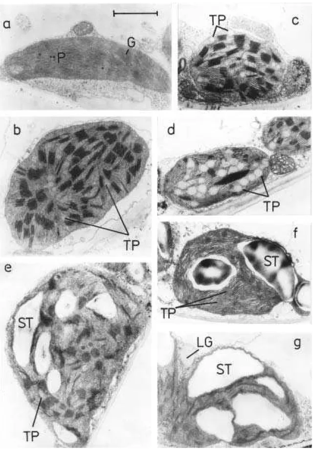
In addition to the decrease in the size and number of chloroplasts, their ultrastructure
Š K Š , Springer et al.
2015, Sobieszczuk-Nowicka et al. 2018). The senescence-associated changes of chloroplast ultrastructure include disappearance of thylakoid membranes, grana unstacking and loosening, swelling of intrathylakoid space, and chloroplast shrinkage. In parallel, a number and size of plastoglobules (PGs) increase (Lichtenthaler and Sprey 1966, Tuquet and Newmann 1980, Ghosh et al. 2001, Biswal et al. 2003, Š , Springer et al. 2015, Tominaga et al. 2018).
Fig. 3 A scheme of autophagy-related forms of chloroplast degradation under different conditions. (a) Darkening of whole plants (sugar starvation) the RCB-pathway; (b) Darkening of individual leaves combination of the RCB-pathway and chlorophagy of shrunken chloroplasts; (c) Excess light intensity (chloroplast photodamage) chlorophagy of damaged chloroplasts prior activation of RCBs.
(Izumi and Nakamura 2018)
PGs are connected to the thylakoid membrane by the outer lipid half-bilayer and arise at the margin of the stromal thylakoid. The PG core contains neutral lipids including prenylquinones, triacylglycerols, fatty acid phytyl esters, carotenoids and others but not glycolipids (for reviews, see e.g. Besagni and Kessler 2013, van Wijk and Kessler 2017). Plastoquinol and tocopherol are the major constituents of the prenylquinones in PGs. For many years, PGs were supposed to be only a passive storage site for thylakoid degradation products
15
(Lichtenthaler 1969, Burke et al. 1984). However, now it is known that PGs contain also small proteome and metabolome (Lundquist et al. 2012, van Wijk and Kessler 2017) and are involved in metabolism of tocopherols, quinones, and carotenoids (van Wijk and Kessler 2017, Ksas et al. 2018). Recently has been shown that PGs play also a role in Chl degradation and phytol recycling (Vom Dorp et al. 2015) and in chloroplast photo-protection (Ksas et al. 2018).
1.3.2 Chloroplast movement
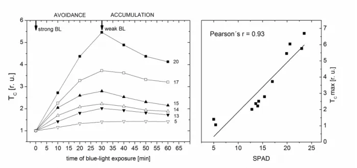
Chloroplast movement is one of the mechanisms by which plants respond to changing light environment (for a review, see e.g. Kong and Wada 2014). In higher plants, the light-induced chloroplast movement is induced by B light via phototropins. There are two types of light-induced chloroplast movement: accumulation and avoidance response. Under low light conditions, chloroplasts accumulate along the cell walls perpendicular to the incident light position). When exposed to high light, chloroplasts migrate to the cell walls parallel to the incoming light into side position (avoidance response). As we have shown in our previous works, the chloroplast movement can be easily detected by measurement of a leaf optical transmittance (N , 2016). During the avoidance response induced by strong light,
the transmittance increases as chl O
decreases (see Fig. 4).
It is thought that avoidance response protects chloroplasts from photo-oxidative
B B D
Hangarter 2012, Cazzaniga et al. 2013). As such conditions commonly occur during senescence, avoidance movement might be important for the controlled and efficient chloroplast degradation. However, almost nothing is known about functioning of the light-induced chloroplast movement in senescing leaves, although its impairment could be expected. In our previous works on tobacco leaves, we have obtained indications that the chloroplast avoidance movement is inhibited during senescence as an extent of the strong B light-induced increase of leaf transmittance decreased with decreasing Chl content (‘ N ) and was lower in older barley leaves compared with younger ones (N ).
Preliminary results with senescing leaves of Arabidopsis (Fig. 4) showing a correlation between extent of the avoidance response and Chl content support this assumption. The inhibition of chloroplast movement in senescing leaves might be related to senescence-associated impairment of cytoskeleton found by Keech et al. (2010). In addition, an abnormal arrangement of chloroplasts has been found in cells of senescing leaves (e.g., Wittenbach et al. 1982, V ) indicating impaired chloroplast anchoring to plasma membrane, which might influence negatively the light-induced chloroplast movement. It cannot be excluded that the observed reduction of light-induced changes in transmittance was associated also with a decrease in the chloroplasts number. Interestingly, our results indicate that chloroplast movement may change after the plant transition from vegetative to generative phase (N al. 2010). Further investigations are needed to elucidate the role of chloroplast movement in leaf senescence.
16
Fig. 4 Relative changes in collimated transmittance (TC Arabidopsis thaliana L. (Col-0) leaves during exposition to strong blue-light ( -2 s-1; avoidance response) and weak blue-light ( -2 s-1; accumulation response) and correlation between the maximal TC value (at time = 30 min) and chlorophyll content measured by SPAD-chlorophyllmeter.
1.4 Senescence-associated pigment changes
1.4.1 Chlorophyll degradation
Loss of green colour, the most apparent sign of leaf senescence, is caused by massive Chl degradation. Due to its light-absorbing ability, Chl has a phototoxic potential and can be dangerous for plant cells (especially when it is not integrated in photosynthetic pigment-protein complexes), therefore its metabolism (including degradation) is highly regulated (for a recent review, see e.g. Zhu et al. 2017).
Breakdown of Chl
three decades. Chl catabolites are now collectively termed phyllobilins and the biochemical
pathway of Chl degradation PAO breakdown step is
catalysed by pheophorbide a oxygenase (PAO) (for an up-to-date review, see Kuai et al. 2018). The PAO/phyllobilin pathway involves two distinct phases (Fig. 5). In the first phase, phototoxic free Chl molecules and their intermediates are degraded to colourless, primary fluorescent Chl catabolites (pFCC) inside chloroplasts, at thylakoid membranes. All reactions involved in the gradual conversion of Chl b to pFCC are common to all plant species in which Chl degradation has been investigated so far (Kuai et al. 2018). Based on the recent identification of C32-hydrolase as a chloroplast inner envelope-located oxygenase (named translocon at the inner chloroplast envelope 55, TIC55) (Hauenstein et al. 2016), and on the common occurrence of formyloxobilin- and dioxobilin-type non-fluorescent Chl catabolites (NCCs and DNCCs, respectively) deriving from C32-hydroxylated pFCC (hydroxyl-pFCC), the hydroxylation of pFCC has been included in the first phase of Chl breakdown (Kuai et al. 2018; Fig. 5).
17
Prior to its degradation by the PAO/phyllobilin pathway, Chl b has to be converted to Chl
a T T T C b
to Chl a is a two-step reaction catalysed by NYC1 (non-yellow coloring1) and NOL (NYC1-LIKE), and by hydroxymethyl Chl a reductase (HCAR).
Since its identification more than a century ago, chlorophyllase (CLH) hydrolysing Chl to chlorophyllide and phytol was assumed to be involved in senescence-associated Chl degradation, despite the fact that CLH proteins were found almost exclusively outside chloroplasts (especially in endoplasmatic reticulum and tonoplast). This was originally explained by extra-chloroplastic Chl dephytylation. Recently, CLH and chlorophyllide were proposed to play a role in plant defence against herbivores: in leaves damaged by herbivore, CLH has access to chloroplast-localized Chl (Hu et al. 2015). Currently, the Chl dechelatation is considered to be the first step of Chl a degradation during leaf senescence (Kuai et al. 2018; Fig. 5).
NYE (non-yellowing) protein (also called stay-green, SGR) has been recently identified as the enzyme degrading Chl a to pheophytin a as their Mg-dechelatase activity was revealed (Shimoda et al. 2016). The next step (dephytylation) is catalysed by pheophytinase (PPH). The next two steps are decisive in the loss of green colour: the ring-opening of pheophorbide a
by PAO (the membrane-localized, nonheme Fe-containing Rieske-type mono-oxygenase) resulting in red Chl catabolite (RCC), and the reduction of RCC by RCC-reductase (RCCR) to pFCC.
The second phase of PAO/phyllobilin pathway involves further side-chain-modifying reactions, which all take place outside chloroplast, and include non-enzymatic isomeration of the fluorescent phyllobilins (pFCC and hydroxyl-pFCC) to non-fluorescent ones (NCCs and DNCCs) in acidic pH (Fig. 5). The occurrence of NCCs and DNCCs varies among plant species (Kuai et al. 2018). In Arabidopsis, CYP89A9 (localized to endoplasmatic reticulum and catalysing oxidative deformylation of FCC to respective DFCC) was identified to be responsible for DNCC formation. Demethylation of FCC was found to be catalysed by methylesterase 16 (MES16) localized in cytosol. Final Chl catabolites are transported and stored in vacuole of senescing cells. The PAO/phyllobilin pathway might not to be the only way of Chl degradation. For instance, peroxidases were reported to be involved, however no respective degradation products have been identified (Kuai et al. 2018).
1.4.2 Carotenoids
Chloroplasts of higher plants usually contain the following carotenoids: -carotene, -carotene (located mainly in reaction centres), and xanthophylls violaxanthin (V), antheraxanthin (A), zeaxanthin (Z), neoxanthin and lutein (mainly associated with light-harvesting complexes). A content of overall carotenoids decreases during leaf senescence, however, a rate of their degradation is lower compared with Chl (Biswal 1995) thereby the ratio of carotenoid and Chl content usually increases during senescence (e.g., J ). In senescing leaves, xanthophyll acyl esters are typically accumulated. It is thought that carotenoids released from thylakoid pigment-protein complexes are stored in PGs.
18
Fig. 5 The PAO/phyllobilin pathway of chlorophyll breakdown in leaves. (A) Reactions of the first part of the pathway from chlorophyll to hydroxy-pFCC that take place inside the chloroplast. (B) pFCC/hydroxy pFCC-modifying reactions of the second part of the pathway that take place outside the chloroplast and lead to the diversity of phyllobilins. Note that MES16 and CYP89A9 are specific Arabidopsis enzymes, while all other enzymes are known from different plant species. Products: DFCCs, dioxobilin-type fluorescent chlorophyll catabolites; DNCCs, dioxobilin-dioxobilin-type non-fluorescent chlorophyll catabolites; FCCs, formyloxobilin-type fluorescent chlorophyll catabolites; NCCs, formyloxobilin-type non-fluorescent chlorophyll catabolites; pFCC, primary fluorescent chlorophyll catabolite; RCC, red chlorophyll catabolite. Enzymes: CYP89A9, specific cytochrome P450 monooxygenase; HCAR, hydroxymethyl Chl a reductase; MES16, methylesterase 16; NOL, NYC1-LIKE; NYC1, non-yellow coloring1; PAO, pheophorbide a oxygenase; PPH, pheophytinase; RCCR, RCC-reductase; SGR, stay-green; TIC55, translocon at the inner chloroplast envelope 55. (Kuai et al. 2018, with permission).
19
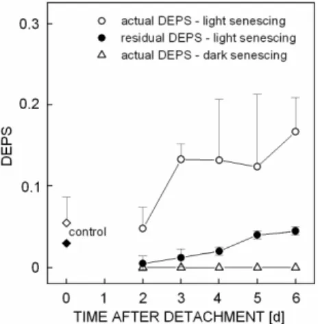
The slower degradation of carotenoids during senescence may be associated with their photo-protective function. One of the important photo-protective mechanisms in senescing leaves is the xanthophyll cycle dissipating excess light energy as heat (Demmig-Adams and Adams 1992). Although the amount of xanthophyll cycle pigments (V, A, Z) decreases during senescence, their de-epoxidation state (DEPS = (A + Z)/(A + Z + V)) usually increases during senescence under field conditions (e.g. G -Plazaola and Becerill 2001, Lu et al. 2001, 2003) as well as in detached leaves senescing under light (Š a, V et al. 2006). The increase in DEPS is higher when senescing leaves are exposed to higher light intensity
G -Plazaola and Becerill 2001, Lu et al. 2001). The protective role of xanthophylls during senescence is indicated also by the increase of (A + Z + V) content relative to content of Chl, which is generally observed in senescing leaves (e.g., Lu et al. 2003, Š a).
1.4.3 Anthocyanins
Unlike Chl and carotenoids, anthocyanins are newly produced during leaf senescence. They are synthesized in cytosol and then transported to vacuoles (Tanaka et al. 2008). The anthocyanins distribution in leaves depends on plant species, they can accumulate in vacuoles of both upper and lower epidermis and also in mesophyll cells (Merzlyak et al. 2008). The anthocyanin synthesis and sequestration represent a considerable metabolic investment for cells. Although the role of anthocyanins has been thoroughly investigated, their function in senescing leaves is still unclear.
Generally, anthocyanins are thought to protect the photosynthetic apparatus in mesophyll cells from excess light intensity and consequent oxidative damage. In vivo, anthocyanins commonly absorb light of wavelengths between 500 600 nm, but their absorption above 600 nm was also reported (Merzlyak et al. 2008, Hlavinka et al. 2013). It has been suggested that synthesis and accumulation of anthocyanins during leaf senescence, when the risk of over-excitation and oxidative damage usually increases, is connected with their photo-protective effect (Hoch et al. 2001). The protection from oxidative damage should allow the efficient nutrients recycling during the re-organization phase of senescence. Consistently with this hypothesis, the anthocyanin content in falling tree leaves negatively correlated with a content of residual nitrogen (Lee et al. 2003).
However, in a number of studies, the photo-protective role of anthocyanins was not confirmed (Gould et al. 2018). There is increasing evidence that the massive biosynthesis of anthocyanins may serve as an alternative way of the decreasing sugar accumulation under conditions of reduced sink strength (Lo Piccolo et al. 2018). The sugar accumulation causes the feed-back inhibition of photosynthesis, down-regulates the expression of photosynthetic genes and it is thought to induce or accelerate leaf senescence (see also parts 1.2.5 and 1.7.3). The accumulation of anthocyanins might therefore delay a sugar-promoted senescence, maintain functional photosynthetic apparatus for a longer time and allow the more efficient nutrient translocation (Lo Piccolo et al. 2018).
20
1.5 Role of ROS in leaf senescence
It is generally thought that aging of an organism or cell is associated with oxidative stress caused
A H
has proved to be inaccurate (see e.g., Pomatto and Davies 2018, Vina et al. 2018), the fact is that a level of oxidative damage increases with age (e.g. M -Bosch and Alegre 2002). Oxidative stress and/or oxidative damage are caused by an accumulation of ROS (singlet oxygen, 1O2;
superoxide anion radical, O2
-; hydroxyl radical, OH -; hydrogen peroxide, H2O2) due to
an imbalance between their generation and elimination. Originally ROS were considered to have only the damaging effect, but now it is clear that ROS have an essential role in the regulation of key cellular processes including senescence.
ROS play multiple roles in senescing leaves. In the initiation phase, ROS participate in induction of re-programming of gene transcription (e.g., Niewiadomska et al. 2009, Bieker et al. 2012, Bresson et al. 2018). At the beginning of re-organization phase, an increase in ROS content (e.g., Smart 1994) leads to pigment-, protein-, and lipid-oxidation that is necessary
for H F F
phase, oxidative processes participate pronouncedly in the final cell and leaf destruction (Zimmermann and Zentgraf 2005). It is obvious that excessive accumulation of ROS should be avoided over the entire senescence process to allow the efficient nutrient recycling and to prevent from premature cell death. To save a low ROS content, an effective and fine-tuned antioxidant protection is needed, involving enzymes as well as low-molecular-weight antioxidant substances.
Several enzyme cycles participate in the antioxidative defence of plants. The enzymes ascorbate-peroxidase (APX) and glutathione reductase (GR) are the main enzymes of the ascorbate-glutathione cycle, a part of the water-water cycle in chloroplasts scavenging O2
and H2O2 (Asada 1999). It is generally assumed that the capacity of antioxidant protection
decreases during leaf senescence. However, the decrease in antioxidant activity occurs mainly in the latest stages of senescence and it is considered to be not the cause but a consequence of senescence (Dertinger et al. 2003). Different changes in activities of antioxidant enzymes and content of non-enzymatic antioxidants were reported in senescing leaves (for a review, see
P W ) reflecting a complexity, time-spatial dependence and specifity of the antioxidant protection during senescence of different plant species under different conditions.
The initiation of senescence was found to coincide with an increase in H2O2 content
in leaves of various species (e.g., Niewiadomska et al. 2009, Bieker et al. 2012, Bresson et al. 2018), which was associated with a temporal decrease in activity of catalase and APX (Zimmermann et al. 2006, Niewiadomska et al. 2009, Bieker et al. 2012). In this case, the decrease in activity of antioxidant enzymes is probably controlled by cell to trigger senescence, as the increase in H2O2 content induces expression of TFs (Miao et al. 2007) and SAGs (Sabater M ). For example, the expression of senescence-associated WRKY and NAC factors is controlled by H2O2 (Miao et al. 2007).
In some cases, especially during stress-induced senescence, the excessive ROS accumulation can cause earlier onset of the terminal phase thus the principal function
21
of senescence, the remobilization of nutrients from the leaf, is not fulfilled. However, it has been shown that the ROS accumulation needs not always to cause oxidative damage in senescing leaves (Vanacker et al. 2006, Niewiadomska et al. 2009, Pilarska et al. 2017). It is possible that in such cases the increased ROS content could activate some defence processes protecting cells from oxidative damage.
Nevertheless, leaf senescence is typically associated with an increased oxidative damage including lipid peroxidation (see part 1.6). The increase in oxidative damage has been found during developmental senescence (e.g., Berger et al. 2001), premature senescence induced by plant shading (Š ) and dark-induced senescence of detached leaves
(Š , Causin et al. 2006, V , J Fig. 6).
Generally, the senescence-associated ROS accumulation is strengthened and level of oxidative damage increases under stress conditions. An origin of ROS can differ in dependence on light conditions during senescence. Under light conditions, chloroplasts can be considered as the main source of ROS (for a review of ROS generation in chloroplasts, see e.g. P
2012). Besides the chloroplasts, other cellular compartments such as mitochondria and peroxisomes also contribute to the ROS generation.
1.6 Leaf senescence and cell membranes
During senescence, cell membranes undergo changes in composition and integrity. These senescence-associated changes occur asynchronously in different types of cellular membranes. The first membranes undergoing degradative changes are thylakoids (e.g., Kolodziejek et al. 2003). The integrity of plasmatic membrane is usually maintained until late stages of leaf senescence as documented by relatively low ion leakage from senescing leaf tissue (e.g., Oda-Yamamizo et al. 2016).
Both main membrane components, lipids and proteins, are degraded during senescence. Phospholipids are extensively catabolised. A content of fatty acid decreases, which leads to enhanced relative concentrations of sterols (Thompson et al. 1998, Hopkins et al. 2007). Senescence-induced changes in lipid compositions result in changed biophysical organization of membrane bilayer (e.g., membrane rigidification, lipid phase separation) and consequent membrane leakiness (Thompson et al. 1998). In contrast to the cytoplasmic membranes, thylakoids do not undergo phase changes during senescence. The enzymatic lipid peroxidation is considered to be a main pathway of senescence-associated degradation of membrane lipids, in which four types of enzymes are involved: phospholipases (PLs), phosphatidic acid phosphatases, lipolytic acyl hydrolases and LOXs (Thompson et al. 1998).
The following scenario is supposed: PLs (activated via Ca2+) release polyunsaturated fatty acids (PUFAs) from membrane phospholipids, the increase of PUFA content activates LOXs and LOXs transform PUFAs to lipid hydroperoxides (LOOHs). Then LOOHs are converted to various secondary products (Spiteller 2003). Galactolipids of thylakoid membranes (mono- and di-galactosyldiacylglycerol) are thought to be degraded by galactolipases, galactosidases and lipolytic acyl hydrolases during leaf senescence (Hopkins et al. 2007). Galactolipids are thought to serve as a source of energy for metabolism of senescent cells (Matile 2001). The released