http://www.cmbl.org.pl
DOI: 10.2478/s11658-007-0027-9 Received: 12 March 2007
Revised form accepted: 30 April 2007
Published online: 03 July 2007 © 2007 by the University of Wrocław, Poland
* Author for correspondence: Yuedi Tang, Department of Otorhinolaryngology, West China Hospital of Sichuan University, No. 37 Guo-Xue-Xiang, Chengdu, Sichuan 610041, P. R. China, e-mail: [email protected]
Abbreviations used: ANP – atrial natriuretic peptide; Ct – cycle threshold; ET – endothelin; ETAR – endothelin type A receptor; ETBR – endothelin type B receptor; NO – nitric oxide; NSt – non-strial tissues; PG – prostaglandins; RQ – relative quantity; RT-PCR – reverse transcription-polymerase chain reaction; StV – strial vascularies
Short communication
THE EXPRESSION OF ENDOTHELIN TYPE A AND B RECEPTORS IN THE LATERAL WALL OF THE MOUSE COCHLEA
YAN LUO1, YUEDI TANG*1, QINGJIE XIA2 and JIN LIU3 1Department of Otorhinolaryngology, West China Hospital of Sichuan
University, Chengdu, Sichuan, China, 2Department of Ophthalmology, Laboratory of Molecular Biology, West China Hospital of Sichuan University,
Chengdu, Sichuan, China, 3Department of Anesthesiology and Critical Care Medicine, West China Hospital of Sichuan University, Chengdu, Sichuan, China
suggest that an endothelin-mediated response via two different receptors, ETAR and ETBR, may play an important role in the physiological functions of the cochlear lateral wall by maintaining the homeostatic environment of the cochlea.
Key words: ETAR, ETBR, Real time quantitative RT-PCR, Cochlea
INTRODUCTION
Endothelin, which was first identified as a potent vasoconstrictive peptide secreted from cultured aortic endothelial cells [1], is now known to consist of a family of 3 mammalian vasoactive peptides that includes ET-1, ET-2 and ET-3 [2]. These ETs are synthesized and released by many different cell types. The pathophysiological actions of endothelin are mediated by two different receptors, the endothelin type A receptor (ETAR) and the endothelin type B receptor (ETBR) [3]. Both are heterogeneously distributed throughout many organs. ET-1 and ET-2 bind to ETAR with high affinity, whereas ET-3 displays extremely low binding affinities by comparison. ETBR has similar binding properties for ET-1, ET-2, and ET-3. The pathophysiological actions of ET-1 have been thoroughly investigated. The release of ET-1 from the vascular endothelial cells activates ETAR, which predominates on the underlining vascular smooth muscle cells. The binding of ET-1 with ETAR activates phospholipase C, leading to the formation of inositol 1,4,5-trisphosphate and diacylglycerol [4]. ET-1 has a different function when it binds to ETBR by promoting nitric oxide and prostacyclin release causing vasodilation in vascellum [5]. ET-1 has been reported to have a wide variety of biological functions, not only in the vascular tissues but also in non-vascular tissues [6]. Considerable evidence suggests that ET-1 is a local autocrine and paracrine factor, in addition to its role in vasoactivity [7-9].
Previous studies have demonstrated an extensive distribution of ETs in many organs, including the inner ear. ET-1 activity was detected in the modiolus, spiral ligament, stria vascularis, spiral prominence, Reissner’s membrane, supporting cells of the organ of Corti, and spiral ganglion cells [10]. In the vestibule of normal guinea pigs, ET-like activity was identified in the sensory epithelial cells, supporting cells, dark cells, transitional cells, vestibular membrane, semicircular wall cells and vestibular ganglion cells [11]. ET-1-like activity was identified in the epithelial cells and subepithelial connective tissue of the endolymphatic sac and duct of normal guinea pigs [12]. Immunoreactivity for ET-1 and ET-3 was localized preferentially in cisterns of the rough endoplasmic reticulum and Golgi apparatus in the intermediate cells of WBB6F1+/+ mice. Immunoreactivity for ET-1 and ET-3 was also seen in lysosomes which were also occasionally found to contain (pre)melanosomes [13]. It was proposed that ET-1 may play an important role in the regulation of inner ear pressure, fluid volume, and ion balance.
ETs, synthesized and released by the intermediate cells, may participate in regulating the function of Na+, K+-ATPase in the production of the endolymph in the margin cells by mediation of ETAR in a paracrine manner [10]. Recently, another study showed that ETAR was expressed in the plasma of the intermediate cells and the capillary walls of the strial vascularis. ETBR activity was detected in the endothelial cells of the capillaries of the strial vascularis in mice. ET-1 expression was found to be distributed in the margin cells in a strong positive staining, and in the intermediate cells of the strial vascularis and in the spiral ligament in a weak positive staining [14].
However, the research thusfar has been limited by the use of the immunohistochemical staining method. The expression of ET receptor subtypes has not been systematically investigated in the lateral wall of the mouse cochlea. There is no available data on the quantity of ET receptors in the StV or NSt of the mouse cochlea. In order to elucidate the importance of the ET receptor in the regulation of inner ear function, it is crucial to quantify the gene expression. Real-time quantitative reverse transcription-polymerase chain reaction (RT-PCR) is a highly sensitive technology that allows high throughput quantification of gene expression [15]. The objective of this study was to determine the levels of mRNA expression of the ET receptor subtypes, ETAR and ETBR, in the strial vascularies and non-strial tissues of the lateral wall of the mouse cochlea using real-time quantitative RT-PCR techniques. We aimed to establish whether the StV and NSt of mice synthesize ETAR and ETBR. We compared the levels of mRNA expression of ETAR and ETBR in the StV and NSt, which are known to be involved in the regulation of inner ear pressure, fluid volume, and ion balance. The side-by-side comparison of our quantitative data on ET receptor expression clearly highlights the crucial roles of these peptides. Further understanding of the roles of these peptides in the cochlear lateral wall should reveal the importance of endothelin in maintaining the homeostatic environment of the cochlea.
MATERIALS AND METHODS
Animals
For this study, we used adult (c. 30 days old) Kunming mice, an outbreed strain of laboratory animal widely utilized in related pharmaceutical and genetic studies in China. The study was approved by the Animal Care and Use Committee at the West China Medical School of Sichuan University. The temporal bone was removed and StV and NSt tissues were dissected at 4ºC, as previously described [17].
RNA isolation and reverse transcription
Inc.) with the addition of random hexamer primers. Briefly, 5 µl of purified RNA was used as template for cDNA synthesis in the presence of 1 µl M-MLV reverse transcriptase (200 U), 1 µl of random hexamer primer, 4 µl of 5 x reaction buffer, 2 µl of hexamer 1 x (Roche), 2 µl of dNTP mix (10 mM each) and 6 µl of RNAse-free water. After incubation for 60 min at 42ºC, the reverse transcriptase was inactivated at 70ºC for 10 min, and the cDNAs were stored at -20ºC until further analysis.
Primers
Primers were designed according to the NCBI Genebank of mice, and were synthesized by the Shanghai SAGON. The primer sequences for RT-PCR were as follows: ETAR, forward primer 5’-CTCCATCTGGATTCTTTTCCTT-3’ and reverse primer 5’-CTTGGTAAAACTCCATGAACT-3’ (a 137-bp product was to be generated); ETBR, forward primer 5’-CAAGTTGCTCGCAGAGGACT-3’ and reverse primer 5’-CAGCTCGATATCTGTCAATACT-3’ (a 128-bp product was to be synthesized); GAPDH, forward primer 5’-TGGGTGTGAACCACG AGAA-3’ and reverse primer 5’-GGCATGGACTGTGGTCATGA-3’ (a 143-bp product was to be generated).
Real-time quantitative PCR
Real-time PCR was used to determine the gene expression profiles of the ET receptor subtypes. cDNAs were amplified by real-time PCR using an FTC-2000 (FUNGLYN, CANADA). Each analysis was performed in a total volume of 30 µl reaction mixture containing 5 µl cDNA sample, 1 µl SYBR Green I, and 2 µl gene-specific forward and reverse primers (10 µM each). Housekeeping genes were included to normalize the data. Amplifications were performed as follows, 45 cycles at 94ºC for 2min, 94ºC for 20 sec and 54ºC for 20 sec, with a final extension at 70ºC for 30 sec and at 80ºC for 20 sec .
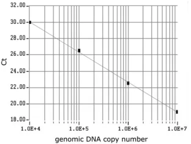
The cycle threshold (Ct) number, defined as the number of PCR amplification cycles required to reach fluorescent intensity above the threshold, was determined for each gene and each developmental time point analyzed. Using serial dilutions of the test sample cDNA, the standard curve was generated on the basis of the linear relationship of existing Ct and the logarithm of the copy number. The slope of the curve was shown to be -3.70, and a strong linear relationship was demonstrated (R2 =1.00; Fig. 1).
Analysis
Fig. 1. The standard curve for the GAPDH set generated from Ct values and plotted against the input standard genomic DNA molecules. The standard curve ranged from 104 copies to 107 copies. Using serial dilutions of the standard cDNA, the standard curve was generated on the basis of the linear relationship between the existing Ct and the logarithm of the copy number.
RESULTS AND DISCUSSION
Via real-time quantitative RT-PCR, we demonstrated that both ETAR and ETBR mRNAs were expressed in the cochlear lateral wall (Fig. 2). We used heart tissue samples, which were previously shown to express ETAR and ETBR, as positive controls. GAPDH was examined as a housekeeping gene.
The results of the analysis of ETAR and ETBR gene expression from the StV and NSt are presented in Fig. 3. ETAR mRNA expression predominated in the StV with a relatively lower level of expression of the ETBR receptor mRNA in the StV. The mRNA levels of ETAR and ETBR were relatively low in the NSt as compared to those in the StV: there was a 1.8 times higher amount of ETAR mRNA in the StV than in the NSt of the lateral wall tissue (p < 0.05), and 8.1 times as much ETBR mRNA in the StV as in the NSt (p < 0.05).
In this study, we demonstrated the mRNA expression of the ET receptor subtypes, ETAR and ETBR, in the cochlear lateral wall. Real-time quantitative RT-PCR provides a specific, sensitive and reliable method for the quantification of mRNA expression in the cochlear lateral wall of mice.
Fig. 2. ETAR and ETBR cDNAs amplified from homogenates of the StV, NSt and heart. The amplified fragments were of the expected sizes of 137 and 128 bp, respectively for the ETAR (A) and ETBR gene (B). The amplification of a 143 bp fragment of GAPDH cDNA as a housekeeping control is also shown (C). M – marker, lane 1– heart, lane 2 – strial vascularies (StV) of the cochlear lateral wall, lane 3 – non-strial tissues (NSt) of the cochlear lateral wall.
Fig. 3. The mRNA expression of ETAR and ETBR by real-time quantitative RT-PCR. The quantification of ETAR and ETBR gene expression in the StV and NSt (n = 7) relative to the heart (n = 9). The mRNA expression of ERAR and ETBR was normalized to the expression of ETBR in the heart (control), which was arbitrarily set to a value of 1. Significantly different expression (p < 0.05) is expressed as the average ± SD.
cells suggest the possibility of their autocrine action on the middle cells [13]. Jinnouchi et al. [10] further investigated the dynamic changes of ET-1 in the epithelium of the endolymphatic sac of guinea pigs and their relationship to the development of endolymphatic hydrops. Their findings suggest that ET-1 may play an important role as one of the regulators maintaining the fluid balance. Here, our results confirm that ETAR mRNA is present in the StV and NSt of the mouse cochlea. This concurs with earlier results obtained through immunoreactivity studies, suggesting that the StV and NSt probably actively synthesize the peptide ETAR.
ET-1 mediates its effect by acting on either the ETAR or ETBR receptor subtypes. The mRNA of both receptor subtypes is found in the StV and NSt, with a predominance of ETAR over ETBR. ET-l may reduce its physiological functions in an autocrine and/or a paracrine manner, as has been shown for other cell types [3]. This study demonstrates that ETAR is the predominant endothelin receptor expressed in the StV and NSt. In addition, our studies demonstrate higher expression levels of ETAR and ETBR in the StV than those in the NSt. It was reported that the endothelins are involved in the activation of the sodium pump of the strial marginal cells by mediation of ETAR [13]. In addition, ETAR mediates vasoconstriction of capillaries in the spiral ligament [20].
In this study, we also determined the expression of ETBR in the cochlear lateral wall of mice. However, the function of ETBR in the StV is uncertain.ET-1 has a different function when it binds to ETBR by promoting nitric oxide and prostacyclin release causing vasodilation in the vascellum [21]. Several studies have demonstrated thatETBR also helps to clear ET-1 from the circulation, via a ligand-receptor internalization mechanism[22]. It was suggested that ET plays an important role in regulating the circulation of the cochlea as a local hormone binding to the ETBR of capillary endotheliocytes. In this study, we further demonstrated the presence of ETBR mRNA in the NSt, implying that ETBR is possibly synthesized in these cells.
In summary, we demonstrated that ETAR and ETBR were both expressed in the StV and NSt. We further obtained data suggesting that ETAR and ETBR receptors were heterogeneously distributed in the mouse cochlear lateral wall. Gene expression of the ETAR and ETBR was predominant in the StV over the NSt. This study provides evidence in support of the action of ET in the cochlear lateral wall. In particular, the high expression of these genes in the StV implies an important role in the homeostasis within the microenvironment of the inner ear.
Acknowledgements. This project was supported by the Natural Science Foundation of China (30271408). We would like to thank Chenliang Xia and Yanfang Chen for their excellent technical assistance.
REFERENCES
1. Yanagisawa, M., Kurihara, H., Kimura, S., Tomobe, Y., Kobayashi, M., Mitui, Y., Azaki, Y., Goto, K. and Masaki, T. A novel potent vasoconstrictor peptide produced by vascular endothelial cells. Nature 33 (1988) 411-415. 2. Hosoda, K., Hammer, R.E., Richardson, J.A., Baynash, A.G., Joto, H.,
McCarron, R.M., Inoue, A., Yanagisawa, M. and Kimura, S. The human endothelin family: three structurally and pharmacologically distinct isopeptides predicted by three separate genes. Proc. Natl. Acad. 86 (1989) 2863-2867.
3. Levin, E.R. Endothelins. N. Engl. J. Med. 333 (1995) 356-363.
4. Masaki, T., Kimura, S., Yanagisawa, M. and Goto, K. Molecular and cellular mechanism of endothelin regulation: implications for vascular function. Circulation 84 (1991) 1457-1468.
5. Haynes, W.G. and Webb, D.J. Endothelin as a regulator of cardiovascular function in health and disease. J. Hypertens. 16 (1998) 1081-1089.
6. Brown, M.A. and Smith, P.L. Endothelin: a potent stimulator of intestinal ion secretion in vitro. Regul. Pept. 36 (1991) 1-19.
7. Shichiri, M., Hirata, Y., Nakajima, T., Ando, K., Imai, T. and Yanagisawa, M. Endothelin-1 is an autocrine/paracrine growth factor for human cancer cell lines. J. Clin. Invest. 87 (1991) 1867-1871.
8. Matsuura, A., Yamochi, W., Hirata, K., Kawashima, S. and Yokoyama, M. Stimulatory interaction between vascular endothelial growth factor and endothelin-1 on each gene expression. Hypertension 32 (1998) 89-95. 9. Evans, J.J., Youssef, A.H., Yandle, T.G., Lewis, L.K. and Nicholls, M.G.
Effects of endothelin-1 on release of adrenomedullin and C-type natriuretic peptide from individual human vascular endothelial cells. J. Endocrinol. 175 (2002) 225-232.
10. Jinnouchi, K., Tomiyama, S. and Pawankar, S. Distribution of endothelin-1-like activity in the cochlea of normal guinea pigs. Acta Otolaryngol. 117 (1997) 41-45.
11. Jinnouchi, K., Tomiyama, S. and Pawankar, R. Distribution of endothelin-1-like activity in the vestibule of normal guinea pigs. ORL J. Otorhinolaryngol. Relat. Spec. 58 (1996) 4-8.
12. Jinnouchi, K., Tomiyama, S. and Pawankar, R. Distribution of endothelin-1 like activity in the endolymphatic sac of normal guinea pigs. Acta Otolaryngol. 115 (1995) 400-404.
14. Xu, X.N., Huang, J.M., Lin, G.J. and Jiang, Z.Z. Contributions of endothelin in the process of the noise-induced in jury of inner ear. Zhonghua Er Bi Yan Hou Tou Jing Wai Ke Za Zhi 40 (2005) 509-512.
15. Higuchi, R., Fockler, C., Dollinger, G. and Watson, R. Kinetic PCR analysis: real-time monitoring of DNA amplification reactions. Mar. Biotechnol. 11 (1993) 1026-1030.
16. Livak, K. and Schmittgen, T. Analysis of relative gene expression data using real-time quantitative PCR and the 2-∆∆Ct method. Methods 25 (2001) 402-408.
17. Marcus, D.C. and Chiba, T. K+ and Na+ absorption by outer sulcus epithelial cells. Hear. Res. 134 (1999) 48-56.
18. Wangemann, P. K+ cycling and its regulation in the Cochlea and the vestibular labyrinth. Audiol. Neurootol. 7 (2002) 199-205.
19. Franz, P., Hauser-Kronberger, C. and Egerbacher, M. Localization of endothelin-1 and endothelin-3 in the cochlea. Acta Otolaryngol. 117 (1997) 358-362.
20. Sadanaga, M., Liu, J. and Wangemann, P. Endothelin-A receptors mediate vasoconstriction of capillaries in the spiral ligament. Hear. Res. 112 (1997) 106-114.
21. Haynes, W., Strachan, F. and Webb, D. Endothelin ETAR and ETBR receptors cause vasoconst riction of human resistance and capacitance vessels in vivo.Circulation 92 (1995) 357-363.