Address for correspondence Dr. Neerja Puri
Dermatologist, Punjab Health Systems Corporation,
Ludhiana, Punjab (India)
Email: neerjaashu @rediffmail.com
Review Article
Etiology and management of leg ulcers – an enigma
Introduction
Chronic ulceration of the lower legs is a relatively common condition amongst adults, one that causes pain and social distress.1 The
condition affects 1% of the adult population and 3.6% of people older than 65 years.2 Chronic
ulceration of the lower legs is a relatively common condition amongst adults, and ulcer symptoms usually include increasing pain, friable granulation tissue, foul odor, and wound breakdown instead of healing.
It has been reported that leg ulcers related to venous insufficiency constitute 70%, arterial disease 10%, and those of mixed etiology 15% of presentations.3 The remaining 5% of leg
ulcers result from less common pathophysiological causes, and this latter group comprises considerable challenges in diagnosis, assessment, and management. Leg ulcers are mainly caused by venous insufficiency, arterial
insufficiency, neuropathy, diabetes (Figure 1), or a combination of these factors.4 Venous ulcers
are the most common type of leg ulcers, accounting for approximately 70% of cases.5,6,7
Arterial disease accounts for another 5% to 10% of leg ulcers; most of the others are due to either neuropathy (usually diabetic) or a combination of those diseases. A study from India shows that etiology of chronic wounds included systemic conditions such as diabetes, atherosclerosis, tuberculosis, and leprosy.8 Other major causes
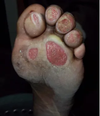
included venous ulcers, pressure ulcers (Figure 2), vasculitis, and trauma. Venous ulcers most commonly occur above the medial or lateral malleoli.9,10 Arterial ulcers often affect the toes
or shin or occur over pressure points. Neuropathic ulcers tend to occur on the sole of the foot or over pressure points.
Discussion
Venous ulceration is the most common type of leg ulceration. Sixty to 80% of leg ulcers have a venous etiology. Venous ulcers arise from
Neerja Puri*, Ashutosh Talwar**
* Dermatologist, Punjab Health Systems Corporation, Punjab, India ** Surgeon, Punjab Health Systems Corporation, Punjab, India
Abstract Chronic leg ulcer is defined as a defect in the skin below the level of knee persisting for more than
six weeks and shows no tendency to heal after three or more months. Leg ulcers are debilitating and greatly reduce patients' quality of life. The common causes are venous disease, arterial disease and neuropathy. Management of patients with chronic ulcers has to be multidisciplinary and should include detailed history, physical examination, investigations, basic and newer treatment modalities, and educating patients on issues of correct foot care and the importance of seeking early medical advice.
Keywords
Figure 1 Figure showing diabetic ulcer
Figure 2 Figure showing multiple pressure foot ulcers.
Figure 3 Figure showing venous ulcer of the lower limb.
venous valve incompetence and calf muscle pump insufficiency which leads to stasis and venous hypertension.11 This results in
microcirculatory changes and localized tissue ischemia. The natural history of the disease is of a continuous cycle of healing and breakdown over decades and chronic venous leg ulcers are associated with considerable morbidity and impaired quality of life. Venous circulation of the lower extremities progresses from the superficial to perforating to deep veins, with valves in each system to ensure unidirectional blood flow.12 As the calf muscles contract, the
pumping action causes the blood to flow from the deep veins into the inferior vena cava. Disease of these pathways results in venous insufficiency. Venous ulcers (Figure 3) are more common in women and older persons. The primary risk factors are older age, obesity, previous leg injuries, deep venous thrombosis, and phlebitis. Venous ulcers are often recurrent, and open ulcers can persist from weeks to many years. Severe complications include cellulitis, osteomyelitis, and malignant change.13
multiple pregnancies, obesity, congenital vein abnormalities, and varicose veins. Venous ulcers are commonly associated with varicose eczema which is characterized by erythema, weeping, scaling and pigmentation, and may be misdiagnosed as infection. Venous ulcers are also prone to being complicated by allergic contact dermatitis. The clinical appearance of varicose eczema and allergic contact dermatitis is similar, but the distribution and response to treatment provide useful diagnostic information.16
The management of leg ulcers should include a detailed history of the onset of the problem, examination of the legs and skin, investigations, and modalities of treatments. The first step toward diagnosis of any leg ulcer is to compile a comprehensive history and assessment of the patient. This should include general health status, social and occupational situation, past and current medical history of relevant diseases (such as deep vein thrombosis, diabetes, autoimmune disorders, inflammatory bowel disease, and connective tissue disease), condition of the skin, current vascular status, limb size and shape, and history and status of the ulcer.17,18 The patient should be asked about
lower extremity pain, paresthesia, anesthesia, and claudication. It is important to determine the duration of ulceration and whether it is a first episode or recurrent. Pain is a major problem for patients with leg ulcers unless there is a neuropathic component. Lack of pain, therefore, suggests a neuropathic etiology. Patients should also be asked about their mobility. The examination of the leg should include palpation of pulses and a search for the signs of venous hypertension, including varicose veins, hemosiderin pigmentation, varicose eczema, atrophie blanche, and lipodermatosclerosis. The ulcer examination should include site, size, appearance, wound base, exudates level, and surrounding skin. The surrounding region should
be examined for pain, edema, erythema, warmth, induration, discoloration, maceration, dryness, scarring from previous wounds, hair pattern, gangrenous digits, clubbing, cyanosis, capillary refill, and varicose veins. It is important to bear in mind that venous and arterial disease may coexist in the same patient. Venous ulcers differ considerably from arterial ulcers and other ulcers of lower extremity. An irregular ulcer border, black necrosis, erythema, or bluish or purple discolorations of adjacent skin are suggestive for ulcer due to vasculitis. A painful leg ulcer with violaceous borders suggests pyoderma gangrenosum.
For management of leg ulcers, the leg should be assessed for signs of venous disease, in particular, varicose veins, venous dermatitis, hemosiderin deposition, lipodermatosclerosis and atrophie blanche.19,20 A venous duplex scan
may aid assessment of the leg. Edema should be assessed and non-venous causes of unilateral and bilateral edema ruled out. Joint mobility, particularly that of the ankle, is an important component of calf muscle pump function and should be carefully recorded. It is important to assess arterial supply with respect to safety of compression therapy, which is the standard treatment for venous leg ulcers. Palpation of pulses alone is not adequate to rule out peripheral arterial disease. Measurement of the ankle brachial pressure index (ABPI) of both lower limbs by hand-held Doppler device is the most reliable way to detect arterial insufficiency.21 Ulcerated legs should be washed
normally in tap water and carefully dried. Necrotic material or slough within a wound margin acts as a medium for bacterial proliferation and therefore should be removed by debridement.22,23 General care of the skin
margins should be coated with a barrier preparation to prevent maceration of surrounding skin. Uncomplicated venous dermatitis usually responds to emollients, but often topical corticosteroids may be required. Failure to respond to a moderately potent steroid is an indication for patch testing.
The effectiveness of graduated compression stockings in achieving and maintaining healing is dependent on the correctness of fit and the pressure generated beneath the stocking.24
Compression bandaging is the most effective treatment for venous leg ulcer. The bandages work by helping push the blood in your leg veins back up to your heart. Different strengths are available and the aim is to find the strongest that patient can wear. A dressing is worn under the bandage. This will be changed when required, usually once a week. When the dressing is changed, leg is washed with warm tap water. Waterproof protectors are available for bathing/showering at home between dressing changes. In clinical and laboratory testing, not all stockings produce an adequate pressure or pressure gradient although they may be described as of a similar class. Patients should be offered the strongest compression which they can tolerate to prevent ulcer recurrence. Patients should be informed that it is likely that compression will be required indefinitely. If a patient finds a stocking uncomfortable, changing the brand of stocking within the same class may improve compliance. Advise patients who experience pain in their calf when walking that this may be an indication of arterial disease and may affect their treatment options. The compression treatment with bandages or with a stocking is the single most important treatment for a leg ulcer, and is far more important than the ulcer dressing.25,26,27 Ensure that patients are
aware of the need for a weekly change of bandage and that more frequent changes may be required in certain circumstances. Encourage
patients to wash their leg gently in warm tap water when bandages are being changed. Advise patients that antibiotics are only needed very occasionally. Pentoxifylline may be used to help improve blood flow which may help leg ulcer to heal. The following advice should be offered to patients and carers during treatment of leg ulcers: when resting, you should try to keep your ankles up higher than your heart. This allows the fluid to drain from your legs. At night time it is important that you keep to ones normal sleeping habits and trying to sleep in your bed rather than in a chair. Raising the foot end of the bed at night if tolerable will help to assist venous return. Dry scaly skin needs to be treated with a non-perfumed moisturizer/emollient to keep the skin moist.
Conclusion
An ulcer which is present for more than three months is considered as chronic ulcer. The majority of chronic leg ulcers are caused by venous insufficiency followed by arterial ulcers. A comprehensive assessment of the patient, limb, and ulcer is required to determine etiology and to formulate an appropriate management plan. The patients should be explained about the benefits of exercise and the need to wear compression hosiery from the time one gets up in the morning until going to bed at night, renewing hosiery every 3-6 months, keeping mobile and elevating their legs when resting.
References
1. Ryan TJ. The epidemiology of leg ulcers. In: Westerhof W, ed. Leg Ulcers: Diagnosis and Treatment. Amsterdam: Elsevier Science Publishers; 1993. P. 19-27.
2. Baker SR, Stacey MC, McKay AG. Epidemiology of chronic venous ulcers. Br J Surg. 1991;78:864-7.
incidence and prevalence in the elderly. J Am Acad Dermatol. 2002;46:381-6. 4. Baker SR, Stacey MC, Singh G et al.
Aetiology of chronic leg ulcers. Eur J Vasc Surg. 1992;6:245-51.
5. Miller OF 3rd, Phillips TJ. Leg ulcers. J Am Acad Dermatol. 2000;43:91-5.
6. Renton EJ. Pharmacological treatments of venous leg ulcers. J Wound Care. 1999;8:195-7.
7. Mekkes JR, Westerhof W. Venous ulceration. Lancet. 1993;342:121-2. 8. Partsch H. Investigations on the
pathogenesis of venous leg ulcers. Acta Chir Scand. 1988;544:25-9.
9. Falanga V, Eaglstein W. The trap hypothesis of venous ulceration. Lancet. 1993;341:1006-8.
10. Valencia IC, Falabella A, Kirsner RS, Eaglstein WH. Chronic venous insufficiency and venous leg ulceration. J Am Acad Dermatol. 2001;44:401-21. 11. Junger M, Steins A, Hahn M, Hafner HM.
Microcirculatory dysfunction in chronic venous insufficiency (CVI). Microcirculation. 2000;7:3-12.
12. Leu HJ. Morphology of chronic venous insufficiency - light and electron microscopic examinations. Vasa. 1991;20:330-42.
13. Singer AJ, Clark RAF. Cutaneous wound healing. N Engl J Med. 1999;341:738-46. 14. Cornwall JV, Dore CJ, Lewis JD. Leg
ulcers: epidemiology and aetiology. Br J Surg. 1986;73:693-6.
15. Nelzen O, Bergqvist D, Lindhagen A. Leg ulcer etiology - a cross sectional population study. J Vasc Surg. 1991;14:557-64. 16. Nelzen O, Bergqvist D, Lindhagen A.
Venous and non-venous leg ulcers: clinical history and appearance in a population study. Br J Surg. 1994;81:182-7.
17. Phillips TJ. Chronic cutaneous ulcers: etiology and epidemiology. J Invest Dermatol. 1994;102:38-41.
18. Kahle B, Hermanns HJ, Gallenkemper G. Evidence-based treatment of chronic leg ulcers. Deutsches Arzteblatt Int. 2011;108:231-7.
19. Meyer FJ, Burnand KG, Lagatolla NRF, Eastham D. Randomized clinical trial comparing the efficacy of two bandaging regimens in the treatment of venous leg ulcers. Br J Surg. 2002;89:40-4.
20. Hafner J, Trueb RM. Management of vasculitic leg ulcers and pyoderma gangrenosum. Curr Probl Dermatol. 1999;27:277-85.
21. Persoon A, Heinen MM, van der Vleuten CJ et al. Leg ulcers: a review of their impact on daily life. J Clin Nurs. 2004;13:341-54.
22. Van Gent WB, Wilschut ED, Wittens C. Management of venous ulcer disease. Br Med J. 2010;341:1092-6.
23. Caputo WJ, Beggs DJ, DeFede JL et al. A prospective randomised controlled clinical trial comparing hydrosurgery debridement with conventional surgical debridement in lower extremity ulcers. Int Wound J. 2008;5:288-94.
24. Lok C, Paul C, Amblard P et al. EMLA cream as a topical anesthetic for the repeated mechanical debridement of venous leg ulcers: a double-blind, placebo-controlled study. J Am Acad Dermatol. 1999;40:208-13.
25. Bello YM, Phillips TJ. Management of venous ulcers. J Cutan Med Surg. 1998;3:6-12.
26. Cullum N, Nelson EA, Fletcher AW, Sheldon TA. Compression for venous leg ulcers (Cochrane Review). The Cochrane Library, Issue 1. Oxford: Update Software, 2001.