Cutting Edge: Transcriptional Profiling Reveals
Multifunctional and Cytotoxic Antiviral Responses of Zika
Virus–Specific CD8
+
T Cells
Alba Grifoni,* Priscilla Costa-Ramos,
†John Pham,* Yuan Tian,*
Sandy L. Rosales,* Gre´gory Seumois,* John Sidney,* Aruna D. de Silva,*
,‡,1Lakshmanane Premkumar,
xMatthew H. Collins,
x,{Mars Stone,
‖Phillip J. Norris,
‖Claudia M. E. Romero,
#Anna Durbin,** Michael J. Ricciardi,
††Julie E. Ledgerwood,
‡‡Aravinda M. de Silva,
xMichael Busch,
‖Bjoern Peters,*
,xxPandurangan Vijayanand,* Eva Harris,
{{Andrew K. Falconar,
#Esper Kallas,
†Daniela Weiskopf,* and Alessandro Sette*
,xxZika virus (ZIKV) constitutes an increasing public health
problem. Previous studies have shown that CD8
+T cells
play an important role in ZIKV-specific protective
im-munity. We have previously defined antigenic targets of
the ZIKV-specific CD8
+T cell response in humans. In
this study, we characterized the quality and phenotypes of
these responses by a combined use of flow cytometry and
transcriptomic methods, using PBMCs from donors
de-riving from different geographical locations collected in
the convalescent phase of infection. We show that
ZIKV-specific CD8
+T cells are characterized by a
polyfunc-tional IFN-
g
signature with upregulation of TNF-
a
,
TNF receptors, and related activation markers, such as
CD69, as well as a cytotoxic signature characterized by
strong upregulation of GZMB and CRTAM. The
signa-ture is stable and not influenced by previous dengue virus
exposure, geographical location, or time of sample
collec-tion postinfeccollec-tion. To our knowledge, this work
eluci-dates the first in-depth characterization of human CD8
+T cells responding to ZIKV infection.
The Journal
of Immunology
, 2018, 201: 3487–3491.
T
he relatively recent spread of Zika virus (ZIKV) in theabsence of any available treatment or preventative vaccine has illustrated the need to better understand a number of issues associated with this previously understudied virus ranging from epidemiology, disease pathogenesis, and
host immune responses. CD8+T cell immune responses seem
to play an important role in ZIKV-specific protective immunity, contributing to protective immunity against
ZIKV. Dengue virus (DENV)–pre-exposed CD8+ T cells
are also able to mount cross-protective responses against ZIKV challenge (1, 2). In this context, it is important to
study and define the features of ZIKV-specific CD8+T cell
responses and ascertain whether these responses are fully functional or somehow altered (especially in DENV–pexposed donors), raising the possibility that these re-sponses may be associated with immunopathology. We previously showed that subjects characterized by prior DENV infection displayed not only increased magnitude of ZIKV responses in the acute phase but also showed a skewed immunodominance pattern of responses toward nonstructural proteins as previously observed in the con-text of DENV infection (3, 4). However, an in-depth
*Division of Vaccine Discovery, La Jolla Institute for Allergy and Immunology, La Jolla, CA 92037;†
Division of Clinical Immunology and Allergy, School of Medicine, University of Sa˜o Paulo, Sa˜o Paulo 01246-903, Brazil;‡
Genetech Research Institute, Colombo 08, Sri Lanka;xDepartment of Microbiology and Immunology, University of North Carolina School of Medicine, Chapel Hill, NC 27516;{The Hope Clinic, Emory Vaccine Center, Division of Infectious Diseases, Department of Medicine, School of Medicine, Emory University, Decatur, GA 30317;‖Blood Systems Research Institute, San Francisco, CA 94118;#Universidad del Norte, Barranquilla 1569, Colombia; **School of Medicine,
University of Vermont, Burlington, VT 05405;††
Department of Pathology, Miller School of Medicine, University of Miami, Miami, FL 33146;‡‡
Vaccine Research Center, National Institute of Allergy and Infectious Diseases, Bethesda, MD 20892;xxUniversity of Califor-nia San Diego, La Jolla, CA 92093; and{{Division of Infectious Diseases and Vaccinology, School of Public Health, University of California, Berkeley, CA 94720
1Current address: Department of Paraclinical Sciences, Kotelawala Defense University,
Ratmalana, Sri Lanka.
ORCIDs:0000-0002-2209-5966(A.G.);0000-0002-8164-6852(G.S.); 0000-0002-0987-405X (J.S.); 0000-0001-5291-9543 (A.D.d.S.); 0000-0001-7974-1933 (M.H.C.); 0000-0001-5619-2767 (M.S.); 0000-0003-0526-2088 (P.J.N.);
0000-0001-7860-5542(C.M.E.R.);0000-0002-6652-2851(A.D.); 0000-0002-1446-125X (M.B.); 0000-0002-8457-6693 (B.P.); 0000-0002-7238-4037 (E.H.); 0000-0002-2066-9487 (A.K.F.); 0000-0003-2026-6925 (E.K.); 0000-0001-7013-2250(A.S.).
Received for publication August 8, 2018. Accepted for publication October 12, 2018.
This work was supported by National Institutes of Health (NIH) Contracts HHSN272200900042CandHHSN27220140045C,andGrantsU19 AI118626-01 toA.S.,HHSN268201100001ItoM.B.,andS10OD016262toP.V.Furthersupport wasprovidedbytheZikaPreparednessLatinAmericanNetwork,whichhasreceived fundingfromtheEuropeanUnion’sHorizon2020researchandinnovationprogram underGrantAgreement734584.BlooddonorsamplesfromPuertoRicoandFlorida werecollected aspartoftheNationalHeart,Lung,andBlood InstituteRecipient EpidemiologyandDonorEvaluationStudy-III.TheLaJollaInstituteforAllergyand Immunology’sFACSAriaIIcellsorterwasacquiredthroughSharedInstrumentation GrantProgramS10RR027366.TheIlluminaHiSeq2500Sequencerwaspurchased throughNIHS10OD016262.
AddresscorrespondenceandreprintrequeststoDr.AlessandroSette,LaJolla,9420 AthenaCircle,LaJolla,CA92037.E-mailaddress:[email protected]
Theonlineversionofthisarticlecontainssupplementalmaterial.
Abbreviationsusedinthisarticle:CRTAM,cytotoxicandregulatoryTcellmolecule; DENV,denguevirus;GZMB,granzymeB;PCA,principalcomponentanalysis;TPM, transcriptpermillion;ZIKV,Zikavirus.
characterization of the functionality of ZIKV-specific
CD8+T cell responses is not available to date.
Materials and Methods
Human blood samples and ZIKV and DENV determination
Blood donations from convalescent donors previously infected with ZIKV were collected (4) in Puerto Rico within the Recipient Epidemiology and Donor Evaluation Study-III or at the University of North Carolina (UNC Institu-tional Review Board no. 08-0895) and University of Miami from United States travelers with Zika symptoms. An additional cohort was identified at the Universidad del Norte in Barranquilla, Colombia. ZIKV infection was confirmed using RT-PCR during the early acute/symptomatic phase (5). Clinical and serological characteristics are summarized in Supplemental Table I. In addition to positive RT-PCR, previous ZIKV infections in con-valescent phase samples were also confirmed using ZIKV neutralization assays, depleting DENV cross-reactive Abs (6). PBMCs were isolated (3), cry-opreserved, and stored in liquid nitrogen until usage. DENV seropositivity was determined by DENV IgG or an inhibition ELISA (7, 8).
IFN-gcapture assay, sorting, and flow cytometry
Cells were stimulated with ZIKV peptide pools (4). Additionally, we used 309 ZIKV 9-mers and 10-mers corresponding to previously identified epitopes supplemented with predicted epitopes based on a 27-allele method (9) using TepiTool, available in the Immune Epitope Database and Analysis Resource
(www.IEDB.org) (10). IFN-g–producing cells were captured using a cytokine
secretion assay (Miltenyi Biotech, Bergisch Gladbach, Germany) (11). Sorting gating strategy is shown in Supplemental Fig. 1. For the intracellular cytokine
staining, PBMCs were cultured 6 h with 1mg/ml peptide pools and BD
GolgiPlug (BD Biosciences, San Diego, CA), then permeabilized, stained, and analyzed using FlowJo software (4). Supplemental Table II shows the Abs used for sorting and flow cytometry experiments.
RNA sequencing, bioinformatics, and statistical data analysis
Two hundred cells for each condition were collected. To generate full-length transcriptomes from the low cells per sample, we adapted the Smart-Seq2 protocol (12). Bioinformatics analysis to obtain the raw counts and dif-ferentially expressed genes were performed as previously described (13). We
considered genes to be differentially expressed with an adjustedpvalue of
,0.05 and the log2 fold change in gene expression.1. Only genes with an
average transcript per million (TPM) count$10 in ZIKV IFN-g+
condi-tion have been considered for further analysis. Principal component analysis (PCA) was performed on the top 500 most variable genes using versust transformation and the “prcomp” function in R, setting “scale” equal to TRUE. The heatmap was generated on versust-transforming data using Qlucore. Statistical analyses for FACS experiments were performed using Prism 7 (GraphPad Software, San Diego, CA). One-tailed Mann–Whitney or Wilcoxon tests were applied when appropriate. The data have been de-posited in the Gene Expression Omnibus (GSE105884, http://www.ncbi. nlm.nih.gov/geo/) and ImmPort (SDY903, http://www.immport.org).
Results and Discussion
To examine the nature of CD8+T cell responses recognizing
ZIKV epitopes, we performed a transcriptomic analysis of PBMCs from ZIKV-infected donors collected at the con-valescent phase. In this study, PBMCs were stimulated ex vivo with ZIKV-specific peptide pools, and Ag-specific
CD8+ T cells were sorted based on their ability to
pro-duce IFN-g. The patients’ CD8+T cells that did not
pro-duce IFN-gin response to epitope stimulation from these
same cultures as well as total unstimulated CD8+ T cells
were also collected for comparison with their ZIKV-specific
IFN-g–producing CD8+T cells. After sorting, microscaled
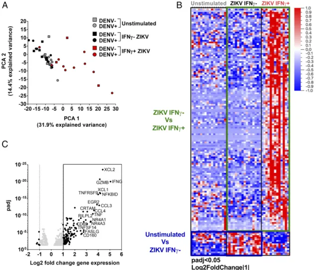
RNA sequence determinations were performed on low cell numbers (12). We then analyzed the global gene expression patterns using PCA of the 500 most variable genes ob-tained after RNA sequencing (Fig. 1A). Unstimulated and
ZIKV-stimulated IFN-g2 CD8+ T cells clustered together
and were clearly separated from the gene expression profiles
of the ZIKV-stimulated IFN-g+ CD8+ T cells. This
sug-gested that the main component of human CD8+ T cell
response to ZIKV epitopes were from their IFN-g– producing
cells, in agreement with previous studies on both ZIKV and the closely related DENV regarding CD8 responses (1, 3, 4,
14, 15). The IFN-g2CD8+T cells may also include some, if
not many, ZIKV-specific T cells, which did not secrete
IFN-g but instead secreted IL-2 or TNF-a under the
stimulation of peptides. Further experiments targeting cell subpopulations secreting other cytokines or selected on the basis of specific activation markers could be performed in the future to broaden the characterization of ZIKV-specific
CD8+T cell responses.
The interplay between ZIKV and DENV is an important issue because ZIKV and DENV not only share a high level of protein sequence homology but are also transmitted by the same mosquitoes, and ZIKV transmission has predominantly occurred in DENV-endemic areas (16). We have previously shown that prior DENV exposure modulates ZIKV responses when comparing acute samples that were DENV naive or previously exposed (4). In this study, we do not observe
dif-ferences both in terms of frequency of IFN-g+cells of DENV
seronegative and seropositive (Supplemental Fig. 1B) as well as in terms of gene expression observed (Fig. 1A), suggesting
that DENV pre-exposure might affect ZIKV CD8+ T cells
only during the acute phase of the infection. These prelimi-nary findings should be further explored; however, they are in agreement with previous work in which small differences in monocyte gene expression profiles were observed in patients who had ZIKV or DENV infections, suggesting similari-ties between the two viral infections at the gene expression level (17).
To identify gene expression patterns associated with
ZIKV-specific CD8+T cell responses, we determined the differential
gene expression profiles of the patients’ IFN-g2 and IFN-g+
cells after ZIKV peptide pools stimulation and on IFN-g2cells
that were unstimulated or stimulated with ZIKV (Fig. 1B). Overall, a total of 153 genes were differentially expressed in either one of these two analyses. Of these, 142 genes were
upregulated at least 2-fold in the IFN-g+ compared with the
IFN-g2ZIKV-stimulated cells, and 32 genes were differentially
upregulated at a more stringent cutoff of a 4-fold level in
IFN-g+compared with IFN-g2cells (Fig. 1C). These results
further emphasize that the main component of CD8+responses
to ZIKV peptides were the IFN-g–producing cells (4).
Next, we more closely inspected the mRNA sequence
profiles of the IFN-g+CD8+T cells to ascertain which genes
at a more stringent 4-fold change cutoff were upregulated in
the ZIKV-specific CD8+ T cells (Fig. 1C). Interestingly,
among the most significantly upregulated genes were those
encoding granzyme B (GZMB) and cytotoxic and regulatory
T cell molecule (CRTAM), implying that the CD8 responses
were not limited to cytokine release but were also associated with the hallmarks of a cytotoxic response (18). Among the other strongly upregulated genes were those associated with cellular activation, such as tumor necrosis receptor
super-family member 9 (TNFRSF9), which encodes the CD137
protein (also known as 4-1BB), important in priming and
promoting CD8+T cell effector and memory functions in the
context of viral infection (19), andCD69, which encodes an
important marker of T cell activation. In addition, significant
upregulation was also noted forNFKBID, which encodes an
system that is activated after TCR stimulation (20). Other upregulated genes that correlated with activation were TNF
ligand superfamily member 6/Fas ligand gene (TNFL6/
FASLG) and member 14 (TNFSF14/CD258), which encode proteins involved in cytotoxic T cell apoptotic processes and
immune regulation, andERG2, encoding an upstream
regu-lator of effector CD4+ and CD8+ T cells linked with
pro-tective responses and limiting immunopathology (21) as well
as with regulating CD8+T cell activation (22).
Other prominently upregulated genes were X-C motif
chemokine ligand 1 and 2 (XCL1and XCL2), which encode
chemokines with inflammatory function that are known to induce leukocyte migration and activation (23). The genes encoding the chemokine (C-C motif) ligand 3 and 4 (CCL3 and CCL4) proteins, associated with inflammation and in the case of CCL3 also with CD8 effector function, were also upregulated (24).
Based on the gene function described above and available in UniProt knowledgebase database (https://www.uniprot.org/), we functionally grouped together the genes more strongly
upregulated in ZIKV-specific CD8+ T cell genes into five
main groups: 1) polyfunctional cytokines, 2) cytotoxicity, 3) T cell activation and regulation, 4) proinflammation, and 5) T cell homing.
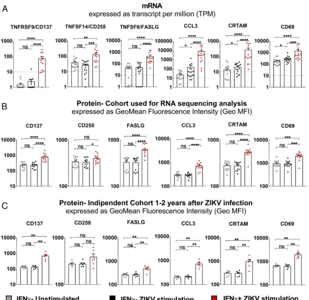
We previously reported, using a different cohort of donors/
samples, that ZIKV-specific CD8+ T cell responses were
characterized by IFN-g, TNF-a, and GZMB release (4). We
then compared the geometric mean of fluorescence intensity
expression of IFN-g, TNF-a, and GZMB in IFN-g2 and
IFN-g+cell groups, reanalyzing our previously published data
(4) (Supplemental Fig. 2B, 2D, 2F), to the TPM count originated by the mRNA sequence determinations performed
in the current study. All genes (IFN-g,TNF-a, andGZMB)
encoding the proteins found to be upregulated by FACS analysis in previous studies were also found to be upregulated at the mRNA level in this study (Supplemental Fig. 2C, 2E,
2G). To further validate the CD8+ T cell ZIKV signature
obtained by the mRNA sequence determinations and analy-ses, we selected six additional proteins (CD137, CD258, FASLG, CCL3, CRTAM, and CD69) based on differential
expression of their corresponding genes (changes of.4-fold,
p-adjusted values ,0.05) (Fig. 1C) and the availability of a
suitable commercial mAb to detect them. The protein-level expression of these markers was determined on a set of 12 donors known to respond to stimulation with ZIKV peptides, as previously reported (4), after a 6-h stimulation period with ZIKV-specific pooled peptides and intracellular staining. Overall, there was a good correlation between
FIGURE 1. Gene expression analysis of ZIKV-specific CD8+T cells. (A) PCA of unstimulated IFN-g2(gray) and ZIKV-stimulated IFN-g2(black) and
IFN-g+(red). DENV pre-exposed (circles) and DENV naive (square) are also shown (B). Heatmap of differentially expressed genes after comparison of IFN-g2and
IFN-g+cells stimulated with ZIKV pools. (C) Differentially expressed genes based on adjustedpvalue and log2 fold change gene expression. Genes with log2 fold
upregulation at the mRNA (Fig. 2A) and protein (Fig. 2B)
expression levels. Our results demonstrated that the CD8+
T cell responses recognizing ZIKV-specific epitopes were
prevalently associated with IFN-g+cells, as expected from our
experimental strategy, and that this response appeared to be fully functional, as demonstrated by the upregulation of genes involved in cytotoxicity, chemotaxis, and inflammation. To prove that these observations were generally applicable re-gardless of the particular cohort analyzed or the time elapsed postinfection, we also analyzed the same markers on PBMCs from a different cohort in Barranquilla (Colombia) that had been collected 1–2 y after ZIKV infection. This independent ZIKV cohort showed patterns of protein upregulation re-markably similar to those detected in the previous cohort (Fig. 2C). These data, therefore, demonstrated that this ex-pression signature was both reproducible and temporally stable after Ag-specific stimulation even 1–2 y after ZIKV infection. In summary, we report, to our knowledge, the
first human immune profile of CD8+ T cells after ZIKV
Ag-specific stimulation. Our data show that ZIKV-specific
CD8+ T cell responses were predominantly associated with
IFN-g responses and that their gene expression signatures
were further associated with cytokine polyfunctionality and cytotoxic activity. These common features were independent of DENV pre-exposure, geographical location, or time of sample collection postinfection and were identified both at the mRNA and protein level, therefore making these suitable candidates to be evaluated in cellular vaccine-induced im-mune responses. Because of the small volume of samples available, we focused our study on immune profiling of
ZIKV-specific CD8+T cells. Future studies will address the
immune profiles of human ZIKV–specific CD4+T cells and
compare the functionalities of CD4+T cells with the ones of
CD8+T cells. Indeed, recent studies in humans and mouse
models have shown the importance of ZIKV-specific CD4+
T cells in protecting from severe ZIKV severe and Ab de-velopment (25–27). Additionally, it would be of interest to test whether disease severity during the acute phase impacts the gene transcription and protein expression in ZIKV-specific T cells. However, the cohorts studied in this study were mostly characterized by a mild form of ZIKV disease and, therefore, this analysis could not be performed based on
FIGURE 2. Correlation of expression at the mRNA and protein levels in CD8+T cells of selected markers. (A) TPM forCD137,CD258,FASLG,CCL3,
1. Elong Ngono, A., E. A. Vizcarra, W. W. Tang, N. Sheets, Y. Joo, K. Kim, M. J. Gorman, M. S. Diamond, and S. Shresta. 2017. Mapping and role of the CD8+T cell response during primary Zika virus infection in mice.Cell Host Microbe
21: 35–46.
2. Wen, J., A. Elong Ngono, J. A. Regla-Nava, K. Kim, M. J. Gorman, M. S. Diamond, and S. Shresta. 2017. Dengue virus-reactive CD8+T cells mediate
cross-protection against subsequent Zika virus challenge.Nat. Commun.8: 1459. 3. Weiskopf, D., M. A. Angelo, E. L. de Azeredo, J. Sidney, J. A. Greenbaum,
A. N. Fernando, A. Broadwater, R. V. Kolla, A. D. De Silva, A. M. de Silva, et al. 2013. Comprehensive analysis of dengue virus-specific responses supports an HLA-linked protective role for CD8+ T cells.Proc. Natl. Acad. Sci. USA110: E2046–E2053. 4. Grifoni, A., J. Pham, J. Sidney, P. H. O’Rourke, S. Paul, B. Peters, S. R. Martini,
A. D. de Silva, M. J. Ricciardi, D. M. Magnani, et al. 2017 Prior dengue virus exposure shapes T cell immunity to Zika virus in humans.J. Virol. DOI: 10.1128/ JVI.01469-17.
5. Waggoner, J. J., L. Gresh, A. Mohamed-Hadley, G. Ballesteros, M. J. Davila, Y. Tellez, M. K. Sahoo, A. Balmaseda, E. Harris, and B. A. Pinsky. 2016. Single-reaction multiplex reverse transcription PCR for detection of Zika, chikungunya, and dengue viruses.Emerg. Infect. Dis.22: 1295–1297.
6. Collins, M. H., E. McGowan, R. Jadi, E. Young, C. A. Lopez, R. S. Baric, H. M. Lazear, and A. M. de Silva. 2017. Lack of durable cross-neutralizing antibodies against Zika virus from dengue virus infection.Emerg. Infect. Dis.23: 773–781. 7. Kanakaratne, N., W. M. Wahala, W. B. Messer, H. A. Tissera, A. Shahani,
N. Abeysinghe, A. M. de-Silva, and M. Gunasekera. 2009. Severe dengue epidemics in Sri Lanka, 2003-2006.Emerg. Infect. Dis.15: 192–199.
8. Ferna´ndez, R. J., and S. Va´zquez. 1990. Serological diagnosis of dengue by an ELISA inhibition method (EIM).Mem. Inst. Oswaldo Cruz85: 347–351. 9. Paul, S., D. Weiskopf, M. A. Angelo, J. Sidney, B. Peters, and A. Sette. 2013. HLA
class I alleles are associated with peptide-binding repertoires of different size, af-finity, and immunogenicity.J. Immunol.191: 5831–5839.
10. Paul, S., J. Sidney, A. Sette, and B. Peters. 2016. TepiTool: a pipeline for computational prediction of T cell epitope candidates.Curr. Protoc. Immunol.
114: 18.19.1–18.19.24.
11. Hinz, D., G. Seumois, A. M. Gholami, J. A. Greenbaum, J. Lane, B. White, D. H. Broide, V. Schulten, J. Sidney, P. Bakhru, et al. 2016. Lack of allergy to timothy grass pollen is not a passive phenomenon but associated with the allergen-specific modulation of immune reactivity.Clin. Exp. Allergy46: 705–719. 12. Rosales, S. L., S. Liang, I. Engel, B. J. Schmiedel, M. Kronenberg,
P. Vijayanand, and G. Seumois. 2018. A sensitive and integrated approach to
profile messenger RNA from samples with low cell numbers.Methods Mol. Biol.
1799: 275–301.
13. Tian, Y., M. Babor, J. Lane, V. Schulten, V. S. Patil, G. Seumois, S. L. Rosales, Z. Fu, G. Picarda, J. Burel, et al. 2017. Unique phenotypes and clonal expansions of human CD4 effector memory T cells re-expressing CD45RA.Nat. Commun.
8: 1473.
14. Lum, F. M., D. C. B. Lye, J. J. L. Tan, B. Lee, P. Y. Chia, T. K. Chua, S. N. Amrun, Y. W. Kam, W. X. Yee, W. P. Ling, et al. 2018. Longitudinal study of cellular and systemic cytokine signatures to define the dynamics of a balanced immune environment during disease manifestation in Zika virus-infected patients.
J. Infect. Dis.218: 814–824.
15. de Alwis, R., D. J. Bangs, M. A. Angelo, C. Cerpas, A. Fernando, J. Sidney, B. Peters, L. Gresh, A. Balmaseda, A. D. de Silva, et al. 2016. Immunodominant dengue virus-specific CD8+ T cell responses are associated with a memory PD-1+ phenotype.J. Virol.90: 4771–4779.
16. World Health Organization. 2017. Situation report: Zika virus microcephaly Guillain-Barre´ syndrome. Available at: http://apps.who.int/iris/bitstream/handle/10665/253604/ zikasitrep20Jan17-eng.pdf;jsessionid=64AF3C2F78E08372CD2E090CC063A504? sequence=1. Accessed: January 20, 2017.
17. Michlmayr, D., P. Andrade, K. Gonzalez, A. Balmaseda, and E. Harris. 2017. CD14+CD16+monocytes are the main target of Zika virus infection in peripheral
blood mononuclear cells in a paediatric study in Nicaragua.Nat. Microbiol.2: 1462–1470.
18. Takeuchi, A., Y. Itoh, A. Takumi, C. Ishihara, N. Arase, T. Yokosuka, H. Koseki, S. Yamasaki, Y. Takai, J. Miyoshi, et al. 2009. CRTAM confers late-stage acti-vation of CD8+ T cells to regulate retention within lymph node.J. Immunol.183: 4220–4228.
19. Yoshimori, M., K. Imadome, H. Komatsu, L. Wang, Y. Saitoh, S. Yamaoka, T. Fukuda, M. Kurata, T. Koyama, N. Shimizu, et al. 2014. CD137 expression is induced by Epstein-Barr virus infection through LMP1 in T or NK cells and me-diates survival promoting signals.PLoS One9: e112564.
20. Schuster, M., M. Annemann, C. Plaza-Sirvent, and I. Schmitz. 2013. Atypical IkB proteins - nuclear modulators of NF-kB signaling.Cell Commun. Signal.11: 23. 21. Miao, T., A. L. J. Symonds, R. Singh, J. D. Symonds, A. Ogbe, B. Omodho,
B. Zhu, S. Li, and P. Wang. 2017. Egr2 and 3 control adaptive immune responses by temporally uncoupling expansion from T cell differentiation.J. Exp. Med.214: 1787–1808.
22. Mauri, D. N., R. Ebner, R. I. Montgomery, K. D. Kochel, T. C. Cheung, G. L. Yu, S. Ruben, M. Murphy, R. J. Eisenberg, G. H. Cohen, et al. 1998. LIGHT, a new member of the TNF superfamily, and lymphotoxin alpha are ligands for herpesvirus entry mediator.Immunity8: 21–30.
23. Lei, Y., and Y. Takahama. 2012. XCL1 and XCR1 in the immune system.Microbes Infect.14: 262–267.
24. Trifilo, M. J., C. C. Bergmann, W. A. Kuziel, and T. E. Lane. 2003. CC chemokine ligand 3 (CCL3) regulates CD8(+)-T-cell effector function and migration following viral infection.J. Virol.77: 4004–4014.
25. Hassert, M., K. J. Wolf, K. E. Schwetye, R. J. DiPaolo, J. D. Brien, and A. K. Pinto. 2018. CD4+T cells mediate protection against Zika associated severe disease in a mouse model of infection.PLoS Pathog.14: e1007237.
26. Lucas, C. G. O., J. Z. Kitoko, F. M. Ferreira, V. G. Suzart, M. P. Papa, S. V. A. Coelho, C. B. Cavazzoni, H. A. Paula-Neto, P. C. Olsen, A. Iwasaki, et al. 2018. Critical role of CD4+T cells and IFNgsignaling in antibody-mediated resistance to Zika virus infection.Nat. Commun.9: 3136.
27. Koblischke, M., K. Stiasny, S. W. Aberle, S. Malafa, G. Tschouchnikas, J. Schwaiger, M. Kundi, F. X. Heinz, and J. H. Aberle. 2018. Structural in-fluence on the dominance of virus-specific CD4 T cell epitopes in Zika virus infection. [Published erratum appears in 2018Front Immunol.9: 2083.]Front. Immunol.9: 1196.
thecurrentcohort.Inconclusion,thecurrentstudyrepresents
abaselineforcomparisonbetweendifferentclinicaloutcomes
ofZIKVinfection intheCD8+Tcellcontext.
Acknowledgments
We thank Sheridan Martini for sample coordination, Ashmitaa Loganda and
Jason Greenbaum of the Bionformatic Core, and Denise Hinz and Cheryl Kim of the Flow Cytometry Core at the La Jolla Institute for Allergy and Immunology.
Disclosures
The authors have no financial conflicts of interest.
1
Supplemental Fig 1. CD8+ T cell sorting and mRNA sequence determinations and analyses. (A)
2
Gating strategy. Lymphocytes have been gated based on FSC-A and SSC-A parameters. Single cells have
3
been derived based first on combination of FSC-A and FSC-W parameters and then SSC-A and SSC-W
4
parameters. The resulting single cells have been gated for being positive for CD3 and negative for
5
CD14/19/Live and Dead to obtain the T cells. The resulting T cells have been further gated based on CD8
6
H[SUHVVLRQ WR REWDLQ&'7FHOOV&'7FHOOV KDYHEHHQIXUWKHUGLYLGHG EDVHGRQ,)1ȖH[SUHVVLRQ
7
Unstimulated cells have been sorted as ,)1Ȗ-&' 7 FHOOV ZKLOH ERWK ,)1Ȗ- DQG ,)1Ȗ &' 7 FHOOV
8
stimulated with ZIKV have been collected.(B)UHTXHQF\RI&'&',)1ȖFHOOVDIWHU=,.9VWLPXODWLRQ
9
in DENV seronegative and DENV seropositive donors used for sorting experiments. Statistical analyses
10
have been performed with Mann-Whitney test.
1
Supplemental Fig 2. Correlations between protein and mRNA expression levels from CD8+ T cells.
2
(A) Gating strategy. (B, D, F) Representative dot plots and Geo MFI calculated for the ,)1Ȗ71)ĮDQG
3
granzyme B markers based on previously published data8. (C, E, G) transcripts per million (TPM) for the
4
expression levels of the ,)1Ȗ,71)Į and granzyme Bencoding genes.
Donor
ID Country of origin Type of experiment
Days after clinical diagnosis
ZIKV status statusDENV a) ObservationClinical
2692 US travellers RNA sequencing 365 convalescent pos not severe 3039 Puerto Rico RNA sequencing 90 convalescent pos not severe 3043 Puerto Rico RNA sequencing 90 convalescent pos not severe 3047 Puerto Rico RNA sequencing 90 convalescent pos not severe 3052 Puerto Rico RNA sequencing 90 convalescent pos not severe 3028 US travellers RNA sequencing 24 convalescent pos not determined 3031 US travellers RNA sequencing 45 convalescent pos not determined 2653 US travellers RNA sequencing 365 convalescent neg not severe 3030 US travellers RNA sequencing 60 convalescent neg not determined 3053 Puerto Rico RNA sequencing 90 convalescent neg not severe 3054 Puerto Rico RNA sequencing 90 convalescent neg not severe
3027 US travellers RNA sequencing /validation 150 convalescent pos not determined
3038 Puerto Rico RNA sequencing /validation 90 convalescent pos not severe
3037 Puerto Rico RNA sequencing /validation 90 convalescent pos not severe
3045 Puerto Rico RNA sequencing /validation 90 convalescent pos not severe
2894 US travellers RNA sequencing /validation 90 convalescent neg not severe
3126 Puerto Rico validation 90 convalescent pos not severe 3127 Puerto Rico validation 90 convalescent pos not severe 3128 Puerto Rico validation 90 convalescent pos not severe 3129 Puerto Rico validation 90 convalescent pos not severe 3130 Puerto Rico validation 90 convalescent pos not severe 3131 Puerto Rico validation 90 convalescent pos not severe 3132 Puerto Rico validation 90 convalescent pos not severe
3421 Colombia validation 475 convalescent pos Guillain-Barré Syndrome
3422 Colombia validation 565 convalescent pos Guillain-Barré Syndrome
3423 Colombia validation 565 convalescent pos not severe 3424 Colombia validation 505 convalescent pos not severe
3428 Colombia validation 535 convalescent pos Guillain-Barré Syndrome
3430 Colombia validation 595 convalescent pos not severe
a) Previous exposure to DENV (Positive or Negative) was determined by the presence of detectable DENV-specific IgG titers.
1
2
Supplementary Table S1. List of ZIKV positive donors in convalescent phase, confirmed either by
RT-3
PCR and/or ZIKV serology after serum depletion.
Target Color Vendor Clone species reactivity Experiment
CD14 V500 BD Biosciences M5E2 mouse human Sorting/Flow cytometry
CD19 V500 BD Biosciences HIB19 mouse human Sorting/Flow cytometry
Live/Dead ef506 eBioscience Sorting/Flow cytometry
CD3 AF700 eBioscience UCHT1 mouse human Sorting/Flow cytometry
CCR7 PerCP Cy5.5 Biolegend G043H7 mouse human Sorting
CD45RA ef450 eBioscience HI100 mouse human Sorting
CD8 BV650 BioLegend RPA-T8 mouse human Sorting/Flow cytometry
CD4 APCef780 eBioscience RPA-T4 mouse human Sorting/Flow cytometry
,)1Ȗ APC-CY7 BioLegend 4S.B3 Mouse human Flow cytometry
CD69 PE Cy7 eBioscience FN50 Mouse human Flow cytometry
CD137 (4-1BB) APC BioLegend 4B4-1 Mouse human Flow cytometry
CD355 (CRTAM) PE/Cy7 ebioscience Cr24.1 Mouse human Flow cytometry
CD178 (Fas-L) PE Biolegend NOK-1 Mouse human Flow cytometry
CD258 (LIGHT) AF647 BD Biosciences 115520 Mouse human Flow cytometry
CCL3 PE ebioscience CR3M Mouse human Flow cytometry
1
Supplementary Table S2. List of monoclonal antibody used for Sorting and Flow cytometry validation
2
experiments.