G
PCRs, which form the largest target class in the druggable genome, are crucial for nearly every physiological pro-cess1. Aminergic GPCRs, including histamine, adrenergic, dopamine, serotonin, and muscarinic receptors, are of particular importance to drug discovery, as they are targeted by one quarter of currently approved drugs2,3. Functional selectivity4, or signaling bias, is a process whereby GPCR ligands can either activate G pro-teins or recruit B-arrestins to activate select downstream signaling pathways at a given receptor5–7. In many instances, one signaling pathway is potentially responsible for therapeutic effects whereas the other is implicated in side effects8–10. Biased ligands that can yield drugs with optimized on-target effects include agonists for the D2 dopamine receptor (D2R)8, D1 dopamine receptor (D1R)11, angiotensin II type 1 receptor (AT1R)10, D-opioid receptor (DOR)12, and the M-opioid receptor (MOR)9. G protein-biased MOR agonists are potentially analgesic and have fewer side effects (for example, respiratory depression and constipation13).The development of biased ligands remains challenging even when using high-throughput screening and extensive interrogation of the signaling properties of existing ligands10,14–17. Recently, our understanding of GPCR ligand recognition and receptor activation dynamics as it pertains to biased signaling has been catalyzed by a ‘golden era’ of GPCR structural biology, with several key aminergic receptor structures being published in the last decade18–23. Despite this wealth of information, no logical process exists for efficiently incorporating insights gleaned from GPCR structures into a design strategy for biased-ligand development.
The D2R remains an essential target for antipsychotic drug discovery24,25, with the newest atypical antipsychotic drugs (for example, aripiprazole, cariprazine) being partial agonists at D2R and other receptors26. We previously conducted extensive medici-nal chemistry exploration of aripiprazole, and although aripip-razole is a partial agonist at multiple GPCRs26, it shows similar
potency and efficacy in Gi/o signaling and B-arrestin recruitment at D2R8,27. Those studies culminated in the discovery of the first D2R B-arrestin-biased ligands8, which show therapeutic potential in animal models of schizophrenia28. Our results suggested that D2R B-arrestin signaling contributes to the antipsychotic efficacy of these drugs, whereas G protein signaling may contribute to extrapy-ramidal side effects8.
In this study, we used D2R as a model system to identify GPCR–ligand contacts that mediate biased signaling, and used this information to develop an approach for the structure-based drug design (SBDD) of B-arrestin-biased ligands for other amin-ergic GPCRs.
RESULTS
Design of indole-aripiprazole hybrid ligands
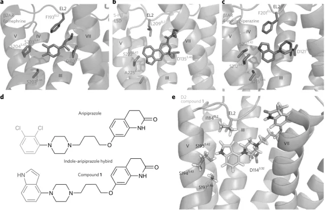
We analyzed prior aminergic GPCR structural and mechanistic data to identify residues implicated in G protein signaling relative to B-arrestin signaling. We focused on the orthosteric site, as this is both the most common and the most well-conserved binding site for class A GPCRs. In the binding pockets of the B1 and B2 adrenergic recep-tors (B1AR and B2AR, respectively), transmembrane helix 5 (TM5) transduces ligand-induced G protein activation via conserved ser-ine residues (5.42, 5.43, and 5.46); these findings are supported by structural21, mutagenesis29, and NMR30 studies. For the nanobody-stabilized B2AR crystallized in complex with epinephrine21, the cat-echol of epinephrine, which is also present on dopamine, forms an extensive hydrogen bond network with these conserved TM5 ser-ines (Fig. 1a), which have been previously posited to form the struc-tural basis of agonist and partial agonist action31 at B1AR and B2AR. D2R also contains TM5 serine residues (Supplementary Results,
Supplementary Fig. 1a), which, as supported by mutagenesis stud-ies, contribute to ligand efficacy and overall G protein activation32,33 and are essential for aripiprazole recognition34.
1National Institute of Mental Health Psychoactive Drug Screening Program, Department of Pharmacology and Division of Chemical Biology and Medicinal Chemistry, University of North Carolina Chapel Hill Medical School, Chapel Hill, North Carolina, USA. 2Center for Chemical Biology and Drug Discovery, Departments of Pharmacological Sciences and Oncological Sciences, Tisch Cancer Institute, Icahn School of Medicine at Mount Sinai, New York, New York, USA. 3Departments of Computer Science and Molecular and Cellular Physiology, Institute for Computational and Mathematical Engineering, and Biophysics Program, Stanford University, Stanford, California, USA. 4Department of Pharmaceutical Chemistry, University of California at San Francisco, Byers Hall, San Francisco, California, USA. 5Neuroscience and Pain Medicinal Chemistry, Pfizer Worldwide R&D, Cambridge, Massachusetts, USA. *These authors contributed equally to this work. *e-mail: [email protected], [email protected] or [email protected]
Structure-inspired design of
B
-arrestin-biased
ligands for aminergic GPCRs
John D McCorvy
1*, Kyle V Butler
2*, Brendan Kelly
3*, Katie Rechsteiner
1, Joel Karpiak
4, Robin M Betz
3,
Bethany L Kormos
5, Brian K Shoichet
4, Ron O Dror
3, Jian Jin
2& Bryan L Roth
1Development of biased ligands targeting G protein-coupled receptors (GPCRs) is a promising approach for current drug dis-covery. Although structure-based drug design of biased agonists remains challenging even with an abundance of GPCR crystal
structures, we present an approach for translating GPCR structural data into B-arrestin-biased ligands for aminergic GPCRs.
Structural clues for binding-pocket residues that mediate arres-tin recruitment are illuminated by the 5-hydroxytryptamine 2B (5-HT2B) receptor structures in complex with ergotamine22 and lysergic acid diethylamide35 (LSD; Fig. 1b). In the 5-HT
2B–LSD structure study, mutation of the conserved hydrophobic EL2 residue Leu209 selectively reduced LSD arrestin recruitment by increasing ligand on- and off-rates at the receptor. EL2 as a structural motif was proposed to function as a ‘lid’ over the binding pocket, thereby enhancing ligand residence time and functioning as a major deter-minant of arrestin recruitment efficacy35. Given that hydrophobic residues located in EL2 are relatively well conserved for aminergic GPCRs (Supplementary Fig. 1a), we posited that targeting the homologous D2 EL2 hydrophobic residue isoleucine 184 (I184EL2) may enhance B-arrestin recruitment at this receptor, thus leading to novel B-arrestin-biased ligands.
First, we required a ligand scaffold to test our hypotheses for the differential involvement of TM5 and EL2 in biased signaling. We recently disclosed B-arrestin-biased ligands that are close structural analogs of aripiprazole8,36, choosing these as starting points. We also required a small fragment predicted to form defined interactions with conserved TM5 serines located in the orthosteric site, which could be substituted in such a way as to disrupt the TM5 serine interactions associated with G protein-dependent activation. Crystal structures of the thermostabilized turkey B1AR in complex with indole-piperazine clearly illustrate the position of the indole group in the orthosteric site near TM5 and EL2, with the indole N–H form-ing a hydrogen bond with TM5 residue S5.42 (ref. 37; Fig. 1c).
Our design strategy, therefore, was to replace the dichlorophenyl- piperazine portion of aripiprazole with the indole-piperazine fragment
found in the B1AR crystal structure, leading to an indole-aripip-razole hybrid, compound 1 (Fig. 1d). To generate reliable assump-tions regarding the binding pose of 1, we constructed hundreds of D2R homology models based on the crystal structure of the D3 receptor19, and subsequently docked compound 1. In the docked D2 structure, the indole-piperazine portion of 1 occupies the orthosteric site, and the indole N–H group forms a hydrogen bond with S1935.42 (Fig. 1e), consistent with D2 docking of aripiprazole38,39 and the B1AR crystal structure pose of the indole-piperazine37. Additionally, we confirmed 1’s docking pose at TM5 serine mutants, in which compound 1’s affinity (Supplementary Fig. 1b) and Gi/o-mediated potency (Supplementary Fig. 1c) were selectively decreased at the TM5 S193A5.42 mutant.
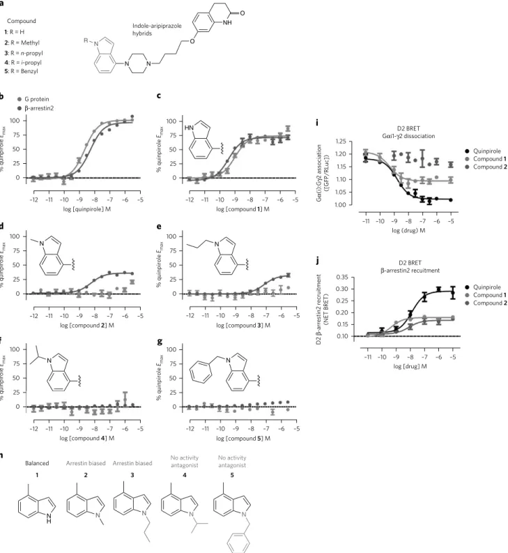
Indole-aripiprazole hybrid D2R SFSR
Next, we evaluated the structure–functional selectivity relationships (SFSR) of indole N-substitutions (for example, methyl, n-propyl,
i-propyl, benzyl) to 1 (Fig. 2a) intended to disrupt interactions with TM5. These substitutions introduce steric repulsion between the ligand and TM5, and are expected to eliminate the S1935.42– ligand hydrogen bond. To assess ligand bias at G protein versus B-arrestin recruitment pathways, ligands were tested by measur-ing Gi/o-mediated cAMP inhibition and B-arrestin2 recruitment assays40 conducted in parallel. D2R expression was similar in both D2 assay platforms (Supplementary Table 1). D2R-mediated cAMP inhibition, but not D2 B-arrestin2 recruitment, was depen-dent on pertussis-toxin-sensitive Gi/o proteins (Supplementary Fig. 2a). Previously, we confirmed that compound 1 is a D2R partial agonist (~75% of quinpirole; Fig. 2b,c) in both Gi/o signaling
EL2
5-HT2B
LSD 4-indole piperazine
1AR
V IV
III
Aripiprazole
Indole-aripiprazole hybird
Compound 1
V
III VII
EL2
V
III
VII I184EL2
S1935.42
S1945.43
S1975.46
D1143.32 IV
III V
EL2
EL2
VII
F193EL2
S2035.42
S2045.43
S2075.46
D1133.32
S2225.43
A2255.46
compound 1
D2
L209EL2
D1355.46
F201EL2
S2115.42
S2125.43
S2155.46
D1213.32
Epinephrine 2AR
N N O
Cl Cl NH
O
N N O
NH O HN
a b c
e d
Figure 1 | Structure-inspired design of indole-aripiprazole hybrid ligands. D2 ligand design based on comparison of three aminergic crystal structures.
N N
O
NH O
N R
HN
N N
N N
N
H N N N N
Compound
c
i
j e
b
d
f g
h a
100 G protein
-arrestin2
75
50
25
% quinpirole
Emax
% quinpirole
Emax 0
100
75
50
25
0
–12 –11 –10 –9 –8
log [compound 2] M log [compound 3] M
–7 –6 –5
Indole-aripiprazole hybrids 1: R = H
2: R = Methyl 3: R = n-propyl 4: R = i-propyl 5: R = Benzyl
100
75
50
25
% quinpirole
Emax
0
100
75
50
25
% quinpirole
Emax
0
1 2 3 4 5
Balanced Arrestin biased Arrestin biased No activityantagonist No activityantagonist
% quinpirole
Emax 100
75
50
25
0
–12 –11 –10 –9 –8 log [quinpirole] M
–7 –6 –5 –12 –11 –10 –9 –8
log [compound 1] M –7 –6 –5
–12 –11 –10 –9 –8 log [compound 4] M
–7 –6 –5
–12 –11 –10 –9 –8 –7 –6 –5
% quinpirole
Emax 100
75
50
25
0
log [compound 5] M –12 –11 –10 –9 –8 –7 –6 –5
Compound 1 Compound 2 Quinpirole
Compound 1 Compound 2 Quinpirole 1.25
G
(i):G
2 association
([GFP/RLuc])
D2 BRET Gi1-2 dissociation
1.20
1.15
1.10
1.05
1.00
–11 –10 –9 –8 –7 –6
log (drug) M
D2 BRET
-arrestin2 recuitment –5
0.35
D2
-arrestin2 recruitment
(NET BRET)
0.30
0.25
0.20
0.15
0.10
–11 –10 –9 –8 –7 –6 log [drug] M
–5
Figure 2 | Indole-aripiprazole hybrid D2R SFSR. Structure–functional selectivity relationships (SFSRs) of indole N1-substituted analogs of
indole-aripiprazole hybrids, which lead to either D2 arrestin-bias or antagonism depending on the substitution. (a) Chemical structures of N1-substituted indole-aripiprazole hybrids. (b–g) Profiling of indole-aripiprazole hybrids measuring D2 G protein activity (GAi/o-mediated cAMP inhibition; red) and
and B-arrestin2 recruitment activity, whereas 1 shows weak prefer-ence for arrestin recruitment over Gi/o signaling (bias factor = 2.5) relative to quinpirole (Fig. 2c).
As predicted, N-alkyl or aryl substitution completely abolished G protein-mediated signaling relative to quinpirole and compound
1 (Fig. 2d–g). However, the N-methyl (2) and N-n-propyl (3) sub-stitution retained arrestin-recruitment efficacy, thus exhibiting arrestin bias relative to quinpirole (Fig. 2d,e). Interestingly, the N-isopropyl (4) and N-benzyl (5) substitutions showed no activ-ity in both assays (Fig. 2f,g), but still retained appreciable affinity for D2R (77 and 22 nM, respectively), as measured by radioligand binding (Supplementary Table 2). In fact, both compounds 4 and
5 are potent and competitive antagonists of quinpirole-stimulated D2R cAMP inhibition (compound 4 KB = 11.3 nM; compound 5KB = 8.1 nM; Supplementary Fig. 2b,c). The added bulk by N-isopropyl or N-benzyl likely avoids hydrogen bonding with TM5 and EL2 engagement, pushing on TM5 and preventing activation, potentially explaining its antagonist activity. In short, a clear D2R SFSR for the indole-aripiprazole hybrids emerged demonstrating either arrestin preference or antagonism, dependent on the indole N-substitution (Fig. 2h).
In addition, because the interpretation of ligand bias can be skewed by system-dependent factors (for example, receptor reserve, cellular background, or assay platforms), we subjected compound 2 to an orthologous assay of D2R G protein activity measuring GAi1-G2 dissociation by bioluminescent resonance energy transfer (BRET). In this assay, compound 2 showed no agonist activity, whereas compound 1 was a partial agonist with respect to quinpirole (Fig. 2i), recapitulating our findings obtained from measuring Gi/o-dependent cAMP inhibition activ-ity. Further confirmation of arrestin bias, employing an ortholo-gous platform for arrestin recruitment using BRET, revealed 2 to be a potent agonist for arrestin recruitment (EC50 = 17 nM; Emax 33% of quinpirole Emax response; Fig. 2j). Although no G protein-mediated agonism could be detected by any method, and therefore no bias factor could be formally calculated, we further tested com-pound 2 as an antagonist of quinpirole-stimulated Gi/o-mediated cAMP inhibition (KB = 3.6 nM; Supplementary Fig. 2d) to dem-onstrate that 2 indeed acts as a competitive antagonist. Finally, in light of recent findings that the kinetic context can influence bias interpretations35,41, we also profiled the kinetics of signaling of 2, which revealed no Gi/o-mediated cAMP inhibition for up to 90 min (Supplementary Fig. 2e) and robust arrestin recruitment peaking between 15–60 min (Supplementary Fig. 2f). In sum-mary, compound 2 was extensively profiled and was confirmed as an arrestin-biased D2 partial agonist.
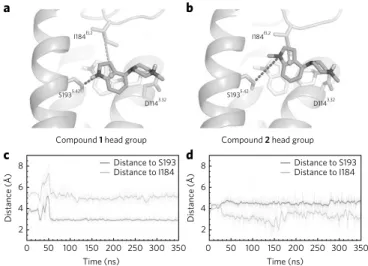
Simulations predict EL2 engagement for arrestin bias
To identify binding pocket residues that lead certain compounds to favor B-arrestin recruitment over G protein signaling, we studied compounds 1 and 2 by molecular dynamics (MD) simulations. The N-methylated compound 2 is incapable of forming a hydrogen bond with S1935.42. Like compound 1, compound 2 will likely position its indole-piperazine portion in the orthosteric site, with the proto-nated nitrogen of the piperazine ring forming a salt bridge with the conserved D1143.32 in TM3. Less clear, however, is how the effect of N-methylation translates to attenuation of G protein signaling with retention of B-arrestin recruitment. We therefore performed MD sim-ulations with the head groups of compounds 1 and 2, i.e., without the dihydroquinolin-2-one and alkyl linker (Supplementary Fig. 3 andSupplementary Table 3). Compounds 1 and 2 are identical aside from the head group moiety, and because of the uncertainty in the orienta-tion of their flexible tail regions, we chose to use the head groups to investigate potential structural features that lead to biased signaling.
Simulations of both head groups were initiated from the same posi-tion in D2R, which was chosen based on the posiposi-tion of 4-(piperazin-1
-yl)-1H-indole (equivalent to the head group of compound 1) in the thermostabilized turkey B1AR crystal structure (3ZPQ). These initial poses incorporated an ionic interaction between the cationic ammonium of the ligand and D1143.32. The head group of 1 retained a stable hydrogen bond with S1935.42 throughout each simulation (Fig. 3a), in agreement with the docked pose of the full-length mol-ecule. The N-methyl indole moiety of 2, on the other hand, moved away from TM5 toward the extracellular surface of the D2 orthosteric site, where it associated closely with I184EL2 (Fig. 3b). These results —which were consistent across several sets of simulations (Fig. 3c,d
and Supplementary Fig. 4)—indicate that the two head groups, which differ by only a single methyl group, prefer substantially dif-ferent positions in the orthosteric site. Compound 1’s head group prefers interaction with TM5 S1935.42, whereas compound 2’s head group prefers interaction with EL2 I184. This pose difference sug-gests that EL2 interaction may be associated with arrestin-biased sig-naling and TM5 interaction with balanced sigsig-naling.
TM5 and EL2 mutants confirm arrestin-biased binding pose
To investigate changes in bias based on ligand contacts with key TM5 and EL2 residues, we tested 2 at the S1935.42 and I184EL2 mutants (Fig. 4a) and quantified Gi/o-mediated cAMP inhibition and B-arrestin2 recruitment. The design of these mutants reflects our previous observations that ligand engagement with S5.42 is required for activation of G protein signaling at B2AR, and the conserved hydrophobic EL2 residue corresponding to I184EL2 spe-cifically dampens LSD’s B-arrestin recruitment at the 5-HT2B and 5-HT2A receptors35.As previously mentioned, the S193A5.42 mutation resulted in a loss of affinity and potency of 1, confirming our prediction that
I184EL2 I184EL2
S1935.42
Compound 1 head group
8
a b
c d
6
4
Distance (Å)
2
8
6
4
Distance (Å)
2
0 50 100 150
Time (ns) Time (ns)
200 250 300 350 0 50 100 150 200 250 300 350 Distance to S193
Distance to I184
Compound 2 head group
D1143.32
S1935.42
D1143.32
Distance to S193 Distance to I184
Figure 3 | D2R MD simulations predict EL2 engagement for arrestin bias.
the indole N–H forms a hydrogen bond with S1935.42, as found in the B1AR crystal structure. Furthermore, we tested the affinity of
2 at TM5 mutants, and observed no substantial affinity changes relative to wild-type D2R for any of the TM5 serine mutations (Supplementary Fig. 5a). By contrast, the G protein-mediated sig-naling of 2 (Fig. 4b) was selectively recovered by the TM5 S193A5.42 mutation, resulting in balanced signaling between G protein and B-arrestin2 activity (Fig. 4c) with respect to quinpirole. We rea-soned that the D2R S193A5.42 mutant creates a hydrophobic space for the N-methyl group of 2 to fit, allowing it to recapitulate the hydrogen bond between compound 1 and S1935.42 at the wild-type D2R, leading to G protein signaling. Docking of 2 to the D2R S193A5.42 model showed that the steric clash between compound 2 and S1935.42 in wild-type D2R is abolished at the D2 S193A5.42 mutant (Supplementary Fig. 5b). In fact, MD simulations of the head group of 2 further support this hypothesis, as at wild-type D2R the head group of 2 moves away from TM5 and interacts with I184EL2. In con-trast, the head group of 2 at the S193A5.42 mutant engages TM5 in a pose within the binding pocket that is almost identical to the com-pound 1 head group in wild-type D2R (Fig. 4d).
Next, we tested compound 2’s arrestin recruitment at the EL2 I184AEL2 mutation and found that arrestin recruitment by 2 was completely abolished in this mutant (Fig. 4e), confirming that EL2 is essential for compound 2’s B-arrestin recruitment. In fact, I184AEL2 resulted in no measureable activity of 2 in either G protein signaling or arrestin recruitment activity (Fig. 4e). By contrast, B-arrestin-recruitment efficacy for the balanced agonists 1 and quinpirole was spared at I184AEL2 (Supplementary Fig. 5c). In addition, 2’s affinity at the I184A mutant was spared (Supplementary Fig. 5d), demon-strating antagonist activity at the D2 I184AEL2 mutant (Fig. 4f and Supplementary Fig. 5e). To confirm that mutations of EL2 may be directly related to 2’s ligand-binding kinetics, we measured a 2.2-fold and 8.7-2.2-fold increase in the on- and off-rate of 2, respectively, at the I184AEL2 mutant compared to wild-type D2R (Supplementary Table 4 and Supplementary Fig. 5f), which is consistent with EL2
mutations decreasing LSD’s residence time at 5-HT2B and 5-HT2A receptors35. Furthermore, MD simulations confirm that compound 2‘s head group is unstable at I184AEL2 D2R and samples many ori-entations within the binding pocket (Fig. 4g and Supplementary Fig. 6a,b), which may partially explain the increased off-rate of 2 at the I184AEL2 mutant. Overall, our mutagenesis and computational studies confirm that I184EL2 and S1935.42 are critical contacts for compound 2’s bias profile.
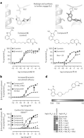
Rational design of arrestin-biased compounds
Based on the signaling profiles of compounds 1 and 2 at the D2R I184EL2A mutant, and MD observations that B-arrestin-biased com-pound 2 preferentially interacts with EL2 over TM5, we designed compounds 6 and 7 to test whether additional EL2 engagement would lead to superior arrestin recruitment efficacy. Compound 7
is an analog of 2 containing a 2-methyl substitution to the indole ring, which would be expected to engage I184EL2 in a hydrophobic contact (Fig. 5a). Compound 6 is the 2-methyl analog of 1, and was proposed as a control compound that has similar properties to 7 but is predicted to form a hydrogen bond with S1935.42 and demonstrate a balanced signaling profile relative to quinpirole. Both 6 and 7
were synthesized and tested at the D2R for bias (Fig. 5a). Consistent with our prediction, 6 displayed no preference for arrestin recruit-ment over G protein activation, again demonstrating that predicted engagement with S1935.42 invariably leads to activation of G protein signaling. Unsurprisingly, simulations indicate that the head groups of 1 and 6 remain closer to TM5 than do those of 2 and 7, because of the presence of a hydrogen bond between S1935.42 and the indole N–H of 1 and 6 (Supplementary Fig. 7).
Compound 7, on the other hand, shows a preference for arrestin recruitment with a calculated bias factor of 20 relative to quinpirole (Fig. 5a), demonstrating much increased arrestin recruitment efficacy (Fig. 5b; Emax = 88% of quinpirole) relative to 2. Although compound 7 still showed G protein-mediated signaling, its G protein activity was much weaker in terms of potency compared
D2 Arrestin-biased compound 2
EL2
100 G protein
-arrestin2 -arrestin2
G protein
-arrestin2
-arrestin2 antagonism
G protein D2 WT
D2 WT
Compound 2 at WT
Compound 1 at WT
Compound 2 at S193A
Compound 2 at I184A 75
50 25
–12 0
100 75 50 25 0
100
-Arr
estin r
ecruitment
(% quinpir
ole-stimulated)
75 50 25 0
100 75 50 25 0
–11 –10 –8 –7 –6 –5
log (compound 2) M log (compound 2) M
–9 –12 –11 –10–9–8 –7 –6 –5
log (compound 2) M –12 –11 –10–9 –7 –6 –5
log (compound 2 + quinpirole) M –12 –11 –10–9–8 –7 –6 –5
% quinpir
ole
Emax
% quinpir
ole
Emax
% quinpir
ole
Emax
III
WT
S1935.42
A184EL2 I184EL2
S1935.42
S193A5.42 I184AEL2
I184AEL2
S1945.43
S1975.46
D1143.32
S/A1935.42 S193A V
I184EL2
–8
a b
d
c e
g
f
Figure 4 | D2 TM5 and EL2 mutants confirm arrestin-bias binding pose. (a) The pose resulting from MD simulation of the head group of the
arrestin-biased N-methyl indole-aripiprazole hybrid (2) places the N-methyl indole moiety in contact with I184 on EL2, having moved away from S193 on TM5. (b) N-Methyl indole-aripiprazole hybrid 2 only shows arrestin recruitment activity in wild-type (WT) D2. Data represent mean and s.e.m. performed in triplicate (Gi/o GloSensor; red; n = 3 independent replicates) and B-arrestin2 recruitment (Tango; blue, n = 3 independent replicates; EC50 = 3.7 nM; Emax = 36%). (c) S193A5.42 transforms arrestin bias of 2 into balanced signaling with respect to quinpirole. Data represent GA
i/o-mediated cAMP inhibition (Gi/o GloSensor; red; n = 3 independent replicates; EC50 = 2.5 nM; Emax = 67%) and B-arrestin2 recruitment (Tango; blue, n = 3 independent replicates; EC50 = 2.6 nM; Emax = 69%). (d) Representative pose of compound 2 head group from simulation at WT and S193A D2R constructs and of compound 1 head group from WT D2R simulation. At S193A, 2 moves to a pose almost identical to that of 1 at WT D2. (e) Mutation of EL2 I184 (I184A) completely abolishes arrestin recruitment for arrestin-biased ligand 2 (Tango; n = 5 independent replicates). (f) I184A mutation transforms 2 into a D2R B-arrestin2 recruitment antagonist as measured in Tango (n = 2 independent replicates, in triplicate), as seen by comparing WT D2 (black, IC50 = 6.3 nM) to EL2 I184A (green, IC50 = 13 nM). (g) Compound 2 head group is unstable throughout simulation at I184A D2R, sampling many orientations within the
to its B-arrestin recruitment activity. To explain the recovery in G protein signaling by 7, simulations with the head groups of compounds 2 and 7 were performed. Although arrestin-biased 2
moves the furthest away from TM5, the additional 2-methyl on 7
hinders this movement and instead shifts the indole ring toward TM5 (Supplementary Fig. 7), enough to engage TM5 and activate G protein signaling to a degree. Despite this, both 7 and 2 moved closer to I184EL2 relative to 1 and 6, potentially explaining their arrestin preference.
To provide evidence for this differential EL2 engagement by 7, newly synthesized ligands were tested at the I184AEL2 mutant. As for compound 2, the I184AEL2 mutation almost completely abol-ishes the arrestin recruitment activity for compound 7 (Fig. 5c) and increases 7’s on- and off-rate by a factor of 6.7 and 6.2-fold, respec-tively (Supplementary Table 4), indicating that I184 is a key inter-action for 7’s enhanced B-arrestin recruitment efficacy. Although the arrestin recruitment of quinpirole and compound 1 are spared by the I184AEL2 mutation, compound 6 showed a partial, but not complete, loss of arrestin recruitment efficacy, indicating that the 2-methyl substitution is sensitive to EL2 mutation, but may retain other ligand–receptor interactions elsewhere in the binding pocket that lead to arrestin recruitment. To provide support for this notion, we tested the previously discovered B-arrestin-biased ligands UNC 9994 and UNC 9975 (ref. 8) and measured no change in arrestin recruitment efficacy at the I184AEL2 mutation (Supplementary Fig. 8), indicating that B-arrestin bias may arise from other ligand–receptor interactions distinct from EL2. In summary, a route to attaining B-arrestin-biased compounds by modification of the head group of aripiprazole-type ligands has emerged: remov-ing interactions with TM5 while enhancremov-ing interactions with EL2 can improve B-arrestin recruitment efficacy to drive arrestin-biased signaling, an SFSR succinctly summarized in a heat map of relative log(T/KA) activities (Fig. 5d and Supplementary Table 5).
Prediction and confirmation of polypharmacologic arrestin
bias
Although aminergic GPCRs bind distinct classes of endogenous ligands (e.g., catecholamines, tryptamines, and histamines), the orthosteric site encompassing TM5 and EL2 residues is relatively well conserved (Supplementary Fig. 1a). We examined whether ligand bias resulting from a lack of interaction with TM5 residues and the retention of hydrophobic engagement with EL2 is conserved for other aminergic GPCRs. Piperazine-containing ligands, such as aripiprazole, have promiscuous activity at aminergic GPCRs and possess substantial affinity at D3, D4, 5-HT, and A- and B-adrenergic receptors29. We hypothesized that the piperazine-containing ligand 2 will bind to the orthosteric site in a similar way for those receptors and will demonstrate arrestin bias at receptors with residues similar to those of D2R at EL2.52 (located 2 residues away from conserved disulfide cysteineEL2.50 that are branched aliphatic; for example, leu-cine and isoleuleu-cine) and TM5 5.42 (polar residues that have hydro-gen bond potential; for example, serine and threonine).
We examined arrestin bias at receptors where 2 has substantial affinity (D3R, D4R, 5-HT7R, 5-HT1AR, 5-HT2Rs, B2AR and B1AR; Supplementary Table 6). The closely related D3R and D4R contain serines at positions 5.42, 5.43, and 5.46 and a branched aliphatic EL2 residue (isoleucine in D3R, leucine in D4R; Fig. 6a,b). Confirming our predictions, 2 demonstrated arrestin bias at D3R (Fig. 6a) and D4R (Fig. 6b) compared to quinpirole, with minimal detected G protein activity below 1 MM. Importantly, the unsubstituted com-pound 1 demonstrated no preference for either G protein or arrestin recruitment at D3R and D4R (Fig. 6a,b). 5-HT7R also has an isoleu-cine present in EL2 and a serine at TM5 5.42; therefore, we expected to observe arrestin bias by 2. Consistent with our prediction, 2 dem-onstrated full agonist arrestin recruitment activity at 5-HT7R rela-tive to 5-HT, but surprisingly exhibited 5-HT7R-GAs inverse agonist
activity (Fig. 6c). Similarly, at D3 and D4, 1 showed 5-HT7R agonist activity in both G protein and arrestin recruitment. In addition, we also tested 2 at 5-HT1AR, which also contains an isoleucine at
log(/KA) log(/KA)
Redesign and synthesis to further engage EL2 a
b
c
d 100 G protein
Bias = 20
–12 –11 –10 –9 –8 –7 –5
log (compound 6) M
log (drug) M
log (drug) M
Quinpirole Quinpirole
7.00
Gi Arr
estin
7.50 9.50
1 (R1 = H)
2 (R1 = Me)
3 (R1 = n-pr)
5 (R1 = Benz)
6 (R1 = Me, R2 = H)
7 (R1 = Me, R2 = Me)
4 (R1 = i-pr)
Compound 7 Compound 6 Compound 2 Compound 1 Compound 7 (N-Me, 2-Me) Compound 2 (N-Me)
Increased -arrestin
recruitment efficacy
Confirm EL2 interaction
I184AEL2
log (compound 7) M
–6
–12 –11 –10 –9 –8 –7 –6 –5
–12 –11 –10 –9 –8 –7 –6 –5
–12 –11 –10 –9 –8 –7 –6 –5
-arrestin2 % quinpir ole Emax -Arr estin2 r ecruitment % quinpir ole Emax 75 50 25 0 100 % quinpir ole Emax 75 50 25 0 100 75 50 25 0 -Arr estin2 r ecruitment (% quinpir ole Emax ) 100 75 50 25 0 EL2 Compound 6 (control) Compound 7 EL2 N H O O N N HN
CH3 CH
3 H3C
N H O O N N N N N O NH O N R1 R2 G protein -arrestin2
Figure 5 | MD-assisted rational design of arrestin-biased compounds.
(a) Mutagenesis data indicating that 2 requires I184EL2 for B-arrestin recruitment and MD findings that 2 preferentially interacts with I184EL2 led to the design of 2-methyl indole derivative 7 to further engage EL2 and enhance B-arrestin recruitment. Compound 6 is the unsubstituted control compound, which can still form a hydrogen bond with S1935.42, and shows balanced D2 signaling with respect to quinpirole (bias factor = 1.3; Gi/o EC50 = 0.49 nM, Emax = 86%; B-arrestin2 EC50 = 0.62 nM, Emax = 78%), but compound 7 shows arrestin bias with respect to quinpirole (bias factor = 20) in comparison to GAi/o-mediated cAMP inhibition (GloSensor; red; n = 3 independent replicates; EC50 = 23 nM; Emax = 60%) to B-arrestin2 recruitment (Tango; blue; n = 3 independent replicates; EC50 = 2.9 nM; Emax = 78%). (b) 2-Methyl substitution (7, purple; Emax = 78%) shows higher D2 B-arrestin2 recruitment efficacy compared to compound 2 (blue; Emax = 36%) with respect to quinpirole, as measured by Tango. Data were normalized to percent quinpirole Emax and represent n = 3 independent replicates. (c) Compound 7 interaction with EL2 was confirmed, with the I184AEL2 mutation selectively abolishing B-arrestin2 recruitment (Tango) for biased ligands 2 and 7, but not for balanced 1 (red) and quinpirole (black). Compound 6 (orange) shows decreased arrestin recruitment by the I184AEL2 mutation, but not complete loss of activity (B-arrestin2 EC50 = 2.7 nM, Emax = 40%). Data were normalized to quinpirole and represent n = 3 independent replicates. (d) Structure–function selectivity relationships for the indole-aripiprazole hybrid series as outlined using a heat map comparing log log(T/KA) activities measuring G protein and
EL2.52 and Ser at 5.42. Compound 2 also showed arrestin bias at 5-HT1AR with a calculated bias factor of 60 with respect to 5-HT, which exhibits Gi/o preference (Supplementary Fig. 9a). Finally, we tested 2 at the 5-HT2 receptors, which all contain a Gly at 5.42; 2 showed no Gq-mediated agonist activity at any of these receptors (Supplementary Fig. 9b–d). However, only at 5-HT2B, which con-tains a Leu at EL2.52, does 2 show weak arrestin recruitment (~25% of 5-HT), indicative of weak arrestin bias relative to that of 5-HT (Supplementary Fig. 9d).
As previously mentioned, the B2AR binding pocket also contains TM5 serines at positions 5.42, 5.43 and 5.46, but con-tains Phe at the EL2.52 residue position (Fig. 6d). Compound
2 demonstrated GAs inverse agonist activity, similar to 5-HT7R, but showed no B-arrestin recruitment at B2AR, consistent with our prediction that smaller aliphatic residues are required for 2
arrestin recruitment efficacy. Although compound 1 showed Gs partial agonism at B2AR, it also showed no arrestin recruitment, comparable to 2 (Fig. 6d). A similar profile for 2 was also found at B1AR, which also contains a Phe at EL2.52 and a Ser at 5.42 (Supplementary Fig. 9e). To test the hypothesis that smaller aliphatic residues present at EL2.52 may be required for com-pound 2’s arrestin recruitment efficacy, we attempted to rescue compound 2’s arrestin recruitment by mutating B2AR F193EL2.52 to either alanine, leucine or isoleucine. Although compound 2
showed no recovered arrestin recruitment activity at any of the B2AR EL2 mutants (Supplementary Fig. 10a), the B2AR EL2 mutation substantially reduced arrestin recruitment for the full reference agonist isoproterenol (Supplementary Fig. 10b), supporting the notion that EL2 plays a prominent role for arrestin recruitment.
N
N O N
H O N
R1 R2
Preclude TM5 engagement EL2.52 TM5 5.42/5.43/5.46
EL2.52 TM5 5.42/5.43/5.46 EL2.52 TM5 5.42/5.43/5.46
EL2.52 TM5 5.42/5.43
D3
compound 2 compound D3 1
D4 compound 1
D4 compound 2
5-HT7 compound 1
5-HT7 compound 2
Gi/o-compound 2 Gi/o-compound 1
100
–12 –11 –10 –9 –8 –7 –6 –5
log (compound 2) M
log (compound 2) M log (compound 2) M
EL2
TM5
log (compound 1) M log (compound 1) M log (compound 1) M
log (compound 1) M log (compound 1) M
Arrestin-bias design template
EL2
Promote EL2 interaction
Ser5.42
Ser5.43
Ser5.46 TM5
EL2.52
R1 = Me
R2 = Me –12 –11 –10 –9 –8 –7 –6 –5 –4
–12 –11 –10 –9 –8 –7 –6 –5 –4 –4
–12 –11 –10 –9 –8 –7 –6 –5 –4
–12 –11 –10 –9 –8 –7 –6 –5 –4
–12 –11 –10 –9 –8 –7 –6 –5 –4 –12 –11 –10 –9 –8 –7 –6 –5 –4 –12 –11 –10 –9 –8 –7 –6 –5 –4 75
50
25
% quinpir
ole r
esponse
% quinpir
ole r
esponse
% quinpir
ole r
esponse
% isopr
oter
enol r
esponse
% isopr
oter
enol r
esponse
% quinpir
ole r
esponse
% 5-HT r
esponse
% 5-HT r
esponse
–25
100
75
50
25
–25
100 75 50
25
–25 0
100
75
50
25
–25 0
0
100
75
50
25
–25 0
0
100
75 50 25
–25 0
100
75 50 25
–25 0 100 75 50
25
–25 0
-Arr-compound 2
Gi/o-compound 2
-Arr-compound 2 Gi/o-compound -Arr-compound 11 Gs-compound -Arr-compound 22 Gs-compound -Arr-compound 1 1
2 compound 1
2 compound 2
Gs-compound 2
-Arr-compound 2
-Arr-compound 1
Gs-compound 1
-Arr-compound 1
a c
b
e
d
100
75
50
25
–25 0
Figure 6 | Prediction and confirmation of polypharmacological arrestin bias. (a–d) Alignments of D2 TM5 and EL2 residues predict that 2 shows arrestin
This result confirms our hypothesis that specific interactions by
2 with smaller aliphatic residues present at EL2, even at other dis-tinct aminergic receptors, can predict arrestin bias. Here, we show that a template can be used to guide biased ligand design at many aminergic receptors, where promoting engagement with aliphatic residues in EL2 and precluding TM5 interaction can induce an arrestin-biased polypharmacological profile (Fig. 6e).
DISCUSSION
Here we illustrate how to design biased ligands by a combined computational, structural, biochemical and molecular dynamics approach. Importantly, our results identify EL2 as a critical con-served region of the receptor that can be targeted to enhance arres-tin bias. We anticipate that this combined strategy will encourage the adoption of MD into SBDD projects.
Our results for the D2 I184AEL2 mutation complement our recent finding that EL2 is important for arrestin bias and slow binding kinetics35. EL2 appears to play an important role in distinguish-ing between B2AR active and inactive states, whereby the activated state of B2AR involves F193EL2.52 and TM7 Y3087.35coming together to form a lid over the ligand21. Here we provide evidence that EL2 of B2AR is also key for arrestin recruitment (Supplementary Fig. 10) and that further study of B2AR arrestin recruitment as it relates to ligand kinetics is warranted. Apart from aminergic GPCRs, mea-surements in structural changes in EL2 of rhodopsin reveal that this region is important for the retinal isomerization42, as mutations of the rod rhodopsin receptor Ile189EL2.52 to proline, which is found at EL2.52 at green cone opsin receptors, directly increased decay rates of the meta II intermediate state of the receptor43. Taken together, these data show that EL2 is an important motif that can ‘lock’ the ligand into the binding site, leading to increased ligand residence times. This increased residence time apparently promotes arrestin recruitment, and this can be exploited for biased drug design.
Structure-inspired drug design supported the hypothesis that orthosteric site TM5 residues are engaged not only in ligand rec-ognition but also in G protein signaling and, further, that these interactions can be exploited to modulate biased signaling. Ligand contacts with residues in TM5 have been regarded as a ‘trigger’ that stabilizes a conformation with a cytoplasmic inward movement of TM5 (ref. 44), which in turn moves intracellular loop 2 and TM6 regions that are involved in G protein activation20,45. Evidence for the involvement of D2R TM5 serines in ligand bias is scant, except for a study suggesting that Ser5.43 may be involved in ligand- dependent arachidonic acid release34. Although we cannot rule out alternative downstream effects stemming from targeting EL2 and avoiding TM5 interaction (for example, arachidonic acid release, pERK1/2), this study is the first to design ligands predicted to avoid TM5-dependent G protein activity entirely.
Importantly, our design strategy yielded a ligand with bias at multiple related GPCRs. Given that the most clinically effective medications for schizophrenia and depression have a complex polypharmacological profile46 targeting multiple aminergic GPCRs47 (i.e., ‘magic shotguns’), it is now possible to design promiscuous drugs that manifest arrestin bias at multiple GPCRs by targeting conserved interactions within the orthosteric site. We thus pro-vide a useful template for the rational design of polypharmacologi-cal drugs incorporating ligand bias (i.e., ‘biased magic shotguns’), and successful design will depend on generating optimal predicted ligand contacts with EL2. One caveat, though, is that this particu-lar strategy may be applicable only to aminergic GPCRs. MOR, for example, was not proposed to trigger G protein signaling through motion of TM5, and thus is not expected to benefit from this SBDD algorithm48. Conceivably, our template for biased ligand design could also be used to design G protein-biased ligands using the reverse approach (i.e., retain TM5 and exclude EL2 engagement); such compounds would represent extremely desirable tools to
dissect the contributions of G protein- versus B-arrestin-dependent signaling at various aminergic GPCRs to uncover favorable thera-peutic and side effect profiles.
The wave of GPCR structures has generated excitement largely because they promise to accelerate the discovery of new and improved drugs49. With knowledge of how ligands can be designed to activate specific signaling pathways, it is apparently possible to leverage GPCR structures to create biased drugs.
Received 20 January 2017; accepted 20 October 2017; published online 11 December 2017
METHODS
Methods, including statements of data availability and any associ-ated accession codes and references, are available in the online ver-sion of the paper.
References
1. Overington, J.P., Al-Lazikani, B. & Hopkins, A.L. How many drug targets are there? Nat. Rev. Drug Discov.5, 993–996 (2006).
2. Rask-Andersen, M., Almén, M.S. & Schiöth, H.B. Trends in the exploitation of novel drug targets. Nat. Rev. Drug Discov.10, 579–590
(2011).
3. Wacker, D., Stevens, R.C. & Roth, B.L. How ligands illuminate GPCR molecular pharmacology. Cell170, 414–427 (2017).
4. Urban, J.D. et al. Functional selectivity and classical concepts of quantitative pharmacology. J. Pharmacol. Exp. Ther.320, 1–13 (2007).
5. DeWire, S.M., Ahn, S., Lefkowitz, R.J. & Shenoy, S.K. B-arrestins and cell signaling. Annu. Rev. Physiol.69, 483–510 (2007).
6. Shukla, A.K., Xiao, K. & Lefkowitz, R.J. Emerging paradigms of B -arrestin-dependent seven transmembrane receptor signaling. Trends Biochem. Sci.36, 457–469 (2011).
7. Violin, J.D., Crombie, A.L., Soergel, D.G. & Lark, M.W. Biased ligands at G-protein-coupled receptors: promise and progress. Trends Pharmacol. Sci. 35, 308–316 (2014).
8. Allen, J.A. et al. Discovery of B-arrestin-biased dopamine D2 ligands for probing signal transduction pathways essential for antipsychotic efficacy. Proc.
Natl. Acad. Sci. USA108, 18488–18493 (2011).
9. Soergel, D.G. et al. Biased agonism of the M-opioid receptor by TRV130 increases analgesia and reduces on-target adverse effects versus morphine: A randomized, double-blind, placebo-controlled, crossover study in healthy volunteers. Pain155, 1829–1835 (2014).
10. Violin, J.D. et al. Selectively engaging B-arrestins at the angiotensin II type 1 receptor reduces blood pressure and increases cardiac performance. J. Pharmacol. Exp. Ther.335, 572–579 (2010).
11. Urs, N.M. et al. Targeting B-arrestin2 in the treatment of L-DOPA-induced dyskinesia in Parkinson’s disease. Proc. Natl. Acad. Sci. USA112, E2517–E2526 (2015).
12. Charfi, I., Audet, N., Bagheri Tudashki, H. & Pineyro, G. Identifying ligand-specific signalling within biased responses: focus on D opioid receptor ligands. Br. J. Pharmacol.172, 435–448 (2015).
13. Manglik, A. et al. Structure-based discovery of opioid analgesics with reduced side effects. Nature537, 185–190 (2016).
14. DeWire, S.M. et al. A G protein-biased ligand at the M-opioid receptor is potently analgesic with reduced gastrointestinal and respiratory dysfunction compared with morphine. J. Pharmacol. Exp. Ther.344, 708–717 (2013). 15. Gesty-Palmer, D. et al.B-arrestin-selective G protein-coupled receptor
agonists engender unique biological efficacy in vivo. Mol. Endocrinol.27, 296–314 (2013).
16. Masri, B. et al. Antagonism of dopamine D2 receptor/B-arrestin 2 interaction is a common property of clinically effective antipsychotics. Proc. Natl. Acad.
Sci. USA105, 13656–13661 (2008).
17. Tchernychev, B. et al. Discovery of a CXCR4 agonist pepducin that mobilizes bone marrow hematopoietic cells. Proc. Natl. Acad. Sci. USA107,
22255–22259 (2010).
18. Cherezov, V. et al. High-resolution crystal structure of an engineered human
B2-adrenergic G protein-coupled receptor. Science318, 1258–1265 (2007).
19. Chien, E.Y. et al. Structure of the human dopamine D3 receptor in complex with a D2/D3 selective antagonist. Science330, 1091–1095 (2010). 20. Rasmussen, S.G. et al. Crystal structure of the B2 adrenergic receptor-Gs
protein complex. Nature477, 549–555 (2011).
21. Ring, A.M. et al. Adrenaline-activated structure of B2-adrenoceptor stabilized by an engineered nanobody. Nature502, 575–579 (2013).
23. Wang, C. et al. Structural basis for molecular recognition at serotonin receptors. Science340, 610–614 (2013).
24. Kapur, S. & Remington, G. Atypical antipsychotics: new directions and new challenges in the treatment of schizophrenia. Annu. Rev. Med.52, 503–517 (2001).
25. Wadenberg, M.L., Soliman, A., VanderSpek, S.C. & Kapur, S. Dopamine D(2) receptor occupancy is a common mechanism underlying animal models of antipsychotics and their clinical effects. Neuropsychopharmacology25, 633–641 (2001).
26. Shapiro, D.A. et al. Aripiprazole, a novel atypical antipsychotic drug with a unique and robust pharmacology. Neuropsychopharmacology28, 1400–1411 (2003).
27. Chen, X. et al. Discovery of G protein-biased D2 dopamine receptor partial agonists. J. Med. Chem.59, 10601–10618 (2016).
28. Park, S.M. et al. Effects of B-arrestin-biased dopamine D2 receptor ligands on schizophrenia-like behavior in hypoglutamatergic mice
.Neuropsychopharmacology41, 704–715 (2016).
29. Ambrosio, C., Molinari, P., Cotecchia, S. & Costa, T. Catechol-binding serines of B2-adrenergic receptors control the equilibrium between active and inactive receptor states. Mol. Pharmacol.57, 198–210 (2000).
30. Isogai, S. et al. Backbone NMR reveals allosteric signal transduction networks in the B1-adrenergic receptor. Nature530, 237–241 (2016).
31. Warne, T. et al. The structural basis for agonist and partial agonist action on a B1-adrenergic receptor. Nature469, 241–244 (2011).
32. Neve, K.A. & Wiens, B.L. Four ways of being an agonist: multiple sequence determinants of efficacy at D2 dopamine receptors. Biochem. Soc. Trans.23, 112–116 (1995).
33. Wiens, B.L., Nelson, C.S. & Neve, K.A. Contribution of serine residues to constitutive and agonist-induced signaling via the D2S dopamine receptor: evidence for multiple, agonist-specific active conformations. Mol. Pharmacol. 54, 435–444 (1998).
34. Fowler, J.C., Bhattacharya, S., Urban, J.D., Vaidehi, N. & Mailman, R.B. Receptor conformations involved in dopamine D(2L) receptor functional selectivity induced by selected transmembrane-5 serine mutations. Mol.
Pharmacol.81, 820–831 (2012).
35. Wacker, D. et al. Crystal structure of an LSD-bound human serotonin receptor. Cell168, 377–389.e12 (2017).
36. Chen, X. et al. Structure-functional selectivity relationship studies of
B-arrestin-biased dopamine D2 receptor agonists. J. Med. Chem.55,
7141–7153 (2012).
37. Christopher, J.A. et al. Biophysical fragment screening of the B 1-adrenergic receptor: identification of high affinity arylpiperazine leads using structure-based drug design. J. Med. Chem.56, 3446–3455 (2013).
38. Kling, R.C., Tschammer, N., Lanig, H., Clark, T. & Gmeiner, P. Active-state model of a dopamine D2 receptor-GAi complex stabilized by aripiprazole-type partial agonists. PLoS One9, e100069 (2014).
39. Luedtke, R.R. et al. Comparison of the binding and functional properties of two structurally different D2 dopamine receptor subtype selective compounds. ACS Chem. Neurosci.3, 1050–1062 (2012).
40. Kroeze, W.K. et al. PRESTO-Tango as an open-source resource for interrogation of the druggable human GPCRome. Nat. Struct. Mol. Biol.22, 362–369 (2015).
41. Klein Herenbrink, C. et al. The role of kinetic context in apparent biased agonism at GPCRs. Nat. Commun.7, 10842 (2016).
42. Ahuja, S. et al. Helix movement is coupled to displacement of the second extracellular loop in rhodopsin activation. Nat. Struct. Mol. Biol.16, 168–175 (2009).
43. Kuwayama, S., Imai, H., Hirano, T., Terakita, A. & Shichida, Y. Conserved proline residue at position 189 in cone visual pigments as a determinant of molecular properties different from rhodopsins. Biochemistry41, 15245–15252 (2002).
44. Warne, T. & Tate, C.G. The importance of interactions with helix 5 in determining the efficacy of B-adrenoceptor ligands. Biochem. Soc. Trans.41, 159–165 (2013).
45. Deupi, X. & Standfuss, J. Structural insights into agonist-induced activation of G-protein-coupled receptors. Curr. Opin. Struct. Biol.21, 541–551 (2011). 46. Roth, B.L., Sheffler, D.J. & Kroeze, W.K. Magic shotguns versus magic
bullets: selectively non-selective drugs for mood disorders and schizophrenia. Nat. Rev. Drug Discov.3, 353–359 (2004).
47. Besnard, J. et al. Automated design of ligands to polypharmacological profiles.
Nature492, 215–220 (2012).
48. Huang, W. et al. Structural insights into M-opioid receptor activation. Nature 524, 315–321 (2015).
49. Kobilka, B. & Schertler, G.F. New G-protein-coupled receptor crystal structures: insights and limitations. Trends Pharmacol. Sci.29, 79–83 (2008).
Acknowledgments
We thank S. Hollingsworth for assistance with simulation analysis and A.J. Venkatakrishnan for assistance with simulation setup. We thank R. Axel at Columbia University for the HTLA cells and M. Bouvier at Université de Montréal for BRET constructs. This work was supported by the National Institutes of Health (NIH) grant U19MH082441 (to B.L.R. and J.J.), R01MH112205 (to B.L.R.), R01NS100930 (to J.J.), the National Institute of Mental Health Psychoactive Drug Screening Program (NIMH PDSP; to B.L.R.), the Michael Hooker Chair for Protein Therapeutics and Translational Proteomics (to B.L.R.), the American Cancer Society postdoctoral fellowship PF-14-021-01-CDD (to K.V.B.), by NIH grant GM59957 (to B.K.S.), by Pfizer, Inc. (R.O.D.), by a Terman Faculty Fellowship (to R.O.D.), and by a National Science Foundation Graduate Research Fellowship (to R.M.B.).
Author contributions
J.D.M. designed experiments, performed mutagenesis, ligand-binding and signaling studies, analyzed the data, and wrote the manuscript. K.V.B. designed and synthesized all ligands, performed analytical chemical analysis and wrote the manuscript. B.K. performed and analyzed MD simulations, used the results to design ligands, and wrote the manuscript. K.R. assisted with mutagenesis and signaling studies. B.K., J.K., and B.L.K. built the D2 homology model. J.K. performed the docking experiments and edited the manuscript. R.M.B. determined ligand parameters and performed preliminary MD simulations. B.K.S. supervised the docking experiments and edited the manuscript. R.O.D. supervised the MD simulation studies and helped prepare the manuscript. J.J. supervised ligand synthesis, designed experiments and edited the manuscript. B.L.R. designed the experiments, was responsible for the overall project strategy and manage-ment and prepared the manuscript.
Competing financial interests
The authors declare no competing financial interests.Additional information
doi:10.1038/nchembio.2527
ONLINE METHODS
General chemistry procedures. All reagents were purchased from Sigma-Aldrich or Fisher Scientific. Anhydrous solvents were used unless otherwise noted. Analytical HPLC method A: equipment: Agilent 6110 series with UV detection at 254 nm; Column: Agilent Eclipse Plus 4.6 mm × 50 mm, 1.8 um C18 column. HPLC solvents: A: 0.1% acetic acid in water; B: 0.1% acetic acid in methanol, with gradient: 10% to 100% B over 5.0 min, followed by 100% B for 2 min, at 1.0 mL/min. Method B: equipment: Agilent Zorbax 300SC-C18 (5 Mm) column with UV detection at 254 nm on an Agilent 1200 Series LC–MSD TOF machine. HPLC solvents: A: 0.1% acetic acid in water; B: 0.1% acetic acid in methanol, with gradient: 1% B for one minute, 1 to 100% B over 3.0 min, followed by 100% B for 4 min, at 1.0 mL/min. LRMS (low resolution mass spectrometry) data were acquired in positive ion mode on an Agilent 6110 single quadrupole mass spectrometer with electrospray ionization (ESI). Nuclear magnetic resonance (NMR) spectra were recorded on either a Varian Mercury spectrometer at 400 MHz for proton (1H NMR) and 100 MHz for carbon (13C NMR), or a Bruker DRX spectrometer at 600 MHz for proton (1H NMR) and 150 MHz for carbon (13C NMR). Preparative HPLC (high pres-sure liquid chromatography) was performed on an Agilent Prep 1200 series with UV detector set to 254 nm, along with a Phenomenex Luna 75 mm × 30 mm, 5 um C18 column with a flow rate of 30 mL/min. High resolution mass spectrometry (HRMS) data was acquired with an Agilent 1200 Series LC–MSD TOF. Medium pressure liquid chromatography (MPLC) was performed on a CombiFlash Isco machine. Final compounds had >95% purity as judged by analytical HPLC. Indole synthesis schemes and compound purification details can be found in Supplementary Note 1.
Drugs and reagents. All compounds and aripiprazole were synthesized as described under ‘General chemistry procedures’. Dopamine hydrochloride, (−)-quinpirole, (+)-butaclamol hydrochloride, 5-hydroxytryptamine creatine sulfate, (−)-isoproterenol bitartrate, and HEPES sodium salt were purchased from Sigma-Aldrich (St. Louis, MO). HBSS (10×) was purchased by Invitrogen, and fatty-acid free BSA was purchased from Akron Biotech.
Cloning and mutagenesis. Mutagenesis was performed according to QuikChange II XL Site-Directed Mutagenesis Kit protocol. Briefly, PCR reac-tions incorporated wild-type D2 long dopamine receptor (pcDNA3.1, cDNA. org) or D2 long-V2-tTA (pcDNA3.1) and primers containing the mutation of interest. Parental wild-type DNA was digested with DpnI (New England BioLabs). PCR products were transformed into supercompetent GC-10 cells, and positive clones were selected by ampicillin resistance. Isolated colonies on the plates were picked, cultured and prepped using QIAprep Spin miniprep and Origene maxiprep kits. DNA was then sequenced (Eton Bioscience) using forward (T7) and reverse (BGHreverse and TEV-REV) sequence primers to verify mutant DNA sequence.
Cell culture. HEK 293T cells (ATCC CRL-11268; 59587035; mycoplasma free) were cultured in Dulbecco’s Modified Eagle Medium (DMEM) containing 10% FBS (Invitrogen) and 0.5% penicillin–streptomycin. HTLA cells express-ing B-arrestin-TEV protease and tTA-driven luciferase (provided by R. Axel at Columbia University) were cultured similarly to HEK293T cells except that media contained selection antibiotics (100 Mg/mL hygromycin B and 5 Mg/mL puromycin). Cells were maintained at 37 °C and 5% CO2.
Radioligand binding assays. D2R radioligand binding assays used [3 H]N-me-thyl Spiperone (NMSP; Perkin Elmer, specific activity = 64.1 Ci/mmol). For competitive binding experiments, assays used [3H]NMSP concentrations rang-ing from 0.7–1.3 nM, unlabeled ligand competitor at concentrations rangrang-ing from 100 MM to 1 pM, and membranes resuspended in binding buffer (50 mM Tris, 10 mM MgCl2, 0.1 mM EDTA, 0.1% BSA, 0.01% ascorbic acid, pH 7.4). Binding assays were incubated at 25 °C for 2 h, and assays were terminated by vacuum filtration using a 96-well Filtermate harvester (Perkin Elmer) onto 0.3% polyethyleneimine presoaked 96-well filter mats A (Perkin Elmer). Filters were washed three times using cold wash buffer (50 mM Tris, pH 7.4), and scintil-lation cocktail (Meltilex) was melted onto dried filters. Radioactivity displace-ment was measured using a Wallac Trilux Microbeta counter (Perkin Elmer).
Counts per minute (c.p.m.) were plotted as a function of unlabeled ligand con-centration and the Ki was calculated using the One-site-Fit Ki using 5.0. Data were normalized to the top (100%, no competitor) and bottom (0%, nonspe-cific binding defined as 5 MM (+)-butaclamol) to represent percent displace-ment. For radioligand binding assays at all other receptors, procedures were similar to those described above, except the radioligand used and membrane sources. For a list of these binding assays, refer to procedures at https://pdspdb. unc.edu/pdspWeb/ for the National Institute of Mental Health Psychoactive Drug Screening Program (NIMH PDSP).
For the determination of kon and koff for unlabeled compounds 2 and 7 membranes of D2 wild-type and I184AEL2 were incubated with at least two concentrations of [3H]NMSP (range 0.08-0.35 nM) and several concentra-tions of 2 or 7 (range 1 MM to 320 pM). On- and off-rates of [3H]NMSP at D2 wild-type and I184AEL2 were previously determined and used to estimate the kon and koff rates of 2 and 7 using “Kinetics of competitive binding” equation in Graphpad Prism 5.0 by Motulsky and Mahan (1984)50.
Gi/o-mediated cAMP inhibition assay. To measure Gi/o-mediated cAMP inhibition, HEK293T cells were co-transfected in a 1:1 ratio with receptor and a split-luciferase-based cAMP biosensor (GloSensor; Promega). After at least 24 h, transfected cells were plated in poly-lysine coated 384-well white clear-bottom cell culture plates with DMEM containing 1% dialyzed FBS at a density of 15,000 cells per 40 ML per well and incubated overnight. On the day of assay, drug dilutions were prepared in filtered fresh assay buffer (20 mM HEPES, 1× HBSS, 0.1% BSA, 0.01% ascorbic acid, pH 7.4) at 3× and 10 ML per well was added to cells containing 20 ML/well of assay buffer. Drug solutions used for G protein-mediated cAMP assays were exactly the same as those used for Tango assays to allow relative within-experiment bias comparisons. After plates were allowed to incubate with drug for 15 min, 10 ML per well of 1 MM (final concentration) forskolin and GloSensor sub-strate was added. Luminescence counts per second (LCPS) were quantified after 15 min using a TriLux microbeta (Perkin Elmer) luminescence counter. LCPS were plotted as a function of drug concentration and normalized to percent quinpirole with 100% as the quinpirole cAMP inhibition Emax and 0% as the forskolin-stimulate cAMP baseline. Data were analyzed using log (agonist) vs. response in GraphPad Prism 5.0 (Graphpad Software Inc., San Diego, CA).
Tango B-arrestin recruitment assays. The human D2Long Tango construct was designed, and assays were performed as previously described8,40. HTLA cells expressing TEV fused B-arrestin2 were transfected with D2 Tango con-struct. For D3 and D4 Tango constructs, GRK2 was co-transfected in a 1:10 ratio of GRK2:receptor. After at least 24 h, cells were plated in DMEM sup-plemented with 1% dialyzed FBS (dFBS) in poly-L-lysine-coated 384-well white clear-bottom cell culture plates at a density of 15,000 cells/well in total of 40 ML. After at least 6 h, media was decanted, and cells were supplemented with 40 ML of 1% dFBS DMEM, and drug solutions (3×) prepared in drug buffer (1× HBSS, 20 mM HEPES, 0.1% BSA, 0.01% ascorbic acid, pH 7.4) were added (20 ML per well) for overnight incubation. Drug solutions used for Tango assay were exactly the same as those used for G protein-mediated cAMP assays to allow relative within experiment bias comparisons. The next day, media and drug solutions were decanted and 20 ML per well of BrightGlo reagents (Promega, 1:20 dilution in drug buffer) was added. The plate was incubated for 20 min at room temperature in the dark before being counted using Wallac TriLux microbeta (Perkin Elmer). LCPS were plot-ted as a function of drug concentration, normalized to percent quinpirole with 100% as the quinpirole Emax and 0% as the baseline, and analyzed using log (agonist) vs. response in GraphPad Prism 5.0 (Graphpad Software Inc., San Diego, CA).