A STOCHASTIC BIOMECHANICAL MODEL FOR RISK AND RISK FOCTORS OF SUSTAINING NON-CONTACT ACL INJURIES
Cheng-Feng Lin, MS
A dissertation submitted to the faculty of the University of North Carolina at Chapel Hill in partial fulfillment of the requirements for the degree of Doctor of Philosophy in the Program of Human Movement Science.
Chapel Hill 2006
Approved by
Chair: Bing Yu
Member: William Garrett
Member: Michael Gross
Member: Chuanshu Ji
Member: Darin Padua
ABSTRACT
Cheng-Feng Lin: A Stochastic Biomechanical Model for Risk and Risk Factors of Sustaining Non-contact ACL Injuries
(Under the direction of Dr. Bing Yu)
Non-contact anterior cruciate ligament injury is one of the most commonly seen injuries in sports with an estimated total cost for treatment over $2 billion per year. To prevent non-contact ACL injuries, several possible risk factors have been proposed. No studies to date, however, have shown conclusive evidence that any of the proposed risk factors are indeed risk factors for sustaining non-contact ACL injuries.
In this study, a sagittal plane knee model was developed to enhance the understanding of the effects of sagittal plane biomechanics on the ACL loading. Knee geometry data were obtained from x-ray films of 10 male and 10 female collegiate aged recreational athletes. The patellar tendon-tibial shaft angle, patella-patellar tendon angle, quadriceps tendon-patella angle, and hamstring tendon-tibial shaft angle were expressed as functions of knee flexion angle, and incorporated into the sagittal plane knee model to estimate the ACL loading. In vivo
lower extremity kinematic and kinetic data of a stop-jump task collected from 80 college aged recreational athletes were used for computer simulation with the sagittal plane knee model. A stochastic biomechanical model was developed and used to determine the risk and risk factors of sustaining non-contact ACL injuries during landing of the stop-jump task.
risk of non-contact ACL injuries than males did. The gender difference in the risk of non-contact ACL injuries was affected by the differences in the lower extremity motion patterns, ACL ultimate load, and knee anatomy between genders. Biomechanical
characteristics during the stop-jump task were significantly different between injured and uninjured jumps or between females and males. During the stop-jump task, the isolated sagittal plane loading alone can injure the ACL while the isolated non-sagittal plane loading alone may not. Knee flexion angle and peak posterior ground reaction force are two important risk factors for the gender difference in the risk of non-contact ACL injuries during the
DEDICATION
ACKNOWLEDGEMENTS
I would like to thank my committee for their critical and constructive suggestions in the conduct and completion of this dissertation. Special thanks to Dr. Bing Yu for his tremendous support and guidance throughout my doctoral study. Thanks for being my academic advisor and chairing my dissertation committee. I also want to express my special thanks to Dr. William Garrett and Dr. Michael Gross for being positive role models as good combination of clinician and researcher. Thanks to Dr. Paul Weinhold for delivery of clear concepts in biomechanics during my doctoral study. Special thanks also to Dr. Chuanshu Ji for ensuring my probability concepts and to Dr. Darin Padua for critical suggestions and careful review of my dissertation.
I also would like to thank Prof. Fong-Chin Su, my academic and thesis advisor while I was in the master program of Biomedical Engineering at the National Cheng Kung University in Taiwan, for opening the door to biomechanics for me and setting the basis for my studies at the doctoral level.
Also, I would like to extend my appreciation to my family and my fiancé Jen-Chieh for their caring and unlimited support and love. Finally, I would like to thank my friends in Taiwan and United States for their support in my life.
Cheng-Feng Connie Lin Chapel Hill, NC.
TABLE OF CONTENTS
Page
ABSTRACT ……….……… ii
DEDICATION………... iv
ACKNOWLEDGEMENTS ………. v
TABLE OF CONTENTS …………..……….……. vi
LIST OF FIGURES ……….……… xi
LIST OF TABLES ……….………... xvii
LIST OF ABBREVIATIONS AND SYMBOLS ………. xix
CHAPTER I. INTRODUCTION ……….………... 1
Statement of Purposes ……….…..……….. 5
II. REVIEW OF LITERATURE ……..….……… 6
ACL Injuries ………...……… 6
ACL Injury Mechanism ………….……… 8
ACL Loading Mechanism …………..……….……… 8
Effects of the Quadriceps Muscle Force on the ACL loading …….….……… 11
Effects of the Knee Flexion Angle on the ACL Loading………. .……… 13
Effects of the Hamstring Co-contraction on the ACL Loading………... 17
Effects of the Gastrocnemius Muscle Force on the ACL loading ………. 21
Effects of the Knee Valgus-varus and Internal-external Rotation Moment on the ACL Loading ……….... 22
Potential Risk Factors …………..……… 27
Non-modifiable Intrinsic Risk Factors ……….. ……….…… 28
Motor Control Related Biomechanical Risk Factors ……..………….……… 28
Effect of the Training Programs on the Prevention of Non-contact ACL Injuries ... 33
Research Methods to Determine Risk Factors for Sustaining Non-contact ACL Injuries ...… 38
Traditional Epidemiological Research Methods ……….………….. 38
Stochastic Biomechanical Modeling ……….………..………. 40
Summary………... 42
III. METHODS ……….……… 44
The Sagittal Plane Knee Model ………..………. 44
Determination of the ACL Loading ………..…….……….………. 45
Determination of the Quadriceps Muscle Force ... 51
Determination of the Patella tendon-tibia shaft angle, Patella-patella tendon angle, Quadriceps tendon-patella angle, and Hamstring tendon-tibial shaft angle ……… 53
Determination of the Moment Arms ………. 56
Determination of the ACL Elevation Angle ……… 57
Collection and Reduction of Lower Extremity Kinetics, Kinematics, and EMG in a Stop-Jump Task ……….………..……… 59
Data Reduction ………. 62 Computer Simulation of the Effects of Sagittal Biomechanics on the ACL
Loading ……….……… 63
The Stochastic Biomechanical Model of the Probability of Sustaining
Non-contact ACL Injuries ……….. 66
Determination of Density Distributions of the Primary Independent
Variables …….……….……….. 66
Determination of Cumulative Distribution Functions of the Primary
Independent Variables ………...……….. 69 Determination of the Secondary Independent Variables……… 71 Monte Carlo Simulation for Estimate of the Probability of Sustaining
Non-contact ACL Injuries ………. 72
Normalization of Moment Arms of Muscles ………. 73 Determination of Mean and Standard Deviation of Muscle Forces…... 73 Determination of the ACL Loading……… 74 Sensitivity Analysis of the ACL Ultimate Load ……… 76 Monte Carlo Simulation for Determining Risk Factors of Sustaining
Non-Contact ACL Injuries ………... 81
Assumptions of Models ……… 81
Assumptions for the Sagittal Plane Inverse Dynamic Knee Model …………. 82 Assumptions for the Stochastic Biomechanical Model ………... 83
IV. RESULTS ………..……… 84
Computer Simulation of Effects of Sagittal Plane Lower Extremity Biomechanics
on Peak ACL Loading during the landing of the Stop-jump Task……… 84 Monte Carlo Simulation of Simulated Non-contact ACL Injuries during the
Goodness-of-fit Test for Independent Variables ……….. 106
Sensitivity Analysis of ACL Ultimate Load during the Stop-jump Task……... 115
Probability and Characteristics of Simulated Injured and Uninjured Jumps … 117 Risk Factors of Simulated Non-contact ACL Injuries ……….. 123
V. DISCUSSION ………..……… 126
Effects of Assumptions on the Validity of the Sagittal Plane Inverse Dynamic Model of the Knee ……….... 126
Effects of Assumptions on the Validity of the Stochastic Biomechanical Model of the Probability of Sustaining Non-contact ACL injuries………... 130
Effects of Sagittal Plane Biomechanics on the Peak ACL loading………... 131
Effect of the Knee Flexion Angle on the ACL Loading……… 131
Effect of the Peak Posterior Ground Reaction Force on the ACL Loading ……….……... 136
Effect of Landing Styles on the ACL Loading………... 140
Effect of Hamstrings Force on the ACL Loading………... 145
Effect of Gastrocnemius Force on the ACL Loading……… 148
Monte Carlo Simulation of the Probability of Sustaining Non-contact ACL Injuries during the Stop-jump Task………... 150
Sensitivity of Simulated Relative Risk of Non-contact ACL injury …………. 150
Biomechanical Characteristics of Simulated Non-contact ACL Injuries……... 153
Comparison of Injured and Uninjured Jumps ………... 153
Comparison of Injured Jumps between Genders ………... 159
Comparison of Uninjured Jumps between Genders ……….... 159
Risk Factors of Simulated Non-contact ACL Injuries………... 161
Significance and Recommended Future Studies….………... 166
Conclusion ………... 172
APPENDICES
Appendix A: Effect of Sagittal Plane Biomechanics on the Peak ACL
Loading ………. 174
Appendix B: Density Distribution of Independent Variables of Stochastic
Biomechanical Models ……… 182
Appendix C: Probability Distribution of Independent Variables of Stochastic
Biomechanical Models ………. 188
LIST OF FIGURES
Figure Page
3.1 The horizontal (FGRF,x) and vertical (FGRF,z) ground reaction forces in the
laboratory reference frame and representation of the tibial tilting angle (σ) … 46 3.2 The knee geometry to show the definition of knee flexion angle (θ), patellar
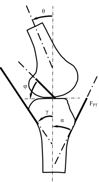
tendon-tibial shaft angle (α), hamstring tendon-tibial shaft angle (γ), and ACL
elevation angle (φ) ……….………... 50
3.3 Definition of the quadriceps tendon-patella angle (φ) and patella-patellar
tendon angle (β) ……… 52
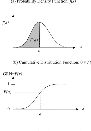
3.4 Relationship between probability density function and cumulative
distribution function. (a) Probability Density Function: f(x). (b) Cumulative
Distribution Function: 0 ≤ F(x) ≤ 1. The integral of f(x) with respect to a (the
gray area under curve) was the cumulative distribution function F(a) (0 ≤ F(a)
≤ 1), which was a randomly generated number. GRN: generated random
number………... 70
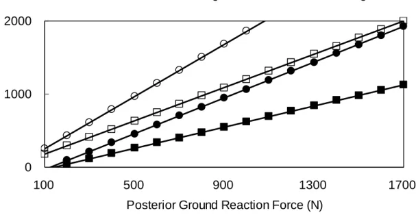
3.5 A flow chart of one iteration of the Monte Carlo Simulation……… 80 4.1 Effect of the knee flexion angle at peak posterior ground reaction force on the
simulated ACL loading for males at different posterior ground reaction forces during the landing of the stop-jump task. The inputs of other independent variables during computer simulation were fixed with the corresponding
values at peak posterior ground reaction force. ……… 90 4.2 Effect of the knee flexion angle at peak posterior ground reaction force on the
simulated ACL loading for females at different posterior ground reaction forces during the landing of the stop-jump task. The inputs of other independent variables during computer simulation were fixed with the corresponding values at peak posterior ground reaction force. The inputs of other independent variables during computer simulation were fixed with the
corresponding values at peak posterior ground reaction force ………. 91 4.3 Effect of the posterior ground reaction force on the simulated ACL loading
for males at different knee flexion angles during the landing of the stop-jump task. The inputs of other independent variables during computer simulation were fixed with the corresponding values at peak posterior ground reaction
for females at different knee flexion angles during the landing of the
stop-jump task. The inputs of other independent variables during computer simulation were fixed with the corresponding values at peak posterior ground
reaction force……… 93
4.5 Effect of landing-on-heels and landing-on-toes on the simulated ACL loading for males during the landing of the stop-jump task. The inputs of other independent variables during computer simulation were fixed with the
corresponding values at peak posterior ground reaction force ………. 94 4.6 Effect of landing-on-heels and landing-on-toes on the simulated ACL loading
for females during the landing of the stop-jump task. The inputs of other independent variables during computer simulation were fixed with the
corresponding values at peak posterior ground reaction force ………. 95 4.7 Effect of the anterior(+)-posterior(-) tibial tilting angle on the simulated ACL
loading for males at different posterior ground reaction forces during the landing of the stop-jump task. The inputs of other independent variables during computer simulation were fixed with the corresponding values at peak
posterior ground reaction force ……… 96 4.8 Effect of the anterior(+)-posterior(-) tibial tilting angle on the simulated ACL
loading for females at different posterior ground reaction forces during the landing of the stop-jump task. The inputs of other independent variables during computer simulation were fixed with the corresponding values at peak
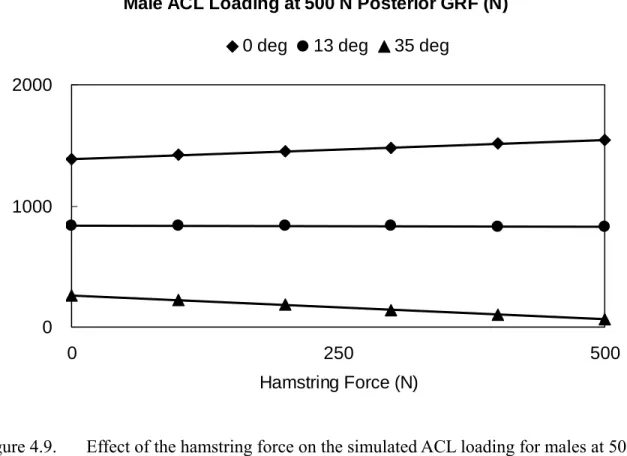
posterior ground reaction force ……… 97 4.9 Effect of the hamstring force on the simulated ACL loading for males at 500
N of posterior ground reaction force and at different knee flexion angles during landing of the stop-jump task. The inputs of other independent variables during computer simulation were fixed with the corresponding
values at peak posterior ground reaction force ………. 98 4.10 Effect of the hamstring force on the simulated ACL loading for males at 1000
N posterior ground reaction force and at different knee flexion angles during the landing of the stop-jump task. The inputs of other independent variables during computer simulation were fixed with the corresponding values at peak
posterior ground reaction force ..………... 99 4.11 Effect of the hamstring force on the simulated ACL loading for females at 500
N of posterior ground reaction force and at different knee flexion angles during the landing of the stop-jump task. The inputs of other independent variables during computer simulation were fixed with the corresponding
1000 N of posterior ground reaction force and at different knee flexion angles during the landing of the stop-jump task. The inputs of other independent variables during computer simulation were fixed with the corresponding
values at peak posterior ground reaction force ………. 101 4.13 Effect of the gastrocnemius force on the simulated ACL loading for males
500 N of posterior ground reaction force and at different knee flexion angles during the landing of the stop-jump task. The inputs of other independent variables during computer simulation were fixed with the corresponding
values at peak posterior ground reaction force……….. 102 4.14 Effect of the gastrocnemius force on the simulated ACL loading for males at
1000 N of posterior ground reaction force and at different knee flexion angles during the landing of the stop-jump task. The inputs of other independent variables during computer simulation were fixed with the corresponding
values at peak posterior ground reaction force ………. 103 4.15 Effect of the gastrocnemius force on the simulated ACL loading for females
at 500 N of posterior ground reaction force and at different knee flexion angles during the landing of the stop-jump task. The inputs of other independent variables during computer simulation were fixed with the
corresponding values at peak posterior ground reaction force………... 104 4.16 Effect of the gastrocnemius force on the simulated ACL loading for females
at 1000 N of posterior ground reaction force and at different knee flexion angles during the landing of the stop-jump task. The inputs of other independent variables during computer simulation were fixed with the
corresponding values at peak posterior ground reaction force……….. 105 4.17 Q-Q plot of independent variables against the test of normal distribution for
males. The abscissa represents the observed value while the ordinate represents the expected normal value. (a) knee varus-valgus moment (BH.BW); (b) knee int.-ext. rotation moment (BH.BW); (c) COP to ankle
distance (m); (d) tibial tilting angle (deg); (e) knee flexion angle (deg)…... 109 4.18 Q-Q plot of independent variables against the test of gamma distribution for
males. The abscissa represents the observed value while the ordinate represents the expected gamma value. (a) posterior GRF (N); (b) standing height (m); (c) normalized linear envelope hamstring EMG ; (d) normalized
linear envelope gastrocnemius EMG ……….... 110 4.19 Q-Q plot of independent variables against the test of normal distribution for
females. The abscissa represents the observed value while the ordinate represents the expected normal value. (a) knee varus-valgus moment (BH.BW); (b) knee int.-ext. rotation moment (BH.BW); (c) COP to ankle
4.20 Q-Q plot of independent variables against the test of gamma distribution for females. The abscissa represents the observed value while the ordinate represents the expected gamma value. (a) posterior GRF (N); (b) knee flexion angle (deg); (c) standing height (m) (d) normalized linear envelope
hamstring EMG ; (e) normalized linear envelope gastrocnemius EMG……… 112 5.1 A flow chart of how the knee flexion angle affects the ACL Loading……….. 133 5.2 A flow chart of how the posterior ground reaction force affects the ACL
Loading ... 138 5.3 A flow chart of how the landing style affects the ACL Loading………... 142 5.4 A flow chart of how the tibial tilting angle affects the ACL Loading………... 144 5.5 A flow chart of how the hamstring force affects the ACL Loading…………... 146 5.6 A flow chart of how the gastrocnemius force affects the ACL Loading ……... 149 A.1 Effect of the knee flexion angle at peak posterior ground reaction force on the
simulated ACL loading for males at different posterior ground reaction forces
during the landing of the stop-jump task………... 174 A.2 Effect of the knee flexion angle at peak posterior ground reaction force on the
simulated ACL loading for females at different posterior ground reaction
forces during the landing of the stop-jump task……… 175 A.3 Effect of the posterior ground reaction force on the simulated ACL
loading for males at different knee flexion angles during the landing of the
stop-jump task ………... 176
A.4 Effect of the posterior ground reaction force on the simulated ACL loading for females at different knee flexion angles during the landing of
the stop-jump task ………. 177 A.5 Effect of the hamstring force on the simulated ACL loading for males at 500
N of posterior ground reaction force and at different knee flexion angles
during the landing of the stop-jump task………... 178 A.6 Effect of the hamstring force on the simulated ACL loading for females at 500
N of posterior ground reaction force and at different knee flexion angles
during the landing of the stop-jump task………... 179 A.7 Effect of the gastrocnemius force on the simulated ACL loading for males at
during the landing of the stop-jump task………... 180 A.8 Effect of the gastrocnemius force on the simulated ACL loading for females
at 500 N of posterior ground reaction force and at different knee flexion
angles during the landing of the stop-jump task ………... 181 B.1 Density distribution of knee flexion angle of males and females. Empirical
indicates the distribution based on the experimental data. ………... 182 B.2 Density distribution of normalized peak posterior ground reaction force of
males and females. Empirical indicates the distribution based on the
experimental data. ………... 183 B.3 Density distribution of normalized internal varus(+)-valgus(-) moment of
males and females. Empirical indicates the distribution based on the
experimental data. ………... 184 B.4 Density distribution of normalized internal internal(+)-external(-) rotation
moment of males and females. Empirical indicates the distribution based on
the experimental data. ………... 185 B.5 Density distribution of anterior(+)-posterior(-) tibial tilting angle of males
and females. Empirical indicates the distribution based on the experimental
data. ………... 186
B.6 Density distribution of COP to ankle joint distance of males and females. (+): prone to land on heel; (-): prone to land on toe. Empirical indicates the
distribution based on the experimental data ………... 187 C.1 Probability distribution of knee flexion angle of males and females.
Empirical indicates the distribution based on the experimental data ………… 188 C.2 Probability distribution of normalized posterior ground reaction force of
males and females. Empirical indicates the distribution based on the
experimental data. ………. 189 C.3 Probability distribution of normalized internal varus(+)-valgus(-) moment of
males and females. Empirical indicates the distribution based on the
experimental data. ………. 190 C.4 Probability distribution of normalized internal internal(+)-external(-) rotation
moment of males and females. Empirical indicates the distribution based on
the experimental data. ………..………... 191 C.5 Probability distribution of anterior(+)-posterior(-) tibial tilting angle of males
data ……… 192 C.6 Probability distribution of COP to ankle joint distance of males and females
(+): prone to land on heel; (-): prone to land on toe. Empirical indicates the
LIST OF TABLES
Table Page
3.1 Pearson correlation coefficients (p-values) of peak posterior ground reaction force with peak vertical ground reaction force and peak knee joint resultants
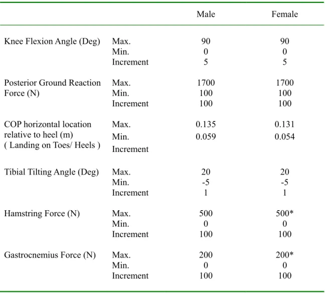
during the landing of the stop-jump task (Yu et al., 2006b) ... 65 3.2 ACL ultimate load (N) in pairs for sensitivity analysis ………... 77 4.1 Anthropometric data of subjects ……….. 85 4.2 Simulated range and increment of each independent variable of the sagittal
plane inverse dynamic knee model……… 86 4.3 Skewness and kurtosis of each independent variable for Monte Carlo
simulation……….. 107 4.4 Types of distribution and p-value of each independent variable for males and
females ……… 108 4.5 Mean and standard deviation (SD) of variables simulated as normal
distribution in the stochastic biomechanical knee model ………. 113 4.6 Shape and scale parameters and mean and standard deviation (SD) of
independent variables with gamma distribution for males ……….. 114 4.7 Shape and scale parameters and mean and standard deviation (SD) of
independent variables as gamma distribution for females ……… 114 4.8 Average (SD) of number of simulated ACL injuries per 100,000 jumps and
female to male non-contact ACL injury rate ratio with different ACL injury
loadings ………... 116
4.9 Mean (SD) of biomechanical characteristics of simulated injured and
uninjured jumps with 2250 N as ACL ultimate load for males ………... 119 4.10 Mean (SD) of biomechanical characteristics of simulated injured and
uninjured jumps with 1800 N as ACL ultimate load for females ……… 120 4.11 Mean (SD) of injured biomechanical characteristics of simulated jumps with
with 2250 N for males and 1800 N for females as ACL ultimate load ……... 122 4.13 Mean of contribution from non-sagittal loadings to total ACL loading in
injured jumps subjected to the change of varus-valgus moment from literature in female stochastic biomechanical model and 1800 N as ACL injury
criterion ………. 123
4.14 Mean of numbers of non-contact ACL injury per 100,000 jumps and female to male non-contact ACL injury rate ratio subjected to the change of each independent variable in female Monte Carlo simulations and 1800 N as ACL
LIST OF ABBREVIATIONS AND SYMBOLS
ACL = Anterior cruciate ligament BW = Body weight
BH = Body height (standing height) COP = Center of pressure
EMG = Electromyography
GRF = Ground reaction force
Int. = Internal
Ext. = External
MCL = Medial collateral ligament
N = Newton
Nm = Newton-meter
PTTS = Patellar tendon-tibial shaft angle
Var = Varus
Val = Valgus
α (alpha) = Patellar tendon-tibial shaft angle β (beta) = Patella-patellar tendon angle
φ (phi) = Quadriceps tendon-patella angle
γ (gamma) = Hamstring tendon-tibial shaft angle θ (theta) = Knee flexion angle
CHAPTER I INTRODUCTION
Anterior cruciate ligament (ACL) injury is one of the most commonly seen injuries in sports. The incidence of ACL injuries is more than three per 1000 athletes over a single season (McCarroll et al., 1995). Arendt and Dick (1995) found that the average ACL injury rate per 1000 athlete-exposures was 0.31 and 0.29 in women soccer and basketball players,
respectively, from National Collegiate Athletic Association (NCAA) database. Gottlob et al. (1999) estimated that approximately 175,000 primary ACL reconstruction surgeries were performed annually in the United States with an estimated cost of over $2 billion.
ACL injuries have devastating influences on patients’ activity levels and quality of life. Complete ACL rupture could induce other knee pathological conditions including knee
instability, damage to menisci and chondral surface, and osteoarthritis. Studies have repeatedly shown that patients with complete ACL rupture had chronic knee instability and secondary damage to menisci and chondral surfaces (Finsterbush et al., 1990; Irvine and Glasgow, 1992; Kannus and Jarvinen, 1987). These debilitating consequences would influence their quality of life and severely impair patients’ functional activities (Smith et al., 1993).
sudden deceleration, landing, and pivoting maneuvers are repeatedly performed (Boden et al., 2000). Female athletes had higher incidence on the ACL injuries compared with their male counterparts (Arendt and Dick, 1995; Ferretti et al., 1992; Huston and Wojtys, 1996). Studies have shown that the incidence of female athletes is two to eight times higher than males in soccer, basketball, and volleyball(Arendt and Dick, 1995; Bjordal et al., 1997; Ferretti et al., 1992; Harmon and Ireland, 2000; Huston and Wojtys, 1996; Huston et al., 2000; Linderfeld et al., 1994).
Risk factors have to be identified to develop intervention strategies to reduce the risk of sustaining non-contact ACL injuries. A variety of risk factors of sustaining non-contact ACL injuries has been proposed in the literature. These proposed risk factors can be categorized into intrinsic and extrinsic risk factors. Intrinsic risk factors are those related to anatomic structure, physiological properties, and motor control related biomechanical factors such as Q-angle, the width of femoral condyle notch (Shambaugh et al., 1991), knee joint laxity (Wojtys et al., 1998), hormonal effects (Arendt et al, 2002), imbalanced lower extremity strength (Boden et al., 2000) and the altered lower extremity motion patterns (Boden et al., 2000; Decker et al., 2003; Malinzak et al., 2001). Extrinsic risk factors include shoe-surface interaction (Garrick and Requa, 1996) and playing surface (Powell and Schootman, 1992). Among these proposed risk factors, risk factors related to neuromuscular control and muscle strength are modifiable through intervention programs.
Extensive research efforts have been made in the last several years to identify
valgus-varus and internal-external rotation moment (Ford et al., 2003; Hewett et al., 2005; McLean et al., 2004), large ground reaction forces (DeVita and Kelly, 1992), and strong quadriceps and weak hamstring muscles activations tend to increase the load on the ACL (Arms et al., 1984; Fleming et al., 2001; Malinzak et al., 2001). Studies also proposed that excessive quadriceps activation along with a small knee flexion angle predisposed the ACL to a great strain which may injure the ACL (Arms et al., 1984; DeMorate et al., 2004; Li et al., 1999, 2004).
Although the studies mentioned previously provide some indirect evidence that the altered movement patterns during athletic task may be risk factors of sustaining non-contact ACL injuries, a recent extensive literature review failed to show conclusive evidence that any proposed risk factors related to neuromuscular control are indeed risk factors of sustaining non-contact ACL injuries. This lack of confirmation of neuromuscular control related risk factors of sustaining non-contact ACL injuries is mainly due to the difficulty in identifying neuromuscular control related risk factors. Longitudinal cohort design and a cross-sectional case-control design are two commonly used traditional epidemiological methods for
identifying risk factors of sustaining an injury or disease. These two traditional epidemiological methods, however, have significant limitations when used to identify
non-contact ACL injuries.
These studies demonstrated the feasibility of using the stochastic biomechanical modeling method to identify risk factors of sustaining non-contact ACL injuries.
Statement of Purposes
The purposes of this study were:
1. to develop a sagittal plane inverse dynamic knee model for studying the effects of lower extremity sagittal plane biomechanics on the ACL loading during landing of the stop-jump task;
2. to determine the effects of the knee flexion angle, posterior ground reaction force, hamstrings and gastrocnemius muscles contractions, tibial tilting angle, and location of center of pressure relative to the ankle joint center on the ACL loading during landing of the stop-jump task; and
CHAPTER II
REVIEW OF LITERATURE
The incidence of ACL injuries, ACL loading mechanism, possible risk factors for non-contact ACL injuries in athletes, training effects on the prevention of non-contact ACL injuries, and current research designs to identify risk factors of sustaining non-contact ACL injuries were reviewed as background information in this chapter to justify the significance of the current study. Studies using stochastic biomechanical modeling were also reviewed in this chapter to further justify the significance and establish the feasibility of the current study.
2.1. ACL Injuries
participated in sports in which they frequently performed pivoting maneuvers had the highest incidence of ACL injury (Griffin et al., 2000).
Female athletes have significantly greater incidence rate of ACL injuries than male athletes do. Henry and Kaeding (2001) reported that female athletes had 4 to 6 times incidence rate of contact ACL injuries when compared with their male counterparts. Huston et al. (2000) reviewed literature and reported that the incidence rate of ACL injuries of female athletes who participated in the sports in which landing and pivoting tasks were frequently performed was 2 to 8 times higher than that of the male counterparts.
The majority of ACL injuries occur with a non-contact mechanism in sports in which sudden deceleration, landing, and pivoting maneuvers are repeatedly performed (Boden et al., 2000; Feagin and Lambert, 1985; Ferretti et al, 1992; Griffin et al., 2000; McNair et al., 1990). ACL injuries that occur without physical contact between athletes at the time of injury are referred to as non-contact ACL injuries (Boden et al., 2000; Ferretti et al., 1992; McNair et al., 1990). McNair et al. (1990) and Boden et al., (2000) reported that 70% to 72% of ACL injuries occurred with non-contact mechanisms. Ferretti et al. (1992) showed that landing from a jump in the attack zone without direct contact was the most frequent injury mechanism in volleyball players.
2.2. ACL Injury Mechanisms
2.2.1. ACL Loading Mechanisms
An ACL injury occurs when an excessive tensile force is applied on the ACL. A non-contact ACL injury occurs when a person generates great forces or moments at the knee that applied excessive loading on the ACL. Research has demonstrated three ACL loading mechanisms: (1) knee anterior shear force loading, (2) knee varus-valgus loading, and (3) knee internal-external rotation loading. An understanding of these ACL loading mechanisms would set the basis for understanding non-contact ACL injury mechanisms and further research to identify the risk factors of sustaining non-contact ACL injuries.
Berns et al. (1992) studied the in vitro strain of the anteromedial bundle of the ACL on
13 cadaver knees subject to pure load of anterior-posterior force, varus-valgus moment, and internal-external rotation torque and combined loads. The pure load was applied when the knee flexion angle were fixed at 0°, 15°, and 30° knee flexion while the combined load were loaded in pairs at 0° and 30° knee flexion. The corresponding range of the pure
anterior-posterior force, varus-valgus moment, and internal-external rotation torque were 150-200 N, 25-30 Nm, and 6-10 Nm. They found that the anterior shear force on the proximal tibia was the primary determinant of the ACL strain. Neither pure axial torque nor pure varus-valgus moment was observed to strain the anteromedial bundle of the ACL at any of the knee flexion angles. The results of this study further showed the ACL strain subject to
combination of the anterior shear force and valgus torque was significantly larger than that of pure anterior shear force.
loading of the cadaver knees. A 100 N anterior shear force and 10 Nm knee valgus, varus, internal rotation, and external rotation moments were applied to cadaver knees. The ACL loading was recorded as the knee was extended from 90 degrees of flexion to 5 degrees hyperextension. The results of this study showed that an anterior shear force on the proximal tibia generated significant ACL loading, while the knee valgus, varus, and internal rotation moments also generated significant ACL loading only when the ACL was loaded by the anterior shear force at the proximal end of the tibia. The results of this study further showed that the ACL loading due to the anterior shear force combined with either valgus or varus moment to the knee was greater than that due to the anterior shear force alone, while that ACL loading due to the anterior shear force combined with knee external rotation moment was lower than that due to anterior shear force alone. Furthermore, the results of this study showed that the ACL loading due to the combined knee varus and internal rotation moment loading was greater than that due to either knee varus moment loading or internal rotation moment loading alone, and that the ACL loading due to the combined knee valgus and external rotation moment loading was lower than that due to either knee valgus or external rotation moment loading alone. Finally, the results of this study showed that the ACL loading due to the anterior shear force and knee valgus, varus, and internal rotation moments increased as the knee flexion angle decreased. The results of this study combined suggest that proximal tibial anterior shear loading is a major ACL loading mechanism, and that non-sagittal plane knee moment loading have significant effects on the ACL loading only when the sagittal plane knee loadings are presented. These results are consistent with those reported by Berns et al. (1992).
implanted a differential variable reluctance transducer to the anterior medial bundle of the ACL of 11 subjects. In vivo ACL strains were measured when a subject’s leg was attached to a
knee loading fixture that allowed independent application of anterior-posterior shear force, valgus-varus moment, and internal-external rotation moment to the tibia and to simulate the weight bearing condition. The anterior shear force was applied on the proximal end of the tibia from 0 N to 130 N in 10 N increments. The valgus-varus moment was applied to the knee from -10 Nm to 10 Nm in 1 Nm increment. The internal-external rotation moments was applied to the knee from -9 Nm to 9 Nm in 1 Nm increment. The knee was fixed at 20 degrees flexion angle during the test. The results of this study showed the ACL strain significantly increased as the anterior shear force at the proximal end of the tibia and the knee internal rotation moment increased while knee valgus-varus and external rotation moments had little effect on the ACL strain under the weight bearing condition.
2.2.2. Effects of the Quadriceps Muscle Force on the ACL loading
Increasing the quadriceps muscle contraction increases the ACL loading by increasing the proximal tibial anterior shear force. Studies have shown that an isolated quadriceps contraction acting through patellar tendon results in a large anterior tibial translation and ACL strain, especially at a small knee flexion angle (Arms et al., 1984; Beynnon et al., 1995; DeMorat et al., 2004; Draganich and Vahey, 1990; Dürselen et al., 1995). Arms et al. (1984) investigated the ACL strain in normal and reconstructed ACL during active and passive knee flexion and simulated isometric and eccentric quadriceps force. The knee flexion angle was moved from 0° to 120° at 15° interval. They implanted the Hall effect transducer to the anteromedial fiber of the ACL. They found that the quadriceps activity did not significantly strain the ACL when the knee flexion angle was above 60°, but significantly strained the ACL from 0° to 45°. Effects of quadriceps on the ACL strain by Arm et al. (1984) were in
conformity with the data from Beynnon et al. (1995), in which the quadriceps activity
significantly strained the ACL from 0° to 45° of knee flexion, but did not strain the ACL when the knee was flexed over 60°.
Draganich and Vahey (1990) studied the in vitro strain of anteromedial bundle of the
ACL during the application of isolated isometric quadriceps force and isometric co-contraction of quadriceps and hamstring forces from 0° to 90° knee flexion at 10° increments. Their results showed that quadriceps force induced significant ACL strain when the knee flexion angle was between 0° and 40°, but no significant ACL strain was seen when the knee flexion angle was between 50° to 90°.
Dürselen et al. (1995) investigated the effects of quadriceps, hamstring, and
knee flexion. A Ω-transducer was attached to the anteromedial bundle of the ACL to record the change of the ACL strain subjected to isolated or combined loadings. The applied loadings included 140 N of quadriceps force, 25 N of hamstring force, and 40 N to 55 N of
gastrocnemius force, 1 Nm of tibial rotation moment, and 5 Nm of varus-valgus moment. Their results demonstrated that an isolated quadriceps loading resulted in a large ACL strain when knee flexion angle was between 0° to 30° and a decreasing trend of ACL strain when the knee flexion angle was between 30° and 70°.
DeMorat et al. (2004) studied the effects of the aggressive quadriceps muscle force on the anterior laxity of the knee and evaluated how the knee kinematics associated with
aggressive quadriceps muscle force simulating a non-contact ACL injury. Thirteen cadaver knees were fixed at 20° knee flexion angle with the quadriceps loading simulated with a ramp loading at a peak load of 4500 N at a rate of 4500 N/s. Anterior tibial displacement was measured with a KT-1000 arthrometer before and after 4,500 N quadriceps muscle force loading. Six out of the 11 specimens had confirmed ACL injuries. All specimens showed increased tibial anterior translation in KT-1000 tests. The result of this study also showed that quadriceps muscle contraction could cause tibial anterior translation and tibial internal
rotation.
During the stop-jump and cutting movements, the quadriceps muscle contracts
eccentrically at the instant of foot contact. The developed eccentric force through the patellar tendon acting on the tibia could be large enough to injure the ACL at a small knee flexion angle.
may not be equal to the quadriceps muscle force. Huberti et al. (1984) and Buff et al. (1988) have shown that the patellar tendon force to quadriceps force ratio changed with knee flexion angles. Both studies found that the patellar tendon force to quadriceps force ratio was greater than unity at knee flexion angle below 30°. These two studies provided information that patellofemoral joint was not a simple pulley system, but instead, a balance beam. Based on their results, the ACL loading can be underestimated if the patellar tendon force is considered equal to the quadriceps muscle force at a small knee flexion angle.
Overall, large quadriceps muscle force could contribute to ACL loading by increasing the patellar tendon force and proximal tibial anterior shear force at small knee flexion angle. A large quadriceps force at a small knee flexion angle, therefore, can be an important contributor to ACL loading.
2.2.3. Effects of the Knee Flexion Angle on the ACL Loading
The results of Nunley et al. (2003) also demonstrated that their female subjects had a mean patellar tendon-tibial shaft angle 3.7° greater than that of their male subjects, which suggest
that females on average had an anterior shear force 13.2% greater than that of males with the same patellar tendon force at the same knee flexion angle. Pflum et al. (2004) also suggested that a large patellar tendon-tibial shaft angle immediately after initial contact could result in a large horizontal component of patellar tendon force and the ACL loading. These results provide a biomechanical explanation for the increased ACL loading with decreased knee flexion angle reported by Markolf et al. (1995).
Decreasing the knee flexion angle increases the ACL loading as well by increasing the ACL elevation angle. Herzog and Read (1993) and Li et al. (2005) expressed the ACL kinematics as a function of the knee flexion angle. Herzog and Read (1993) determined the line of action of the ACL as a function of the knee flexion angle. The line of action of the ACL was determined using the most anterior attachment point on the tibia and femur. Results of Herzog and Read (1993) showed that the line of action of the ACL changed with the knee flexion angle. An increase of knee flexion angle results in a more horizontal line of action of the ACL and decreases the ACL elevation angle. Li et al. (2005) determined the in vivo ACL
and knee flexion angle and again provided the biomechanical explanation for Markolf et al. (1995).
2.2.4. Effects of the Ground Reaction Forces on the ACL Loading
Large ground reaction forces during the landing phase of athletic tasks can affect ACL loading by increasing the quadriceps muscle contraction. Increased posterior ground reaction force applied to the foot during the landing task can result in a large external knee flexion moment and cause the knee to flex. The increased external knee flexion moment needs to be balanced by the internal knee extension moment to avoid knee buckling. The quadriceps muscle force is the major contributor to the internal knee extension moment. As mentioned previously, a large quadriceps force results in a large patellar tendon force and therefore increases the anterior shear force on the proximal tibia to increase the ACL loading at a small knee flexion angle. A vertical ground reaction force also affects the knee extension moment. The effect of vertical ground reaction force on the knee extension moment, however, depends on the landing style. Landing on the heels and landing with less posterior-tilted tibial angle exaggerate the external knee flexion moment and require large quadriceps muscle force to balance this exaggerated external knee flexion moment. The increased quadriceps force due to ground reaction forces can possibly threaten the ACL.
Cerulli et al. (2003) and Lamontagne et al. (2005) recently demonstrated the
Cerulli et al. (2003). Force plate, EMG, and in vivo ACL strain were recorded simultaneously.
Both studies showed the maximal ACL strain occurred almost at the same time as occurrence of peak vertical ground reaction force. Large ACL strain was maintained throughout the landing phase with small knee flexion angle. Simonsen et al. (2000) demonstrated the first peak of knee extension moment after initial contact was in conjunction with the onset of peak posterior ground reaction force during a side-cutting. A computer simulation with a 3-D model to calculate the ACL loading of ACL-deficient knee and ACL-intact knee during walking showed peak ACL loading, peak anterior shear force, peak patellar tendon force, and peak quadriceps force were concurrent (Shelburne et al., 2004a, b). A recent study by Yu et al. (2006b) demonstrated that peak posterior ground reaction force and peak knee extension moment occurred essentially at the same time as peak vertical ground reaction force occurred during the stop-jump task.
result is consistent with a study by Yu et al. (2006b), in which the peak proximal tibial anterior shear force was significantly correlated with peak vertical and posterior ground reaction force during landing of the stop-jump task.
In summary of the studies on the ACL loading and lower extremity kinetics, the peak ACL strain occurs at the peak vertical ground reaction force while peak posterior ground reaction force and peak knee extension moment occur essentially at the same time as the vertical ground reaction force occurs. The peak ground reaction forces, peak anterior shear force, and peak knee extension moment are significantly correlated to each other. These results demonstrated an association of the ACL loading with peak posterior ground reaction force, peak quadriceps force, peak proximal tibial anterior shear force, and peak knee extension moment.
2.2.5. Effects of the Hamstring Co-contraction on the ACL Loading
The effect of the hamstring muscle co-contraction on the ACL has been investigated in
vivo (Beynnon et al., 1995; Kingma et al., 2004), in vitro (Draganich and Vahey, 1990;
Dürselen et al., 1995; Li et al., 1999; MacWilliams et al., 1999), or by computer modeling (O’Connor, 1993; Shelburne and Pandy, 1997; Yu and Garrett, 2005). Beynnon et al. (1995) investigated the in vivo ACL strain during selected rehabilitation exercise by implantation of
condition at 15° knee flexion, but no significant increases were observed in rest of knee flexion angles. The co-contraction of quadriceps and hamstring muscles produced significantly greater ACL strains at 15° and 30° of knee flexion than those at 60° and 90° of knee flexion. This study showed that the effect of the hamstring on the ACL strain is knee flexion angle dependent.
Kingma et al. (2004) studied the knee flexor and extensor electromyography during a quasi-isometric task for a range of 5° to 50° in a 5° interval with the external flexion moments of 20, 50, and 80 Nm. Their results showed that the hamstring activation increased only from 1.3 to 2.0 times the activation at the lowest extension moment level while knee extension moment increased from 2.7 to 3.4 times the lowest level of extension moment. They concluded that the hamstring muscle activation did not increase proportionally with the anterior shear force induced by the quadriceps muscle and the hamstring activation may not be protective for the ACL during isometric extension efforts. The increase in co-contraction of hamstrings between the ranges of 20 to 50 degrees irrespective of the effect of moment levels, however, could support that the hamstring muscle is protective for the ACL.
that was the reason the hamstring force was not protective at small knee flexion angle. Simulated results further showed that the co-contraction of the quadriceps, hamstring, and gastrocnemius muscles could unload the ACL when the knee flexion angle was greater than 22°. Shelburne and Pandy (1997) used a sagittal plane knee model to simulate the ligament force in three tasks: quadriceps leg raise, maximum isometric knee extension, and maximum isometric knee flexion. They found that the ACL forces were much lower for maximum isometric flexion than for extension. They also reported that isolated hamstring force could not unload the ACL when the knee flexion angle was below 10°. Yu and Garrett (2005) also investigated the effects of hamstring contraction by computer simulation with a sagittal plane knee model. They considered relevant knee geometries as functions of knee flexion angle in their sagittal plane knee model and had empirical data of 60 subjects as inputs. They found that the ACL loading increased as the hamstring co-contraction increased at knee flexion angle smaller than 15° for males and 20° for females. All these three studies (O’Connor, 1993; Shelburne and Pandy, 1997; Yu and Garrett, 2005) investigated the ACL loading by using computer model and computer simulation and all showed that the hamstring muscle could not reduce the ACL loading at small knee flexion angle.
of hamstring muscle on the ACL strain by Dürselen et al. (1995) was unable to be compared with other studies because the hamstring muscle was assumed not active at knee flexion angle below 70 degree.
A study by Li et al. (1999) also investigated the effects of the quadriceps and hamstring muscle loadings on the tibial kinematics and ACL loading in 10 cadaver knees. Their results showed that the in situ ACL loading at 15° was significantly higher than that at other knee
flexion angles when subjected to either isolated quadriceps force or combined quadriceps and hamstring forces. They also reported that addition of hamstring force significantly reduced the
in situ ACL loading at 15°, 30°, and 60° and the tibial anterior and lateral translation and tibial
internal rotation when the knee flexion angle is greater than 30° when compared to isolated quadriceps contraction. The in situ ACL loading increased as knee flexion angle decreased
when the quadriceps muscle was loaded regardless of the hamstring muscle loading conditions. Some other studies also demonstrated that significant reduction in tibial internal rotation, anterior displacement, and anterior shear force when the hamstring load was added to the isolated quadriceps activation during knee flexion (MacWilliams et al., 1999; More et al., 1993; Yanagawa et al., 2002).
McNair and Marshall (1994) showed that higher activation of lateral hamstring muscle had lower vertical ground reaction force during the landing task and lower vertical ground reaction force had lower anterior tibial acceleration. Their results indicate that the activation of hamstring muscle reduces anterior tibial translation.
was an average of 520 ± 68 N. They claimed that such a force was unlikely to injure the ACL
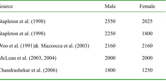
compared to the 2000 N ultimate strength of the ACL (Woo et al., 1991). This underestimated ACL loading maybe due to the assumption in their 2D model, in which the ACL loading was assumed to equal proximal tibial shear force. Lack of consideration of the ACL elevation angle in their 2-D model may underestimate the ACL force.
2.2.6. Effects of the Gastrocnemius Muscle Force on the ACL loading
The contribution of gastrocnemius muscle to the ACL loading is inconsistent in current literature. Some studies hypothesized that its contraction could pull tibia anteriorly, load the ACL, and increase the ACL strain because of its geometrical orientation (Fleming et al., 2001; O’Connor, 1993; Pflum et al., 2004). Fleming et al. (2001) studied in vivo ACL strain of 6
could increase the ACL strain, especially when the knee was near extension. O’Connor (1993) showed the identical results that co-contraction of quadriceps and gastrocnemius muscles loaded the ACL throughout the knee flexion angles. Pflum et al. (2004) used a 3-D model of body to simulate a drop-landing task. The input of muscle excitations were adjusted until the performance of the model matched the in vivo data. They reported that the gastrocnemius
muscle applied an anteriorly directed force on the tibia, but may have limited effects on the ACL loading because the gastrocnemius muscle applied a relatively small anterior shear force to the lower leg.
Several other studies, however, showed opposite effects of the gastrocnemius muscle on the ACL loading (Dürselen et al., 1995; Shelburne and Pandy, 1997). A computer modeling showed that isolated gastrocnemius muscle force could reduce the ACL loading at the knee flexions angle from 0° to 90° (Shelburne and Pandy, 1997). Although Dürselen et al. (1995) found that the gastrocnemius muscle force had no significant effect on the ACL strain when compared to the strain at 60° knee flexion without muscle force, the activation of the
gastrocnemius muscle could significantly strain the posterior cruciate ligament. Based on their results, this posterior pull of the tibia due to the contraction of gastrocnemius muscle may reduce the ACL loading. The controversial findings of the effects of the gastrocnemius muscle contraction on the ACL loading highlight the need of more research on this topic.
2.2.7. Effects of the Knee Valgus-varus and Internal-external Rotation Moment on the ACL Loading
simulation results showed that on average the sagittal plane biomechanics contributed about 1,000 N. The authors, however, claimed that the sagittal plane biomechanics cannot injure the ACL based on their simulation results, and concluded that the knee valgus moment loading was the major ACL loading mechanism. This conclusion was apparently subjective without careful considerations of the limitations and validity of their model and methods. Their results showed that sagittal plane biomechanics on average could generate 1,000 N ACL loading. Compared to the 2,160 N ACL ultimate strength (Woo et al., 1991), the reported ACL loading from sagittal plane biomechanics was about 46% of ACL injury loading and should have been considered as a significant contribution to ACL loading. The authors ignored this result and subjectively claimed that sagittal plane biomechanics could not injure the ACL. Further, the ACL loading in this study was significantly underestimated. The ACL loading in this study was approximated as the knee shear force balanced by the ACL without consideration of the ACL elevation angle. With a 55 degree ACL elevation angle and 1,000 N knee shear force balanced by the ACL, the axial force on the ACL would be over 1,700 N or 79% of the ACL ultimate strength. In addition, the validity of the forward dynamic model in this study was questionable. The results of simulation showed a posterior knee resultant shear force greater than 1,000 N while the horizontal ground reaction force was also in the posterior direction, which was inconsistent to in vivo results of other studies. The mass of the lower leg plus the
foot segment is less than 5 kg. This means that the posterior knee resultant shear force observed in this study alone could generate a lower leg posterior linear acceleration greater than 22 g, which appeared to be far away from reality.
importance of lower limb alignment on the ACL injury threshold which was defined as the maximal sustainable axial force that the limb could tolerate before the knee joint opened medially or laterally more than 8°. A three-link frontal plane model was used to investigate the consequences of dynamic alignment and muscle contraction on the ACL injury threshold. The results indicated that valgus or varus alignment reduced the injury threshold, but contraction of hip abductors and adductors increased the injury threshold. Chaudhari and Andriachii (2006) claimed that a publication by Woo et al. (1999) showed that the ACL became tensed before the MCL was tensed when the knee is subject to a knee valgus moment loading. A careful review of the publication by Woo et al. (1999) and the original study by Inoue et al. (1987) failed to show any direct and indirect evidence to support Chaudhari and Andriachii’s claim.
The results of the two above described studies of McLean et al. (2004) and Chaudhari and Andriachii (2006) were not supported by other studies. As mentioned previously, Markolf et al. (1995) investigated effects of anterior shear force at the proximal end of the tibia and knee valgus, varus, internal rotation, and external rotation moments on the ACL loading of the cadaver knees. The results of this study not only demonstrated that the proximal tibial anterior shear loading as a major ACL loading mechanism, but also showed that each of the knee valgus, varus, and internal rotation moments alone could not generate significant ACL loading unless combined with proximal tibia anterior shear loading. Fleming et al. (2001) studied the effects of weight bearing and tibial external loading on in vivo ACL strain and obtained results
similar to those of Markolf et al. (1995).
pattern of ACL strain showed a minimum strain between 30 and 35° of knee flexion. The passive varus moment increased ACL strain throughout the range of knee flexion angle tested. The passive internal rotation torque markedly increased the strain when compared to the passive normal strain curve. The passive external rotation torque resulted in less strain during the first few degrees of flexion. They also found that the ACL strain measured at combined eccentric quadriceps force and varus moment or combined eccentric quadriceps force and valgus moment had greater strain than the eccentric quadriceps force alone when the knee flexion angle was at around 30°. They concluded that the varus and internal rotation moments increased the ACL strain and the ACL strain was significantly different depending on active or passive flexion of the knee. The results of Arms et al. (1984), to certain extent, agree with the results of Markolf et al. (1995) in which the varus moment and internal rotation torque can increase the ACL loading.
Bendjaballah et al. (1997) studied knee ligaments loading responses to knee valgus-varus moment of up to 15 Nm using a finite element model of the knee joint. The loading was applied to the femur while the tibia was remained fixed. Their results
demonstrated that the collateral ligaments were the primary knee valgus-varus load bearing structures, and cruciate ligaments were the primary knee valgus-varus load bearing structures only when collateral ligaments were injured. The simulated ACL loading due to a 15 Nm knee valgus moment loading in this study was similar to that obtained in the study by Markolf et al. (1995).
experimental and control group. The cadaver knees in the experimental group were held at 30 degrees of flexion while under a 200 N valgus force applied a displacement rate of 2
mm/second. Control specimens were not subjected to valgus loading. These six unpaired femur-ACL-tibia specimens were then aligned vertically with a tensile preload of 10 N applied. The ACL was loaded to failure at a displacement rate of 2 mm/second. A differential variable reluctance transducer was implanted into ACL to measure the ACL strain. The study by Mazzocca et al. (2003) showed that minimal ACL strain occurred during valgus loading before MCL rupture, but the ACL strain increased substantially after MCL rupture in the cadaver study. Their results showed that ACL still have about 60% of its original strength after complete MCL ruptures due to knee valgus loading. The results of this study also
demonstrated that the ACL is the major constraint to valgus loading only when the MCL is ruptured. The findings of Bendjaballah et al. (1997) and Mazzocca et al. (2003) together demonstrate that it is unlikely that the cruciate ligaments were tensed before the collateral ligaments were tensed.
Based on this extensive review of literature of the effects of non-sagittal plane loading on the ACL loading and ACL strain, we need to be careful when interpreting the non-sagittal plane loading alone as the major ACL loading mechanism. Although the role of knee
valgus-varus and internal-external rotation moments in non-contact ACL injuries may be still open to debate and need further research, current literature does not support these non-sagittal plane knee moments as major ACL loading mechanisms.
2.3. Potential Risk Factors
As an attempt to prevent non-contact ACL injuries, many investigations have been conducted to identify risk factors of sustaining non-contact ACL injuries. A variety of
potential risk factors of sustaining non-contact ACL injuries have been proposed in literature. These potential risk factors have been categorized into two groups: intrinsic and extrinsic (Arendt and Dick, 1995; Boden et al., 2000). Intrinsic risk factors include anatomic,
2.3.1. Non-modifiable Intrinsic Risk Factors
A number of investigations have demonstrated the association between intrinsic risk factors and non-contact ACL injuries. Anderson et al. (2001) and Chandrashekar et al. (2005) showed that females had smaller ACL size in comparison to that of males. Several other investigators have reported that females had wider pelvis, greater Q-angle and knee joint laxity than did males, which might contribute to the gender differences in the non-contact ACL injury rate (Hewett, 2000; Wojtys et al., 1998). Wojtys et al. (1998) and Hewett (2000) both
observed that female athletes had increased ligament laxity and altered neuromuscular
performance during menstruation. Arendt et al. (2002) studied the association of ACL injuries in female athletes with menstrual cycles, and found that most of the ACL injuries occurred mainly in follicular and luteal phases. These results suggest that hormonal effect might play a role in the ACL injuries.
A recent extensive literature review, however, failed to reveal any epidemiological studies that established the cause-and-effect relationships between the non-modifiable intrinsic risk factors and the non-contact ACL injuries. Risk factors other than the non-modifiable intrinsic risk factors, therefore, need to be considered and investigated.
2.3.2. Motor Control Related Biomechanical Risk Factors
activation than did male athletes during the stance phase of each activity. Chappell et al. (2002) investigated the knee kinetics and kinematics of male and female recreational athletes during the stop-jump task. They found women exhibited larger proximal tibial anterior shear force, knee extension moment, and valgus moment, and smaller knee flexion angle at the peak proximal tibial anterior shear force during the landing phase of the stop-jump task than men did. Lephart et al. (2002) evaluated kinematics and vertical ground reaction force in single leg forward hop and drop landing tasks, and strength measures of healthy collegiate female basketball, volleyball, and soccer players and the height, age, and activity level matched male athletes. Subjects were asked to perform a single-leg forward hop and a single-leg drop landing from 20 cm platform with their dominant leg. Results of this study showed that female athletes had less knee flexion angle and lower leg internal rotation angle in both tasks
female athletes exhibited a smaller knee flexion angle at initial contact, smaller maximal knee flexion angle, and larger ground reaction forces at the maximal knee flexion than male athletes did during the stance phase of the cutting maneuver.
Ford et al. (2003) studied knee valgus motion of 81 high school basketball players. Subjects were asked to perform a drop vertical jump off a 31 cm height box. The drop vertical jump task consisted of dropping off a box, landing and immediately performing a maximum vertical jump. Only the first landing phase off the box was analyzed. Their results showed that female subjects had greater valgus angle through the stance phase of the drop-jump and the significantly greater maximal valgus angle than male counterparts. No significant differences in knee flexion angle at initial contact and at peak were observed in this study. Kernozek et al. (2005) investigated the gender differences in the lower extremity kinematics and kinetics for age-matched and skill-matched college recreational athletes during the drop-jump task. Their results showed female subjects had greater valgus angle through stance phase of the drop-jump and significantly larger maximal valgus angle than male counterparts, which were partly consistent with Ford et al. (2003). Both studies showed no significant differences in knee flexion angle at initial contact and at peak. Kernozek et al. (2005), however, found male subjects had significantly lower ground reaction forces than did females.
A small hip flexion angle during landing triggers a large knee extension torque to accelerate the tibia anteriorly to injure the ACL (Griffin et al., 2000), especially when it is seen simultaneously with the large transferred ground reaction forces at a small knee flexion angle. The more extended hip position maximizes the quadriceps demand and minimizes the
countering effect of the hamstrings forces on the proximal tibial anterior shear force. A study revealed the knee isometric Hamstrings/Quadriceps ratio was significantly increased from 38% to 99% when the hip angle was increased from 10° to 90° for the 10° to 90° knee
conditions (Ball et al., 1999). The authors concluded that the flexed hip and knee postures can be implied to facilitate the ACL-protective hamstring tension.
Landing on the heels requires large quadriceps demand as well to bring the trunk forward (Griffin et al., 2000) and to balance the exaggerated external knee flexion moment. This exerted large quadriceps force transferred to the patellar tendon can contribute to a large proximal tibial anterior shear force to likely injure the ACL, especially at a small knee flexion angle. It has been shown that the more anteriorly placed center of pressure predicted greater plantar flexion moment and less knee extensor moment. Landing on the toes, therefore, may be a protective landing style of the non-contact ACL injuries while landing on the heels may possibly increase the ACL loading.
contribute to ACL loading during athletic tasks is still unclear and needs further research. The developmental physical change along with the age may associate with non-contact ACL injuries. Yu et al. (2005) and Swartz et al. (2005) both investigated the effects of the age and gender on the lower extremity motion patterns during landing. Yu et al. (2005) found that female adolescent recreational soccer athletes have decreased knee and hip flexion angle at initial foot contact and decreased knee and hip motions during landing of the stop-jump task when compared to their male counterparts. The significant gender differences in the knee and hip motion patterns of the youth recreational soccer players occur after 12 years of age and increase with age before 16 years of age. The results of Yu et al. (2005) demonstrated the selected lower extremity motion patterns may be risk factors of sustaining non-contact ACL injuries because literature has indicated that the ACL injury rate in adolescents increases linearly after 12 years of age and that adolescent at 17 and 18 years of age have the highest ACL injury rate (Shea et al., 2004).
2.4. Effect of the Training Programs on the Prevention of Non-contact ACL Injuries Training programs based on the proposed possible risk factors for the non-contact ACL injuries in current literature were developed and applied in the clinical practice. The effect of the developed training programs on the prevention of the non-contact ACL injuries has been investigated and demonstrated the effect in altering the lower extremity motion patterns.
Neuromuscular and proprioception trainings alter the lower extremity motion patterns in athletic tasks. Hewett et al. (1996) investigated the effects of a plyometric training program on the peak vertical ground reaction force in landing after a volleyball block jump. They reported a 22% decrease in the peak vertical ground reaction force and a 50% decrease in the knee valgus-varus moment after training. Irmischer et al. (2004) studied the effects of a nine-week and low-intensity plyometric training program on the peak vertical ground reaction force and rate of the development of the peak vertical ground reaction force during a vertical jump-landing-jump task. The authors found that the training group significantly decreased their peak vertical ground reaction force and the rate of development of the peak ground reaction force during landing. Lephart et al. (2005) compared the effects of a plyometric training program and a basic resistance training program on the lower extremity
decreased the maximum valgus (28%) and varus (38%) moments for their right knee during the stance phase of the drop landing-vertical jump task. This study showed that the
combination of multiple-injury prevention-exercise components into a comprehensive program improved performance and modified lower extremity motion patterns. Myer et al. (2006) further studied the effects of the plyometric exercise on the dynamic stabilization and the balance exercises on the lower extremity motion patterns in vertical and medial drop landing tasks. The results of this study further demonstrated that subjects in the plyometric training group significantly increased knee flexion angle during the vertical drop jump task but did not significantly change the knee flexion angle during the medial drop landing task. In contrast, subjects in the balance training group significantly increased knee flexion angle during the medial drop landing task but did not significantly change the knee flexion angle during the drop landing task. Pollard et al. (2006) investigated how the practice combined with the injury prevention programs affects the lower extremity motion patterns in a drop-landing task in a longitudinal pre-post intervention study. The injury prevention program included warm-up, stretching, strengthening, plyometrics, and agilities drills. They reported that the significantly decreased hip internal rotation angle and increased hip abduction angle were observed, but no differences in the knee flexion angle and valgus angle between the pre-intervention and post-intervention were observed.