The Roles of Actin and Microtubule-based Motor Proteins in T Cell Activation
Chang K. Yi
A dissertation submitted to the faculty of the University of North Carolina at Chapel Hill in partial fulfillment of the requirements for the degree of Doctor of Philosophy in the
Department of Biology
Chapel Hill 2012
Approved by,
Dr. John A. Hammer III Dr. Xufeng S. Wu Dr. Kerry S. Bloom Dr. Stephen Rogers
ABSTRACT
Chang K. Yi: The Roles of Actin and Microtubule-based Motor Proteins in T Cell Activation (Under the direction of Dr. John A. Hammer III)
T cell activation is a critical process in our body’s fight against infection and disease. The activation process begins upon encounter with stimulatory antigen presenting cells (APCs), which leads to the rapid polarization of various cellular components in T cells. At the membrane of the contact site, T cell receptors (TCRs), adhesion molecules, and other accessory components become differentially clustered and rearranged to form a supra-molecular structure called the immunological synapse. While both actin retrograde flow and myosin IIA activity have been implicated in immunological synapse formation, their relative organization and dynamics at the immunological synapse, and their relative contributions toward the reorganization of receptor clusters have not been fully understood. In this study, we show that the immunological synapse is essentially a radially symmetric migrating cell, as previously hypothesized, and provide direct proof that classical lamellipodial and lamellar actin networks localize at dSMAC and pSMAC regions, respectively. Moreover, our
experiments using actin and myosin II inhibitors reveal that actin retrograde flow and actomyosin II contraction work coordinately to drive the inward movements of receptor clusters at the immunological synapse.
Concomitant with immunological synapse formation, the microtubule organizing center (MTOC) polarizes rapidly toward the contact site with the APC to position the
secretory apparatus for directed secretion. Due to the lack of spatial and temporal control in most previous studies, the kinetics of MTOC repositioning have not been well characterized in T cells. In addition, the specific roles of dynein and dynein regulatory proteins during T
cell activation have not been defined. In this study, we show using an optical trap-controlled T cell activation method that TCR and LFA-1 signaling are both required for robust MTOC repositioning. Moreover, we report that the repositioning process is bi-phasic, with distinct kinetics at each phase of MTOC movement. Lastly, inhibition experiments using dominant negative constructs show that dynein and LIS1 are required for both phases of MTOC repositioning. In conclusion, the actin and microtubule cytoskeletal systems mediate many distinct aspects of T cell activation, and are critical components of the overall adaptive immune response.
To my loving parents
With deepest admiration and gratitude
ACKNOWLEDGEMENTS
We shall not cease from exploration And the end of all our exploring Will be to arrive where we started And know the place for the first time.
- T.S. Eliot
It has been an incredible honor to have received training at NIH, one of the premier research institutions in the world, and to have interacted with many gifted scientists through the UNC partnership program. First, I would like to thank the members of my committee, Drs. Kerry Bloom, Stephen Rogers, Lawrence Samelson, and Kevin Slep for their invaluable input and support through the years. Also, I thank my mentor, Dr. John Hammer, for his guidance and friendship. His passion for scientific rigor and integrity, and his deep sense of humanity have impacted me greatly. Next, I would like to thank the chairperson of my committee, Dr. Xufeng Wu, for her innumerable contributions to my graduate experience. Her magnanimous heart, adventurous spirit, and keen scientific mind have inspired me on a regular basis. Our exuberant, ad lib discussions over “cool” results will surely be missed.
This dissertation would not have been possible without the constant support and care from my family. Words cannot express the deep respect and appreciation I feel toward my parents, Kwang Taek and Chong Hee Yi, who have dedicated their lives toward my growth as a student and as an individual. I’m inspired by their immigrant journey, and the many sacrifices they’ve made along the way. This dissertation has been, in no small part, a labor of love in celebration of their journey. Lastly, and most importantly, I thank my wife, Chanmi Yi, for her support and encouragement throughout my graduate career. She has
been a constant source of warmth and joy, enriching the often monotonous experience of basic research. Thank you for making the journey worthwhile.
TABLE OF CONTENTS
LIST OF FIGURES………..…………xi
LIST OF ABBREVIATIONS………..…xiii
CHAPTER 1: INTRODUCTION……….1
I. Overview………..1
II. Lymphocytes and adaptive immunity………..1
III. Signaling pathways of T cell activation……….…..………2
a. TCR signal transduction……….………2
b. Integrin regulation and signaling……….………..3
IV. Immunological synapse……….5
c. Identity and function of SMAC domains………..….…5
d. TCR MCs and the regulation of TCR signaling………...…...6
V. The role of actin and myosin IIA in T cells……….8
e. Non-muscle myosin IIA………...8
f. Actin networks in migrating cells………...8
g. The role of actin and myosin IIA in T cell function………10
VI. The role of dynein in MTOC repositioning in T cells………..……12
h. Cytoplasmic dynein………...12
i. Dynein adaptor proteins………...12
j. MTOC repositioning in T cells……….14
k. TCR-proximal events involved in MTOC repositioning………14
l. Cortical pulling model of MTOC repositioning in T cells……….16
VII. References………21
CHAPTER 2: ACTIN RETROGRADE FLOW AND ACTO-MYOSIN II ARC CONTRACTION DRIVE RECEPTOR CLUSTER DYNAMICS AT THE IMMUNOLOGICAL SYNAPSE IN JURKAT T CELLS………26
I. Abstract………...26
II. Introduction………...27
III. Results………...30
a. The region of the IS corresponding to the LM/pSMAC contains concentric actin arcs that are rich in myosin IIA………...30
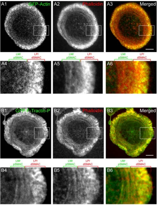
b. A prototype of F-Tractin, a novel reporter for F-actin, but not GFP-actin, localizes to both LP and LM actin networks at the IS………..36
c. Quantitation of F-actin dynamics using F-Tractin-P reveals a striking difference in centripetal flow rates between the LP/dSMAC and LM/pSMAC………...42
d. Myosin IIA moves inward with the actin arcs in the LM/pSMAC………..45
e. TCR MCs move inward at the speed of actin retrograde flow in the LP/dSMAC and at the speed of acto-myosin IIA arc contraction in the LM/pSMAC………...49
f. Inhibition of myosin IIA with blebbistatin slows TCR MC movement in the LP/dSMAC and disrupts both the organization of actin arcs and the directed transport of TCR MCs in the LM/pSMAC………....52
g. Inhibition of actin retrograde flow causes the F-actin network and associated TCR MCs in the LP/dSMAC to retract at a speed that corresponds to slowed acto-myosin II arc contraction in the LM/pSMAC……….58
h. Simultaneous inhibition of both actin retrograde flow and acto-myosin II arc contraction blocks the vast majority of centripetal TCR MC movements at the IS……….68
i. Acto-myosin II contraction is required for the accumulation of LFA-1 clusters at the inner aspect of the LM/pSMAC……….69
IV. Discussion……….72 a. Correspondence between LP and LM actin networks and
the SMAC regions of the IS……….73
b. Novel observation of contracting acto-myosin II arcs in the LM/pSMAC………..74
c. Kinetic coupling between TCR MC movement and cortical actin network flow at the IS………..75
d. The role of myosin IIA at the IS………...76
e. Regulation and dynamics of F-actin networks at the IS…………...77
f. Role of microtubules and dynein-based TCR MC transport at the IS………..78
g. Conclusion………..79
V. Materials and methods………...….80
VI. Acknowledgements………..86
VII. References………88
CHAPTER 3: CORTICAL FORCE GENERATION BY DYNEIN AND LIS1 DRIVES THE BI-PHASIC REPOSITIONING OF THE MTOC IN T CELLS………...92
I. Abstract……….92
II. Introduction………93
III. Results………...95
a. Optical trap-controlled activation induces robust MTOC repositioning in Jurkat T cells………..95
b. TCR and LFA-1 signaling are both required for robust MTOC repositioning………..98
c. MTOC repositioning in T cells is bi-phasic………101
d. Large invagination of the synaptic interface is observed when MTOC movement is impeded……….104
e. Cortical dynein spots are uniformly distributed and transiently bound at the IS……….105
f. Inhibition of dynein impairs MTOC movement during the polarization phase and blocks entry into the docking phase of MTOC repositioning ………...109
g. Inhibition of LIS1 impairs MTOC movement during the polarization phase and disrupts prolonged centrosome
docking during the docking phase of MTOC repositioning………..112
IV. Discussion………..114
a. Optical trap-controlled activation of T cells……….115
b. The two phases of MTOC repositioning in T cells……….116
c. The role of LFA-1 integrins in MTOC repositioning………...117
d. Generation of cortical pulling force at the IS………...118
e. The localization of dynein at the IS………..118
f. The role of dynein in MTOC repositioning in T cells ……….120
g. The role of dynein regulatory proteins in MTOC repositioning in T cells………..121
h. Conclusion………..122
V. Materials and methods………..123
VI. Acknowledgements………128
VII. References………..129
CHAPTER 4: CONCLUDING REMARKS AND FUTURE DIRECTIONS………...132
I. Overview……….132
II. What is the role of formin-generated actin filaments at the IS?……….132
III. How does F-actin reorganize at the border between the LP/dSMAC and LM/pSMAC?...133
IV. What is the “diffusion” barrier for LFA-1 clusters at the pSMAC-cSMAC border?………..135
V. How is dynein anchored at the IS cortex?……….136
VI. What is the mechanism of cortical force generation at the IS?...137
VII. References……….139
LIST OF FIGURES Figure
1. Illustration of the TCR signaling cascade ………..4 2. Model of MTOC repositioning in T cells……….17 3. Mobility of ICAM-1 in planar bilayers and efficiency of IS formation
in Jurkat T cells………..31 4. Localization of endogenous F-actin structures and corresponding
LP/LM markers at the IS………..33 5. Comparison of F-actin structures at the IS reported by GFP-actin
and mGFP-F-Tractin-P……….38
6. Lack of effect of mGFP-F-Tractin-P expression on cellular F-actin
content and dynamics, and on TCR signaling………..39 7. Characterization of F-actin dynamics in the LP/dSMAC and
LM/pSMAC regions of the IS………...43 8. Rates of actin retrograde flow and acto-myosin II arc contraction in
DMSO-treated Jurkat cells engaged on coverslip and planar bilayer substrates, and imaging of actin arcs in the LM/pSMAC using only
dynamic probes for myosin IIA………...46 9. Characterization of TCR MC dynamics at the LP/dSMAC and
LM/pSMAC regions of the IS………...50 10. Effect of Blebbistatin on the dynamics of F-actin and TCR MCs in the
LP/dSMAC and LM/pSMAC regions of the IS………..53 11. Testing the effects of CD, Jas, and CD-Jas treatments on actin
retrograde flow in cells engaged on coverslips………59 12. Inhibition of actin retrograde flow and acto-myosin II arc contraction
using combinations of CD, Jas, and BB………65 13. The effect of myosin II inhibition on the accumulation of LFA-1
clusters at the inner aspect of the LM/pSMAC……….71 14. Optical trap-controlled method of T cell activation……...96 15. Contribution of TCR and LFA-1 signaling pathways to MTOC
repositioning……….99 16. Kinetics of MTOC movement during the two phases of repositioning
and evidence of cortical pulling force at the IS membrane………102
17. Localization of cortical dynein at the IS………...107 18. Effect of dynein and LIS1 inhibition on MTOC repositioning ………..111
LIST OF ABBREVIATIONS ADAP Adhesion and degranulation-promoting protein ADP Adenosine diphosphate
APC Antigen presenting cell ATP Adenosine triphosphate
BB Blebbistatin
CC1 Coiled coil region 1
CD Cytochalasin D
CN Control
cSMAC Central SMAC
CTL Cytotoxic T Lymphocyte DAG Diacylglycerol
DNS Data not shown
dSMAC Distal SMAC
ELC Essential light chain FMNL1 Formin-like 1
FRAP Fluorescence recovery after photobleaching GEF Guanine exchange factor
GFP Green flourescent protein
HC Heavy chain
IC Intermediate chain
ICAM-1 Intercellular adhesion molecule-1 IS Immunological synapse
ITAM Immunoreceptor-based tyrosine activation motif Jas Jasplakinolide
LC Light chain
LFA-1 Lymphocyte function-associated antigen-1 LIC Light intermediate chain
LM Lamella
LP Lamellipodia
MC Microcluster
MPM Modulated polarization microscopy MT Microtubule
MTOC Microtubule organizing center
pMHC Peptide-bound major histocompatibility complex
pSMAC Peripheral SMAC
PTK Protein tyrosine kinase RFP Red flourescent protein SH2 Src-homology 2
shRNA Short hairpin ribonucleic acid siRNA Short interference ribonucleic acid SMAC Supra-molecular activation cluster TCR T cell receptor
WT Wild type
CHAPTER 1 INTRODUCTION
I. Overview
The actin and microtubule cytoskeleton are known to play critical roles in T cell function. They are involved in signaling of antigen receptors, formation of the immunological synapse, and focused delivery of effector molecules, which are necessary for targeted cell death and communication between lymphocytes. In addition, these cytoskeletal systems play important roles in numerous fundamental cellular processes such as directed cell migration and asymmetric cell division. Thus, the investigation of the role of actin and MTs in T cells is relevant not just for the study of cell-mediated immunity, but also for the
discovery of common biological mechanisms in cytoskeletal function and regulation.
II. Lymphocytes and adaptive immunity
1
Lymphocytes are essential for adaptive immunity against infection from pathogens, and the emergence of immune-related diseases, such as AIDS, has served to underscore their importance. Adaptive immunity refers to the body’s ability to generate a specific immune response toward particular microbes and microbial toxins, and its ability to mount an amplified response upon future exposure to those same pathogens. In adaptive immunity, the response is largely carried out by two types of cells: B and T lymphocytes. The B
T Lymphocytes are equipped with specialized receptors, T cell receptors (TCRs), which engage peptide-bound major histocompatibility complexes (pMHCs) on the surface of antigen presenting cells (APCs). There are two subsets of T cells, distinguished by the presence of either CD4 or CD8 glycoprotein on the cell surface. CD4+ T cells are called
helper T cells because of their role in helping B cells produce antibodies, as well as in helping phagocytes destroy harmful microbes, CD8+ T cells are called cytotoxic T
lymphocytes (CTL’s) because of their role in lysing infected cells (Abbas and Lichtman, 2006).
All T cells have their origin in the thymus, where progenitors differentiate into T cells after migration from the bone marrow. These “naïve” T cells then leave the thymus and circulate between peripheral lymphoid organs where they encounter multiple APCs. After recognition of antigen, naive T cells proliferate and differentiate into two different types of T cells: effector and memory cells. Effector helper T cells secrete cytokines to activate macrophages and B cells, whereas effector CTLs develop the ability to kill infected cells. Effector T cells are usually short-lived while memory T cells can continue to survive long after differentiation. Although memory cells are functionally “silent,” they can give rise to a large population of effector cells upon re-encountering the same antigen, which is the basis for the rapid, amplified nature of the secondary immune response (Abbas and Lichtman, 2006).
III. Signaling pathways of T cell activation a. TCR signal transduction
Underlying T cell activation is the outside-in signal transduction mediated by the TCR signaling complex. The TCR signaling complex consists of α-, β-, and ζ- TCR chains and a repertoire of CD3 subunits, which associate with the TCR to form the functional signaling complex (Chan et al., 1994; Samelson, 2002; Guy and Vignali, 2009). Recognition and
binding of pMHC by the α-and β- TCR chains initiates the signal transduction cascade by recruiting Src-family of protein tyrosine kinases (PTKs) Lck and Fyn to phosphorylate the immunoreceptor-based tyrosine activation motifs (ITAMs) on the CD3 subunits and the ζ- chain of the TCR complex (Fig. 1) (Chan et al., 1994; Samelson, 2002; Billadeau et al., 2007; Guy and Vignali, 2009). This leads to the binding and activation of a Syk-family PTK, ZAP-70, which in turn phosphorylates key adaptor proteins, including the transmembrane protein LAT (linker for activation of T cells) (Fig. 1) (Samelson, 2002; Billadeau et al., 2007). LAT also contains several ITAMs which can bind Src homology (SH2)-containing molecules such as PLCγ1 and SLP76, and functions as a protein scaffold for the recruitment of
multiple downstream effectors involved in TCR signal transduction (Fig. 1) (O'Shea, 2000; Samelson, 2002; Billadeau et al., 2007).
b. Integrin regulation and signaling
In addition to TCR signaling, full activation of T cells requires the engagement of co-stimulatory and adhesion molecules (Guy and Vignali, 2009). A major example is the β2
-integrin, LFA-1 (lymphocyte function-associated antigen-1; αLβ2), which binds ICAM-1
(intercellular adhesion molecule-1) on the surface of APCs (Kinashi, 2005). LFA-1 is involved in adhesion to APCs, and is critical for the effector function of T cells during activation (Kinashi, 2005). In contrast to the TCR, LFA-1 integrins are initially regulated by inside-out signaling, and require T cell stimulation by antigen or cytokines to become activated (Baker and Koretzky, 2008). In non-stimulated T cells, LFA-1 integrins adopt a low-affinity, bent conformation, and lack the ability to form multivalent clusters. However, engagement of the T cell antigen receptor alters the avidity of LFA-1 by opening the heterodimer into a high-affinity ligand binding state, as well as allowing the formation of multivalent LFA-1 clusters (Kinashi, 2005). This TCR-dependent increase in LFA-1 avidity requires LAT recruitment of PLCγ1, which leads to calcium flux and diacylglycerol (DAG)
Figure 1. Illustration of the TCR signaling cascade (Billadeau et al., 2007).
production, and SLP76, which activates VAV1, an important guanine exchange factor (GEF) involved in actin reorganization (Fig 1) (Billadeau et al., 2007; Baker and Koretzky, 2008). Also, SLP76 associates with ADAP (adhesion and degranulation-promoting protein), an adaptor molecule required for integrin function in T cells (Baker and Koretzky, 2008). Once activated, LFA-1 contributes to the outside-in co-stimulatory signal to enhance T cell
activation and effector function.
IV. Immunological Synapse
a. Identity and function of SMAC domains
APC engagement also leads to dramatic changes at the cellular level in T lymphocytes. Upon binding their respective ligands on the surface of the APC, antigen receptors and adhesion proteins undergo differential clustering and rearrangement at the synaptic junction to form segregated, concentric domains known as supra-molecular activating clusters (SMACs) (Monks et al., 1998; Grakoui et al., 1999; Fooksman et al., 2010). The resulting bull’s-eye pattern of SMACs is a hallmark of the immunological
synapse (IS), and provides the structural basis for signaling and secretion at the T cell-APC interface. The center area of the IS, known as the central SMAC (cSMAC), is marked by the accumulation of TCRs, which are bound to pMHCs on the surface of the APC (Monks et al., 1998). The surrounding ring of the bull’s eye, known as the peripheral SMAC (pSMAC), is marked by LFA-1 clusters, which are bound to ICAM-1 receptors present on the APC surface (Monks et al., 1998). The outermost ring, known as the distal SMAC (dSMAC), is marked by CD45, as well as an enrichment of F-actin (Freiberg et al., 2002; Sims et al., 2007).
Recent studies argue that TCR signaling is degraded at the cSMAC, and that active signaling actually takes place at the periphery of the IS (Yokosuka et al., 2005; Varma et al., 2006; Vardhana et al., 2010). Thus, the pSMAC region may serve dual functions during T
cell activation: as a zone of adhesion between the T cell and the APC, and as a zone of active TCR signaling at the IS. Meanwhile, the dSMAC is thought to function as a zone of exclusion from the IS, owing to the localization of receptors with large ecto-domains and/or inhibitory effect on TCR signaling (Freiberg et al., 2002).
Substitution of the APC surface with a supported planar lipid bilayer displaying stimulatory molecules has been shown to replicate the signaling activity and spatial organization of the IS, and has become an important tool in the study of T cell activation (Grakoui et al., 1999; Dustin, 2009). Alternatively, glass coverslips coated with stimulatory molecules can be used to engage and activate T cells (Bunnell et al., 2003). However, since the stimulatory ligands are immobilized on the coverslip substrate, antigen receptors and adhesion molecules on the T cell surface cannot rearrange to form the bull’s eye pattern of SMACs, as observed after engagement to APC and planar bilayer substrates. This difference between the coverslip and planar bilayer substrates has been utilized in our imaging studies to distinguish the reorganization of molecules occurring at the membrane from that of the synaptic cortex.
b. TCR MCs and the regulation of TCR signaling
T cells display remarkable sensitivity to antigen despite the relatively weak affinity of TCRs for pMHCs and low numbers of available ligand on the APC surface (Shaw and Dustin, 1997; Huppa and Davis, 2003). It is known that stimulation by a single pMHC on the APC surface is sufficient for transient activation of T cells, and that engagement of ~10 pMHC molecules can result in sustained activation (Irvine et al., 2002). This sensitivity is, in part, enabled by the T cell co-receptor, as helper T cells lacking the CD4 co-receptor require higher numbers of pMHC for activation and show reduced ability to form a mature IS (Irvine et al., 2002).
However, sensitization of individual TCRs cannot account for the prolonged TCR signaling observed during T cell activation (on the order of hours), as their association with antigen is short-lived (on the order of seconds) (Huppa and Davis, 2003). Rather, this requires amplification of signal from the transiently engaged TCRs through a multi-protein structure at the membrane called the TCR microcluster (MC). Within seconds of T cell engagement to the anti-CD3-bound substrate, TCR MCs form at the contact site (Grakoui et al., 1999; Bunnell et al., 2002). These MCs are estimated to contain ~100 TCR molecules,
and act as a platform for the recruitment and activation of downstream signaling molecules (Campi et al., 2005; Varma et al., 2006). Studies using artificial substrates have shown concentrated tyrosine phosphorylation activity, as well as dynamic localization of ZAP70, Lck, and LAT to these MCs, indicating that the TCR MC functions as a basic signaling unit during T cell activation (Bunnell et al., 2002; Campi et al., 2005; Yokosuka et al., 2005; Varma et al., 2006). Paradoxically, either signal amplification or degradation can be observed at TCR MCs, depending on the strength of antigenic peptide utilized (Lee et al., 2003). Based on this result, A. Shaw and colleagues propose that TCR signaling is actively tuned at the IS, allowing amplification of weak signals and dampening of strong signals (Cemerski and Shaw, 2006).
Further regulation of TCR signaling occurs via active transport of TCR MCs. During IS formation, TCR MCs form at the periphery and are transported toward the center of the IS through an actin-dependent mechanism (Campi et al., 2005; Varma et al., 2006; Kaizuka et al., 2007). However, once accumulated at the cSMAC, these MCs disassociate from
downstream signaling molecules, lose their tyrosine phosphorylation activity, and are internalized for recycling back to the IS periphery (Yokosuka et al., 2005; Varma et al., 2006). Thus, it is through the continuous formation and cycling of TCR MCs that TCR signaling is sustained for full T cell activation.
V. The role of actin and myosin IIA in IS formation a. Non-muscle myosin IIA
Non-muscle myosin II (referred to here as myosin II), a subclass of myosin motor proteins, is expressed in all eukaryotic non-muscle cells and is involved in many crucial functions of the cell, including cell polarity, migration, and cytokinesis. It is composed of two heavy chains (HCs), two regulatory light chains (RLCs), and two essential light chains (ELCs). The globular N-terminal head region of the HCs contains the binding site for actin and catalytic sites for ATP hydrolysis, and generates the force required for myosin II contractile activity. Adjacent to the head is a neck region that binds the regulatory and essential LCs, and acts as a level arm to amplify the small ATP-hydrolysis induced conformational changes in the head, allowing the myosin to step. Following the neck is a long coiled-coil rod domain that mediates dimerization of the HCs, and a short-helical tail that contain multiple phosphorylation sites for regulation of myosin II activity. The rod domains of multiple myosin II dimers self-associate to form bipolar filaments, which can then bind and exert tension on actin filaments to form contractile structures in cells (Vicente-Manzanares et al., 2009).
Three myosin II HC genes exist in mammalian cells (MYH9, MYH 10, and MYH14), giving rise to three myosin II isoforms (myosin IIA, IIB, and IIC, respectively) that differ in structure, regulation, localization and function. In vitro experiments have shown that myosin IIA displays the highest rate of ATPase hydrolysis and the fastest rate of actin filament displacement. Myosin IIA plays critical roles in leading edge protrusion and adhesion in migrating cells and is the dominant isoform expressed in mammalian T cells (Jacobelli et al., 2004; Morin et al., 2008; Vicente-Manzanares et al., 2009).
b. Actin networks in migrating cells
The actin cytoskeleton is required for numerous fundamental cellular processes and has been studied in a broad spectrum of cellular systems (Gardel et al., 2010). In particular, characterization of actin networks at the leading edge of migrating cells has yielded
important understanding of their dynamics and function in recent decades. At the leading edge is the lamellipodia (LP), where a branched network of dendritic F-actin filaments undergoes rapid actin retrograde flow, driven by actin polymerization at the front and depolymerization at the rear of the LP (Pollard and Borisy, 2003; Ponti et al., 2004).
Polymerization of actin requires addition of ATP-bound actin monomers to the “barbed” ends of growing filaments, and Arp2/3, which creates free barbed ends by de novo nucleation of actin filaments off the sides of pre-existing filaments (Pollard and Borisy, 2003). On the other hand, ADF cofilin (which can also create free barbed ends by severing existing actin filaments) promotes depolymerization of actin at the “pointed” ends of actin filaments (Pollard and Borisy, 2003). The resulting actin retrograde flow provides the protrusive ‘pushing’ force against the leading edge membrane when the cell is engaged to a substrate, and is the kinetic signature of the LP actin network.
Proximal to the LP is the lamella (LM), where transverse arcs containing actin and myosin IIA filaments undergo slow contraction toward and disassembly at the cell rear (Medeiros et al., 2006; Wilson et al., 2010). The contraction of acto-myosin IIA filaments provides the ‘pulling’ force for actin networks at the cell cortex, and is the kinetic signature of the LM actin network. Focal adhesion complexes enriched with integrins and associated signaling proteins also form in the LM, and are known to mediate the linkage between the cortical actin networks and the extracellular matrix (Gardel et al., 2010). In combination with actin polymerization-driven protrusion in the LP, contraction of acto-myosin IIA filaments against these focal adhesion complexes results in the net forward movement of the cell body during cell migration.
c. The roles of actin and myosin IIA in T cell function
Actin networks also regulate various aspects of T cell activation. In vivo studies have shown that the initial stages of migration, polarization, and synapse formation require the actin cytoskeleton (Billadeau et al., 2007). More recently, in vitro experiments utilizing supported planar bilayer and immobilized coverslip substrates have revealed an intricate link between receptor-ligand complexes and cortical F-actin at the IS (Barda-Saad et al., 2005; Campi et al., 2005). Upon contact with a stimulatory surface, sub-micron-sized TCR MCs form and signal downstream to activate actin polymerization (Bunnell et al., 2001; Varma et al., 2006). This leads to increased cell spreading and further nucleation of TCR and LFA-1
clusters. On laterally mobile substrates, these newly formed TCR and LFA-1 clusters subsequently undergo actin-dependent centripetal transport to accumulate at their respective SMAC regions, forming the bull’s eye pattern of the IS (Kaizuka et al., 2007). However, the rate of actin retrograde flow is reported to be two-fold higher than that of receptor cluster movements (Kaizuka et al., 2007), and TCR MCs are able to move around physical barriers in the bilayer substrate without cortical F-actin reorganization (DeMond et al., 2008), suggesting an inherent “slippage” in the mechanism of coupling between receptor
clusters and cortical actin . Moreover, receptor clusters appear to be differentially linked to cortical actin networks as pre-formed LFA-1, but not TCR, clusters are destabilized by Latrunculin B treatment (Campi et al., 2005; Kaizuka et al., 2007). While the molecular interactions mediating these linkages are unknown, centripetal flow of cortical actin has emerged as the likely driving mechanism behind receptor transport at the IS.
The synaptic interface of T lymphocytes is analogous to the leading edge of
migrating cells, as previously noted by M. Dustin and colleagues. Based on the motility of T cells on the planar bilayer substrate, as well as the localization of LP and LM markers at the IS, the dSMAC and pSMAC regions were predicted to be functionally equivalent to the LP and LM networks, respectively (Dustin et al., 2007; Sims et al., 2007). There is evidence of
LP network involvement in IS formation. Regulators of dendritic actin nucleation, such as Arp2/3 and the WAVE/WASP complex, are known to be recruited and activated by VAV1 during TCR signal transduction (Fig. 1) (Billadeau et al., 2007). Dynamic ruffling has been described at the contact site between T cells and APCs, and rapid actin treadmilling has been observed at the IS as imaged by ectopic expression of GFP-actin (Bunnell et al., 2001; Kaizuka et al., 2007). Additionally, inhibition of actin polymerization is known to disrupt the inward movement of receptor clusters at the IS (Campi et al., 2005; Varma et al., 2006; Kaizuka et al., 2007). In contrast, it is not known whether a classical LM network functions at the IS, as no one has described acto-myosin II arcs or focal adhesion complexes at the IS. Consistent with these observations, R. Vale and colleagues have proposed that a single LP actin network exists at the IS, and that actin retrograde flow drives the inward movement of receptor clusters at the IS (Kaizuka et al., 2007).
Myosin IIA is known to regulate the migration and adhesion of T cells (Jacobelli et al., 2004; Morin et al., 2008), and has been implicated in the secretion of lytic granules in
natural killer cells (Andzelm et al., 2007). However, the role of myosin IIA in T cell activation is unclear as conflicting reports exist regarding the regulation and function of myosin IIA in IS formation. Jacobelli et al. reported that IS formation does not require myosin IIA, as TCR and LFA-1 receptors reorganized into the bull’s eye pattern of cSMAC and pSMAC regions in blebbistatin-treated T cells (Jacobelli et al., 2004). On the other hand, recent report by M. Dustin and colleagues showed that myosin IIA is required for IS formation, as inhibition of myosin IIA, using blebbistatin and siRNA knockdown of myosin IIA heavy chain, disrupted the directed movement of TCR MCs as well as cSMAC formation (Ilani et al., 2009). However, actin and myosin IIA were not dynamically imaged at the IS, and the relative contribution of actin retrograde flow and myosin II contractility to IS formation was not quantitatively analyzed in these studies, thus giving very little information about how actin and myosin II are involved in IS formation.
VI. The role of dynein in MTOC repositioning in T cells a. Cytoplasmic Dynein
Dynein is a class of minus end-directed MT motor proteins involved in a wide range of cellular functions including cargo transport, asymmetric cell division, and cell polarity (Dujardin and Vallee, 2002; Kardon and Vale, 2009; Allan, 2011). In contrast to other classes of cytoskeletal motors, only a single dynein, cytoplasmic dynein, accommodates these diverse functions in non-axonemal cells. Cytoplasmic dynein (referred to here as dynein) is a large complex composed of a heavy chain (HC) and several non-catalytic subunits. The C-terminal portion of the HC contains a coiled-coil “stalk” extension that mediates binding to the MT and a ring-shaped “head” consisting of six AAA domains that functions as the catalytic site for ATP hydrolysis. In addition to the AAA domains, the “head” region contains a linker domain that shifts its position on the catalytic ring during the ATPase cycle, and possibly serves as the mechanical element in the dynein power stroke. The N-terminal “tail” portion is involved in the homo-dimerization of HCs and functions as a scaffold to recruit dimers of non-catalytic subunits: TCTEX 1, light chain 8 (LC8), LC7, intermediate chain (IC), and light intermediate chain (LIC). These non-catalytic subunits are implicated in the binding of selective cargo, and are known to associate with different adaptor proteins that regulate the activity of dynein in cells (Kardon and Vale, 2009).
b. Dynein adaptor proteins
Among the dynein adaptor proteins, two complexes have become of interest: dynactin and LIS1-NUDE/NUDEL. Like dynein, dynactin is a large complex comprised of multiple subunits, including p150 which mediates binding of dynactin to the dynein complex through its N-terminal coiled coil region (CC1). Dynactin is involved in the targeting of dynein to specific cellular regions, binding of dynein to cargo, and modulates the
processivity of dynein’s movement on MTs. Regarding the latter role of dynactin, in vitro experiments showed that dynein processivity along MT tracks increased almost two-fold in the presence of dynactin. Initially, this enhancement in processivity was attributed to the increased tethering of the dynein-dynactin complex to MTs via the CAP-Gly MT-binding domain of p150, but subsequent in vivo studies showed that the CAP-Gly domain was dispensable for modulation of dynein processivity. At present, the mechanism by which dynactin affects dynein motility remains unclear (Kardon and Vale, 2009).
In addition to dynactin, LIS1 plays an important role in the regulation of dynein activity. LIS1 is thought to be an important cofactor for dynein, and forms a tripartite complex with dynein and the LIS1-binding partner, NUDE or NUDEL (in metazoans). LIS1 is composed of an N-terminal coiled-coil domain that mediates dimerization, and a C-terminal WD40 domain that binds to an ATPase domain (AAA1) in the head region of the
dynein HC, and possibly to the tail region of the HC as well as the p50 subunit of dynactin. In budding yeast, homologs of LIS1 and NUDE (Pac1 and Ndl1, respectively) are involved in the localization of dynein at the plus ends of MTs, and in the cortical anchoring of dynein at the bud cell during cell division. In metazoans, LIS1, NUDE, and NUDEL are required for dynein-mediated positioning of the nucleus, centrosome and the mitotic spindle, and are involved in the cortical anchoring of dynein during spindle repositioning (Kardon and Vale, 2009).
Furthermore, recent reports from R. Vallee and colleagues show that NUDE recruits LIS1 to the dynein complex to modulate force generation, distinct from dynactin-regulated dynein activity. Specifically, LIS1-NUDE associates with dynein to induce a sustained state of force generation under high load conditions (McKenney et al., 2010). Association with LIS1-NUDE also improved the ensemble activity of multiple dynein motors, promoting dynein activity under high load conditions (McKenney et al., 2010). Furthermore, NUDE and dynactin share a common binding site on the dynein IC subunit, suggesting that association
of dynein with LIS1-NUDE and dynactin is mutually exclusive (McKenney et al., 2011). While it is unclear whether LIS1-NUDE and dynactin compete with other adaptors for association with dynein, these results are consistent with the emerging idea that differential binding to adaptor proteins allows dynein to accommodate a wide range of cargos and functions in the cell (Kardon and Vale, 2009).
c. MTOC repositioning in T cells
Concomitant with IS formation, APC engagement leads to rapid repositioning of the MTOC toward the contact site in T cells (Kupfer and Dennert, 1984). It is believed that MTOC repositioning serves as a key step in the directed secretion of effector molecules in T lymphocytes (Sancho et al., 2002). In the case of target cell killing by CTLs, antigen
recognition and subsequent TCR signaling leads to rapid MTOC polarization to the IS (Stinchcombe et al., 2006). Lytic granules, then, move to the centrosome via dynein-mediated intracellular transport, and undergo docking and fusion at the synaptic membrane (Stinchcombe and Griffiths, 2007). Imaging studies by Stinchcombe et al. suggest that lytic granules localize to a specialized secretory domain of the IS and that the lytic contents are secreted into a sealed cleft between the CTL and target cell to cause lysis of the target cell (Stinchcombe et al., 2006). In addition to the known functional role in directed secretion, a recent publication by Sanchez-Madrid and colleagues suggests that MTOC repositioning contributes to sustained T cell signaling and proper IS maturation (Martin-Cofreces et al., 2008).
d. TCR-proximal events involved in MTOC repositioning
While MTOC repositioning is known to be dependent on TCR engagement, the downstream signaling pathways and mechanisms leading to repositioning are poorly understood. Among the known molecules required for MTOC repositioning, which include
Lck, ZAP70, LAT, SLP76, VAV1, and CDC4, most are involved in signaling immediately downstream of the TCR (Stowers et al., 1995; Kuhne et al., 2003; Billadeau et al., 2007). Studies have also shown that TCR-proximal events, including calcium flux, are involved in MTOC repositioning in T cells (Kuhne et al., 2003; Billadeau et al., 2007). However, the cascade of TCR-proximal events leading to MTOC repositioning is unclear as M. Huse and colleagues reported, using a photoactivateable pMHC ligand system, that DAG, and not calcium flux, is required for MTOC repositioning (Quann et al., 2009). In their signaling model, PLCγ1-directed cleavage of PIP2 produces DAG, which then signals through atypical
PKCs to recruit dynein to the contact site, leading to MTOC repositioning (Quann et al., 2011). Further experiments using T cell-APC conjugates are needed to confirm the distinct roles of DAG and calcium flux in T cell activation.
In addition to DAG/ calcium influx, F-actin reorganization is thought to play an important role in MTOC repositioning (Sancho et al., 2002; Billadeau et al., 2007). CDC42, a key regulator of Arp2/3-mediated actin polymerization, is required for MTOC polarization in T cells (Stowers et al., 1995), and has been implicated in the tethering of MT plus ends to actin filaments via its interaction with IQGAP1 in non-T cell systems (Fukata et al., 2002). Based on these and other observations, G. Griffiths and colleagues proposed that the ring of lamellipodial actin in the dSMAC drives MTOC repositioning through CDC42-mediated tethering of MTs to the spreading actin ring (Stinchcombe et al., 2006). However, Gomez et al. report that MTOC repositioning is independent of CDC42 and Arp2/3, and that MTOC
repositioning occurs in the absence of a lamellipodial actin ring (Gomez et al., 2007). Instead, they suggest that the formins mDIA1 and FMNL1 mediate the association between filopodial actin and MTs during actin-dependent MTOC repositioning. Further complicating the role of F-actin in MTOC repositioning is a study from Sedwick et al. They show utilizing a live-cell competition assay that the MTOC consistently repositions toward the site of TCR stimulation while F-actin polarizes toward the site of LFA-1 stimulation (Sedwick et al., 1999).
They conclude that the signal pathways leading to actin reorganization contribute to MTOC repositioning by amplifying TCR signaling, rather than through a direct association between actin filaments and MTs. Given the conflicting reports regarding the role of actin in MTOC repositioning, more experiments are needed to understand the crosstalk between the actin and MT cytoskeleton during MTOC repositioning in T cells.
e. Cortical pulling model of MTOC repositioning in T cells
The most attractive model of MTOC repositioning in T cells to date is the one proposed by Martin Poenie’s lab (Fig. 2). Using a technique called modulated polarization microscopy (MPM), they observe sliding of MTs near the CTL-target cell contact site coincident with MTOC movement toward the synapse (Kuhn and Poenie, 2002). When engaged by multiple target cells, the CTL’s centrosome appears to oscillate back and forth between the targets in a linear fashion, suggesting that MTs become pulled at both sites of contact. Furthermore, MTs are seen anchored at and looping through the pSMAC region in CTL-target cell conjugates (Kuhn and Poenie, 2002). Based on these observations, the authors postulate that MTs become captured and pulled by a minus end-directed MT motor present in the pSMAC ring, resulting in MT sliding and net centrosome polarization to the IS. Since dynein is the major minus end-directed MT motor in cells, they speculate that
cortically anchored dynein captures and pulls MTs in the pSMAC, analogous to its function in sliding MTs along the bud cortex in budding yeast (Adames and Cooper, 2000). However, the detailed localization of cortical dynein, the mechanism of its cortical anchoring, and the regulation of cortical dynein activity at the IS are still unknown.
Regarding the cortical pulling model of MTOC repositioning, questions are raised in light of other experimental results. First, there is a difference between the time frames involved in centrosome polarization and IS maturation. Studies utilizing planar lipid bilayers to engage T cells have shown that a mature IS, containing a defined pSMAC ring, forms 5-
Figure 2. Model of MTOC repositioning in T cells (Billadeau et al., 2007).
10 minutes after contact with stimulatory molecules (Grakoui et al., 1999; Kaizuka et al., 2007). In contrast, the MTOC is known to reposition to the contact site within 2-3 minutes after engagement with APC (Poenie et al., 2004), suggesting that the centrosome
movement precedes pSMAC formation. Second, a number of reports show that TCR ligation alone is enough to induce MTOC repositioning in T cells. T cells engaged by polystyrene beads as well as coverslip substrates coated with anti-TCR antibodies result in MTOC localization toward the contact area (Stowers et al., 1995; Kuhne et al., 2003). Therefore, MTOC repositioning does not require LFA-1 engagement, nor the lateral movement of antigen and adhesion receptors to form a segregated IS at the contact site.
Third, a follow-up study by M. Poenie’s group implicated the involvement of ADAP, not LFA-1, in anchoring dynein at the contact site in T cells (Combs et al., 2006). LFA-1-deficient Jurkat T cells were observed to reposition their MTOC normally, while inhibition of ADAP using shRNA knockdown disrupted MTOC translocation. Furthermore, cortically associated MTs localized to the dSMAC region of the IS along with ADAP, in contrast to the pSMAC localization observed previously. Lastly, ADAP was shown to associate with subunits of dynein in pull-down experiments, suggesting that ADAP serves as the cortical anchor for recruitment of dynein during MTOC repositioning. However, the functional role of ADAP is inconclusive, as mouse T cells do not require ADAP for MTOC repositioning and ADAP-/- mice do not exhibit significant immune defects. Identifying the cortical receptor for
dynein at the synapse, therefore, will be an important pursuit in the overall goal of characterizing MTOC repositioning in T lymphocytes.
f. General mechanism of MTOC repositioning
Centrosome positioning is essential for many fundamental cellular processes. In interphase cells, MTOC positioning is known to play an important role in establishing overall cellular architecture and in the distribution of various organelles, including the endoplasmic
reticulum, Golgi apparatus, and endosomes. Additionally, MTOC polarization is thought to play a critical role in the differentiation of neural and epithelial cells, mitotic spindle
organization in both symmetric and asymmetric cell division, wound healing, and in directed migration of fibroblasts, epithelial cells, astrocytes, and neurons (Manneville and Etienne-Manneville, 2006).
In most cells, the centrosome is thought to be physically linked to the nucleus near the center of the cell (Manneville and Etienne-Manneville, 2006). Two possible forces acting on MTs are thought to maintain the MTOC at that position. The first is a pushing force produced either by MT plus-end growth or centripetal actin retrograde flow (Burakov et al., 2003). The second is a pulling force produced either by plus end-attachment coupled MT depolymerization or minus end-directed MT motor activity at the cortex (Burakov et al., 2003). Laser ablation and RNAi experiments in single cell C. elegans embryos, as well as localized nocodazole experiments in mammalian interphase cells, have strongly suggested that MT motor-dependent pulling forces dominate in positioning the centrosome (Burakov et al., 2003; Manneville and Etienne-Manneville, 2006).
Dynein has been shown to be a critical component of this cortical pulling force. Function blocking antibodies injected into interphase cells and wounded fibroblasts, respectively, show that dynein inhibition causes displacement of the centrosome from the cell center (Burakov et al., 2003; Gomes et al., 2005; Manneville and Etienne-Manneville, 2006). Furthermore, the recent report by Tsai et al. shows, using siRNA knockdown of dynein HC and LIS1, that dynein and LIS1 are required for pulling the MT network from the leading process during neural progenitor cell migration (Tsai et al., 2007). The mechanism of MT pulling by cortical dynein is especially well characterized during cell division in
budding yeast. Translocation of the nucleus and spindle into the bud neck is known to occur through a ‘MT capture and sliding’ mechanism, driven by dynein localized at the bud cortex via the cortical receptor, Num1 (Adames and Cooper, 2000; Gundersen, 2002). As
emerging evidence continues to solidify the conserved role of dynein in centrosome positioning, it seems plausible, then, that cortically targeted dynein at the IS could likewise drive MTOC repositioning in T lymphocytes.
VII. References
Abbas, A.K., and Lichtman, A.H. (2006). Basic immunology : functions and disorders of the immune system. Elsevier Saunders: Philadelphia, PA.
Adames, N.R., and Cooper, J.A. (2000). Microtubule interactions with the cell cortex causing nuclear movements in Saccharomyces cerevisiae. J Cell Biol 149, 863-874.
Allan, V.J. (2011). Cytoplasmic dynein. Biochem Soc Trans 39, 1169-1178.
Andzelm, M.M., Chen, X., Krzewski, K., Orange, J.S., and Strominger, J.L. (2007). Myosin IIA is required for cytolytic granule exocytosis in human NK cells. J Exp Med 204, 2285-2291.
Baker, R.G., and Koretzky, G.A. (2008). Regulation of T cell integrin function by adapter proteins. Immunol Res 42, 132-144.
Barda-Saad, M., Braiman, A., Titerence, R., Bunnell, S.C., Barr, V.A., and Samelson, L.E. (2005). Dynamic molecular interactions linking the T cell antigen receptor to the actin cytoskeleton. Nat Immunol 6, 80-89.
Billadeau, D.D., Nolz, J.C., and Gomez, T.S. (2007). Regulation of T-cell activation by the cytoskeleton. Nat Rev Immunol 7, 131-143.
Bunnell, S.C., Barr, V.A., Fuller, C.L., and Samelson, L.E. (2003). High-resolution multicolor imaging of dynamic signaling complexes in T cells stimulated by planar substrates. Sci STKE 2003, PL8.
Bunnell, S.C., Hong, D.I., Kardon, J.R., Yamazaki, T., McGlade, C.J., Barr, V.A., and Samelson, L.E. (2002). T cell receptor ligation induces the formation of dynamically regulated signaling assemblies. J Cell Biol 158, 1263-1275.
Bunnell, S.C., Kapoor, V., Trible, R.P., Zhang, W., and Samelson, L.E. (2001). Dynamic actin polymerization drives T cell receptor-induced spreading: a role for the signal transduction adaptor LAT. Immunity 14, 315-329.
Burakov, A., Nadezhdina, E., Slepchenko, B., and Rodionov, V. (2003). Centrosome positioning in interphase cells. J Cell Biol 162, 963-969.
Campi, G., Varma, R., and Dustin, M.L. (2005). Actin and agonist MHC-peptide complex-dependent T cell receptor microclusters as scaffolds for signaling. J Exp Med 202, 1031-1036.
Cemerski, S., and Shaw, A. (2006). Immune synapses in T-cell activation. Curr Opin Immunol 18, 298-304.
Chan, A.C., Desai, D.M., and Weiss, A. (1994). The role of protein tyrosine kinases and protein tyrosine phosphatases in T cell antigen receptor signal transduction. Annu Rev Immunol 12, 555-592.
Combs, J., Kim, S.J., Tan, S., Ligon, L.A., Holzbaur, E.L., Kuhn, J., and Poenie, M. (2006). Recruitment of dynein to the Jurkat immunological synapse. Proc Natl Acad Sci U S A 103, 14883-14888.
DeMond, A.L., Mossman, K.D., Starr, T., Dustin, M.L., and Groves, J.T. (2008). T cell receptor microcluster transport through molecular mazes reveals mechanism of translocation. Biophys J 94, 3286-3292.
Dujardin, D.L., and Vallee, R.B. (2002). Dynein at the cortex. Curr Opin Cell Biol 14, 44-49. Dustin, M.L. (2009). Supported bilayers at the vanguard of immune cell activation studies. J Struct Biol 168, 152-160.
Dustin, M.L., Starr, T., Varma, R., and Thomas, V.K. (2007). Supported planar bilayers for study of the immunological synapse. Curr Protoc Immunol Chapter 18, Unit 18 13.
Fooksman, D.R., Vardhana, S., Vasiliver-Shamis, G., Liese, J., Blair, D.A., Waite, J., Sacristan, C., Victora, G.D., Zanin-Zhorov, A., and Dustin, M.L. (2010). Functional anatomy of T cell activation and synapse formation. Annu Rev Immunol 28, 79-105.
Freiberg, B.A., Kupfer, H., Maslanik, W., Delli, J., Kappler, J., Zaller, D.M., and Kupfer, A. (2002). Staging and resetting T cell activation in SMACs. Nat Immunol 3, 911-917.
Fukata, M., Watanabe, T., Noritake, J., Nakagawa, M., Yamaga, M., Kuroda, S., Matsuura, Y., Iwamatsu, A., Perez, F., and Kaibuchi, K. (2002). Rac1 and Cdc42 capture microtubules through IQGAP1 and CLIP-170. Cell 109, 873-885.
Gardel, M.L., Schneider, I.C., Aratyn-Schaus, Y., and Waterman, C.M. (2010). Mechanical integration of actin and adhesion dynamics in cell migration. Annu Rev Cell Dev Biol 26, 315-333.
Gomes, E.R., Jani, S., and Gundersen, G.G. (2005). Nuclear movement regulated by Cdc42, MRCK, myosin, and actin flow establishes MTOC polarization in migrating cells. Cell 121, 451-463.
Gomez, T.S., Kumar, K., Medeiros, R.B., Shimizu, Y., Leibson, P.J., and Billadeau, D.D. (2007). Formins regulate the actin-related protein 2/3 complex-independent polarization of the centrosome to the immunological synapse. Immunity 26, 177-190.
Grakoui, A., Bromley, S.K., Sumen, C., Davis, M.M., Shaw, A.S., Allen, P.M., and Dustin, M.L. (1999). The immunological synapse: a molecular machine controlling T cell activation. Science 285, 221-227.
Gundersen, G.G. (2002). Evolutionary conservation of microtubule-capture mechanisms. Nat Rev Mol Cell Biol 3, 296-304.
Guy, C.S., and Vignali, D.A. (2009). Organization of proximal signal initiation at the TCR:CD3 complex. Immunol Rev 232, 7-21.
Huppa, J.B., and Davis, M.M. (2003). T-cell-antigen recognition and the immunological synapse. Nat Rev Immunol 3, 973-983.
Ilani, T., Vasiliver-Shamis, G., Vardhana, S., Bretscher, A., and Dustin, M.L. (2009). T cell antigen receptor signaling and immunological synapse stability require myosin IIA. Nat Immunol 10, 531-539.
Irvine, D.J., Purbhoo, M.A., Krogsgaard, M., and Davis, M.M. (2002). Direct observation of ligand recognition by T cells. Nature 419, 845-849.
Jacobelli, J., Chmura, S.A., Buxton, D.B., Davis, M.M., and Krummel, M.F. (2004). A single class II myosin modulates T cell motility and stopping, but not synapse formation. Nat Immunol 5, 531-538.
Kaizuka, Y., Douglass, A.D., Varma, R., Dustin, M.L., and Vale, R.D. (2007). Mechanisms for segregating T cell receptor and adhesion molecules during immunological synapse formation in Jurkat T cells. Proc Natl Acad Sci U S A 104, 20296-20301.
Kardon, J.R., and Vale, R.D. (2009). Regulators of the cytoplasmic dynein motor. Nat Rev Mol Cell Biol 10, 854-865.
Kinashi, T. (2005). Intracellular signalling controlling integrin activation in lymphocytes. Nat Rev Immunol 5, 546-559.
Kuhn, J.R., and Poenie, M. (2002). Dynamic polarization of the microtubule cytoskeleton during CTL-mediated killing. Immunity 16, 111-121.
Kuhne, M.R., Lin, J., Yablonski, D., Mollenauer, M.N., Ehrlich, L.I., Huppa, J., Davis, M.M., and Weiss, A. (2003). Linker for activation of T cells, zeta-associated protein-70, and Src homology 2 domain-containing leukocyte protein-76 are required for TCR-induced microtubule-organizing center polarization. J Immunol 171, 860-866.
Kupfer, A., and Dennert, G. (1984). Reorientation of the microtubule-organizing center and the Golgi apparatus in cloned cytotoxic lymphocytes triggered by binding to lysable target cells. J Immunol 133, 2762-2766.
Lee, K.H., Dinner, A.R., Tu, C., Campi, G., Raychaudhuri, S., Varma, R., Sims, T.N., Burack, W.R., Wu, H., Wang, J., Kanagawa, O., Markiewicz, M., Allen, P.M., Dustin, M.L.,
Chakraborty, A.K., and Shaw, A.S. (2003). The immunological synapse balances T cell receptor signaling and degradation. Science 302, 1218-1222.
Manneville, J.B., and Etienne-Manneville, S. (2006). Positioning centrosomes and spindle poles: looking at the periphery to find the centre. Biol Cell 98, 557-565.
Martin-Cofreces, N.B., Robles-Valero, J., Cabrero, J.R., Mittelbrunn, M., Gordon-Alonso, M., Sung, C.H., Alarcon, B., Vazquez, J., and Sanchez-Madrid, F. (2008). MTOC translocation modulates IS formation and controls sustained T cell signaling. J Cell Biol 182, 951-962. McKenney, R.J., Vershinin, M., Kunwar, A., Vallee, R.B., and Gross, S.P. (2010). LIS1 and NudE induce a persistent dynein force-producing state. Cell 141, 304-314.
McKenney, R.J., Weil, S.J., Scherer, J., and Vallee, R.B. (2011). Mutually exclusive
cytoplasmic dynein regulation by NudE-Lis1 and dynactin. J Biol Chem 286, 39615-39622.
Medeiros, N.A., Burnette, D.T., and Forscher, P. (2006). Myosin II functions in actin-bundle turnover in neuronal growth cones. Nat Cell Biol 8, 215-226.
Monks, C.R., Freiberg, B.A., Kupfer, H., Sciaky, N., and Kupfer, A. (1998).
Three-dimensional segregation of supramolecular activation clusters in T cells. Nature 395, 82-86. Morin, N.A., Oakes, P.W., Hyun, Y.M., Lee, D., Chin, Y.E., King, M.R., Springer, T.A., Shimaoka, M., Tang, J.X., Reichner, J.S., and Kim, M. (2008). Nonmuscle myosin heavy chain IIA mediates integrin LFA-1 de-adhesion during T lymphocyte migration. J Exp Med 205, 195-205.
O'Shea, J.J. (2000). Something happens; a brief history of TCR signal transduction. Methods Mol Biol 134, 197-207.
Poenie, M., Kuhn, J., and Combs, J. (2004). Real-time visualization of the cytoskeleton and effector functions in T cells. Curr Opin Immunol 16, 428-438.
Pollard, T.D., and Borisy, G.G. (2003). Cellular motility driven by assembly and disassembly of actin filaments. Cell 112, 453-465.
Ponti, A., Machacek, M., Gupton, S.L., Waterman-Storer, C.M., and Danuser, G. (2004). Two distinct actin networks drive the protrusion of migrating cells. Science 305, 1782-1786. Quann, E.J., Liu, X., Altan-Bonnet, G., and Huse, M. (2011). A cascade of protein kinase C isozymes promotes cytoskeletal polarization in T cells. Nat Immunol 12, 647-654.
Quann, E.J., Merino, E., Furuta, T., and Huse, M. (2009). Localized diacylglycerol drives the polarization of the microtubule-organizing center in T cells. Nat Immunol 10, 627-635. Samelson, L.E. (2002). Signal transduction mediated by the T cell antigen receptor: the role of adapter proteins. Annu Rev Immunol 20, 371-394.
Sancho, D., Vicente-Manzanares, M., Mittelbrunn, M., Montoya, M.C., Gordon-Alonso, M., Serrador, J.M., and Sanchez-Madrid, F. (2002). Regulation of microtubule-organizing center orientation and actomyosin cytoskeleton rearrangement during immune interactions.
Immunol Rev 189, 84-97.
Sedwick, C.E., Morgan, M.M., Jusino, L., Cannon, J.L., Miller, J., and Burkhardt, J.K. (1999). TCR, LFA-1, and CD28 play unique and complementary roles in signaling T cell cytoskeletal reorganization. J Immunol 162, 1367-1375.
Shaw, A.S., and Dustin, M.L. (1997). Making the T cell receptor go the distance: a topological view of T cell activation. Immunity 6, 361-369.
Sims, T.N., Soos, T.J., Xenias, H.S., Dubin-Thaler, B., Hofman, J.M., Waite, J.C., Cameron, T.O., Thomas, V.K., Varma, R., Wiggins, C.H., Sheetz, M.P., Littman, D.R., and Dustin, M.L. (2007). Opposing effects of PKCtheta and WASp on symmetry breaking and relocation of the immunological synapse. Cell 129, 773-785.
Stinchcombe, J.C., and Griffiths, G.M. (2007). Secretory mechanisms in cell-mediated cytotoxicity. Annu Rev Cell Dev Biol 23, 495-517.
Stinchcombe, J.C., Majorovits, E., Bossi, G., Fuller, S., and Griffiths, G.M. (2006).
Centrosome polarization delivers secretory granules to the immunological synapse. Nature 443, 462-465.
Stowers, L., Yelon, D., Berg, L.J., and Chant, J. (1995). Regulation of the polarization of T cells toward antigen-presenting cells by Ras-related GTPase CDC42. Proc Natl Acad Sci U S A 92, 5027-5031.
Tsai, J.W., Bremner, K.H., and Vallee, R.B. (2007). Dual subcellular roles for LIS1 and dynein in radial neuronal migration in live brain tissue. Nat Neurosci 10, 970-979.
Vardhana, S., Choudhuri, K., Varma, R., and Dustin, M.L. (2010). Essential role of ubiquitin and TSG101 protein in formation and function of the central supramolecular activation cluster. Immunity 32, 531-540.
Varma, R., Campi, G., Yokosuka, T., Saito, T., and Dustin, M.L. (2006). T cell receptor-proximal signals are sustained in peripheral microclusters and terminated in the central supramolecular activation cluster. Immunity 25, 117-127.
Vicente-Manzanares, M., Ma, X., Adelstein, R.S., and Horwitz, A.R. (2009). Non-muscle myosin II takes centre stage in cell adhesion and migration. Nat Rev Mol Cell Biol 10, 778-790.
Wilson, C.A., Tsuchida, M.A., Allen, G.M., Barnhart, E.L., Applegate, K.T., Yam, P.T., Ji, L., Keren, K., Danuser, G., and Theriot, J.A. (2010). Myosin II contributes to cell-scale actin network treadmilling through network disassembly. Nature 465, 373-377.
Yokosuka, T., Sakata-Sogawa, K., Kobayashi, W., Hiroshima, M., Hashimoto-Tane, A., Tokunaga, M., Dustin, M.L., and Saito, T. (2005). Newly generated T cell receptor
microclusters initiate and sustain T cell activation by recruitment of Zap70 and SLP-76. Nat Immunol 6, 1253-1262.
CHAPTER 2
ACTIN RETROGRADE FLOW AND ACTO-MYOSIN II ARC CONTRACTION DRIVE RECEPTOR CLUSTER DYNAMICS AT THE IMMUNOLOGICAL
SYNAPSE IN JURKAT T CELLS
I. Abstract
Actin retrograde flow and acto-myosin II contraction have both been implicated in the inward movement of TCR microclusters and immunological synapse formation, but no study has integrated and quantified their relative contributions. Using Jurkat T cells expressing fluorescent myosin IIA heavy chain and F-Tractin, a novel reporter for F-actin, we now provide direct evidence that the dSMAC and pSMAC correspond to lamellipodial (LP) and lamellar (LM) actin networks, respectively, as hypothesized previously. Importantly, our images reveal concentric and contracting acto-myosin II arcs/rings at the LM/pSMAC. Moreover, the speeds of centripetally moving TCR microclusters correspond very closely to the rates of actin retrograde flow in the LP/dSMAC and acto-myosin II arc contraction in the LM/pSMAC. Using cytochalasin D and jasplakinolide to selectively inhibit actin retrograde flow in the LP/dSMAC, and blebbistatin to selectively inhibit acto-myosin II arc contraction in the LM/pSMAC, we demonstrate that both forces are required for centripetal TCR
microcluster transport. Finally, we show that LFA-1 clusters accumulate over time at the inner aspect of the LM/pSMAC, and that this accumulation is dependent on acto-myosin II contraction. Thus, actin retrograde flow and acto-myosin II arc contraction coordinately drive receptor cluster dynamics at the immunological synapse.
II. Introduction
The activation of T lymphocytes involves antigen receptors, adhesion molecules, and other accessory components, all of which polarize rapidly toward the site of contact with the antigen presenting cell (Fooksman et al., 2010). Upon binding their respective ligands on the surface of the APC, these proteins undergo differential clustering and rearrangement at the synaptic junction to form two segregated, concentric domains known as SMACs (Monks et al., 1998; Grakoui et al., 1999). The resulting bull’s-eye pattern of SMACs is a hallmark of
the IS, and provides the structural basis for signaling and secretion at the T cell-APC interface. The center area of the IS, known as the cSMAC, is marked by the accumulation of TCR MCs, which are bound to major histocompatibility complex proteins displaying antigenic peptide present on the surface of the APC (Campi et al., 2005; Yokosuka et al., 2005). The surrounding ring of the bull’s eye, known as the pSMAC, is marked by clusters of the β2 integrin, LFA-1, which are bound to ICAM-1 receptors present on the APC surface (Monks et al., 1998; Grakoui et al., 1999). Recent studies argue that TCR signaling is degraded at the cSMAC, and that active signaling actually takes place at the periphery of the IS (Varma et al., 2006; Vardhana et al., 2010). Thus, the pSMAC region may serve dual functions during T cell activation: as a zone of adhesion between the T cell and the APC, and as a zone of active TCR signaling at the IS. Substitution of the APC surface with a glass-supported planar lipid bilayer displaying stimulatory molecules has been shown to replicate the signaling activity and spatial organization of the IS, and has become an important tool for studying T cell activation (Grakoui et al., 1999; Dustin, 2009).
The creation of the bull’s eye pattern exhibited by the mature IS requires the centripetal transport of both TCR MCs and integrin clusters, as well as their differential sorting at the pSMAC-cSMAC boundary. The vast majority of previous studies (see the Discussion for one recent exception) point to the inward flow of cortical F-actin at the IS as the major if not sole driving force behind centripetal receptor cluster movement (Billadeau et
al., 2007; Kaizuka et al., 2007; DeMond et al., 2008; Hartman et al., 2009; Yu et al., 2010).
First, dynamic imaging of F-actin at the IS using GFP-actin as the reporter reveals very robust actin polymerization-driven retrograde actin flow at the perimeter of the IS (Bunnell et al., 2001; Kaizuka et al., 2007; Yu et al., 2010). Moreover, this flow is radial symmetric, fully
consistent with a symmetric centering force. Second, the inward movement of TCR MCs does not begin until leading edge actin polymerization converts from initial cell spreading to retrograde flow upon completion of spreading (DeMond et al., 2008). Third, the centripetal movement of preformed TCR MCs completely ceases upon depolymerization of F-actin using latrunculin (Varma et al., 2006; Kaizuka et al., 2007).
Consistent with centripetal actin flow driving receptor cluster movement,
simultaneous imaging of TCR MCs, integrin clusters and F-actin (using GFP-actin) at the periphery of bilayer-engaged Jurkat T cells showed that both types of clusters move inward with actin flow (Kaizuka et al., 2007). Interestingly, the speed of centripetal TCR MC movement was reported to be ~40% that of actin retrograde flow, indicating significant slippage between cluster movement and actin flow (Kaizuka et al., 2007). As predicted from previous images of the mature IS, TCR MCs were seen to accumulate at the cSMAC, while the inward movement of integrin clusters ceased at the pSMAC/cSMAC boundary (Kaizuka et al., 2007; Hartman et al., 2009). These two observations highlight three important
questions regarding SMAC formation: what molecules link receptor clusters to actin flow, what are the physical/mechanical properties of this linkage, and how are TCR MCs and integrin clusters sorted at the pSMAC/cSMAC boundary (Hartman and Groves, 2011)? Regarding the second question, the apparent slippage between TCR MCs and actin flow observed by Kaizuka et al. (2007) was interpreted as evidence that the clusters spend variable periods of time completely detached from actin flow, by analogy with the duty cycle of a motor protein. Perhaps a more robust interpretation of slippage has come from elegant studies employing physical barriers placed within bilayers (DeMond et al., 2008; Yu et al.,
2010), which argue strongly for a dissipative or frictional coupling mechanism in which numerous, transient, weak interactions between individual receptors within a cluster and actin keeps the cluster attached to actin but allows slippage.
Importantly, the peripheral ring of robust actin retrograde flow discussed above has been shown to lie immediately outside of the pSMAC, and as a consequence has been named the distal SMAC (dSMAC) (Freiberg et al., 2002; Sims et al., 2007). Based on this observation, and on the staining of the IS with various antibodies, M. Dustin proposed that the IS is in essence a symmetrical version of the actin cytoskeleton at the front of a
migrating cell, where the dSMAC corresponds to the lamellipodium (LP) and the pSMAC corresponds to the lamellum (LM) (Dustin, 2007). Implicit in this comparison, therefore, is that the centripetal movement of receptor clusters may well be driven by a combination of the pushing force provided by polymerization-based actin retrograde actin flow in the LP and the pulling force provided by myosin II-based contraction of transverse actin bundles in the LM (Medeiros et al., 2006; Gardel et al., 2010; Wilson et al., 2010).
With regard to the possible role of myosin II in the centripetal transport of TCR MCs, an early study using blebbistatin (BB) to inhibit myosin II argued that the myosin is not required for IS formation (Jacobelli et al., 2004). In contrast, a subsequent study using both BB and siRNA knockdown of myosin IIA reported a dramatic inhibition of inward TCR MC movement, SMAC formation and IS stability (Ilani et al., 2009). While convincing in many aspects, this study did not image the dynamics of F-actin or myosin II, determine the effect of myosin II inhibition on the rate of actin flow, define the organization of F-actin within the LM/pSMAC, or pinpoint the site of action of myosin II within the IS. Moreover, it did not parse out the relative contributions made by actin retrograde flow and myosin II-based contraction to the centripetal transport of TCR MCs. Armed with a novel reporter for F-actin, we sought here to address these and related, unresolved questions regarding the role of the actin cytoskeleton in IS formation.
III. Results
a. The region of the IS corresponding to the LM/pSMAC contains concentric actin arcs that are rich in myosin IIA
To examine in greater detail the organization of cortical F-actin at the IS, we utilized E6.1 Jurkat T cells stimulated by glass-supported planar lipid bilayers containing anti-CD3ε
antibody and ICAM-1 (Kaizuka et al., 2007) (see Methods). Anti-CD3ε antibody labeled with rhodamine-X and attached to biotinylated lipids in the bilayer via a streptavidin bridge
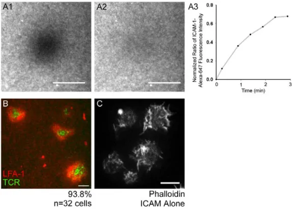
distributes evenly in bilayers (data not shown [DNS]). Moreover, use of Fluorescence Recovery After Photobleaching (FRAP) to assess the lateral mobility of ICAM-1 tagged with Alexa-647 and attached to the bilayer via NTA-conjugated lipids indicated that the lipids in these bilayers are diffusing freely and uniformly (Fig. 3 A1-A3). Finally, after 5 min of engagement with the bilayer, the vast majority (93.8%, n=32 cells; Fig. 3 B) of Jurkat T cells formed the central accumulation of TCR MCs, as inferred from the distribution of the anti-CD3ε antibody in the bilayer, and the peripheral accumulation of the integrin LFA-1, as inferred from the distribution of ICAM-1 in the bilayer, that is characteristic of the bull’s eye-patterned IS formed by primary T cells bound to bilayers containing peptide-MHC (Grakoui et al., 1999).
To image the endogenous network of cortical F-actin at the plane of the IS, Jurkat T cells were stained with rhodamine-phalloidin (Fig. 4 A1-A4; two representative cells are shown; unless indicated otherwise, all fixations were performed 5 min after engagement with the bilayer). This staining revealed three visually distinct rings or zones of F-actin at the IS: an outer ring characterized by very intense F-actin staining interrupted by streaks, a middle ring characterized by concentric arcs of F-actin, and a central zone relatively free of F-actin (Fig. 4 A1 and A3, and the corresponding insets in A2 and A4, respectively: the cell in A1 shows the outer ring most clearly, while the cell in A3 shows the middle ring best).