Non-infectious environmental antigens as a trigger for the
initiation of an autoimmune skin disease
Ye Qiana,*, Donna A. Cultona, Joseph S. Jeonga, Nicole Trupianoa, Jesus G. Valenzuelab,
and Luis A. Diaza
aDepartment of Dermatology, University of North Carolina at Chapel Hill, Chapel Hill, NC 27599,
USA
bVector Molecular Biology Section, LMVR, National Institute of Allergy and Infectious Diseases,
NIH, Rockville, MD 20852, USA
Abstract
Pemphigus represents a group of organ specific autoimmune blistering disorders of the skin mediated by pathogenic autoantibodies with well-defined antigenic targets. While most of these diseases are sporadic, endemic forms of disease do exist. The endemic form of pemphigus foliaceus (also known as fogo selvagem, FS) exhibits epidemiological features that suggest exposure to hematophagous insect bites are a possible precipitating factor of this autoimmune disease, and provides a unique opportunity to study how environmental factors contribute to autoimmune disease development. FS patients and healthy individuals from endemic regions show an autoreactive IgM response that starts in early childhood and becomes restricted to IgG4 autoantibodies in FS patients. In searching for triggering environmental antigens, we have found that IgG4 and IgE autoantibodies from FS patients cross-react with a salivary antigen from sand flies. The presence of these cross-reactive antibodies and antibody genetic analysis confirming that these antibodies evolve from the same naïve B cells provides compelling evidence that this non-infectious environmental antigen could be the initial target of the autoantibody response in FS. Consequently, FS serves as an ideal model to study the impact of environmental antigens in the development of autoimmune disease.
Keywords
Pemphigus foliaceus; Autoantibodies; Environmental antigens; IgG4; IgE
Introduction
Pemphigus encompasses a group of autoimmune blistering diseases of the skin and mucosa. Pemphigus foliaceus (PF) and pemphigus vulgaris (PV) are the two major phenotypes of
*Corresponding author at: Ye Qian, Department of Dermatology, CB# 7287, University of North Carolina at Chapel Hill, Chapel Hill,
NC 27599, USA. Phone#: 919-843-7227. Fax#: 919-966-3898. [email protected].
Publisher's Disclaimer: This is a PDF file of an unedited manuscript that has been accepted for publication. As a service to our
customers we are providing this early version of the manuscript. The manuscript will undergo copyediting, typesetting, and review of
HHS Public Access
Author manuscript
Autoimmun Rev
. Author manuscript; available in PMC 2017 September 01.Published in final edited form as:
Autoimmun Rev. 2016 September ; 15(9): 923–930. doi:10.1016/j.autrev.2016.07.005.
A
uthor Man
uscr
ipt
A
uthor Man
uscr
ipt
A
uthor Man
uscr
ipt
A
uthor Man
uscr
pemphigus [1, 2]. Clinically, PF affects only the skin, whereas PV initially affects mucous membranes (mucosal PV) and later involves the skin (mucocutaneous PV) [1, 3, 4]. The hallmark of all pemphigus phenotypes is the presence of intraepidermal vesicles [1] and anti-epidermal autoantibodies [2, 3, 5-7]. The anti-epidermal autoantibodies are detected bound to the surface of detached keratinocytes in lesional epidermis and circulating in the serum of patients [7]. It has been well demonstrated that these anti-epidermal autoantibodies recognize desmogleins (Dsg), a family of desmosomal cell adhesion glycoproteins [1, 2, 5-7]. The desmosome is a cell-cell adhesion organelle present in keratinocytes and other squamous epithelial cells [8], and consists of core and plaque structural components. The core contains transmembrane glycoproteins including desmogleins and desmocollins that belong to the cadherin superfamily [9, 10]. In humans, four desmogleins have been identified, namely Dsg 1, 2, 3, and 4 and they share high degree of sequence homology [9-16]. Immunologically, the sera of PF patients show anti-Dsg1 antibodies while the sera of PV patients contain antibodies to both Dsg1 and Dsg3 [2, 3, 6]. PV and PF in North
America are sporadic [1, 6], but an endemic form of PF is described in certain states of Brazil, where it is known as Fogo Selvagem (FS) [17, 18]. FS shows similar clinical, histological and immunological features to those observed in non-endemic PF [19, 20].
The Terena Reservation of Limao Verde (LV), Brazil, in particular, has a 3.4% prevalence rate of FS [21]. Epidemiologic and immunogenetic studies suggest that both genetic and environmental factors contribute to the development of FS [17, 22, 23]. Genetically, FS exhibits a strong association with the HLA-DRB1*0102, 0404 and 1402 alleles (p<0.005, RR: 14) and affects people of many races and ethnic backgrounds [22]. Inbreeding, which is a common practice in the endemic areas, may compound the likelihood that genetically predisposed individuals will pass these traits to their offspring. Approximately 93% of familial cases of FS were found in genetically related individuals [24]. While the genetic factors that contribute to disease development are known, the environmental antigens triggering disease development is an evolving field of study.
The term “Exposome” has recently been introduced to describe the environmental factors that are associated with the initiation of autoimmune diseases [25, 26]. Environmental factors include airborne, oral, and percutaneous exposures and may be infectious or noninfectious in nature. The association of infectious agents with the development of autoimmune diseases has been well studied [25, 27-30], and the term “infectome” has been proposed to describe the link between external environmental infectious antigens and the initiation of autoimmune diseases [25-27]. Compared to the studies of the infectious agents, the study of non-infectious environmental antigens and their association with the
development of autoimmune diseases is limited. Unlike infectious antigens that induce a robust IgG immune response, low doses of non-infectious antigens, e.g. allergens, usually induce IgE and IgG4 responses, especially if the routes of exposure are via skin or mucosa. Using the endemic autoimmune skin disease FS as a model, we have identified a possible non-infectious antigen(s) that may initiate this autoimmune skin disease. We propose a model in which non-infectious environmental antigens which cross-react with Dsg1 first induce an IgE response and then trigger the IgG4 autoantibody response that eventually leads to FS in these genetically susceptible individuals.
A
uthor Man
uscr
ipt
A
uthor Man
uscr
ipt
A
uthor Man
uscr
ipt
A
uthor Man
uscr
IgG4 and IgE autoantibodies in autoimmune skin diseases
Since the initial observation of Jones et al [31] on the IgG subclass restriction of PV autoantibodies to the IgG4 and IgG1 subclasses, several studies have confirmed and extended these findings using indirect immunofluorescence (IF) assays [32-34]. The IgG subclass restriction has been additionally confirmed by ELISA studies using recombinant Dsg3 and Dsg1 [35-38]. Studies on PV suggest that IgG4 anti-Dsg3 autoantibodies are pathogenic [37, 39]. This IgG subclass restriction is also observed in PF and FS. In 1989 Rock et al demonstrated through passive transfer experiments in mice that the pathogenic IgG response in FS is predominantly IgG4 [40]. A subsequent study using FS sera collected longitudinally showed that progression from preclinical to clinical stage of the disease is associated with a dramatic rise in IgG4 anti-Dsg1 autoantibodies [41]. Testing of larger cohorts of FS and normal control sera confirmed this observation and established a novel “IgG4-based classifier” [42]. The positive predictive value of the “IgG4-based classifier” is approximately 50%, and can identify individuals in the preclinical stage of FS.
It is well known that increased levels of IgE antibodies are found in human allergic responses, but the role that IgE plays in autoimmune skin diseases has only recently been investigated. Increased levels of IgE antibodies have been well described in patients with bullous pemphigoid (BP), another autoimmune blistering disorder with distinct histology, immunofluorescence, and pathophysiology [43-47]. The pathogenic role of autoantigen-specific IgE in BP has been systematically studied by the Fairley [48, 49] and Zone [50] groups. However, early studies on the IgE autoantibody response in pemphigus are sparse. These initial studies do suggest that IgE autoantibodies are present in some pemphigus patients [43, 51]. In FS patients, potential infestation by intestinal parasites was suggested as an explanation for the presence of IgE antibodies among these patients [51]. Later, Spaeth et al [52] tested 41 PV patients’ sera for IgG subclasses, IgA and IgE autoantibodies against the recombinant Dsg3 ectodomain by immunoblotting and found IgE anti-Dsg3
autoantibodies in 4 patients. Nagel et al. [53] conducted a comprehensive study on 93 PV patients regarding their IgE and IgG4 Dsg3 levels. They found that IgE and IgG4 anti-Dsg3 correlate with disease activity in PV patients and closely associate with active PV [53]. Taken together, these findings along with the known restriction of pathogenic autoantibodies to the IgG4 subclass suggest a possible relationship between the IgG4 and IgE responses in FS.
Indeed, our studies have revealed that FS patients have significantly higher levels of IgE anti-Dsg1 antibodies compared to healthy control individuals from both FS endemic regions and the US, and the levels of IgE anti-Dsg1 correlate with those of anti-Dsg1 IgG4 [54]. Interestingly, the IgE anti-Dsg1 levels from FS patients are also significantly higher than those of PF patients from US and Japan, suggesting there is a difference between FS endemic regions from non-FS endemic regions [54]. Moreover, the sera of FS patients during the pre-clinical and clinical stages of the disease have higher than normal levels of IgE cross-reactive antibodies against self Dsg1 and a sand fly antigen [54-56]. These findings collectively bolster the argument that an environmental exposure may trigger the development of FS.
A
uthor Man
uscr
ipt
A
uthor Man
uscr
ipt
A
uthor Man
uscr
ipt
A
uthor Man
uscr
The link between allergy and autoimmunity
Aside from immunodeficiency, the other two main outcomes of a dysfunctional immune system are allergy and autoimmunity. It is well accepted that different mechanisms govern these two harmful immune responses. However, growing evidence suggests there may be some connections between the development of these two abnormalities [57]. Allergic and autoimmune conditions can coincide in some patients, suggesting the two abnormalities share a common pathophysiologic mechanism [57]. For example, children with rheumatoid arthritis and celiac disease have higher incidence of asthma [58, 59]. The allergens that IgE target in atopic patients can be either exogenous (allergens) or endogenous (autoantigens) [60, 61]. IgE antibodies from atopic dermatitis patients react with human protein [62], and several allergens display shared epitopes with human self-proteins [63, 64]. Using serum from atopic dermatitis patients, IgE reactive human autoantigens have been isolated from a human epithelial cDNA expression library. Importantly, these autoantigens are expressed predominantly in the target organs of atopic dermatitis (skin, lung, etc.) [65, 66].
A link between allergy and autoimmunity are the antibodies, i.e. IgG4 and IgE that are involved in these two processes. The presence of autoantibodies in allergic diseases suggests that an autoimmune response may be playing a role in the pathogenesis of these illnesses [57]. An IgE antibody response is the hallmark in allergic diseases, whereas a parallel IgG4 response may be an attempt to block the allergen-induced inflammation, which is observed during the course of allergen-specific immunotherapy [67]. These observations were confirmed and extended by Gleich et al [68] demonstrating the reciprocal serum values of IgE and IgG4 antibodies in pollen-sensitive patients during the course of immunotherapy [69]. The levels of IgE decreased as the levels of IgG4 increased during the months following immunotherapy. Similar observations have been made from immunotherapy of patients with bee venom hypersensitivity [70] and from heavily exposed beekeepers where there is chronic antigenic stimulation [71]. It has been hypothesized that IgG4 “blocking” antibodies may exert their beneficial effect by mechanisms, such as: a) competition with IgE for antigenic sites on the allergen [72, 73], b) binding Fc receptors on immune cells
(macrophages, B cells, neutrophils, mast cells and basophils) thus triggering
immunomodulatory events [74], and c) behaving as anti-idiotypic antibodies. These diverse effector functions of blocking antibodies may depend on the avidity for the allergen of these molecules. Production of IgG4 antibodies and the decrease of allergen-specific IgE may be modulated by IL-10, which is produced by T cells exposed to the allergen during
immunotherapy [75, 76]. Interestingly, IgG4 antibodies are the main pathogenic
autoantibodies in autoimmune skin diseases, such as pemphigus foliaceus/Fogo selvagem [40], pemphigus vulgaris [77], bullous pemphigoid as detailed above [78]. Importantly, these IgG4 anti-Dsg1 and anti-Dsg3 autoantibodies in PF/FS and PV are pathogenic by passive transfer into mice, recapitulating the human skin disease in the animals [37, 39, 40]. In addition, IgG4 antibodies are also present in numerous autoimmune diseases, including autoimmune pancreatitis [79], Mikulicz’s disease (primary Sjogren’s syndrome) [80], as well as the newly defined IgG4-related diseases [81-83]. Besides the link between IgE and IgG4 antibodies in allergy and autoimmunity, the regulation of the immune system may also connect allergy and autoimmunity. It is suggested that allergy may “relax” the regulation of self-reactive B cells and lead to the autoimmune response [84]. It was also determined that
A
uthor Man
uscr
ipt
A
uthor Man
uscr
ipt
A
uthor Man
uscr
ipt
A
uthor Man
uscr
the polymorphism and dysfunction of transcription factor BACH2 is associated with both allergic and autoimmune diseases [85].
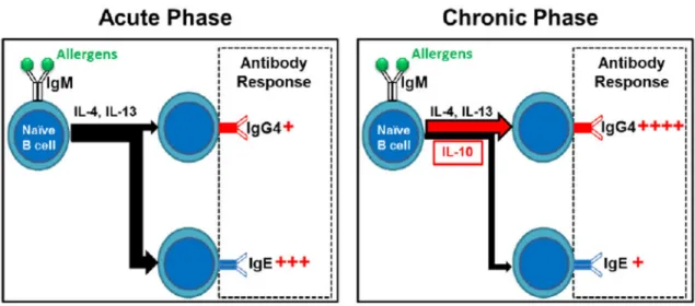
The close relationship between the development of IgE and IgG4 antibodies in humans may be due to the sequential isotype switch from IgM through IgG4 to IgE [86]. The class switch of both isotypes is promoted by Th2 cytokines, such as IL-4 and IL-13. However, chronic antigenic stimulation induces secretion of IL-10 which shifts the balance of the class switch towards IgG4 [87] (Fig. 1). Divergence of IgE to IgG4 is beneficial to patients with allergies. However, in autoimmune skin diseases such as FS, this process may trigger a more robust IgG response, which ultimately leads to the generation of pathogenic IgG4 autoantibodies. Thus, it is possible that an environmental “allergen” triggers IgE response in FS, and the antibodies directed against this “allergen” cross-react with epidermal Dsg1.
Hematophagous insect bites as environmental risk factors in FS
The most intriguing question in autoantibody-mediated autoimmune diseases is what triggers the generation of self-reactive antibodies. FS affects people of all races and ethnicities and is clustered in rural and impoverished communities. FS is endemic to Brazil, but endemic forms of PF have been reported in other countries such as Colombia, and Tunisia [23]. In Brazil, people settled in endemic areas and living in rustic houses with thatched roofs and walls, tend to develop FS [23]. Two Amerindian reservations in Brazil (the Terena reservation of Limao Verde located in the state of Mato Grosso do Sul, and the Xavante Reservation located in the state of Mato Grosso) show a high prevalence of FS. The majority of individuals in these communities are dedicated to outdoor activities such as farming, shrub and tree removal. These human settlements are usually live in close proximity to rivers [23]. The inhabitants are constantly exposed to hematophagous insect bites, such as black flies (simuliids), sand flies (phlebotomines), or kissing bugs
(triatomines). Interestingly, the endemic regions where FS occurs, such as Limao Verde, overlap with endemic regions of Leishmaniasis (sand flies) and Chagas disease [88].
To further study the link between hematophagous insect bites and the occurrence of autoantibodies against Dsg1, we tested several cohorts of sera from FS and other tropical diseases including onchocerciasis, leishmaniasis, Chagas disease and blastomycosis [88]. We found that sera from patients where an insect vector is involved in the transmission of the disease, show anti-Dsg1 antibodies, as compared with other tropical diseases [88]. It should be noted that sera from FS patients do not recognize antigens from the parasites causing the respective tropical diseases (leishmaniasis and Chagas), suggesting that anti-Dsg1 antibodies found in these patients may recognize antigens from the saliva of the insect vector [88]. Based on the results we hypothesized that the saliva from the hematophagous insects could be the source of antigens that trigger cross-reactive autoantibody responses to Dsg1 in FS patients.
Antigen-selection of autoantibody development during and before the onset of FS
Analysis of the V gene sequences of the autoantibodies in pemphigus has yielded important clues for the development of anti-Dsg1 and anti-Dsg3 antibodies in these patients [89-94]. For example, our group and others have found that the development of anti-Dsg3 antibodies
A
uthor Man
uscr
ipt
A
uthor Man
uscr
ipt
A
uthor Man
uscr
ipt
A
uthor Man
uscr
in PV is an antigen-driven response [90, 94]. We also analyzed the H and L chain V genes of anti-Dsg1 IgM and IgG antibodies from 8 FS patients and an healthy individual before the onset of clinical FS (pre-FS) [92]. Besides the overwhelming evidence that the development of anti-Dsg1 autoantibodies in FS are antigen-driven, we have also found that the generation of anti-Dsg1 autoantibodies in pre-FS individuals is also antigen selected [92]. This is evident not only from the shared VH chain V gene usage and shared amino acid replacement mutations among these anti-Dsg1 autoantibodies, but also that 10 out of 20 autoantibody H chains show significant evidence of antigen selection in the pre-FS individual [92] based on the multinomial model for antigen selection analysis [95]. Because this individual did not have active disease at the time of the blood sample collection, the source of the selecting antigens were most likely from the exposure to the environmental antigens, further suggesting the presence of FS associated environmental antigens in FS endemic regions. It should be noted that V genes of these anti-Dsg1 autoantibodies from both FS patients and the pre-FS individual shared some of the same replacement mutations, suggesting that they may be selected by the same environmental- or self-antigen(s) [92].
Distinctive autoreactive IgM antibodies in FS
IgM antibodies are considered to be responsible for the primary immune response against infectious and noninfectious exogenous antigens, and increased levels of IgM (hyper-IgM) in autoimmune diseases are well documented in the literature [96]. It is interesting that a significant number of FS patients and healthy individuals living in endemic areas have IgM anti-Dsg1 as compared with PF patients and healthy controls from either US or Japan [97]. There is no evidence that PF or FS patients have a defect in immunoglobulin class switch [98].
The higher frequency of IgM anti-Dsg1 in FS patients and healthy individuals from endemic areas is likely due to the environmental differences between FS endemic and non-FS endemic regions. It has been reported that FS patients transferred to urban hospitals away from their native environment, show a lower frequency of IgM anti-Dsg1, possibly due to the elimination of environmental antigenic exposure [97]. Ongoing prospective sero-epidemiological studies of three cohorts composed of children and adolescents from Limao Verde show that IgM anti-Dsg1 antibodies are present in children as young as five years of age [97], but IgM anti-Dsg1 antibodies are absent in the cord sera of neonates from mothers from Limao Verde [99]. We were able to document the rise in IgG and pathogenic IgG4 anti-Dsg1 autoantibodies during the transition from the pre-clinical to the clinical stage of FS. These findings suggest the existence of an environmental antigen(s) in endemic areas that sensitizes and triggers anti-Dsg1 autoantibody formation in these individuals.
There are numerous possibilities that could explain the presence of persistent IgM
autoantibodies in these individuals, such as the presence of IgM memory B cells [100]. Due to the unique environment that surrounds the individuals settled in these communities, it is likely that the high level of anti-Dsg1 IgM in FS patients and healthy individuals is due to polyclonal activation of IgM memory B cells [101]. This observation appears to be
confirmed in our studies regarding the antigen selection of anti-Dsg1 antibodies in FS and a
A
uthor Man
uscr
ipt
A
uthor Man
uscr
ipt
A
uthor Man
uscr
ipt
A
uthor Man
uscr
pre-FS individual. We find that less IgM anti-Dsg1 antibodies are antigen selected as compared to those of IgG anti-Dsg1 antibodies in FS patients and a pre-FS individual [92].
IgE and IgG4 responses to a non-infectious environmental antigen in FS endemic regions
Detection of high levels of IgE anti-Dsg1 in FS patients suggest an ongoing exposure to an environmental sensitizing antigen(s) which is present in regions where these patients live [54]. Based on our serological studies on anti-Dsg1 response in patients with insect-borne tropical diseases such as leishmaniasis and Chagas disease [88], we have begun dissect the IgG reactivity of FS IgG and IgG4, containing anti-Dsg1 antibodies, against
well-characterized salivary antigens for Lutzomya longipalpis [102-104]. Our studies demonstrate that FS patients have both IgE and IgG4 antibodies against sand fly salivary gland antigens (SGLL). The levels of IgG4 anti-SGLL antibodies significantly correlate with the that of IgG4 anti-Dsg1 autoantibodies, suggesting that IgG4 reacts to both exogenous and
endogenous antigens might be due to the cross-reactivity of these IgG4 antibodies [55]. The levels of IgG4 anti-SGLL also significantly correlate with that of IgE anti-SGLL, implying that there is a parallel development of IgG4 and IgE against the same environmental antigens which is similar to those observed in the chronic allergen stimulation and allergy
immunotherapy [71]. Further testing using FS sera against main components from SGLL recognized by human, LJM11, LJM17, and LJL143 indicated that the LJM11 component from sand fly is the primary target for FS IgG4 antibodies [55]. The LJM11 antigen, a member of the “yellow family” of salivary proteins is the most immunogenic component among the sand fly saliva proteins and is also a marker for human exposure to sand fly bites [103]. Two IgG4 monoclonal anti-Dsg1 antibodies isolated from FS patients cross-react to both Dsg1 and LJM11 antigens [55]. Furthermore, mice immunized with LJM11, but not LJM17 and LJL143, develop a strong humoral response to human Dsg1 [55]. All these evidence strongly suggest the existence of a “cross-reactive” epitope(s) on both LJM11 and Dsg1. Thus, it is possible that IgE and IgG4 responses directed against the “cross-reactive” epitope(s) initiates the generation of IgG4 autoantibodies in FS susceptible individuals due to the chronic stimulation of non-infectious antigen (LJM11) present in FS endemic regions.
IgE antibodies against environmental antigens may be an early marker for the FS development
It is apparent that IgE anti-LJM11 antibodies are widely present in FS patients [55]. We next explored whether IgE anti-Dsg1 and anti-LJM11 antibodies develop in individuals before the onset of their FS. If it were the case, it would be an indication that they may play an etiological role in the development of autoantibodies and subsequent FS. The development of IgE anti-environmental antigen before the onset of FS would be a useful marker for early detection of individuals at risk of developing FS. We studied samples collected from individuals living in the FS endemic region over many years. We tested serum samples from 12 FS individuals prior to their onset of clinical FS (1 to 4 years), as well as after the development of clinical FS, to determine their serum anti-Dsg1 and anti-LJM11 IgE levels. Compared to normal controls from both LV and US, individuals before and after their onset of FS have significantly higher IgE anti-LJM11 antibodies [56]. There are also significantly higher anti-Dsg1 IgE levels in individuals before and after their onset of FS than normal controls from both LV and US. These results suggest that the IgE antibodies develop well
A
uthor Man
uscr
ipt
A
uthor Man
uscr
ipt
A
uthor Man
uscr
ipt
A
uthor Man
uscr
before the onset of FS (as early as 4 years before) among FS susceptible individuals. However, in contrast to the comparable IgE anti-LJM11 levels from individuals before and after the onset of FS, pre-FS individuals have significantly lower levels of anti-Dsg1 IgE before their onset of FS than after [56]. It indicates that anti-LJM11 antibodies developed earlier in those individuals than that of anti-Dsg1 IgE, suggesting that LJM11 might be the initial target of IgE response. Interestingly, even there is no significant difference between IgE anti-Dsg1 levels of normal controls from both FS (LV) and non-FS (US) endemic regions, normal controls from LV have significantly higher IgE anti-LJM11 antibodies than those from US (p<0.01) [56]. These findings again support that there are environmental differences between these two geographic areas.
Coinciding development of IgG4 antibodies reactive to environmental antigen and autoantigen in FS
A critical question that needs to be answered in any given immune response is whether the response represents non-specific activation of the immune system or an antigen-selected response [105]. Our earlier studies on the autoreactive IgM and IgG antibodies from FS patients indicate that most of the IgG anti-Dsg1 antibodies are antigen selected [92]. However, only two IgG4 monoclonal antibodies, the major subclass of pathogenic autoantibodies in FS, were identified using the hybridoma method, despite the fact that several thousands of hybridomas being screened. Thus, we cannot conclusively state IgG4 autoantibodies are also antigen selected. To further dissect the IgG4 antibody repertoires and the nature of the IgG4 antibody response in FS, we modified the original antibody phage display method [106] to isolate only IgG4 antibodies from FS patient blood samples [107]. Since the antibody phage display libraries that we generate comprise only IgG4 subclass from patients, we can focus on isolating only the potential pathogenic IgG4 antibodies from FS patients. Using this modified antibody phage display method, 136 IgG4 monoclonal antibodies were isolated from three FS patient samples, including 14 clonally independent IgG4 monoclonal antibodies [107]. Among them, two IgG4 monoclonal antibodies originally isolated by hybridoma method were also identified [92, 107]. This method may prove to be a very useful tool for the study of IgG4 antibody repertoires and the mechanism by which IgG4 antibodies develop in patients with the newly categorized IgG4-related diseases [81-83].
As demonstrated, FS patients have IgG4 anti-LJM11 antigen from sand fly saliva, in addition to IgG4 anti-Dsg1 [55, 56]. We sought to further determine if the IgG4 anti-Dsg1 and – LJM11 responses in FS patients are associated. We panned the IgG4 antibody phage display libraries against both Dsg1 and LJM11 antigens [107]. The IgG4 antibody responses towards these two antigens could yield three possible outcomes (Fig. 2). The most likely scenario is that immune responses to either Dsg1 or LJM11 have their own population of IgG4 antibodies, but that the two populations have significant overlap (Fig. 2, outcome 1). There are also two other possible outcomes. First, IgG4 responses to Dsg1 and LJM11 are independent (Fig. 2, outcome 2). Our previous studies [55] indicate that there are cross-reactive antibodies from FS patients, making it unlikely that IgG4 responses to both molecules are completely independent. Second, the IgG4 responses to Dsg1 and LJM11 are the same and all the IgG4 antibodies isolated from panning against Dsg1 or LJM11 are
A
uthor Man
uscr
ipt
A
uthor Man
uscr
ipt
A
uthor Man
uscr
ipt
A
uthor Man
uscr
cross-reactive (Fig. 2, outcome 3). Dsg1 and LJM11 are evolutionary distant molecules and share little primary sequence homology. Furthermore, the cross-reactive monoclonal antibodies from FS bind both molecules via conformational epitope [55]. Therefore, the chance that all the conformational epitopes on both molecules are the same seems unlikely. The analysis of the IgG4 antibodies directed against either Dsg1 or LJM11 enabled us to disclose the degree of the overlap between these two IgG4 responses, and as a result, the significance of how IgG4 response to environmental antigens contributes to the development of IgG4 autoreactive antibodies in FS.
Based on the H chain complementary determining region 3 (CDR3) sequence identity, or “clonal signature” [108], whether a group of IgG4 antibodies are derived from the same parental B cells, or whether they are clonally related, could be conclusively determined [107]. The analyses of the H chain V genes and the clonal signature of all identified IgG4 monoclonal antibodies from the panning of these three IgG4 antibody phage display libraries indicated that they could be grouped into a 14 lineages of clones (that originated from the same parental B cells) according to their CDR3 clonal signatures [107]. All of these IgG4 monoclonal antibodies are cross-reactive to both Dsg1 and LJM11 as determined by either Dsg1/LJM11 alternate panning, anti-Dsg1 and anti-LJM11 ELISA, or both [107]. It seems that the findings from panning of the IgG4 antibody phage display libraries fit the Outcome 3 shown in Fig 2. However, as elucidated above, it is unlikely that all IgG4 antibodies from FS are cross-reactive to both Dsg1 and LJM11. The possible explanations are, 1. The panning of IgG4 phage display libraries only captures those abundant IgG4 antibodies, while rare clones (Dsg1 or LJM11 specific or not cross-reactive) escape detection [107]; 2. The IgG4 antibodies identified from IgG4 phage display libraries only represent those IgG4 committed B cells in patient blood samples, but not those terminally differentiated plasma cells in FS patients that secrete IgG4 autoantibodies that represent the IgG4 antibodies in patients’ sera.
Another finding is that all monoclonal antibodies isolated from the libraries are extensively mutated as their homology to their germline V genes are low. Replacement mutations in the complementary determining regions are significantly higher compared to the framework regions, which harbor mostly silent mutations [107]. Thus, similar to our earlier studies that anti-Dsg1 IgG autoantibodies are antigen selected [92], these potential pathogenic IgG4 antibodies in FS are also antigen selected [107]. Furthermore, revertant monoclonal antibodies, which represent the germline configuration (expressed by naïve B cells) of these cross-reactive monoclonal antibodies, also recognize both Dsg1 and LJM11 [107].
Collectively, this evidence strongly suggests that LJM11 is an inciting environmental antigen that activates the naïve B cells for the succeeding autoantibody development and ensuing FS among FS-susceptible individuals in FS endemic regions. However, our findings regarding the role of LJM11 as an environmental antigen does not exclude in any way that other environmental antigens present in FS endemic regions could also serve as inciting antigens for the development of FS.
A
uthor Man
uscr
ipt
A
uthor Man
uscr
ipt
A
uthor Man
uscr
ipt
A
uthor Man
uscr
Conclusion
Taken together, our extensive studies on FS patients and healthy individuals living in endemic areas allow us to draw several lines of evidence that point to salivary gland protein components from the sand fly, as the trigger of FS. These studies, using FS as a model of an environmentally induced autoimmune disease, demonstrate that non-infectious antigens can precipitate harmful autoantibodies to the host. Antigen-mimicry may play an important role. The possible mechanism by which environmental antigens could trigger the development of anti-Dsg1 in FS-susceptible individuals is depicted in Fig. 3. The environmental antigen (such as LJM11) as an inciting antigen stimulates naïve B cells to undergo class switch and generates the initial IgE response directed against LJM11. Because of the presence of “cross-reactive” epitopes on LJM11 and Dsg1, the initial antibody response, such as IgE response, also reacts to Dsg1. The chronic stimulation of LJM11 antigen and the production of IL10 by the immune system promotes the development of IgG4 antibodies, which are also cross-reactive to both LJM11 and Dsg1. Those IgG4 antibodies are either pathogenic in FS, or they trigger the further development of IgG4 autoantibodies via epitope spreading [109] to develop pathogenic autoantibodies and subsequent FS in susceptible individuals. Our findings support the hypothesis that non-infectious environmental antigens may trigger autoimmune disease and pave the way for similar studies in other autoimmune diseases.
Acknowledgments
This work was supported by NIH grants, K01-AR056378, R01 AR067315 (to YQ) and R01-AR32599 (to LAD).
References
1. Lever W. Pemphigus. Medicine (Baltimore). 1953; 32:1–123. [PubMed: 13024494]
2. Amagai M. Desmoglein as a target in autoimmunity and infection. J Am Acad Dermatol. 2003; 48:244–52. [PubMed: 12582396]
3. Ding X, Aoki V, Mascaro JM Jr, Lopez-Swiderski A, Diaz LA, Fairley JA. Mucosal and mucocutaneous (generalized) pemphigus vulgaris show distinct autoantibody profiles. J Invest Dermatol. 1997; 109:592–6. [PubMed: 9326396]
4. Mustafa MB, Porter SR, Smoller BR, Sitaru C. Oral mucosal manifestations of autoimmune skin diseases. Autoimmun Rev. 2015; 14:930–51. [PubMed: 26117595]
5. Anhalt G, Diaz L. Prospects for autoimmune disease: Research advances in pemphigus. JAMA. 2001; 285:652–4. [PubMed: 11176877]
6. Udey MC, Stanley JR. Pemphigus--diseases of antidesmosomal autoimmunity. JAMA. 1999; 282:572–6. [PubMed: 10450720]
7. Beutner EH, Jordon RE. Demonstration of Skin Antibodies in Sera of Pemphigus Vulgaris Patients by Indirect Immunofluorescent Staining. Proc Soc Exp Biol Med. 1964; 117:505–10. [PubMed: 14233481]
8. Garrod DR, Merritt AJ, Nie Z. Desmosomal adhesion: structural basis, molecular mechanism and regulation (Review). Mol Membr Biol. 2002; 19:81–94. [PubMed: 12126234]
9. Goodwin L, Hill J, Raynor K, Raszi L, Manabe M, Cowin P. Desmoglein shows extensive homology to the cadherin family of cell adhesion molecules. Biochem Biophys Res Commun. 1990;
173:1224–30. [PubMed: 1702628]
10. Koch P, Franke W. Desmosomal cadherins: another growing multigene family of adhesion molecules. Curr Opin Cell Biol. 1994; 6:682–7. [PubMed: 7833048]
11. Wheeler G, Parker A, Thomas C, Ataliotis P, Poynter D, Arnemann J, Rutman A, Pidsley S, Watt F, Rees D. Desmosomal Glycoprotein DGI, a Component of Intercellular Desmosome Junctions, is
A
uthor Man
uscr
ipt
A
uthor Man
uscr
ipt
A
uthor Man
uscr
ipt
A
uthor Man
uscr
Related to the Cadherin Family of Cell Adhesion Molecules. Proc Natl Acad Sci U S A. 1991; 88:4796–4800. [PubMed: 1711210]
12. Nilles LA, Parry DA, Powers EE, Angst BD, Wagner RM, Green KJ. Structural analysis and expression of human desmoglein: a cadherin-like component of the desmosome. J Cell Sci. 1991; 99(Pt 4):809–21. [PubMed: 1770008]
13. Schafer S, Koch P, Franke W. Identification of the ubiquitous human desmoglein, Dsg2, and the expression catalogue of the desmoglein subfamily of desmosomal cadherins. Exp Cell Res. 1994; 211:391–9. [PubMed: 8143788]
14. Amagai M, Klaus-Kovtun V, Stanley J. Autoantibodies against a novel epithelial cadherin in pemphigus vulgaris, a disease of cell adhesion. Cell. 1991; 67:869–877. [PubMed: 1720352] 15. Kljuic A, Bazzi H, Sundberg J, Martinez-Mir A, O’Shaughnessy R, Mahoney M, Levy M,
Montagutelli X, Ahmad W, Aita V. Desmoglein 4 in Hair Follicle Differentiation and Epidermal Adhesion Evidence from Inherited Hypotrichosis and Acquired Pemphigus Vulgaris. Cell. 2003; 113:249–260. [PubMed: 12705872]
16. Whittock N, Bower C. Genetic Evidence for a Novel Human Desmosomal Cadherin, Desmoglein 4. J Invest Dermatol. 2003; 120:523. [PubMed: 12648213]
17. Diaz LA, Sampaio SA, Rivitti EA, Martins CR, Cunha PR, Lombardi C, Almeida FA, Castro RM, Macca ML, Lavrado C, et al. Endemic pemphigus foliaceus (Fogo Selvagem): II. Current and historic epidemiologic studies. J Invest Dermatol. 1989; 92:4–12. [PubMed: 2642512]
18. Empinotti J, Diaz L, Martins C, Rivitti E, Sampaio S, Lombardi C, Sanches J. Endemic pemphigus foliaceus in western Parana, Brazil (1976-1988). Cooperative Group for Fogo Selvagem Research. Br J Dermatol. 1990; 123:431–7. [PubMed: 2095173]
19. Diaz L, Sampaio S, Rivitti E, Martins C, Cunha P, Lombardi C, Almeida F, Castro R, Macca M, Lavrado C. Endemic pemphigus foliaceus (fogo selvagem). I. Clinical features and
immunopathology. J Am Acad Dermatol. 1989; 20:657–69. [PubMed: 2654208]
20. Stanley J, Klaus-Kovtun V, Sampaio S. Antigenic Specificity of Fogo Selvagem Autoantibodies Is Similar to North American Pemphigus Foliaceus and Distinct from Pemphigus Vulgaris
Autoantibodies. J Invest Dermatol. 1986; 87:197–201. [PubMed: 3525686]
21. Hans-Filho G, dos Santos V, Katayama JH, Aoki V, Rivitti EA, Sampaio SA, Friedman H, Moraes JR, Moraes ME, Eaton DP, Lopez AL, Hoffman RG, Fairley JA, Giudice GJ, Diaz LA. An active focus of high prevalence of fogo selvagem on an Amerindian reservation in Brazil. Cooperative Group on Fogo Selvagem Research. J Invest Dermatol. 1996; 107:68–75. [PubMed: 8752842] 22. Moraes ME, Fernandez-Vina M, Lazaro A, Diaz LA, Hans-Filho G, Friedman H, Rivitti EA, Aoki
V, Stastny P, Moraes JR. An epitope in the third hypervariable region of the DRB1 gene is involved in the susceptibility to endemic pemphigus foliaceus (fogo selvagem) in three different Brazilian populations. Tissue Antigens. 1997; 49:35–40. [PubMed: 9027963]
23. Aoki V, Millikan RC, Rivitti EA, Hans-Filho G, Eaton DP, Warren SJ, Li N, Hilario-Vargas J, Hoffmann RG, Diaz LA. Environmental risk factors in endemic pemphigus foliaceus (fogo selvagem). J Investig Dermatol Symp Proc. 2004; 9:34–40.
24. Aoki V, Huang MH, Perigo AM, Fukumori LM, Maruta CW, Santi CG, Oliveira ZN, Rivitti E. Endemic pemphigus foliaceus (fogo selvagem) and pemphigus vulgaris: immunoglobulin G heterogeneity detected by indirect immunofluorescence. Rev Hosp Clin Fac Med Sao Paulo. 2004; 59:251–6. [PubMed: 15543395]
25. Bogdanos DP, Smyk DS, Invernizzi P, Rigopoulou EI, Blank M, Pouria S, Shoenfeld Y. Infectome: a platform to trace infectious triggers of autoimmunity. Autoimmun Rev. 2013; 12:726–40. [PubMed: 23266520]
26. Colafrancesco S, Agmon-Levin N, Perricone C, Shoenfeld Y. Unraveling the soul of autoimmune diseases: pathogenesis, diagnosis and treatment adding dowels to the puzzle. Immunol Res. 2013; 56:200–5. [PubMed: 23733136]
27. Bogdanos DP, Smyk DS, Invernizzi P, Rigopoulou EI, Blank M, Sakkas L, Pouria S, Shoenfeld Y. Tracing environmental markers of autoimmunity: introducing the infectome. Immunol Res. 2013; 56:220–40. [PubMed: 23592050]
28. Miller FW, Alfredsson L, Costenbader KH, Kamen DL, Nelson LM, Norris JM, De Roos AJ. Epidemiology of environmental exposures and human autoimmune diseases: findings from a
A
uthor Man
uscr
ipt
A
uthor Man
uscr
ipt
A
uthor Man
uscr
ipt
A
uthor Man
uscr
National Institute of Environmental Health Sciences Expert Panel Workshop. J Autoimmun. 2012; 39:259–71. [PubMed: 22739348]
29. Germolec D, Kono DH, Pfau JC, Pollard KM. Animal models used to examine the role of the environment in the development of autoimmune disease: findings from an NIEHS Expert Panel Workshop. J Autoimmun. 2012; 39:285–93. [PubMed: 22748431]
30. Fierabracci A. Unravelling the role of infectious agents in the pathogenesis of human autoimmunity: the hypothesis of the retroviral involvement revisited. Curr Mol Med. 2009; 9:1024–33. [PubMed: 19747107]
31. Jones CC, Hamilton RG, Jordon RE. Subclass distribution of human IgG autoantibodies in pemphigus. J Clin Immunol. 1988; 8:43–9. [PubMed: 3284896]
32. Brooks W, Lee Y, Abell E, Deng J. Comparison of IgG subclasses and complement binding activity of autoantibodies from patients with bullous pemphigoid and pemphigus. J Clin Lab Anal. 1989; 3:307–11. [PubMed: 2681622]
33. Yamada H, Hashimoto T, Nishikawa T. IgG Subclasses of Intercellular and Basement Membrane Zone Antibodies: The Relationship to the Capability of Complement Fixation. J Invest Dermatol. 1989; 92:585–587. [PubMed: 2649595]
34. David M, Katzenelson V, Hazaz B, Ben-Chetrit A, Sandbank M. Determination of IgG subclasses in patients with pemphigus with active disease and in remission. Arch Dermatol. 1989; 125:787– 90. [PubMed: 2658844]
35. Bhol K, Natarajan K, Nagarwalla N, Mohimen A, Aoki V, Ahmed A. Correlation of Peptide Specificity and IgG Subclass with Pathogenic and Nonpathogenic Autoantibodies in Pemphigus Vulgaris: A Model for Autoimmunity. Proc Natl Acad Sci U S A. 1995; 92:5239–5243. [PubMed: 7761479]
36. Hacker MK, Janson M, Fairley JA, Lin MS. Isotypes and antigenic profiles of pemphigus foliaceus and pemphigus vulgaris autoantibodies. Clin Immunol. 2002; 105:64–74. [PubMed: 12483995] 37. Yeh SW, Cavacini LA, Bhol KC, Lin MS, Kumar M, Duval M, Posner MR, Ahmed AR.
Pathogenic human monoclonal antibody against desmoglein 3. Clin Immunol. 2006; 120:68–75. [PubMed: 16635589]
38. Torzecka JD, Wozniak K, Kowalewski C, Waszczykowska E, Sysa-Jedrzejowska A, Pas HH, Narbutt J. Circulating pemphigus autoantibodies in healthy relatives of pemphigus patients: coincidental phenomenon with a risk of disease development? Arch Dermatol Res. 2007; 299:239– 43. [PubMed: 17534636]
39. Parlowsky T, Welzel J, Amagai M, Zillikens D, Wygold T. Neonatal pemphigus vulgaris: IgG4 autoantibodies to desmoglein 3 induce skin blisters in newborns. J Am Acad Dermatol. 2003; 48:623–5. [PubMed: 12664033]
40. Rock B, Martins CR, Theofilopoulos AN, Balderas RS, Anhalt GJ, Labib RS, Futamura S, Rivitti EA, Diaz LA. The pathogenic effect of IgG4 autoantibodies in endemic pemphigus foliaceus (fogo selvagem). N Engl J Med. 1989; 320:1463–9. [PubMed: 2654636]
41. Warren S, Arteaga L, Rivitti E, Aoki V, Hans-Filho G, Qaqish B, Lin M, Giudice G, Diaz L. The Role of Subclass Switching in the Pathogenesis of Endemic Pemphigus Foliaceus. J Invest Dermatol. 2003; 120:104–108. [PubMed: 12535205]
42. Qaqish BF, Prisayanh P, Qian Y, Andraca E, Li N, Aoki V, Hans-Filho G, dos Santos V, Rivitti EA, Diaz LA. Development of an IgG4-based predictor of endemic pemphigus foliaceus (fogo selvagem). J Invest Dermatol. 2009; 129:110–8. [PubMed: 18704107]
43. Arbesman C, Wypych J, Reisman R, Beutner E. IgE levels in sera of patients with pemphigus or bullous pemphigoid. Arch Dermatol. 1974; 110:378–81. [PubMed: 4217592]
44. Bowszyc-Dmochowska M, Dmochowski M. Immediate hypersensitivity phenomena in bullous pemphigoid: critical concepts. J Med. 2002; 33:189–98. [PubMed: 12939118]
45. Christophoridis S, Budinger L, Borradori L, Hunziker T, Merk HF, Hertl M. IgG, IgA and IgE autoantibodies against the ectodomain of BP180 in patients with bullous and cicatricial pemphigoid and linear IgA bullous dermatosis. Br J Dermatol. 2000; 143:349–55. [PubMed: 10951144]
A
uthor Man
uscr
ipt
A
uthor Man
uscr
ipt
A
uthor Man
uscr
ipt
A
uthor Man
uscr
46. Delaporte E, Dubost-Brama A, Ghohestani R, Nicolas JF, Neyrinck JL, Bergoend H, Janin A, Capron M. IgE autoantibodies directed against the major bullous pemphigoid antigen in patients with a severe form of pemphigoid. J Immunol. 1996; 157:3642–7. [PubMed: 8871665]
47. Dimson OG, Giudice GJ, Fu CL, Van den Bergh F, Warren SJ, Janson MM, Fairley JA. Identification of a potential effector function for IgE autoantibodies in the organ-specific autoimmune disease bullous pemphigoid. J Invest Dermatol. 2003; 120:784–8. [PubMed: 12713582]
48. Fairley JA, Burnett CT, Fu CL, Larson DL, Fleming MG, Giudice GJ. A Pathogenic Role for IgE in Autoimmunity: Bullous Pemphigoid IgE Reproduces the Early Phase of Lesion Development in Human Skin Grafted to nu/nu Mice. J Invest Dermatol. 2007
49. Fairley JA, Fu CL, Giudice GJ. Mapping the binding sites of anti-BP180 immunoglobulin E autoantibodies in bullous pemphigoid. J Invest Dermatol. 2005; 125:467–72. [PubMed: 16117787] 50. Zone JJ, Taylor T, Hull C, Schmidt L, Meyer L. IgE basement membrane zone antibodies induce
eosinophil infiltration and histological blisters in engrafted human skin on SCID mice. J Invest Dermatol. 2007; 127:1167–74. [PubMed: 17235329]
51. Bruns GR, Ablin RJ, Guinan PD. Serum immunoglobulin E in pemphigus. J Invest Dermatol. 1978; 71:217–8. [PubMed: 690486]
52. Spaeth S, Riechers R, Borradori L, Zillikens D, Büdinger L, Hertl M. IgG, IgA and IgE autoantibodies against the ectodomain of desmoglein 3 in active pemphigus vulgaris. Br J Dermatol. 2001; 144:1183–1188. [PubMed: 11422039]
53. Nagel A, Lang A, Engel D, Podstawa E, Hunzelmann N, de Pita O, Borradori L, Uter W, Hertl M. Clinical activity of pemphigus vulgaris relates to IgE autoantibodies against desmoglein 3. Clin Immunol. 2009; 134:320–30. [PubMed: 20015693]
54. Qian Y, Prisayanh P, Andraca E, Qaqish BF, Aoki V, Hans-Filhio G, Rivitti EA, Diaz LA. IgE, IgM, and IgG4 anti-desmoglein 1 autoantibody profile in endemic pemphigus foliaceus (fogo selvagem). J Invest Dermatol. 2011; 131:985–7. [PubMed: 21191415]
55. Qian Y, Jeong JS, Maldonado M, Valenzuela JG, Gomes R, Teixeira C, Evangelista F, Qaqish B, Aoki V, Hans G Jr, Rivitti EA, Eaton D, Diaz LA. Cutting Edge: Brazilian Pemphigus Foliaceus Anti-Desmoglein 1 Autoantibodies Cross-React with Sand Fly Salivary LJM11 Antigen. J Immunol. 2012; 189:1535–9. [PubMed: 22798673]
56. Qian Y, Jeong JS, Abdeladhim M, Valenzuela JG, Aoki V, Hans-Filhio G, Rivitti EA, Diaz LA. IgE Anti-LJM11 Sand Fly Salivary Antigen May Herald the Onset of Fogo Selvagem in Endemic Brazilian Regions. J Invest Dermatol. 2015; 135:913–5. [PubMed: 25285921]
57. Rottem M, Gershwin ME, Shoenfeld Y. Allergic disease and autoimmune effectors pathways. Dev Immunol. 2002; 9:161–7. [PubMed: 12885156]
58. Kero J, Gissler M, Hemminki E, Isolauri E. Could TH1 and TH2 diseases coexist? Evaluation of asthma incidence in children with coeliac disease, type 1 diabetes, or rheumatoid arthritis: a register study. J Allergy Clin Immunol. 2001; 108:781–3. [PubMed: 11692104]
59. Simpson CR, Anderson WJ, Helms PJ, Taylor MW, Watson L, Prescott GJ, Godden DJ, Barker RN. Coincidence of immune-mediated diseases driven by Th1 and Th2 subsets suggests a common aetiology. A population-based study using computerized general practice data. Clin Exp Allergy. 2002; 32:37–42. [PubMed: 12002734]
60. Valenta R, Natter S, Seiberler S, Grote M. Isolation of cDNAs coding for IgE autoantigens: a link between atopy and autoimmunity. Int Arch Allergy Immunol. 1997; 113:209–12. [PubMed: 9130525]
61. Valenta R, Seiberler S, Natter S, Mahler V, Mossabeb R, Ring J, Stingl G. Autoallergy: A pathogenetic factor in atopic dermatitis? J Allergy Clin Immunol. 2000; 105:432–437. [PubMed: 10719290]
62. Valenta R, Maurer D, Steiner R, Seiberler S, Sperr W, Valent P, Spitzauer S, Kapiotis S, Smolen J, Stingl G. Immunoglobulin E Response to Human Proteins in Atopic Patients. J Invest Dermatol. 1996; 107:203–208. [PubMed: 8757763]
63. Crameri R, Faith A, Hemmann S, Jaussi R, Ismail C, Menz G, Blaser K. Humoral and cell-mediated autoimmunity in allergy to Aspergillus fumigatus. J Exp Med. 1996; 184:265–70. [PubMed: 8691141]
A
uthor Man
uscr
ipt
A
uthor Man
uscr
ipt
A
uthor Man
uscr
ipt
A
uthor Man
uscr
64. Valenta R, Duchene M, Pettenburger K, Sillaber C, Valent P, Bettelheim P, Breitenbach M, Rumpold H, Kraft D, Scheiner O. Identification of profilin as a novel pollen allergen; IgE autoreactivity in sensitized individuals. Science. 1991; 253:557–60. [PubMed: 1857985] 65. Natter S, Seiberler S, Hufnagl P, Binder BR, Hirschl AM, Ring J, Abeck D, Schmidt T, Valent P,
Valenta R. Isolation of cDNA clones coding for IgE autoantigens with serum IgE from atopic dermatitis patients. FASEB J. 1998; 12:1559–69. [PubMed: 9806765]
66. Valenta R, Natter S, Seiberler S, Wichlas S, Maurer D, Hess M, Pavelka M, Grote M, Ferreira F, Szepfalusi Z, Valent P, Stingl G. Molecular characterization of an autoallergen, Hom s 1, identified by serum IgE from atopic dermatitis patients. J Invest Dermatol. 1998; 111:1178–83. [PubMed: 9856836]
67. Lichtenstein L, Holtzman N, Burnett L. A Quantitative in Vitro Study of the Chromatographic Distribution and Immunoglobulin Characteristics of Human Blocking Antibody. J Immunol. 1968; 101:317. [PubMed: 4174497]
68. Gleich G, Zimmermann E, Henderson L, Yunginger J. Effect of immunotherapy on
immunoglobulin E and immunoglobulin G antibodies to ragweed antigens: a six-year prospective study. J Allergy Clin Immunol. 1982; 70:261–71. [PubMed: 6811645]
69. Rossi R, Monasterolo G, Coco G, Silvestro L, Operti D. Evaluation of serum IgG4 antibodies specific to grass pollen allergen components in the follow up of allergic patients undergoing subcutaneous and sublingual immunotherapy. Vaccine. 2007; 25:957–964. [PubMed: 17045368] 70. Golden D, Lawrence I, Hamilton R, Kagey-Sobotka A, Valentine M, Lichtenstein L. Clinical
correlation of the venom-specific IgG antibody level during maintenance venom immunotherapy. J Allergy Clin Immunol. 1992; 90:386–93. [PubMed: 1527321]
71. Aalberse RC, van der Gaag R, van Leeuwen J. Serologic aspects of IgG4 antibodies. I. Prolonged immunization results in an IgG4-restricted response. J Immunol. 1983; 130:722–6. [PubMed: 6600252]
72. Larche M, Akdis C, Valenta R. Immunological mechanisms of allergen-specific immunotherapy. Nat Rev Immunol. 2006; 6:761–71. [PubMed: 16998509]
73. Larche M. Update on the current status of peptide immunotherapy. J Allergy Clin Immunol. 2007; 119:906–9. [PubMed: 17418662]
74. Strait R, Morris S, Finkelman F. IgG-blocking antibodies inhibit IgE-mediated anaphylaxis in vivo through both antigen interception and Fc?RIIb cross-linking. J Clin Invest. 2006; 116:833–841. [PubMed: 16498503]
75. Nouri-Aria K, Wachholz P, Francis J, Jacobson M, Walker S, Wilcock L, Staple S, Aalberse R, Till S, Durham S. Grass Pollen Immunotherapy Induces Mucosal and Peripheral IL-10 Responses and Blocking IgG Activity 1. J Immunol. 2004; 172:3252–3259. [PubMed: 14978133]
76. Akdis CA, Blaser K, Akdis M. Mechanisms of allergen-specific immunotherapy. Chem Immunol Allergy. 2006; 91:195–203. [PubMed: 16354960]
77. Bhol K, Natarajan K, Nagarwalla N, Mohimen A, Aoki V, Ahmed AR. Correlation of peptide specificity and IgG subclass with pathogenic and nonpathogenic autoantibodies in pemphigus vulgaris: a model for autoimmunity. Proc Natl Acad Sci U S A. 1995; 92:5239–43. [PubMed: 7761479]
78. Bernard P, Prost C, Aucouturier P, Durepaire N, Denis F, Bonnetblanc JM. The subclass
distribution of IgG autoantibodies in cicatricial pemphigoid and epidermolysis bullosa acquisita. J Invest Dermatol. 1991; 97:259–63. [PubMed: 2071938]
79. Kamisawa T, Okamoto A. Autoimmune pancreatitis: proposal of IgG4-related sclerosing disease. J Gastroenterol. 2006; 41:613–25. [PubMed: 16932997]
80. Yamamoto M, Takahashi H, Sugai S, Imai K. Clinical and pathological characteristics of Mikulicz’s disease (IgG4-related plasmacytic exocrinopathy). Autoimmun Rev. 2005; 4:195–200. [PubMed: 15893711]
81. Stone JH, Zen Y, Deshpande V. IgG4-related disease. N Engl J Med. 2012; 366:539–51. [PubMed: 22316447]
82. Brito-Zeron P, Ramos-Casals M, Bosch X, Stone JH. The clinical spectrum of IgG4-related disease. Autoimmun Rev. 2014; 13:1203–10. [PubMed: 25151972]
A
uthor Man
uscr
ipt
A
uthor Man
uscr
ipt
A
uthor Man
uscr
ipt
A
uthor Man
uscr
83. Islam AD, Selmi C, Datta-Mitra A, Sonu R, Chen M, Gershwin ME, Raychaudhuri SP. The changing faces of IgG4-related disease: Clinical manifestations and pathogenesis. Autoimmun Rev. 2015; 14:914–22. [PubMed: 26112170]
84. Astrakhan A, Omori M, Nguyen T, Becker-Herman S, Iseki M, Aye T, Hudkins K, Dooley J, Farr A, Alpers CE, Ziegler SF, Rawlings DJ. Local increase in thymic stromal lymphopoietin induces systemic alterations in B cell development. Nat Immunol. 2007; 8:522–31. [PubMed: 17401368] 85. Roychoudhuri R, Hirahara K, Mousavi K, Clever D, Klebanoff CA, Bonelli M, Sciume G, Zare H,
Vahedi G, Dema B, Yu Z, Liu H, Takahashi H, Rao M, Muranski P, Crompton JG, Punkosdy G, Bedognetti D, Wang E, Hoffmann V, Rivera J, Marincola FM, Nakamura A, Sartorelli V, Kanno Y, Gattinoni L, Muto A, Igarashi K, O’Shea JJ, Restifo NP. BACH2 represses effector programs to stabilize T(reg)-mediated immune homeostasis. Nature. 2013; 498:506–10. [PubMed: 23728300] 86. Jabara H, Loh R, Ramesh N, Vercelli D, Geha RS. Sequential switching from mu to epsilon via
gamma 4 in human B cells stimulated with IL-4 and hydrocortisone. J Immunol. 1993; 151:4528– 33. [PubMed: 8409416]
87. Jeannin P, Lecoanet S, Delneste Y, Gauchat J, Bonnefoy J. IgE Versus IgG4 Production Can Be Differentially Regulated by IL-10. J Immunol. 1998; 160:3555–3561. [PubMed: 9531318] 88. Diaz LA, Arteaga LA, Hilario-Vargas J, Valenzuela JG, Li N, Warren S, Aoki V, Hans-Filho G,
Eaton D, dos Santos V, Nutman TB, de Mayolo AA, Qaqish BF, Sampaio SA, Rivitti EA. Anti-desmoglein-1 antibodies in onchocerciasis, leishmaniasis and Chagas disease suggest a possible etiological link to Fogo selvagem. J Invest Dermatol. 2004; 123:1045–51. [PubMed: 15610512] 89. Payne AS, Ishii K, Kacir S, Lin C, Li H, Hanakawa Y, Tsunoda K, Amagai M, Stanley JR, Siegel
DL. Genetic and functional characterization of human pemphigus vulgaris monoclonal
autoantibodies isolated by phage display. J Clin Invest. 2005; 115:888–99. [PubMed: 15841178] 90. Qian Y, Diaz LA, Ye J, Clarke SH. Dissecting the anti-desmoglein autoreactive B cell repertoire in
pemphigus vulgaris patients. J Immunol. 2007; 178:5982–90. [PubMed: 17442983] 91. Ishii K, Lin C, Siegel DL, Stanley JR. Isolation of pathogenic monoclonal anti-desmoglein 1
human antibodies by phage display of pemphigus foliaceus autoantibodies. J Invest Dermatol. 2008; 128:939–48. [PubMed: 18007588]
92. Qian Y, Clarke SH, Aoki V, Hans-Filhio G, Rivitti EA, Diaz LA. Antigen selection of anti-DSG1 autoantibodies during and before the onset of endemic pemphigus foliaceus. J Invest Dermatol. 2009; 129:2823–34. [PubMed: 19571823]
93. Yamagami J, Payne AS, Kacir S, Ishii K, Siegel DL, Stanley JR. Homologous regions of autoantibody heavy chain complementarity-determining region 3 (H-CDR3) in patients with pemphigus cause pathogenicity. J Clin Invest. 2010; 120:4111–7. [PubMed: 20978359] 94. Cho MJ, Lo AS, Mao X, Nagler AR, Ellebrecht CT, Mukherjee EM, Hammers CM, Choi EJ,
Sharma PM, Uduman M, Li H, Rux AH, Farber SA, Rubin CB, Kleinstein SH, Sachais BS, Posner MR, Cavacini LA, Payne AS. Shared VH1-46 gene usage by pemphigus vulgaris autoantibodies indicates common humoral immune responses among patients. Nat Commun. 2014; 5:4167. [PubMed: 24942562]
95. Lossos IS, Tibshirani R, Narasimhan B, Levy R. The inference of antigen selection on Ig genes. J Immunol. 2000; 165:5122–6. [PubMed: 11046043]
96. Duarte-Rey C, Bogdanos DP, Leung PS, Anaya JM, Gershwin ME. IgM predominance in autoimmune disease: genetics and gender. Autoimmun Rev. 2012; 11:A404–12. [PubMed: 22178509]
97. Diaz LA, Prisayanh PS, Dasher DA, Li N, Evangelista F, Aoki V, Hans-Filho G, dos Santos V, Qaqish BF, Rivitti EA. The IgM anti-desmoglein 1 response distinguishes Brazilian pemphigus foliaceus (fogo selvagem) from other forms of pemphigus. J Invest Dermatol. 2008; 128:667–75. [PubMed: 17960181]
98. Notarangelo LD, Lanzi G, Peron S, Durandy A. Defects of class-switch recombination. J Allergy Clin Immunol. 2006; 117:855–64. [PubMed: 16630945]
99. Hilario-Vargas J, Vitorio IB, Stamey C, Culton DA, Prisayanh P, Rivitti EA, Aoki V, Filho GH, Dos Santos V, Qaqish B, Diaz LA. Analysis of Anti-desmoglein 1 Autoantibodies in 68 Healthy Mother/Neonate Pairs from a Highly Endemic Region of Fogo Selvagem in Brazil. J Clin Exp Dermatol Res. 2014; 5
A
uthor Man
uscr
ipt
A
uthor Man
uscr
ipt
A
uthor Man
uscr
ipt
A
uthor Man
uscr
100. Reynaud CA, Descatoire M, Dogan I, Huetz F, Weller S, Weill JC. IgM memory B cells: a mouse/ human paradox. Cell Mol Life Sci. 2012; 69:1625–34. [PubMed: 22481437]
101. Bernasconi NL, Traggiai E, Lanzavecchia A. Maintenance of serological memory by polyclonal activation of human memory B cells. Science. 2002; 298:2199–202. [PubMed: 12481138] 102. Oliveira F, Kamhawi S, Seitz AE, Pham VM, Guigal PM, Fischer L, Ward J, Valenzuela JG. From
transcriptome to immunome: identification of DTH inducing proteins from a Phlebotomus ariasi salivary gland cDNA library. Vaccine. 2006; 24:374–90. [PubMed: 16154670]
103. Teixeira C, Gomes R, Collin N, Reynoso D, Jochim R, Oliveira F, Seitz A, Elnaiem DE, Caldas A, de Souza AP, Brodskyn CI, de Oliveira CI, Mendonca I, Costa CH, Volf P, Barral A, Kamhawi S, Valenzuela JG. Discovery of markers of exposure specific to bites of Lutzomyia longipalpis, the vector of Leishmania infantum chagasi in Latin America. PLoS Negl Trop Dis. 2010; 4:e638. [PubMed: 20351786]
104. Xu X, Oliveira F, Chang BW, Collin N, Gomes R, Teixeira C, Reynoso D, My Pham V, Elnaiem DE, Kamhawi S, Ribeiro JM, Valenzuela JG, Andersen JF. Structure and function of a “yellow” protein from saliva of the sand fly Lutzomyia longipalpis that confers protective immunity against Leishmania major infection. J Biol Chem. 2011; 286:32383–93. [PubMed: 21795673] 105. Link JM, Schroeder HW Jr. Clues to the etiology of autoimmune diseases through analysis of
immunoglobulin genes. Arthritis Res. 2002; 4:80–3. [PubMed: 11879542]
106. Barbas, CFI.; Burton, DR.; Scott, JK.; Silverman, GJ. Book Phage display: a laboratory manual. Cold Spring Harbor Laboratory Press; Cold Spring Harbor, New York, USA: 2001. Phage display: a laboratory manual.
107. Qian Y, Jeong JS, Ye J, Dang B, Abdeladhim M, Aoki V, Hans-Filhio G, Rivitti EA, Valenzuela JG, Diaz LA. Overlapping IgG4 Responses to Self- and Environmental Antigens in Endemic Pemphigus Foliaceus. J Immunol. 2016; 196:2041–50. [PubMed: 26826247]
108. Snow RE, Chapman CJ, Holgate ST, Stevenson FK. Clonally related IgE and IgG4 transcripts in blood lymphocytes of patients with asthma reveal differing patterns of somatic mutation. Eur J Immunol. 1998; 28:3354–61. [PubMed: 9808205]
109. Li N, Aoki V, Hans-Filho G, Rivitti EA, Diaz LA. The role of intramolecular epitope spreading in the pathogenesis of endemic pemphigus foliaceus (fogo selvagem). J Exp Med. 2003; 197:1501– 10. [PubMed: 12771179]
A
uthor Man
uscr
ipt
A
uthor Man
uscr
ipt
A
uthor Man
uscr
ipt
A
uthor Man
uscr
Take-home messages
• Non-endemic pemphigus foliaceus (PF) and endemic pemphigus foliaceus (Fogo Selvagem, FS) are epidermal-specific autoimmune skin diseases mediated by IgG4 autoantibodies.
• Insect bites are a major risk factor for the development of FS. The salivary components of hematophagous insects could serve as the environmental trigger for the development of autoantibodies and subsequent FS among susceptible individuals in endemic regions.
• IgG4 and IgE autoantibodies from FS patients cross-react with LJM11 antigen, a salivary protein component from sand fly. Sand fly is the vector of leishmaniasis that is also prevalent in FS endemic regions.
• Salivary components, such as LJM11 from sand fly, from indigenous insects could serve as non-infectious environmental antigens that induce IgE response to individuals living in FS endemic regions.
• Both IgE and IgG4 responses are promoted by Th2 cytokines. IgG4 autoantibody development in FS patients could be the result of initial IgE response to the non-infectious environmental antigens, due to the developmental association between IgG4 and IgE antibodies during chronic antigenic stimulation.
A
uthor Man
uscr
ipt
A
uthor Man
uscr
ipt
A
uthor Man
uscr
ipt
A
uthor Man
uscr
Figure 1. IgE to IgG4 shift during acute-to-chronic transition of immune response to environmental antigens or allergens
Sequential class switch from IgM to IgE via IgG4 under the regulation of Th2 cytokines. Under the chronic stimulation of allergen, IL-10 shifts the class switch in favor of IgG4.
A
uthor Man
uscr
ipt
A
uthor Man
uscr
ipt
A
uthor Man
uscr
ipt
A
uthor Man
uscr
Figure 2.
Three possible outcomes of IgG4 responses to Dsg1 autoantigen and LJM11 environmental antigen.
A
uthor Man
uscr
ipt
A
uthor Man
uscr
ipt
A
uthor Man
uscr
ipt
A
uthor Man
uscr
Figure 3. Environmental antigens (such as LJM11) as inciting antigens trigger the development of cross-reactive antibody response and subsequent FS in susceptible individuals