Complete genome sequence of Geodermatophilus
obscurus type strain (G-20
T)
Natalia Ivanova1, Johannes Sikorski2, Marlen Jando2, Christine Munk3, Alla Lapidus1, Tijana Glavina Del Rio1, Alex Copeland1, Hope Tice1, Jan-Fang Cheng1, Susan Lucas1, Feng Chen1, Matt Nolan1, David Bruce1,3, Lynne Goodwin1,3, Sam Pitluck1, Konstantinos Mavromatis1, Natalia Mikhailova1, Amrita Pati1, Amy Chen4, Krishna Palaniappan4, Miriam Land1,5, Loren Hauser1,5, Yun-Juan Chang1,5, Cynthia D. Jeffries1,5, Linda Meincke1,3, Thomas Brettin1,3, John C. Detter1,3, Manfred Rohde7, Markus Göker2, Jim Bristow1, Jonathan A. Eisen1,8, Victor Markowitz4, Philip Hugenholtz1, Nikos C. Kyrpides1, and Hans-Peter Klenk2*
1 DOE Joint Genome Institute, Walnut Creek, California, USA
2 DSMZ – German Collection of Microorganisms and Cell Cultures GmbH, Braunschweig, Germany
3 Los Alamos National Laboratory, Bioscience Division, Los Alamos, New Mexico, USA 4 Biological Data Management and Technology Center, Lawrence Berkeley National
Laboratory, Berkeley, California, USA
5 Oak Ridge National Laboratory, Oak Ridge, Tennessee, USA
6 Lawrence Livermore National Laboratory, Livermore, California, USA 7 HZI – Helmholtz Centre for Infection Research, Braunschweig, Germany 8 University of California Davis Genome Center, Davis, California, USA
*Corresponding author: Hans-Peter Klenk
Keywords: aerobic, non-pathogenic, soil and rock varnish, morphogenetic growth cycle of C-form and R-C-form, Frankineae, Actinobacteria, GEBA
Geodermatophilus obscurus Luedemann 1968 is the type species of the genus, which is the type genus of the family Geodermatophilaceae. G. obscurus is of interest as it has frequently been isolated from stressful environments such as rock varnish in deserts, and as it exhibits interesting phenotypes such as lytic capability of yeast cell walls, UV-C resistance, strong production of extracellular functional amyloid (FuBA) and manganese oxidation. This is the first completed genome sequence of the family Geodermatophilaceae. The 5,322,497 bp long genome with its 5,161 protein-coding and 58 RNA genes is part of the GenomicE ncyc-lopedia ofBacteria andArchaea project.
Introduction
Strain G-20
T(= DSM 43160 = ATCC 25078 = JCM
3152) is the type strain of the species
Geodermato-philus obscurus
, which is the type genus in the family
Geodermatophilaceae
[1,2]. The species name
de-rives from the Latin word ‘obscurus’ meaning dark,
obscure, indistinct, unintelligible [1]. The genus
Geo-dermatophilus
and family
Geodermatophilaceae
were originally proposed in 1968 by Luedemann [1].
The genus
Geodermatophilus
was first described as a
genus closely related to genus
Dermatophilus
, but
being isolated from soil, as indicated by the prefix
‘geo’, which derives from Greek ‘Gea’ meaning Earth
[1]. In contrast, members of the genus
Dermatophi-lus
originated from skin lesions of cattle, sheep,
horses, deer, and man [3], as the meaning of the
ge-nus name is ‘skin-loving’. Yet, on the basis of 16S
rRNA gene sequences,
Geodermatophilus
proved to
be only distantly related to
Dermatophilus
[4] and
was thus included in 1989 in the family
Frankiaceae
[5], together with the genera
Blastococcus
and
Fran-kia
. In 1996, the genera
Dermatophilus
and
Blasto-coccus
were excluded again from the family
Frankia-ceae
[6] and finally formally combined with the
ge-nus
Modestobacter
in the family
Geodermatophila-ceae
again [2].
G. obscurus
is the only validly
de-scribed species in the genus
Geodermatophilus
[7],
The type strain G-20
T, together with other strains,
has been isolated from soil in the Amargosa Desert
of Nevada, USA [3]. Further
Geodermatophilus
strains were isolated from limestone [8,9] and rock
varnish [10] in the Negev Desert, Israel, from
mar-ble in Delos, Greece [8,9], from chestnut soil in
Gar-dabani, Central Georgia [11], from rock varnish in
the Whipple Mountains, California, USA [12], from
orange patina of calcarenite in Noto, Italy [13],
from gray to black patinas on marble in Ephesus,
Turkey [13], and from high altitude Mount Everest
soils [14,15]. Here we present a summary
classifi-cation and a set of features for
G. obscurus
G-20
T,
together with the description of the complete
ge-nomic sequencing and annotation.
Classification and features
Cells of
Geodermatophilus
produce densely packed
cell aggregates [8], which are described as a
muri-form, tuber-shaped, noncapsulated, holocarpic
thal-lus consisting of masses of cuboid cells averaging 0.5
to 2.0 µm in diameter (Table 1 and Figure 1) [1]. The
thallus breaks up, liberating cuboid or coccoid
non-motile cells and elliptical to lanceolate zoospores [1].
The single cell can differentiate further into polar
flagellated motile zoospores [15]. Thus, cells of
Der-matophilus
may express a morphogenetic growth
cycle in which it switches between a thalloid C-form
and a motile zoosporic R-form [15]. It has been
sup-posed that tryptose (Difco) contains an unidentified
factor, M, which controls morphogenesis in
Geoder-matophilus
[15], though others could not observe
the motile, budding zoospores of the R-form [8]. As
colonies, strains of
Geodermatophilus
strains exhibit
usually a dark brownish, greenish, or black
pigmen-tation with a smooth to rough surface and in most
cases a solid consistency, including minor variations
in colony shape [8]. Young colonies are almost
color-less, having smooth edges which become distorted
and lobed in older colonies, where the colony
consis-tency becomes somewhat crumby [8]. The colonies
become darkly pigmented immediately when they
started to protrude upwards in the space above the
agar [8].
Geodermatophilus
does not produce
hy-phae, vesicles, outer membranous spore layers or
capsules [5].
Strain G-20
Tutilizes
L-arabinose,
D-galactose,
D-glucose, glycerol, inositol,
D-levulose,
D-mannitol,
sucrose, and
D-xylose as single carbon sources for
growth, but not
D-
arabinose, dulcitol, β
-lactose,
me-lezitose, α
-melibiose, raffinose,
D-ribose, and
etha-nol [1,23]. Growth with
L-rhamnose is only poor
[1]. Strain G-20
Tis negative for β
-hemolysis of
blood agar (10% human blood) [1]. Also, nitrate
reduction occurs only sporadically with both
inor-ganic or orinor-ganic nitrate broth [1]. Strain G-20
Thy-drolyses starch, is weakly positive for gelatin
lique-faction and negative for casein utilization [23].
Strain G-20
Tshowed a remarkable production of
extracellular functional bacterial amyloid (FuBA),
which is accessible to WO2 antibodies without
sapo-nification [24]. The WO2 antibody has been shown to
bind only to amyloid and not to other kinds of protein
aggregates [20,24]. One strain of
G. obscurus
was
de-scribed as having a lytic activity on yeast cell walls
[12]. Another strain from rock varnish was shown to
exhibit very strong resistance to UV-C light (220 J×m
-2) [12]. Two strains from rock varnish in the Negev
Desert were able to oxidize manganese [10].
Only three
G. obscurus
isolates have 16S rRNA gene
sequences with >98% sequence similarity to strain
G-20
T: isolate G18 from Namibia, 99.1% [2], isolate
06102S3-1 from deep-sea sediments of the East
Pacif-ic and Indian Ocean (EU603760) 98.5%, and
G.
obscu-rus
subspecies
utahensis
DSM 43162, 98.03% [8]. The
highest degree of sequence similarity in
environmen-tal metagenomic surveys, 93.3% was reported from a
marine metagenome (AACY020064011) from the
Sargasso Sea [25]. (January 2010).
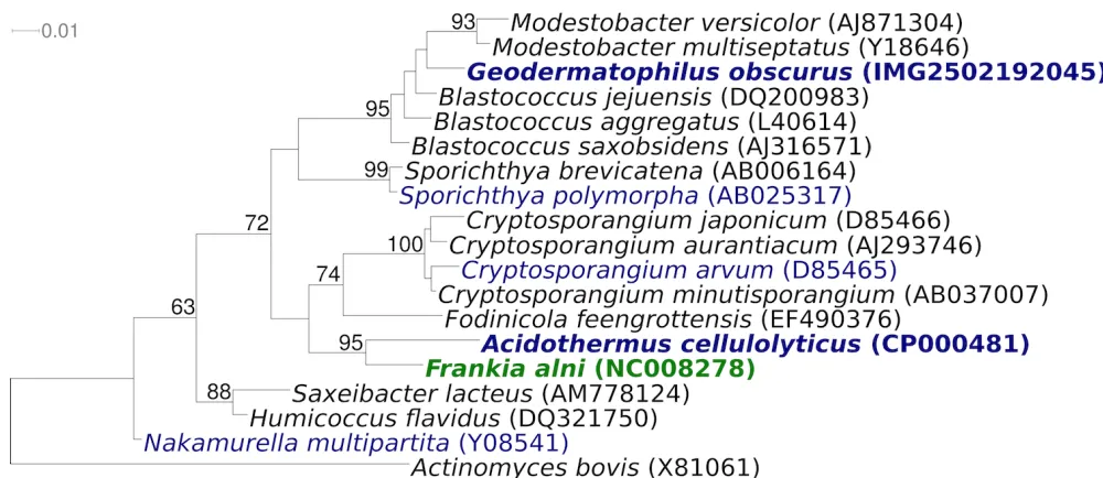
Figure 2 shows the phylogenetic neighborhood of for
G. obscurus
G-20
Tin a 16S rRNA based tree. The
se-quences of the three 16S rRNA gene copies in the
ge-nome of
G. obscurus
G-20
Tdo not differ from each
other, but differ by 24 nucleotides from the
previous-ly published 16S rRNA sequence obtained from DSM
43160 (X92356). These considerable discrepancies
are most likely due to sequencing errors in the latter
sequence. Genbank accession L40620, which was
ob-tained from ATCC 25078, differs by only one single
nucleotide from the 16S rRNA gene copies in the
ge-nome obtained from DSM 43160.
Chemotaxonomy
Figure 1. Scanning electron micrograph of G. obscurus G-20T
Figure 2. Phylogenetic tree highlighting the position of G. obscurus G-20T relative to the other type strains within the
Table 1. Classification and general features of G. obscurus G-20T according to the MIGS recommendations [16]
MIGS ID Property Term Evidence code
Current classification
Domain Bacteria TAS [17]
Phylum Actinobacteria TAS [18]
Class Actinobacteria TAS [19]
Subclass Actinobacteridae TAS [19]
Order Actinomycetales TAS [19]
Suborder Frankineae TAS [19]
Family Geodermatophilaceae TAS [2]
Genus Geodermatophilus TAS [1]
Species Geodermatophilus obscurus TAS [1]
Type strain G-20 TAS [1]
Gram stain gram positive TAS [1]
Cell shape cuboid or coccoid nonmotile cells and elliptical to lanceolate zoospores TAS [1]
Motility motile zoospores TAS [1]
Sporulation unknown TAS [1]
Temperature range 18°C–37°C TAS [20]
Optimum temperature 24°C-28°C TAS [20]
Salinity does not grow at 3% or more NaCl TAS [20]
MIGS-22 Oxygen requirement aerobic TAS [20]
Carbon source soluble sugars TAS [1]
Energy source chemoorganotroph TAS [8]
MIGS-6 Habitat worldwide distribution in soil, on rock surfaces, and deep sea marine sediments TAS [2,8]
MIGS-15 Biotic relationship free-living TAS [1,8,10,12,14]
MIGS-14 Pathogenicity no NAS
Biosafety level 1 TAS [21]
Isolation soil TAS [1]
MIGS-4 Geographic location Amargosa Desert, Nevada, USA TAS [1]
MIGS-5 Sample collection time 1968, or before TAS [1]
MIGS-4.1
MIGS-4.2 Latitude Longitude 36.48 -116.50 NAS
MIGS-4.3 Depth unknown
MIGS-4.4 Altitude unknown
Evidence codes - IDA: Inferred from Direct Assay (first time in publication); TAS: Traceable Author Statement (i.e., a direct report exists in the literature); NAS: Non-traceable Author Statement (i.e., not directly observed for the living, isolated sample, but based on a generally accepted property for the species, or anecdotal evidence). These evidence codes are from of the Gene Ontology project [22]. If the evidence code is IDA, then the property was directly observed by one of the authors or an expert mentioned in the acknowledgements.
Genome sequencing and annotation
Genome project history
This organism was selected for sequencing on the
basis of its phylogenetic position, and is part of the
Genomic
Encyclopedia of
Bacteria and
Archaea
project. The genome project is deposited in the
Genome OnLine Database [30] and the complete
genome sequence is deposited in GenBank.
Se-quencing, finishing and annotation were
per-formed by the DOE Joint Genome Institut
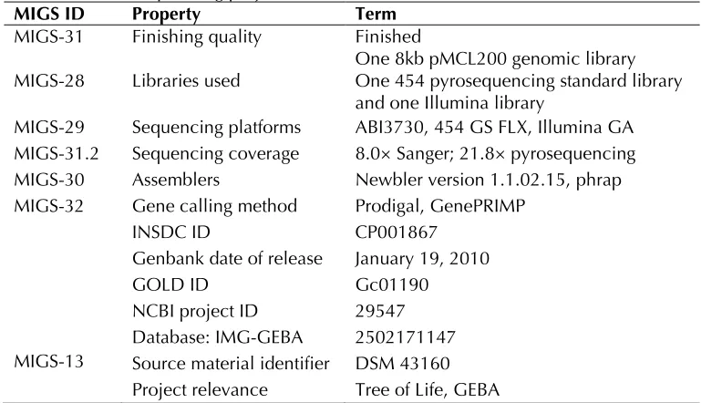
Table 2. Genome sequencing project information
MIGS ID Property Term
MIGS-31 Finishing quality Finished
MIGS-28 Libraries used One 8kb pMCL200 genomic library One 454 pyrosequencing standard library and one Illumina library
MIGS-29 Sequencing platforms ABI3730, 454 GS FLX, Illumina GA MIGS-31.2 Sequencing coverage 8.0× Sanger; 21.8× pyrosequencing MIGS-30 Assemblers Newbler version 1.1.02.15, phrap MIGS-32 Gene calling method Prodigal, GenePRIMP
INSDC ID CP001867
Genbank date of release January 19, 2010
GOLD ID Gc01190
NCBI project ID 29547 Database: IMG-GEBA 2502171147 MIGS-13 Source material identifier DSM 43160
Project relevance Tree of Life, GEBA
Growth conditions and DNA isolation
G. obscurus
G-20
T, DSM 43160, was grown in
DSMZ medium 65 [36] at 28°C. DNA was isolated
from 0.5-1 g of cell paste using Qiagen Genomic
500 DNA Kit (Qiagen, Hilden, Germany) with a
modified protocol for cell lysis, (procedure st/L),
and one hour incubation at 37°C, according to Wu
et al
. [37].
Genome sequencing and assembly
The genome was sequenced using a combination
of Sanger and 454 sequencing platforms. All
gen-eral aspects of library construction and
sequenc-ing performed at the JGI can be found at the JGI
websit
quencing reads were assembled using the
Newb-ler assembNewb-ler version 1.1.02.15 (Roche). Large
Newbler contigs were broken into 5,725
overlap-ping fragments of 1,000 bp and entered into
sembly as pseudo-reads. The sequences were
as-signed quality scores based on Newbler consensus
q-scores with modifications to account for overlap
redundancy and adjust inflated q-scores. A hybrid
454/Sanger assembly was made using the parallel
phrap assembler (High Performance Software,
LLC). Possible misassemblies were corrected with
Dupfinisher or transposon bombing of bridging
clones [38]. A total of 1,530 Sanger finishing reads
were produced to close gaps, to resolve repetitive
regions, and to raise the quality of the finished
se-quence. Illumina reads were used to improve the
final consensus quality using an in-house
devel-oped tool (the Polisher). The error rate of the
completed genome sequence is less than 1 in
100,000. Together, the combination of the Sanger
and 454 sequencing platforms provided 29.8×
coverage of the genome. The final assembly
con-tains 48,209 Sanger reads and 353,553
pyrose-quencing reads.
Genome annotation
Genes were identified using Prodigal [39] as part
of the Oak Ridge National Laboratory genome
an-notation pipeline, followed by a round of manual
curation using the JGI GenePRIMP pipeline [40].
The predicted CDSs were translated and used to
search the National Center for Biotechnology
In-formation (NCBI) nonredundant database,
Uni-Prot, TIGR-Fam, Pfam, PRIAM, KEGG, COG, and
In-terPro databases. Additional gene prediction
anal-ysis and functional annotation was performed
within the Integrated Microbial Genomes - Expert
Review (IMG-ER) platform [41].
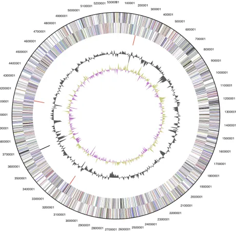
Genome properties
Figure 3. Graphical circular map of the genome. From outside to the center: Genes on forward strand (color by COG categories), Genes on reverse strand (color by COG categories), RNA genes (tRNAs green, rRNAs red, other RNAs black), GC content, GC skew.
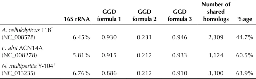
Comparison with closest related genomes
Table 5 provides an overall comparison of the
ge-nomes of
G. obscurus
strain G-20
Twith the closest
available genomes, that is,
Acidothermus
celluloly-ticus
11B
T,
Frankia alni
ACN14A and
N.
multiparti-ta
Y-104
T. The total length of (non-overlapping)
high-scoring segment pairs (HSPs) and the
num-ber of identical base pairs within these HSPs were
determined using the GGDC web server [42] by
directly applying NCBI Blastn to the genomes
represented as nucleotide sequences [43].
Number and proportion of shared homologs were
determined using the 'Phylogenetic Profiler'
func-tion of the
While the relative order of 16S rRNA difference
does not correspond to the genomic similarities,
the four genome-based measures uniformly
indi-cate that
N. multipartita
Y-104
Tpossesses the
ge-nome most similar to the one of
G. obscurus
G-20
T,
followed by
F. alni
ACN14A and
A. cellulolyticus
Table 3. Genome Statistics
Attribute Value % of Total
Genome size (bp) 5,322,497 100.00%
DNA coding region (bp) 4,756,139 89.36%
DNA G+C content (bp) 3,937,802 73.98%
Number of replicons 1
Extrachromosomal elements 0
Total genes 5,219 100.00%
RNA genes 58 1.11%
rRNA operons 3
Protein-coding genes 5,161 98.89%
Pseudo genes 350 6,71%
Genes with function prediction 3,640 69.75%
Genes in paralog clusters 896 17.17%
Genes assigned to COGs 3,408 65.30%
Genes assigned Pfam domains 3,584 68.67%
Genes with signal peptides 793 15.19%
Genes with transmembrane helices 1,105 21.17%
CRISPR repeats 0
Table 4. Number of genes associated with the general COG functional categories Code Value %age Description
J 166 3.2 Translation, ribosomal structure and biogenesis A 1 0.0 RNA processing and modification
K 309 6.0 Transcription
L 196 3.8 Replication, recombination and repair B 1 0.0 Chromatin structure and dynamics D 27 0.5 Cell cycle control, mitosis and meiosis Y 0 0.0 Nuclear structure
V 51 1.0 Defense mechanisms
T 242 4.7 Signal transduction mechanisms M 213 4.1 Cell wall/membrane biogenesis N 43 0.8 Cell motility
Z 0 0.0 Cytoskeleton
W 0 0.0 Extracellular structures
U 52 1.0 Intracellular trafficking and secretion
O 96 1.9 Posttranslational modification, protein turnover, chaperones C 277 5.4 Energy production and conversion
G 267 5.2 Carbohydrate transport and metabolism E 313 6.1 Amino acid transport and metabolism F 87 1.7 Nucleotide transport and metabolism H 180 3.5 Coenzyme transport and metabolism
I 188 3.6 Lipid transport and metabolism P 164 3.2 Inorganic ion transport and metabolism
Q 127 2.5 Secondary metabolites biosynthesis, transport and catabolism R 552 10.7 General function prediction only
Table 5. Percent-wise 16S rRNA sequence divergence 1
16S rRNA formula 1 GGD formula 2 GGD formula 3 GGD
Number of shared
homologs %age A. cellulolyticus 11BT
(NC_008578) 6.45% 0.930 0.231 0.946 2,309 44.7%
F. alni ACN14A
(NC_008278) 5.81% 0.915 0.212 0.933 3,124 60.5%
N. multipartita Y-104T
(NC_013235) 6.76% 0.886 0.212 0.910 3,300 63.9%
1Percent-wise 16S rRNA sequence divergence compared to genomic similarity for the three closest
available genomes to G. obscurus strain G-20T. GGD formulas: formula 1, length of sequence
fragments not in HSPs per average total genome length; formula 2, number of non-identical bases per total HSP length; formula 3, number of non-identical bases within HSPs per average total genome length.
Acknowledgements
We would like to gratefully acknowledge the help of Susanne Schneider (DSMZ) for DNA extraction and quality analysis. This work was performed under the auspices of the US Department of Energy's Office of Science, Biological and Environmental Research Pro-gram, and by the University of California, Lawrence Berkeley National Laboratory under contract No.
DE-AC02-05CH11231, Lawrence Livermore National La-boratory under Contract No. DE-AC52-07NA27344, Los Alamos National Laboratory under contract No. DE-AC02-06NA25396, and Oak Ridge National Laboratory under contract DE-AC05-00OR22725, as well as Ger-man Research Foundation (DFG) INST 599/1-1 and SI 1352/1-2.
References
1. Luedemann GM. Geodermatophilus, a new genus of the Dermatophilaceae (Actinomycetales). J Bac-teriol 1968; 96:1848-1858
2. Normand P. Geodermatophilaceae fam. nov., a formal description. Int J Syst Evol Microbiol 2006;
56:2277-2278
3. Gordon MA. The genus Dermatophilus. J Bacte-riol 1964; 88:509-522
4. Stackebrandt E, Kroppenstedt RM, Fowler VJ. A phylogenetic analysis of the family Dermatophila-ceae. J Gen Microbiol 1983; 129:1831-1838
5. Hahn D, Lechevalier MP, Fischer A, Stackebrandt E. Evidence for a close phylogenetic relationship between members of the genera Frankia, Geo-dermatophilus and Blastococcus and emendation of the family Frankiaceae.Syst Appl Microbiol 1989; 11:236-242.
6. Normand P, Orso S, Cournoyer B, Jeannin P. Chapelon, Dawson J, Evtushenko L, Misra AK. Molecular phylogeny of the genus Frankia and re-lated genera and emendation of the family
Fran-kiaceae. Int J Syst Bacteriol 1996; 46:1-9
7. Garrity GM, Lilburn TG, Cole JR, Harrison SH, Euzéby J, Tindall BJ. Taxonomic outline of the Bacteria and Archaea, Release 7.7 March 6, 2007. Part 10 - The Bacteria: Phylum Actinobac-teria: Class "Actinobacteria".
8. Eppard M, Krumbein WE, Koch C, Rhiel E, Staley JT, Stackebrandt E. Morphological, physiological, and molecular characterization of actinomycetes isolated from dry soil, rocks, and monument sur-faces. Arch Microbiol 1996; 166:12-22
9. Salazar O, Valverde A, Genilloud O. Real-Time PCR for the detection and quantification of Geo-dermatophilaceae from stone samples and identi-fication of new members of the genus Blastococ-cus. Appl Environ Microbiol 2006; 72:346-352
in desert varnish formation. Can J Microbiol 1987; 33:939-943.
11. Kudukhashvili PG, Gurielidze MA, Pataraya DT. Study of the lytic activities of actinomycetes iso-lated from different soils in Georgia. Appl Bio-chem Microbiol 2001; 37:251-252.
12. Kuhlman KR, Allenbach LB, Ball CL, Fusco WG, La Duc MT, Kuhlman GM, Anderson RC, Stueck-er T, Erickson IK, Benardini J, et al. Enumeration, isolation, and characterization of ultraviolet (UV-C) resistant bacteria from rock varnish in the Whipple Mountains, California. Icarus 2005;
174:585-595
13. Urzì C, Brusetti L, Salamone P, Sorlini C, Stacke-brandt E, Daffonchio D. Biodiversity of Geoder-matophilaceae isolated from altered stones and monuments in the Mediterranean basin. Environ Microbiol 2001; 3:471-479
14. Ishiguro EE, Fletcher DW. Characterization of Geodermatophilus strains isolated from high alti-tude Mount Everest soils. Mikrobiologika 1975; 12:99-108.
15. Ishiguro EE, Wolfe RS. Control of morphogenesis in Geodermatophilus: ultrastructural studies. J Bacteriol 1970; 104:566-580
16. Field D, Garrity G, Gray T, Morrison N, Selengut J, Sterk P, Tatusova T, Thomson N, Allen MJ, An-giuoli SV, et al. The minimum information about a genome sequence (MIGS) specification. Nat Biotechnol 2008; 26:541-547
17. Woese CR, Kandler O, Wheelis ML. Towards a natural system of organisms: proposal for the do-mains Archaea, Bacteria, and Eucarya.Proc Natl Acad Sci USA 1990; 87:4576-4579
18. Garrity GM, Holt JG. The Road Map to the Ma-nual. In: Garrity GM, Boone DR, Castenholz RW (eds), Bergey's Manual of Systematic Bacteriology, Second Edition, Springer, New York, 2001, p. 119-169.
19. Stackebrandt E. rainey FA, Ward-Rainey NL. Pro-posal for a new hierarchic classification system, Actinobacteria classis nov. Int J Syst Bacteriol
1997; 47:479-491
20. O'Nuallain B, Wetzel R. Conformational Abs re-cognizing a generic amyloid fibril epitope. Proc
Natl Acad Sci USA 2002; 99:1485-1490
21. Classification of Bacteria and Archaea in risk groups www.baua.de TRBA 466.
22. Ashburner M, Ball CA, Blake JA, Botstein D, But-ler H, Cherry JM, Davis AP, Dolinski K, Dwight SS, Eppig JT, et al. Gene Ontology: tool for the unification of biology. Nat Genet 2000; 25:25-29
23. Mevs U, Stackebrandt E, Schumann P, Gallikows-ki C, Hirsch P. Modestobacter multiseptatus gen. nov., sp. nov., a budding actinomycete from soils of the Asgard Range (Transantarctic Mountains). Int J Syst Evol Microbiol 2000; 50:337-346
24. Jordal PB, Dueholm MS, Larsen P, Petersen SV, Enghild JJ, Christiansen G, Hojrup P, Nielsen PH, Otzen DE. Widespread abundance of functional bacterial amyloid in mycolata and other gram-positive bacteria. Appl Environ Microbiol 2009; 75:4101-4110
25. Venter JC, Remington K, Heidelberg JF, Halpern AL, Rusch D, Eisen JA, Wu D, Paulsen I, Nelson KE, Nelson W, et al. Environmental genome shot-gun sequencing of the Sargasso Sea. Science 2004; 304:66-74
26. Castresana J. Selection of conserved blocks from multiple alignments for their use in phylogenetic analysis. Mol Biol Evol 2000; 17:540-552
27. Lee C, Grasso C, Sharlow MF. Multiple sequence alignment using partial order graphs. Bioinformat-ics 2002; 18:452-464
28. Stamatakis A, Hoover P, Rougemont J. A Rapid Bootstrap Algorithm for the RAxML Web Servers. Syst Biol 2008; 57:758-771
29. Pattengale ND, Alipour M, Bininda-Emonds ORP, Moret BME, Stamatakis A. How Many Bootstrap Replicates Are Necessary? Lect Notes Comput Sci
2009; 5541:184-200
Acids Res 2010; 38:D346-D354
31. Tice H, Mayilraj S, Sims D, Lapidus A, Nolan M, Lucas S, Glavina Del Rio T, Copeland A, Cheng JF, Meincke L, et al. Complete genome sequence of Nakamurella multipartita type strain (Y-104T). Stand Genomic Sci 2010; 2:168-175.
32. Normand P, Lapierre P, Tisa LS, Gogarten JP, Al-loisio N, Bagnarol E, Bassi CA, Berry AM, Bick-hart DM, Choisne N, et al. Cenome characteris-tics of facultatively symbiontic Frankia sp. strains reflect host range and host plant biogeography. Genome Res 2007; 17:7-15
33. Mirza MS, Janse JD, Hahn D, Akkermans ADL. Identification of atypical Frankia strains by fatty acid analysis. FEMS Microbiol Lett 1991; 83
:91-98
34. Wink JM
35. Lechevalier MP, Lechevalier H. Chemical compo-sition as a criterion in the classification of aerobic actinomycetes. Int J Syst Bacteriol 1970; 20
:435-443
36. List of growth media used at DSMZ:
37. Wu D, Hugenholtz P, Mavromatis K, Pukall R, Dalin E, Ivanova N, Kunin V, Goodwin L, Wu M, Tindall BJ, et al. A phylogeny-driven genomic en-cyclopedia of Bacteria and Archaea. Nature
2009; 462:1056-1060
38. Sims D, Brettin T, Detter J, Han C, Lapidus A, Co-peland A, Glavina Del Rio T, Nolan M, Chen F, Lucas S, et al. Complete genome sequence of Ky-tococcus sedentarius type strain (541T). Stand Genomic Sci 2009; 1:12-20 39. Hyatt D, Chen GL, Locascio PF, Land ML,
Lari-mer FW, Hauser LJ. Prodigal Prokaryotic Dynam-ic Programming Genefinding Algorithm. BMC Bioinformatics 2010; 11:119
40. Pati A, Ivanova N, Mikhailova N, Ovchinikova G, Hooper SD, Lykidis A, Kyrpides NC. GenePRIMP: A Gene Prediction Improvement Pipeline for mi-crobial genomes. Nat Methods (In press). 41. Markowitz VM, Ivanova NN, Chen IMA, Chu K,
Kyrpides NC. IMG ER: a system for microbial ge-nome annotation expert review and curation. Bio-informatics 2009; 25:2271-2278
42. Auch AF, Klenk HP, Göker M. Standard operating procedure for calculating genome-to-genome dis-tances based on high-scoring sequence pairs. Stand Genomic Sci 2010; 2:142-148.
43. Auch AF, von Jan M, Klenk HP, Göker M. Digital DNA-DNA hybridization for microbial species delineation by means of genome-to-genome se-quence comparison. Stand Genomic Sci 2010;
![Table 1. Classification and general features of G. obscurus G-20T according to the MIGS recommendations [16]](https://thumb-us.123doks.com/thumbv2/123dok_us/675414.2066212/4.612.70.546.74.546/table-classification-general-features-obscurus-according-migs-recommendations.webp)