CONFERENCE SUMMARY
Improvements and Limitations of Humanized
Mouse Models for HIV Research:
NIH/NIAID ‘‘Meet the Experts’’ 2015 Workshop Summary
Ramesh Akkina,1 Atef Allam,2,3 Alejandro B. Balazs,4 Joel N. Blankson,5John C. Burnett,6 Sofia Casares,7J. Victor Garcia,8Kim J. Hasenkrug,9Fatah Kashanchi,10Scott G. Kitchen,11
Florian Klein,12,13Priti Kumar,14Andrew D. Luster,15Larisa Y. Poluektova,16Mangala Rao,3 Brigitte E. Sanders-Beer,17Leonard D. Shultz,18and Jerome A. Zack11
Abstract
The number of humanized mouse models for the human immunodeficiency virus (HIV)/acquired
immunode-ficiency syndrome (AIDS) and other infectious diseases has expanded rapidly over the past 8 years. Highly
immunodeficient mouse strains, such as NOD/SCID/gamma chain
null(NSG, NOG), support better human
hematopoietic cell engraftment. Another improvement is the derivation of highly immunodeficient mice,
transgenic with human leukocyte antigens (HLAs) and cytokines that supported development of HLA-restricted
human T cells and heightened human myeloid cell engraftment. Humanized mice are also used to study the HIV
reservoir using new imaging techniques. Despite these advances, there are still limitations in HIV immune
responses and deficits in lymphoid structures in these models in addition to xenogeneic graft-versus-host
responses. To understand and disseminate the improvements and limitations of humanized mouse models to the
scientific community, the NIH sponsored and convened a meeting on April 15, 2015 to discuss the state of
knowledge concerning these questions and best practices for selecting a humanized mouse model for a
par-ticular scientific investigation. This report summarizes the findings of the NIH meeting.
Introduction
T
he plenary lecture, entitled ‘‘Of Next Generation Humanized Mice and Men,’’ was presented byDr. Leo-nard D. Shultz. Preclinical testing in humanized mice has beenemployed for studies of human immunodeficiency virus-1 (HIV-1) pathogenesis and latency, drug therapy, vaccine de-velopment, gene therapy, and mucosal immunity in preparation for clinical trials.1Infectious agents that have been investigated in humanized mice so far include HIV, human T lymphotropic
1
Department of Microbiology, Immunology and Pathology, Colorado State University, Fort Collins, Colorado.
2
Henry M. Jackson Foundation for the Advancement of Military Medicine, Silver Spring, Maryland.
3
U.S. Military HIV Research Program, Walter Reed Army Institute of Research, Silver Spring, Maryland.
4
The Ragon Institute of MGH, MIT & Harvard, Cambridge, Massachusetts.
5Department of Medicine, Center for AIDS Research, Johns Hopkins University School of Medicine, Baltimore, Maryland. 6
Department of Molecular and Cellular Biology, Beckman Research Institute of City of Hope, Duarte, California.
7U.S. Military Malaria Vaccine Program, Naval Medical Research Center, Silver Spring, Maryland. 8
Division of Infectious Diseases, Department of Medicine, UNC Center for AIDS Research, University of North Carolina at Chapel Hill, Chapel Hill, North Carolina.
9Laboratory of Persistent Viral Diseases, Rocky Mountain Laboratories, NIAID, NIH, Hamilton, Montana. 10
School of Systems Biology, National Center for Biodefense and Infectious Diseases, George Mason University, Manassas, Virginia.
11
Departments of Medicine and Microbiology, Immunology and Molecular Genetics, UCLA AIDS Institute, Los Angeles, California.
12
First Department of Internal Medicine, University Hospital of Cologne, Cologne, Germany.
13Center for Molecular Medicine Cologne (CMMC), University of Cologne, Cologne, Germany. 14
School of Medicine, Infectious Diseases/Internal Medicine, Yale University, New Haven, Connecticut.
15Center for Immunology and Inflammatory Diseases, Division of Rheumatology, Allergy and Immunology, Massachusetts General
Hospital, Harvard Medical School, Boston, Massachusetts.
16Department of Pharmacology and Experimental Neuroscience, University of Nebraska Medical Center, Omaha, Nebraska. 17
National Institute of Allergy and Infectious Diseases, National Institutes of Health, Bethesda, Maryland.
18
The Jackson Laboratory, Bar Harbor, Maine.
Volume 32, Number 2, 2016
ªMary Ann Liebert, Inc. DOI: 10.1089/aid.2015.0258
virus 1, influenza, Rift Valley fever, dengue virus, Epstein Barr virus, Ebola virus, cytomegalovirus (CMV), measles virus, Salmonella typhi,Mycobacterium tuberculosis,Neisseria gon-orrhea, andPlasmodium falciparum.2
The CB17 strain carrying the spontaneous severe com-bined immunodeficiency (Prkdcscid, abbreviated as scid) mouse mutation was the first model in the chronological development path of humanized mouse models, followed by the NOD/Lt-scid mouse and the highly immunodeficient NOD.Cg-PrkdcscidIl2rgtm1Wjl/Sz(NSG) mouse.3–6The NOD/ Lt background confers defects in innate immunity, im-paired macrophage activation, lack of hemolytic com-plement, and human-like polymorphism in the signal regulatory protein alpha (Sirpa) gene. The scid mutation prevents development of mature T and B cells. The in-terleukin 2 (IL-2) receptor common gamma chain-targeted mutation (IL2rgnull) inhibits the expression of receptors for IL-2, -4, -7, -9, -15, and -21 and prevents natural killer (NK) cell development.
Described hereunder are the hematolymphoid engraftment methods for various humanized mouse models (see also Table 1):
Human peripheral blood lymphocytes-SCID (Hu-PBL-SCID): Human peripheral blood leukocytes are injected
intraperitoneally (IP) or intravenously (IV) into an immu-nodeficient mouse. This procedure leads to efficient T-cell engraftment, but the mice develop lethal xenogeneic graft-versus-host disease (GvHD).
Hu-SRC-SCID (SRC=‘‘scidrepopulating cell’’): Human hematopoietic stem cells (HSCs) from bone marrow, um-bilical cord blood, mobilized peripheral blood, or fetal liver are injected IV into sublethally irradiated neonatal or adult immunodeficient mice. This leads to the development of multiple hematopoietic lineages. The human T cells are ed-ucated in the mouse thymus.
SCID-hu: Human fetal liver and thymus fragments are transplanted under the kidney capsule of CB17-scidmice. A functional human thymus develops; however, there is mini-mal peripheral immune system development.
Bone marrow, liver, thymus (BLT) mouse: Human fetal liver and thymus fragments are implanted under the kidney capsule of sublethally irradiated NSG or NOD-scid mice, and in addition autologous fetal HSCs are in-jected IV. The results are robust development of multiple hematopoietic lineages and T-cell education in the human thymus. One disadvantage is the development of lethal GvHD in some animals after more than 20 weeks of engraftment.
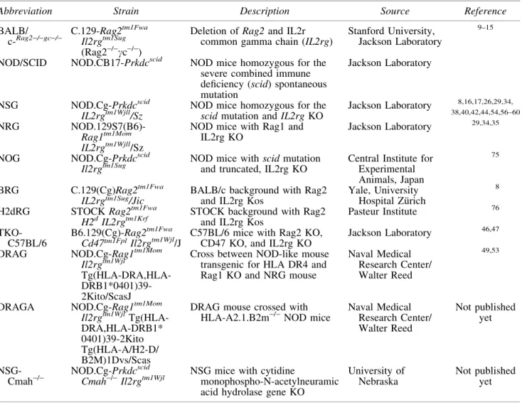
Table1. Major Strain Platforms for Humanized Mice
Abbreviation Strain Description Source Reference
BALB/ c-
Rag2-/-gc-/-C.129-Rag2tm1Fwa Il2rgtm1Sug (Rag2-/-cc-/-)
Deletion of Rag2and IL2r common gamma chain (IL2rg)
Stanford University, Jackson Laboratory
9–15
NOD/SCID NOD.CB17-Prkdcscid NOD mice homozygous for the severe combined immune deficiency (scid) spontaneous mutation
Jackson Laboratory
NSG NOD.Cg-Prkdcscid
IL2rgtm1Wjll/Sz
NOD mice homozygous for the scidmutation andIL2rgKO
Jackson Laboratory 8,16,17,26,29,34, 38,40,42,44,54,56–60
NRG
NOD.129S7(B6)-Rag1tm1Mom IL2rgtm1Wjll/Sz
NOD mice with Rag1 and IL2rg KO
Jackson Laboratory 29,34,35
NOG NOD.Cg-Prkdcscid
Il2rgtm1Sug
NOD mice withscidmutation and truncated, IL2rg KO
Central Institute for Experimental Animals, Japan
75
BRG C.129(Cg)Rag2tm1Fwa
IL2rgtm1Sug/Jic BALB/c background with Rag2and IL2rg Kos Yale, UniversityHospital Zu¨rich
8
H2dRG STOCKRag2tm1Fwa
H2dIL2rgtm1Krf
STOCK background with Rag2 and IL2rg Kos
Pasteur Institute 76
TKO-C57BL/6
B6.129(Cg)-Rag2tm1Fwa
Cd47tm1FplIl2rgtm1Wjl/J C57BL/6 mice with Rag2 KO,CD47 KO, and IL2rg KO Jackson Laboratory
46,47
DRAG NOD.Cg-Rag1tm1Mom
Il2rgtm1Wjl
Tg(HLA-DRA,HLA- DRB1*0401)39-2Kito/ScasJ
Cross between NOD-like mouse transgenic for HLA DR4 and Rag1 KO and NRG mouse
Naval Medical Research Center/ Walter Reed
49,53
DRAGA NOD.Cg-Rag1tm1Mom
Il2rgtm1Wjl Tg(HLA-DRA,HLA-DRB1* 0401)39-2Kito Tg(HLA-A/H2-D/ B2M)1Dvs/Scas
DRAG mouse crossed with HLA-A2.1.B2m-/-NOD mice
Naval Medical Research Center/ Walter Reed
Not published yet
NSG-Cmah-/
-NOD.Cg-Prkdcscid Cmah-/-Il2rgtm1Wjl
NSG mice with cytidine
monophospho-N-acetylneuramic acid hydrolase gene KO
University of Nebraska
The main limitations of current humanized mouse model are as follows7:
1. Engraftment with mature human T cells leads to xe-nogeneic GvHD.
2. Human leukocyte antigen (HLA) molecules are re-quired for appropriate T-cell selection following hu-man HSC engraftment.
3. Many human cytokines and other factors are species specific.
4. Remaining innate immunity impairs engraftment. 5. Impaired humoral immune responses, low level of
immunoglobulin production, and impaired immuno-globulin class switching.
6. Impaired lymph node development, poorly developed germinal centers.
Next generation humanized mice
Mouse major histocompatibility complex (MHC) class I (NSG-B2mtm1Unc) and (H2D1tm1Bpe H2K1tm1Bpe
(KD)null or class II (NSG-H2-Ab1tm1Gru) (abbreviated as I-Anull) and H2dlAb1-Ea (abbreviated as I-A/I-E)null) knockout (KO) mice have been developed to reduce xenogeneic GvHD. Progress has also been made in generating a panel of human HLA class I and II transgenic NSG mice. NSG class I transgenic mice available include expression of HLA-A2, A11, A24, B7, B27, and Cw3 alleles, and NSG class II transgenic NSG mice available include HLA-DR1, DR2, DR3, DR4, and DQ8 alleles. For example, NSG-HLA-A2 transgenic mice engrafted with HLA-A2+HSCs develop HLA-A2-restricted human cytotoxic T cells.
Another new approach is the transgenic expression of human cytokines in immunodeficient mouse strains and tar-geting of mouse genes.8The new NSG-KitW-41mice support human HSC engraftment without X-ray preconditioning. NSG-Ifnar1tm1Agt[type I interferon (IFN) receptor KO] mice show increased sensitivity to dengue virus infection and in-creased sensitivity for testing neutralizing activity of anti-dengue human IgM antibodies. Another emerging model for humanized mice is the implantation of human-induced pluripotent stem cells-derived thymic epithelial cells into NSG-Foxn1nu(nude) mice.
Preexposure Prophylaxis and Novel Treatment Strategies for HIV/Acquired
Immunodeficiency Syndrome
Dr. Ramesh Akkina’spresentation highlighted the util-ity of humanized mice in multiple areas of HIV/acquired immunodeficiency syndrome (AIDS) research in generating important preclinical data. Hu-HSC mice derived by inject-ing human CD34+ HSCs into neonatal C.129-Rag2tm1Fwa Il2rgtm1Sug(Rag2-/-cc-/-) mice (Rag-hu mice) were primarily used in these studies.9Multilineage human hematopoiesis is seen in these mice, which permits efficient HIV infection. Long-term viremia lasting more than 1 year could be estab-lished with both CXCR4- and CCR5-tropic viruses.9With the presence of HIV-susceptible cells in mucosal tissues, this model also permits HIV transmission through both vaginal and rectal routes; thus it is suitable for the evaluation of HIV preexposure prophylactic (PrEP) strategies.9
The integrase inhibitor raltegravir (RAL) and CCR5 in-hibitor maraviroc (MVC) were tested as oral PrEP in
hu-manized mice and were shown to protect 100% of the mice from intravaginal challenge.10 In the context of topical mi-crobicide application, protection was also achieved with MVC and a broadly neutralizing antibody (bNAb) VRC01 against vaginal HIV-1 challenge.11,12A major question for PrEP is the optimal concentration of combination anti-retroviral therapy (cART) (which is designed for therapeutic purposes) that needs to be reached in mucosal tissues for conferring full protection against viral exposure. In this con-text, the hu-mice were found to be suitable for conducting pharmacokinetics-pharmacodynamics (PK-PD) studies.13 ART drugs tenofovir (TFV), MVC, and RAL both individually and in combination (TFV+RAL, TFV+MVC, RAL+MVC) were tested. Higher concentrations were found in mucosal tissues than in plasma, and the drug interactions and syner-gistic effects are currently being evaluated.
In the second part of the talk, anti-HIV small interfer-ing RNAs (siRNAs) were shown to be highly effective in suppressing viral replicationin vitro, but specific delivery to HIV-infected cells in vivohas been difficult. To overcome this hurdle, siRNAs conjugated with an HIV-1 gp120-specific aptamer were tested in HIV-infected hu-mice.14 Marked suppression of viral loads was seen with concomitant protection against viral-mediated CD4 T-cell loss.
The third part of the talk focused on potentiating antiviral immune responses through PD-1 blockade.15 Mice treated with human anti-PD-L1 monoclonal antibody (mAb) showed decreased viral loads and improved T-cell function, sug-gesting its potential application for immuno-enhancement therapy. The advantages of the Hu-HSC model over BLT mice for some studies are summarized as follows9:
Easy to prepare and lower cost
More animals can be generated per cohort
Negligible GvHD and longer life span
Chronic HIV infection lasting more than 1 year
Long-term safety and toxicity assessments possible
Dr. Alejandro B. Balazspresented a novel approach for delivery of bNAbs, called vectored immunoprophylaxis (VIP). Adeno-associated virus (AAV; serotype 8) is used as a vector, because it is nonpathogenic in humans, nonintegrat-ing, and has excellent expression characteristics in vivo. Despite the limited capacity to carry foreign genes (4.8 kb), a transgene for the expression of antibody from muscle cells was presented.16 In the first experiment, BLT mice were administered a vector resulting in expression of 100lg/ml of VRC07G54W antibody and challenged intravaginally 21 times with the transmitted founder strain of HIV REJO.c. VIP protected against CD4 T-cell loss in splenic lymphocytes, gut intraepithelial lymphocytes (IEL), gut lamina propria lym-phocytes (LPL), and vaginal LPL. All treated mice were entirely resistant to mucosal challenge with the REJO.c founder strain by sensitive viral load assays.17
virus. Dr. Balazs described a third experiment, in which AAV vectors expressing either PG9, PGT121, or VRC07 were prophylactically administered, and the mice challenged with 107human CD4 cells from an infected patient suppressed with ART drugs. The blood was monitored for signs of virus emerging from infected cells. High concentrations were achieved for PGT121 (219lg/ml) and PG9 (58lg/ml) and lower concentrations for VRC07 (1.6lg/ml), but only PGT121 protected the mice from HIV infection and CD4 cell loss. In contrast to PG9 and VRC07, the PGT121-transfused mice had undetectable viral load (below 103RNA copies/ml), but the PG9- and VRC01-treated animals became viremic to similar levels as the control mice. In conclusion, VIP enables the long-lived expression of any desired antibody after a single intramuscular injection.
Dr. Fatah Kashanchi’stalk discussed the use of irradiation to activate viral transcription and induce apoptosis in HIV-1-infected cells. It has previously been shown that HIV-1 long terminal repeat (LTR) can be induced with various transcrip-tion activators including irradiatranscrip-tion,18which can be reversed by specific peptide or cyclin-dependent kinase inhibitors in humanized mouse models.19–22 Here, total body irradiation was used to better define the latent tissue reservoirs in animals and also potentially use low-level irradiation as part of a ‘‘kick and kill’’ strategy activating HIV-1 cells under ART. Total body irradiations are frequently used to treat lymphomas, in-cluding solid epithelial tumors, lymphomas, breast, neck, and head cancers, and Hodgkin and non-Hodgkin lymphomas. Total body radiotherapy was also applied for the ‘‘Berlin’’ patient before CCR5D32/D32 transplantation.
Using an NSG ‘‘latent HIV mouse model’’ under a cocktail of cART, Dr. Kashanchi’s laboratory was able to show that latent viruses could be activated using low-level whole body irradiation.23These low doses were not lethal or cancerous to animals at least up to 6 months. The question is whether T cells are equally affected by low-level irradiation compared to myeloid cells. It was shown that X-ray irradiation activates HIV-1 transcription by removing negative inhibitors from HIV-1 DNA and promotes transcriptional elongation. The effect may be further enhanced by proteasome inhibitors (to improve Tat stability in vivo). Latently infected T cells are more sensitive to cell death (as compared with uninfected T cells) by low-level irradiation than myeloid cells, possibly by activation of the wild-type p53 pathway in cells. The effect of cell death was not observed in myeloid-infected (or uninfect-ed) cells with low-level irradiation, pointing to a different mechanism of apoptosis in T cells versus myeloid cells. Im-portantly, most tissues (blood, liver, lung, brain, and spleen) from NSG-humanized mice contain latent HIV-1 that can be activated with low-level irradiation alone. Reverse phase protein microarray on few infected cells can show the status of latently infected cells (i.e., senescence vs. exhaustion) in the presence or absence of activators. Irradiation plus ALLN (proteasome inhibitor) or bryostatin enhances transcription activity more than low-level irradiation alone. Collectively, the data indicate that latent HIV-1 can be transcriptionally activated with low-level irradiation, which is not toxic to the host, and potentially ‘‘purge’’ the virus from various secluded reservoirs including brain.
Dr. Scott G. Kitchenreported on the characterization of chronic immune activation and type I IFN signaling during HIV infection in humanized mice. The progression from naive
to exhausted T cells involves chronic antigen stimulation during chronic infection, triggering the expression of exhaus-tion markers PD-1, LAG-3, CD244 (2B4), and Tim-3.24,25 Similar to what is observed in humans, HIV-infected BLT mice have elevated expression of cellular activation and ex-haustion markers. In particular, the expression of exex-haustion (PD-1, Tim-3) and activation markers (HLA-DR) is increased in chronically infected BLT mice. T cells from infected mice have higher Tim-3 expression and an impaired ability to pro-duce IL-2 and IFN-cupon stimulation. After HIV infection of humanized mice containing human cells genetically modified with a molecularly cloned anti-HIV T-cell receptor (TCR),26 Tim-3+HIV antigen-specific cells produce less IFN-cand IL-2 in response to immunodominant HIV Gag p17 (SL9) peptide stimulation. It was also found that Tim-3 is elevated on HIV nonspecific T cells in chronically infected mice.
Type I IFNs (IFNaandb) have both antiviral and immu-nomodulatory effects and could contribute to chronic immune activation and exhaustion.27HIV-infected mice have chroni-cally elevated levels of type I IFN signature gene expression than uninfected animals. In addition, HIV infection upregu-lates immunoregulatory dendritic cells,28as characterized by high levels of expression of both PD-L1 and CD95. Type I IFN receptor blockade significantly reduced type I IFN signaling, led to a reduction of activation and exhaustion markers on T cells, and improved their cytokine production upon stimula-tion. The BLT mouse model can be used to examine the mechanisms of immune defects during HIV infection and for testing therapies aimed at reversing immune suppression.
Dr. Florian Kleinpresented data from monocloncal an-tibody therapy studies in HIV-1YU2-infected humanized mice. NOD.129S7(B6)-Rag1tm1Mom/Sz (NRG) mice were irradiated with 1.0–3.6 Gray (Gy) and 4–6 h later injected intrahepatically with human HSCs. HIV-1-infected hu-mice demonstrated stable HIV-1 infection for more than 100 days, had a decreasing CD4+/CD8+T-cell ratio, and the virus di-versified within the host (2.2·10-3 mutations/bp 21 days after infection).
Antibody-mediated therapy with a single bNAb (e.g., 45– 46G54W) led to a transient decrease viremia, but escape oc-curred rapidly mediated by HIV envelope mutations (e.g., N279H and N280Y for antibody 45–46G54W).29In contrast, antibody combinations (e.g., 3BNC117+10-1074+PG16) led to a sustained decrease of HIV-1 RNA.29,30
In contrast to the experiments in hu-mice, monotherapy appeared to be more effective in nonhuman primates: A single infusion with 3BNC117 or 10-1074 resulted in a transient suppression and although classical escape mutations were detected after 10-1074 monotherapy (N332K, S334N), no apparent escape was seen after 3BNC117 infusion.31In conclusion, antibody monotherapy can transiently suppress viremia in HIV-1-infected hu-mice, but escape and rebound occur rapidly. However, escape can be prevented by com-bining bNAbs that target different epitopes. Single bNAbs can effectively decrease viremia in nonhuman primates and in some cases maintain suppression until antibody serum levels decay.31,32
of the viral load was detected up to 28 days after antibody infusion.34
Finally, experiments in humanized mice demonstrated that bNAbs can synergize with non- or weakly neutralizing an-tibodies by inducing HIV escape variants. As shownin vitro, escape variants can be more susceptible to commonly gen-erated HIV antibodies (e.g., V3-loop antibodies). When the bNAb 10-1074 was injected with one of the V3-loop anti-bodies 10-188 or 1-79 into HIV-1YU2-infected humanized mice, viremia was suppressed more effectively.35Therefore, HIV-1 infection is, in part, more readily controlled during immunotherapy because escape from bNAbs can create holes in the glycan shield that render the virus susceptible to oth-erwise ineffective antibodies that are present in nearly all HIV-infected individuals.
Dr. Jerome A. Zack presented modeling of cell-based therapeutics in BLT mice. His laboratory has found that anti-MART-1 HLA class I-restricted TCR (F5) introduced into human stem cells results in the generation of mature CD8 T cells, which can eliminate human melanomas in BLT mice.36,37 In addition, an anti-HIV Gag class I-restricted TCR introduced into human stem cells results in the gen-eration of mature CD8 T cells that can greatly reduce HIV replication in BLT mice.26,38 However, HLA restriction renders TCR-based approaches difficult to use clinically because multiple anti-HIV TCRs would be required. In addition, rapid resistance of HIV would be likely because of the rapid mutation rate.
A previously established approach created a CD4-zeta chimeric antigen receptor (CAR), which contained the ex-tracellular and transmembrane domains of the human CD4 molecule, and the CD3-zeta signaling domain. The CAR recognizes HIVgp120 independent of HLA restriction. Li-gation induces TCR signaling and activation. This construct demonstrated stable, safe engraftment with a persistence of greater than 10 years and has been used in peripheral T cells in multiple clinical trials.39 It has modest clinical efficacy because of functional defects in modified peripheral T cells. The new CD4-zeta CAR Triple Vector expresses a CCR5sh1005 gene for CCR5 knock down, a sh516 gene, which targets the LTR R region of HIV-1 and a CD4-zeta gene.40 NSG mice were implanted with the thy-liv ‘‘orga-noid,’’ irradiated with 270 cGy, and CD34+cells transduced either with a control vector or with the Triple CD4CAR vector, and then infected with HIV. CD4-zeta CAR-modified stem cells develop into multiple hematopoietic lineages, such as T cells, macrophages, and NK cells. CD4 CAR-expressing cells have decreased expression of CD3, TCRab, and TCR excision circles in thymocytes,40 suggesting that the CAR leads to allelic exclusion of endogenous TCRs in some cells. CAR-expressing T cells developed into effector cells and were activated in response to HIV. CD4-zeta CAR-modified mice have reduced HIV viral loads and preserved CD4+ T-cell counts than control mice. Engineered immunity is potentially a feasible approach in eliminating chronic viral infections.
Finally, human CD34+cells can be engineered with TCRs plus a nonimmunogenic positron emission tomography (PET) reporter gene through lentiviral vectoring. Addition of the proper PET tracer allows localization of antigen-specific cells in vivoin BLT mice.41This approach may be useful in tracking engineered immune cells during clinical trials.
Mouse Viral Outgrowth Assay
Dr. Joel N. Blanksonreported that NSG mice have a high level of human T-cell engraftment and a high degree of im-mune activation because of GvHD. For the mouse viral outgrowth assay (MVOA), 25–55 million peripheral blood mononuclear cells (PBMCs) from patients on suppressive cART regimens (1–6 years) were IP injected into NSG mice. Seven days later, anti-CD8 mAb was administered. The mice were bled weekly to evaluate viral load and CD4 T-cell count. All mice became viremic after PBMC transfer from patients on suppressive cART regimens.42
Next, elite suppressors were studied to answer the question whether MVOA is more sensitive than the quantitative viral outgrowth assay (QVOA). Elite suppressors have much lower frequencies of latently infected cells than patients on cART.43 However, HIV isolates from HLA-B57 elite sup-pressors replicate vigorously in BLT-humanized mice.44 Elite suppressors were selected who had a frequency of la-tently infected cells ranging from 1 in 15 million to 1 in 25 million CD4+ T cells. Sixty-six million PBMCs from one elite suppressor or 20–26 million purified CD4 T cells from four other elite suppressors were injected IP into NSG mice. On day 7 anti-CD8 mAb was administered IP to the mice engrafted with PBMCs, and for the mice engrafted with pu-rified CD4s, anti-CD8 mAb was given as needed. Two mice were treated with anti-CD3 IP. The mice were bled weekly to evaluate viral load and CD4 T-cell count. The NSG mice became viremic after PBMC or CD4 transfer from elite controllers. Elite suppressor CD8 T cells have potent antiviral activity,45and the expansion of these cells may potentially explain why virus does not continue to replicatein vivo.
The advantages of the MVOA are as follows:
Recapitulates what happensin vivowhen cART is in-terrupted.
Continuous stimulation of cells for up to 6 weeks be-cause of xenogeneic response.
Can assay very large number of cells.
Can be used for nonhuman primate (simian immuno-deficiency virus) and human (HIV) studies.
At least as sensitive as the QVOA; sensitivity may increase with anti-CD3 and/or anti-CD28 mAbs treat-mentin vivo.
Future directions are to validate the sensitivity with a lar-ger number of patients to optimizein vivostimulation with mAbs, to determine whether low-level viremia represents replication-competent virus, and to determine whether NSG mice can be engrafted with CD4+ T cells from lymphoid tissue [gut-associated lymphoid tissue (GALT), lymph nodes].
Novel Mouse Models
HIV infection with pathogenic loss of CD4+ T cells. HLA class II-expressing cells and both CD4+ and CD8+ human cells are present in lymphoid tissues. Spleens and mesenteric lymph nodes form B-cell follicles with interspersed T cells.
TKO-BLT mice have functional immune systems. HIV-specific IFNcEnzyme-Linked ImmunoSpot responses from splenocytes could be detected 7 weeks postinfection (p.i.) and could be mapped to different Gag, Pol, and Env peptides. Antibodies (IgG) to gp120JR-CSF were present 8 weeks p.i. although at levels lower than that seen in HIV-infected patients. In vaccine experiments, no antibody responses were detectable after inoculation of Ad5 HIV vaccine vectors or DTP (diphtheria, tetanus, pertussis) vaccination. Experi-ments to improve B-cell responses by transfusing mesen-chymal stem cells isolated from human fetal lung to provide human stromal cells in lymphoid tissues showed trafficking to multiple tissues. However, there was no improvement to vaccine-induced antibody responses. As seen with other models, there is an age-dependent loss of B cells with a concomitant increase in T cells and stable monocytes and dendritic cells.
Evidence of possible GvHD was seen in only 2 of 55 co-horts (more than 2,000 mice). Humanized mice in the TKO background are healthy for more than 35 weeks with no signs of GvHD. Thus, this model is an excellent platform for long-term studies, such as HIV latency and cure.
Dr. Sofia Casares discussed the generation of NOD-Rag1tm1Mom Il2rgtm1Wjl Tg(HLA-DRA,HLA-DRB1*0401) 39-2Kito/ScasJ (DRAG) mice.48 HLA-DR4 Tg RagKO (C57BL/6) mice were crossed with NOD-Rag1tm1Mom (RagKO mice). The hybrid mouse was backcrossed into the NOD background for 12 generations. The resulting HLA DR4 Tg Rag1 KO mouse was crossed with the NRG mouse, resulting in the DRAG mouse. Compared with NRG mice, DRAG mice express human HLA-DR4 molecules in cells from spleen, thymus, and bone marrow. For humanization, the DRAG mice were irradiated with 3.5 Gy and then infused with human HSCs from umbilical cord blood that was en-riched for CD34+cells. The expression of HLA-DR4 mole-cules favors engraftment of human pro-T cells in the mouse thymus. The rate of human T-cell reconstitution is much higher in the DRAG mice than in the NRG mice at 25 weeks post-transfusion. In addition, all four human IgG subclasses (1–4), human IgA, and human IgE can be found in a number of the DRAG mice. The DRAG mice also elicit specific IgG antibodies upon immunization with tetanus toxoid vaccine. In addition, antibody responses to malaria parasites were found (IgM and IgG).
DRAG mice can also be used as a resource to generate mAbs. The DRAG mice are vaccinated with a protein, toxin, or infectious agent. Upon euthanasia of the DRAG mice, human B cells are recovered from the spleen, and B cell hybridomas generated producing human mAbs. For example, the 8F1-2C9 human IgG2 antibody inhibits malaria parasite growthin vitroand oocyst development on Anopheles ste-phensimosquitos.49
Another new transgenic-humanized mouse is the DRAGA mouse (NOD.HLA-A2.HLA-DR4.RagKO.IL2RgcKO). The DRAGA mice are generated by crossing HLA-DR4 Tg/RagKO.IL2RgcKO (DRAG) mice with HLA-A2 trans-genic mice. The resulting F1 generation is intercrossed, and the F2 generation is selected for the
HLA-A2.HLA-DR4.RagKO.IL2RgcKO phenotype. DRAG and DRAGA mice reconstitute similar number of human CD4 and CD8 cells. Human CD8 T cells from DRAGA mice are HLA-A2 restricted and functional, as shown by MHC dextramer staining. Immunization with radiation-attenuated sporozoites or live sporozoites under chloroquine prophylaxis confers
>80% malaria protection in humans, which can be experi-mentally reproduced in the DRAGA mouse model. There-fore, DRAGA mice may be useful for testing malaria vaccines.
Dr. Mangala Rao discussed the immune response in mucosal tissues of humanized DRAG mice. Mucosal mem-ory CD4 T cells are of effector memmem-ory phenotype and en-riched for CCR5 expression.50–52Human cells reconstitute the female reproductive tract (FRT) of humanized DRAG mice. There is a distinct distribution of B and T cells in the gut of humanized DRAG mice. CD4+and CD4+CD8+T cells are highly enriched for CCR5 anda4b7 expression.
Concentrated stocks of primary HIV-1 were prepared for mouse inoculation by ultracentrifugation and removal of microsomes and exosomes by antiacetylcholinesterase beads and anti-CD45 beads. The stocks were titrated on P4R5 HeLa-CD4-LTR-b-gal (MAGI) cells and had a TCID50 of more than 105/ml. As little as 500 infectious units of primary HIV-1 BaL were sufficient to infect a DRAG mouse in-travaginally with HIV-1 BaL.
In conclusion, humanized DRAG mice show a high level of reconstitution of human T and B cells in the gut, FRT, and spleen. The majority of CD4+T cells (79%–96%) exhibited a memory phenotype. The majority of CD4+T cells in the FRT, IEL, LPL, and Peyer’s Patches (PP) expressed CCR5 (50%– 80%). Mucosal tissues, which are the primary sites of HIV-1 transmission, had CD4+a4b7+T cells in varying frequencies. The proportion ofa4b7+CD4+CD8+T cells in IEL and LPL was higher than that ofa4b7+CD4+T cells. Finally, a single low-dose intravaginal challenge with primary HIV-1 BaL resulted in 100% infectivity of humanized DRAG mice. Future studies include (1) imaging humanized mice infected with luciferase-expressing HIV-1 to determine the localiza-tion of the virus in the different organs/tissues and (2) im-munization studies with subtype C gp145 adsorbed to AL(OH)3gel and added to unilamellar liposomes containing monophosphoryl lipid A.
In conclusion, TFH cells accumulate in the PP and FRT during the chronic phase of HIV-1 infection. TFH cells are present in the endo- and ectocervix of humans and in the FRT of humanized DRAG mice and are highly permissive to HIV-1 with impaired IL-2HIV-1 production over the course of infec-tion. Owing to the high frequency of TFHcells in gut and FRT, DRAG mice are a suitable model for testing HIV-1 vaccines.
Latency and Viral Dynamics
Dr. Andrew D. Lusterpresented data about the cellular and viral dynamics of intravaginal HIV-1 transmission in humanized NOD-scid BLT (BLT-NS) and NSG BLT (BLT-NSG).54,55 One week before intravaginal infection, BLT mice were pretreated with progesterone, followed by an atraumatic intravaginal application of 105TCID50HIV-1 JR-CSF. The viral dissemination and cytokine/chemokine ex-pression in the BLT mice were characterized as follows: at day 2 p.i., local amplification occurred in the cervico-vaginal tract; hCXCL9, hCXCL10, and hTNFa were expressed. Between days 2 and 6 p.i., the virus spread to the draining and nondraining lymph nodes. Between days 8 and 15 p.i., hCXCL9, hCXCL10, and hIFNb were expressed in the draining lymph nodes. Between days 10 and 15 p.i., the virus could be detected in the mesenteric lymph nodes, the gut, and plasma. At day 15 p.i., 85 of 90 (95%) NS and BLT-NSG mice from 13 different cohorts were clearly positive for plasma HIV RNA. Lymphocytes and pDCs accumulated in the cervicovaginal tissue following HIV infection. In con-clusion, the paradigm described for SIV infection of ma-caques appears to be mirrored during HIV infection of humanized BLT mice.56
The next study investigated the various stages of GvHD. The clinical phenotype is divided into grade I (conjunctivitis and/or blepharitis only), grade II (thinning hair and/or alo-pecia at their surgical incision sites), and grade III (general-ized alopecia and/or dermatitis). The pathologic phenotype involves the following tissues:
Skin (grade I: minimum infiltrates in epithelium and/or hair follicles, grade II: important inflammation with thicker epithelium, but preserved hair follicles, grade III: complete loss of hair follicles).
Cervico-vaginal tract (infiltration of CD45+and CD3+ cells).
Large intestine (infiltrates in the epithelium and ab-normal cell death).
The number of histological changes in skin and cervico-vaginal tract correlated directly with the clinical severity and grade (I to III) of GvHD. The number of CD45+and CD3+ cells increased with the clinical grade of GvHD, and may render humanized mice more susceptible to HIV infection.
Dr. J. Victor Garciapresented work to develop the BLT-humanized mouse as a flexible platform for the study of HIV latency and persistence.57,58The first step was to establish an efficient ART regimen that results in consistent reduction of plasma viral RNA (vRNA) levels in BLT mice. After 50 days of infection, BLT mice were administered a combination of RAL, emtricitabine, and TFV that reduced viral loads to below detection levels (*670 copies/ml).59 To determine whether bona fide HIV latency had been established, a
QVOA was performed using resting CD4+ T cells isolated from multiple pooled organs from suppressed BLT mice. Using this assay, an average of eight infectious units per million cells (IUPM) were found in this model.59 Like in humans, HIV plasma viral load rebounded after analytical treatment interruption. Further analysis of the number of vRNA+cells that occur during ART demonstrated a durable reduction in the levels of HIV-infected cells after treatment in all tissues analyzed, including thymic organoid, spleen, lymph nodes, liver, and lungs. But these results also dem-onstrated the presence of a significant number of vRNA+cells in the tissues of suppressed mice.
The presence of vRNA+cells despite therapy represents a significant problem for eradication studies. Therefore, an immunotherapeutic approach for the destruction of the ‘‘ac-tive’’ reservoir of HIV-infected cells was evaluated. The 3B3-PE38 immunotoxin consisting of the Pseudomonas aeruginosaexotoxin A translocation and cytotoxic domains linked to an Env-targeting moiety was testedin vivofor its ability to kill HIV-infected cells expressing vRNA that re-main despite ART.60 The results demonstrated effective in vivo killing of residual HIV+ cells in tissues of BLT-humanized mice. Based on these results, the BLT mouse model was presented as providing an experimental platform for in vivoevaluation of ‘‘Kick and Kill’’ HIV eradication strategies. Dr. Garcia also presented data regarding another humanized animal model developed in his laboratory, the T-cell only mouse (ToM).61ToM mice are similar to BLT mice, but they only receive the Thy-Liv implant and not the CD34+ HSC transplant. In this model, his laboratory demonstrated the efficient establishment of HIV latency and reactivation that occurs in the complete absence of human monocytes, macrophages, and dendritic cells. Progress was also men-tioned in the development of another model reconstituted with human B cells and myeloid cells referred to as myeloid-only mice, because these are the myeloid-only HIV targets in this model.
Gene Therapy
virus envelope protein (SIN), in which the IgG-binding do-main of protein A (ZZ) was inserted into the E2 region for binding to CD7. Human T cells of Hu-PBL mice are selec-tively transduced with the ZZ-SIN:aCD7 construct. Mice treated with the ZZ-SIN:aCD7 construct in combination with shCCR5 resist HIV challenge and selectively expand shCCR5-transduced cells.
An alternative gene-editing approach for mutating the CCR5 gene uses triplex-forming peptide nucleic acids (PNAs) that stimulate site-specific genome modification by forming triplex structures that induce cellular pathways of nucleotide excision repair and homology-dependent recom-bination. Importantly, PNAs are very safe as they are not themselves associated with nuclease activity and display highly specific binding that does not tolerate even a 2 bp mismatch obliterating off-target activity. PNAs can stimulate the recombination of short 50–60 bp donor DNA fragments for containing a stop codon into the human CCR5 gene. PLGA nanoparticles have been created for codelivery of PNA and donor DNA. These nanoparticles, upon injection through the tail vein, can render hematopoietic cells HIV resistant. HIV-1 can, therefore, be controlled in Hu-PBL mice by mediated gene editing. Furthermore, PNA-edited CD34+HSCs from Hu-HSC mice can be transplanted into secondary recipient mice to confer HIV resistance.
Dr. John C. Burnett presented combinatorial anti-HIV RNA-based therapeutics in the NSG-humanized mouse model. Two-day-old pups were irradiated with 100cGy and intrahepatically injected with 5·105CD34+HSC from fetal liver tissues. The distribution of CD45 cells was 33% human origin and 67% mouse origin. Of the human CD45 cells, 38% were hCD19, 17% hCD14, 29% hCD4, 9% hCD8, and 34% hCD3 (more information on engraftment data is included in62,63). Hu-NSG mice have central memory CD4+ T cells (CD27, CD45RO) that are known to support latent infection. Three groups of humanized mice were challenged with HIV-1 BaL or NL4-3 together with a control group. The highest plasma viremia was detected after 2 weeks with a challenge dose of 200 ng BaL p24, followed by 100 ng BaL p24, and then 200 ng NL4-3 p24. The infected mice were placed on an ART regimen of 300 mg TFV disoproxil fumarate, 200 mg Emtricitabine, and 400 mg RAL in drinking water mixed with MediDrop Sucralose. HIV RNA levels were reduced nearly 100-fold after 2 weeks of ART and were undetectable after 5 weeks, as measured by qRT-PCR. ARV-treated animals also exhibited concurrent increases in the percentage of CD4+T cells in peripheral blood.
Next, he gave an overview of anti-HIV RNA-based ther-apeutics. He evaluated a combinatorial RNA-based gene therapy for HIV using lentiviral vector delivery with anti-HIV siRNAs to silence CCR5 in an attempt to create an anti- HIV-resistant immune system.64
Specifically, the four constructs delivered individually were a combination of small RNAs targetingtat,rev, CCR5, and a conserved U5 region within the HIV-1 LTR and transactiva-tion response element decoys in an MCM7 (DNA replicatransactiva-tion licensing factor) platform. After HIV-1 JRFL challenge, the combinatorial lentivectors suppressed viral replication and protected CD4+T lymphocytes and CD4+monocytes.64
The next part of the talk focused on aptamers, which are in vitroevolved nucleic acids that bind to selected ligands (proteins, carbohydrates, and other nucleic acids) with high
affinities similar to antibodies. They are composed of single-stranded DNA or RNA molecules, often with chemically modified nucleotides. The discovery process involves the selection of high-affinity binding aptamers out of as many as 1014 molecules from random sequence libraries. Six novel CCR5 aptamer candidates were selected after eight rounds of whole cell-based systematic evolution of ligands by expo-nential enrichment. Aptamers can be used for targeted de-livery of anti-HIV siRNAs (HIV gp120 envelope, CD4, CD7, CCR5). Anex vivoHIV-1 challenge to test a CCR5-specific aptamer (G-3) in humanized mouse spleen cells was per-formed.65
Brain Models
The first neuroAIDS mouse model was developed by Drs. William Tyor, Howard E. Gendelman, and Yuri Persidsky, who transplanted infected human macrophages into the SCID mouse brain to induce HIV encephalitis.66,67 When NOD/ SCID mice became available, HIV-infected macrophages were injected intracranially and simultaneously Hu-PBL IP. The model was useful for 2–4-week-long experiments.68 Later, central nervous system involvement was studied in the CD34+HSC-reconstituted NOD/scid-IL-2rcnull(CD34-NSG model) during natural HIV-1 progression of disease.69The CD34-NSG model became a valuable tool for long-acting antiretroviral drug(s) development (nanomedicine program led by Dr. Gendelman).70,71
Another investigator at the University of Nebraska Medi-cal Center, Dr. Mike Boska, conducted magnetic resonance imaging (MRI), proton magnetic resonance spectroscopy, and microstructural diffusion tensor MRI evaluations of the humanized mouse brain. These models also provided the possibility to observe longitudinal behavior, brain imaging, and metabolite changes.72 The manganese (Mn)-enhanced MRI strategy to see changes in neuronal activities by Mn2+ uptake was also tested on HIV-1-infected CD34-NSG mice.73
brain pathology of HIV-associated opportunistic infections, like JC virus, CMV, and other human-specific members of theHerpesviridaefamily.
The University of Nebraska Medical Center was awarded a Center for Humanized Mice grant (R24 OD 018546). The Center is creating five new mouse strains with improved background for human immune system function (NSG-Cmah-/-), human macrophages distribution (CD11b- and CD11c-DRT-NSG, CD18mt-NSG, i.e., Joker), and human-type liver metabolism (PXR-CAR-CYP3A4/3A7-NOG). The new Center will evaluate the new strains for their suitability for human infections, immunity, drug interactions, and vac-cine studies.
Acknowledgments
The authors wish to thank Sandra Bridges, Tony Conley, Roger Miller, Frosso Voulgaropoulou, and Diana Finzi for their help in planning and moderating the workshop, as well as the many participants who posed probing questions and added insights.
The workshop was funded in whole with Federal funds from the National Institute of Allergies and Infectious Diseases, National Institutes of Health, Department of Health and Human Services, under contract no. HHSN272201100001G.
The views expressed in this publication are those of the authors and do not necessarily reflect the official policies of the Department of Health and Human Services, nor does mention of trade names, commercial practices, or organiza-tions imply endorsement by the U.S. government.
Author Disclosure Statement
Dr. Leonard D. Shultz is a faculty member and Professor at The Jackson Laboratory, an independent, nonprofit organi-zation focusing on mammalian genetics research to advance human health. The Jackson Laboratory also manages a re-pository of mouse models and distributes mouse strains to researchers at academic institutions and companies. No other competing financial interests exist.
References
1. Duyne RV, Narayanan A, K-Hall K, Saifuddin M, Shultz L, Kashanchi F: Humanized mouse models of HIV-1 latency. Curr HIV Res 2011;9:595–605.
2. Brehm MA, Jouvet N, Greiner DL, Shultz LD: Humanized mice for the study of infectious diseases. Curr Opin Im-munol 2013;25:428–435.
3. McCune JM, Namikawa R, Kaneshima H, Shultz LD, Lieberman M, Weissman IL: The SCID-hu mouse: Murine model for the analysis of human hematolymphoid differ-entiation and function. Science 1988;241:1632–1639. 4. McCune JM, Shultz LD: Humanized mice as models for
human disease. In: Humanized Mice for HIV Research
(Poluektove LY, Garcia JV, Koyanagi Y, Manz MG, Tager AM, eds.) Springer-Verlag, New York, 2014, pp. 15–24. 5. Namikawa R, Kaneshima H, Lieberman M, Weissman IL,
McCune JM: Infection of the SCID-hu mouse by HIV-1. Science 1988;242:1684–1686.
6. Brehm MA, Wiles MV, Greiner DL, Shultz LD: Generation of improved humanized mouse models for human infec-tious diseases. J Immunol Methods 2014;410:3–17.
7. Brehm MA, Shultz LD, Luban J, Greiner DL; Overcoming current limitations in humanized mouse research. J Infect Dis 2013;208 Suppl 2:S125–S130.
8. Shultz LD, Brehm MA, Garcia-Martinez JV, Greiner DL: Humanized mice for immune system investigation: Pro-gress, promise and challenges. Nat Rev Immunol 2012;12: 786–798.
9. Akkina R: New generation humanized mice for virus re-search: Comparative aspects and future prospects. Virology 2013;435:14–28.
10. Neff CP, Ndolo T, Tandon A, Habu Y, Akkina R: Oral pre-exposure prophylaxis by anti-retrovirals raltegravir and maraviroc protects against HIV-1 vaginal transmission in a humanized mouse model. PLoS One 2010;5:e15257. 11. Neff CP, Kurisu T, Ndolo T, Fox K, Akkina R: A topical
microbicide gel formulation of CCR5 antagonist maraviroc prevents HIV-1 vaginal transmission in humanized RAG-hu mice. PLoS One 2011;6:e20209.
12. Veselinovic M, Neff CP, Mulder LR, Akkina R: Topical gel formulation of broadly neutralizing anti-HIV-1 mono-clonal antibody VRC01 confers protection against HIV-1 vaginal challenge in a humanized mouse model. Virology 2012;432:505–510.
13. Veselinovic M, Yang KH, LeCureux J, et al.: HIV pre-exposure prophylaxis: Mucosal tissue drug distribution of RT inhibitor Tenofovir and entry inhibitor Maraviroc in a humanized mouse model. Virology 2014;464–465:253– 263.
14. Neff CP, Zhou J, Remling L, et al.: An aptamer-siRNA chimera suppresses HIV-1 viral loads and protects from helper CD4(+) T cell decline in humanized mice. Sci Transl Med 2011;3:66ra66.
15. Palmer BE, Neff CP, Lecureux J,et al.:In vivoblockade of the PD-1 receptor suppresses HIV-1 viral loads and im-proves CD4+T cell levels in humanized mice. J Immunol 2013;190:211–219.
16. Balazs AB, Chen J, Hong CM, Rao DS, Yang L, Baltimore D: Antibody-based protection against HIV infection by vectored immunoprophylaxis. Nature 2012;481:81–84. 17. Balazs AB, Ouyang Y, Hong CM, et al.: Vectored
im-munoprophylaxis protects humanized mice from mucosal HIV transmission. Nat Med 2014;20:296–300.
18. Clark E, Santiago F, Deng L, et al.: Loss of G(1)/S checkpoint in human immunodeficiency virus type 1-infected cells is associated with a lack of cyclin-dependent kinase inhibitor p21/Waf1. J Virol 2000;74:5040–5052. 19. Dubrovsky L, Van Duyne R, Senina S, et al.: Liver X
receptor agonist inhibits HIV-1 replication and prevents HIV-induced reduction of plasma HDL in humanized mouse model of HIV infection. Biochem Biophys Res Commun 2012;419:95–98.
20. Van Duyne R, Cardenas J, Easley R, et al.: Effect of transcription peptide inhibitors on HIV-1 replication. Vir-ology 2008;376:308–322.
21. Van Duyne R, Guendel I, Jaworski E, et al.: Effect of mimetic CDK9 inhibitors on HIV-1-activated transcription. J Mol Biol 2013;425:812–829.
22. Van Duyne R, Pedati C, Guendel I,et al.: The utilization of humanized mouse models for the study of human retroviral infections. Retrovirology 2009;6:76.
24. Khaitan A, Unutmaz D: Revisiting immune exhaustion during HIV infection. Curr HIV/AIDS Rep 2011;8:4–11. 25. Wherry EJ: T cell exhaustion. Nat Immunol 2011;12:
492–499.
26. Kitchen SG, Bennett M, Galic Z, et al.: Engineering antigen-specific T cells from genetically modified human hematopoietic stem cells in immunodeficient mice. PLoS One 2009;4:e8208.
27. Wilson EB, Brooks DG: Decoding the complexity of type I interferon to treat persistent viral infections. Trends Mi-crobiol 2013;21:634–640.
28. Mueller SN, Vanguri VK, Ha SJ,et al.: PD-L1 has distinct functions in hematopoietic and nonhematopoietic cells in regulating T cell responses during chronic infection in mice. J Clin Invest 2010;120:2508–2515.
29. Klein F, Halper-Stromberg A, Horwitz JA, et al.: HIV therapy by a combination of broadly neutralizing antibodies in humanized mice. Nature 2012;492:118–122.
30. Horwitz JA, Halper-Stromberg A, Mouquet H,et al.: HIV-1 suppression and durable control by combining single broadly neutralizing antibodies and antiretroviral drugs in humanized mice. Proc Natl Acad Sci U S A 2013;110: 16538–16543.
31. Shingai M, Nishimura Y, Klein F, et al.: Antibody-mediated immunotherapy of macaques chronically infected with SHIV suppresses viraemia. Nature 2013;503:277–280. 32. Barouch DH, Whitney JB, Moldt B, et al.: Therapeutic efficacy of potent neutralizing HIV-1-specific monoclonal antibodies in SHIV-infected rhesus monkeys. Nature 2013; 503:224–228.
33. Scheid JF, Mouquet H, Ueberheide B,et al.: Sequence and structural convergence of broad and potent HIV antibodies that mimic CD4 binding. Science 2011;333:1633–1637. 34. Caskey M, Klein F, Lorenzi JC,et al.: Viraemia suppressed
in HIV-1-infected humans by broadly neutralizing antibody 3BNC117. Nature 2015;522:487–491.
35. Klein F, Nogueira L, Nishimura Y,et al.: Enhanced HIV-1 immunotherapy by commonly arising antibodies that target virus escape variants. J Exp Med 2014;211:2361–2372. 36. Vatakis DN, Arumugam B, Kim SG, Bristol G, Yang O, Zack
JA: Introduction of exogenous T-cell receptors into human hematopoietic progenitors results in exclusion of endogenous T-cell receptor expression. Mol Ther 2013;21:1055–1063. 37. Vatakis DN, Koya RC, Nixon CC,et al.: Antitumor activity
from antigen-specific CD8 T cells generated in vivo from genetically engineered human hematopoietic stem cells. Proc Natl Acad Sci U S A 2011;108:E1408–E1416. 38. Kitchen SG, Levin BR, Bristol G,et al.:In vivosuppression
of HIV by antigen specific T cells derived from engineered hematopoietic stem cells. PLoS Pathog 2012;8:e1002649. 39. Scholler J, Brady TL, Binder-Scholl G,et al.: Decade-long
safety and function of retroviral-modified chimeric antigen receptor T cells. Sci Transl Med 2012;4:132ra153. 40. Zhen A, Kamata M, Rezek V,et al.: HIV-specific
immu-nity derived from chimeric antigen receptor-engineered stem cells. Mol Ther 2015;23:1358–1367.
41. McCracken MN, Vatakis DN, Dixit D, McLaughlin J, Zack JA, Witte ON: Noninvasive detection of tumor-infiltrating T cells by PET reporter imaging. J Clin Invest 2015;125: 1815–1826.
42. Metcalf Pate KA, Pohlmeyer CW, Walker-Sperling VE,
et al.: A murine viral outgrowth assay to detect residual HIV type 1 in patients with undetectable viral loads. J In-fect Dis 2015;212:1387–1396.
43. Blankson JN, Bailey JR, Thayil S, et al.: Isolation and characterization of replication-competent human immuno-deficiency virus type 1 from a subset of elite suppressors. J Virol 2007;81:2508–2518.
44. Salgado M, Swanson MD, Pohlmeyer CW,et al.: HLA-B*57 elite suppressor and chronic progressor HIV-1 iso-lates replicate vigorously and cause CD4+T cell depletion in humanized BLT mice. J Virol 2014;88:3340–3352. 45. Hersperger AR, Migueles SA, Betts MR, Connors M:
Qualitative features of the HIV-specific CD8+ T-cell re-sponse associated with immunologic control. Curr Opin HIV AIDS 2011;6:169–173.
46. Lavender KJ, Messer RJ, Race B, Hasenkrug KJ: Produc-tion of bone marrow, liver, thymus (BLT) humanized mice on the C57BL/6 Rag2(-/-)gammac(-/-)CD47(-/-) back-ground. J Immunol Methods 2014;407:127–134.
47. Lavender KJ, Pang WW, Messer RJ, et al.: BLT-humanized C57BL/6 Rag2-/-gammac-/-CD47-/- mice are resistant to GVHD and develop B- and T-cell immunity to HIV infection. Blood 2013;122:4013–4020.
48. Danner R, Chaudhari SN, Rosenberger J,et al.: Expression of HLA class II molecules in humanized NOD.Rag1KO .IL2RgcKO mice is critical for development and function of human T and B cells. PLoS One 2011;6:e19826. 49. Wijayalath W, Majji S, Villasante EF, Brumeanu TD,
Richie TL, Casares S: Humanized HLA-DR4.RagKO.IL2 RgammacKO.NOD (DRAG) mice sustain the complex verte-brate life cycle of Plasmodium falciparummalaria. Malar J 2014;13:386.
50. Wieczorek L, Krebs SJ, Kalyanaraman V,et al.: Compar-able antigenicity and immunogenicity of oligomeric forms of a novel, acute HIV-1 subtype C gp145 envelope for use in preclinical and clinical vaccine research. J Virol 2015; 89:7478–7493.
51. Alving CR, Peachman KK, Rao M, Reed SG: Adjuvants for human vaccines. Curr Opin Immunol 2012;24:310–315. 52. Alving CR, Rao M, Steers NJ, Matyas GR, Mayorov AV:
Liposomes containing lipid A: An effective, safe, generic adjuvant system for synthetic vaccines. Expert Rev Vac-cines 2012;11:733–744.
53. Allam A, Majji S, Peachman K,et al.: TFH cells accu-mulate in mucosal tissues of humanized-DRAG mice and are highly permissive to HIV-1. Sci Rep 2015;5:10443. 54. Deruaz M, Luster AD: BLT humanized mice as model to
study HIV vaginal transmission. J Infect Dis 2013;208 Suppl 2:S131–S136.
55. Li Q, Estes JD, Schlievert PM,et al.: Glycerol monolaurate prevents mucosal SIV transmission. Nature 2009;458: 1034–1038.
56. Deruaz M, Luster AD: Chemokine-mediated immune re-sponses in the female genital tract mucosa. Immunol Cell Biol 2015;93:347–354.
57. Denton PW, Estes JD, Sun Z, et al.: Antiretroviral pre-exposure prophylaxis prevents vaginal transmission of HIV-1 in humanized BLT mice. PLoS Med 2008;5:e16. 58. Melkus MW, Estes JD, Padgett-Thomas A,et al.:
Huma-nized mice mount specific adaptive and innate immune re-sponses to EBV and TSST-1. Nat Med 2006;12:1316–1322. 59. Denton PW, Olesen R, Choudhary SK,et al.: Generation of HIV latency in humanized BLT mice. J Virol 2012;86: 630–634.
61. Honeycutt JB, Wahl A, Archin N, Choudhary S, Margolis D, Garcia JV: HIV-1 infection, response to treatment and establishment of viral latency in a novel humanized T cell-only mouse (TOM) model. Retrovirology 2013;10:121. 62. Ramer PC, Chijioke O, Meixlsperger S, Leung CS, Munz
C: Mice with human immune system components asin vivo
models for infections with human pathogens. Immunol Cell Biol 2011;89:408–416.
63. Denton PW, Nochi T, Garcia JV: Impact of the mouse IL-2Rg chain on lymphoid tissue development and human reconstitution in immunodeficient mice. In: Humanized Mice for HIV Research (Poluektova LY, Garcia JV, Koyanagi Y, Manz MG, Tager AM, eds.) Springer-Verlag, New York, 2014, pp. 61–73.
64. Chung J, Scherer LJ, Gu A, et al.: Optimized lentiviral vectors for HIV gene therapy: Multiplexed expression of small RNAs and inclusion of MGMT(P140K) drug resis-tance gene. Mol Ther 2014;22:952–963.
65. Zhou J, Satheesan S, Li H,et al.: Cell-specific RNA ap-tamer against human CCR5 specifically targets HIV-1 susceptible cells and inhibits HIV-1 infectivity. Chem Biol 2015;22:379–390.
66. Tyor WR, Power C, Gendelman HE, Markham RB: A model of human immunodeficiency virus encephalitis in scid mice. Proc Natl Acad Sci U S A 1993;90:8658–8662. 67. Persidsky Y, Limoges J, McComb R,et al.: Human im-munodeficiency virus encephalitis in SCID mice [see comments]. Am J Pathol 1996;149:1027–1053.
68. Poluektova LY, Munn DH, Persidsky Y, Gendelman HE: Generation of cytotoxic T cells against virus-infected human brain macrophages in a murine model of HIV-1 encephalitis. J Immunol 2002;168:3941–3949.
69. Dash PK, Gorantla S, Gendelman HE,et al.: Loss of neuronal integrity during progressive HIV-1 infection of humanized mice. J Neurosci 2011;31:3148–3157.
70. Dash PK, Gendelman HE, Roy U, et al.: Long-acting nanoformulated antiretroviral therapy elicits potent anti-retroviral and neuroprotective responses in HIV-1-infected humanized mice. AIDS 2012;26:2135–2144. 71. Puligujja P, Araı´nga M, Dash P,et al.: Pharmacodynamics of
folic acid-receptor targeted antiretroviral nanotherapy in HIV-1-infected humanized mice. Antiviral Res 2015;120:85–88. 72. Boska MD, Dash PK, Knibbe J, et al.: Associations
be-tween brain microstructures, metabolites, and cognitive deficits during chronic HIV-1 infection of humanized mice. Mol Neurodegener 2014;9:58.
73. Bade AN, Gorantla S, Dash PK,et al.: Manganese-enhanced magnetic resonance imaging reflects brain pathology during progressive HIV-1 infection of humanized mice. Mol Neu-robiol 2015; [Epub ahead of print]; DOI:10.1007/s12035-015-9258-3.
74. Poluektova LY, Epstein AA, Gorantla S: Brain HIV-1 infec-tion modeling in humanized mice. In:Humanized Mice for HIV Research(Poluektova LY, Garcia JV, Koyanagi Y, Manz MG, Tager AM, eds.) Springer, New York, 2014, pp. 305–312. 75. Sato K, Nie C, Misawa N, Tanaka Y, Ito M, Koyanagi Y: Dynamics of memory and naive CD8+T lymphocytes in humanized NOD/SCID/IL-2Rgammanull mice infected with CCR5-tropic HIV-1. Vaccine 2010;28 Suppl 2:B32–B37. 76. Shultz LD, Ishikawa F, Greiner DL: Humanized mice in
translational biomedical research. Nat Rev Immunol 2007; 7:118–130.
Address correspondence to: Brigitte E. Sanders-Beer BSP, DAIDS, NIAID, NIH, DHHS 5601 Fishers Lane, Room 9G58 Rockville, MD 20852-9830