THE ROLE OF TRPA1 AND AUTONOMIC IMBALANCE IN THE CARDIAC RESPONSE TO AIR POLLUTION
Nicole Anne Kurhanewicz
A dissertation submitted to the faculty of the University of North Carolina at Chapel Hill in partial fulfillment of the requirements for the degree of Doctor of Philosophy in the
Curriculum of Toxicology.
Chapel Hill 2016
© 2016
ABSTRACT
Nicole Anne Kurhanewicz: The Role of TRPA1 and Autonomic Imbalance in the Cardiac Response to Air Pollution.
(Under the direction of Mehdi Hazari)
PREFACE
The first manuscript presented in this dissertation (Chapter 2) is a pre-copy-editing, author produced version of an article accepted for publication in Particle and Fibre
Toxicology. The definitive publisher-authorized version of this manuscript is available online at http://particleandfibretoxicology.biomedcentral.com/articles/10.1186/s12989-014-0054-4 and is cited as follows:
ACKNOWLEDGEMENTS
Although it is my name that appears on the cover of this dissertation, a great many people have contributed to its creation. I owe my gratitude to all who have made this achievement possible. First and foremost I would like to express my sincere appreciation to my advisor Dr. Mehdi Hazari, who saw fit to take a chance on an untrained, unruly student. He has truly been a mentor, both as a superb researcher and as a well-balanced person. Through his patience, kindness, and immense knowledge he has guided me through turbulent years. A special thanks is due to Dr. Aimen Farraj as well for his welcome advice on
family for their never ending love and support, my husband Everett for his unyielding
TABLE OF CONTENTS
LIST OF TABLES………..……….……….………...x
LIST OF FIGURES...………..…………..…………...xi
LIST OF ABBREVIATIONS..………...……….…..…xiii
Chapter I. INTRODUCTION………..1
Background………..………..………1
Specific Aims………..………..………...24
II. OZONE CO-EXPOSURE MODIFIES CARDIAC RESPONSES TO FINE AND ULTRAFINE AMBIENT PARTICULATE MATTER IN MICE: CONCORDANCE OF ELECTROCARDIOGRAM AND MECHANICAL RESPONSES …………..…...28
Overview………..………28
Introduction………..………..……..29
Materials and Methods………....…….32
Results………..………..……..38
Discussion………..………..……41
Tables………..………..…...50
Figures………..………..…. 56
III. TRPA1 MEDIATES CHANGES IN HEART RATE VARIABILITY AND CARDIAC MECHANICAL FUNCTION IN MICE EXPOSED TO ACROLEIN………...…...62
Overview………..……62
Materials and Methods………..………...66
Results………..………..……..71
Discussion………..………….……….……….…...…73
Tables………..………….………... 81
Figures………..………...………..……….. 85
IV. EXPOSURE TO ACROLEIN PRODUCES CARDIAC EFFECTS MEDIATED BY PARASYMPATHETIC DOMINANCE BUT ALSO SYMPATHETIC MODULTION IN MICE………..………..……. 88
Overview……….……… 88
Introduction………...…..………….. 89
Materials and Methods………..…………..…………...………. 92
Results………..……..………..96
Discussion………...………...99
Tables………...………. 108
Figures………....………...……… 109
V. IMPLICATIONS AND CONCLUSIONS………..…………..………... 116
Principle Conclusions………119
Summary and Broad Perspectives……….125
LIST OF TABLES
2.1 Chamber and Exposure Characteristics………...50 2.2 Elemental Composition of Particulate Matter in Exposure
Groups………..……....51 2.3 Baseline Hemodynamic Properties and the Onset
Time to Ischemic Contracture………..52 2.4 Biochemical Markers in the Bronchoalveolar Lavage
and Serum………..……..53
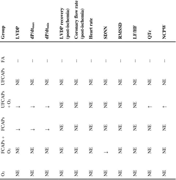
2.5 Summary of Effects……….55
3.1 The Effect of Acrolein and Ozone Exposure on Heart Rate………81 3.2 The Effect of Acrolein or Ozone Exposure on
Electrocardiogram Parameters………...81 3.3 Cardiac Mechanical Effects of Exposure to Acrolein or Ozone………..83
3.4 Summary of Exposure Effects……….84
4.1 Summary of Acrolein Exposure and Pharmacological Treatment Effects on Measures of Heart Rate Variability and Occurances
of Arrhythmia in Wild-type and TRPA1 KO Mice………...…………108 4.2 Summary of Acrolein Exposure and Pharmacological
Treatment on Ventilatory Parameters in Wild-type and
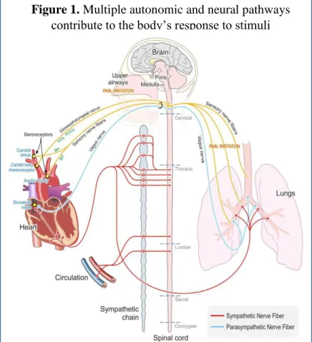
LIST OF FIGURES 1.1 Multiple Autonomic and Neural Pathways Contribute
to the Body’s Response to Stimuli………..……..16 1.2 Acute Air Pollution Exposure Produces Cardiovascular
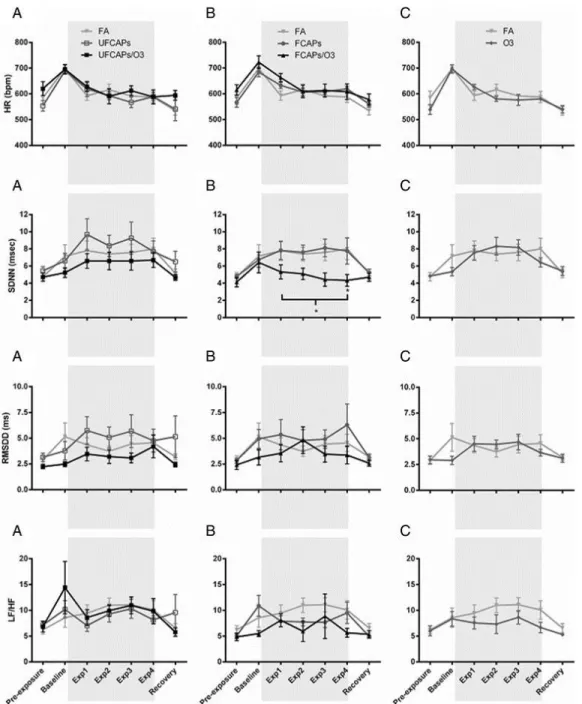
Dysfunction via Autonomic Imbalance Mediated by TRPA1………..………24 2.1 The Effect of CAPs With and Without Ozone on Heart Rate
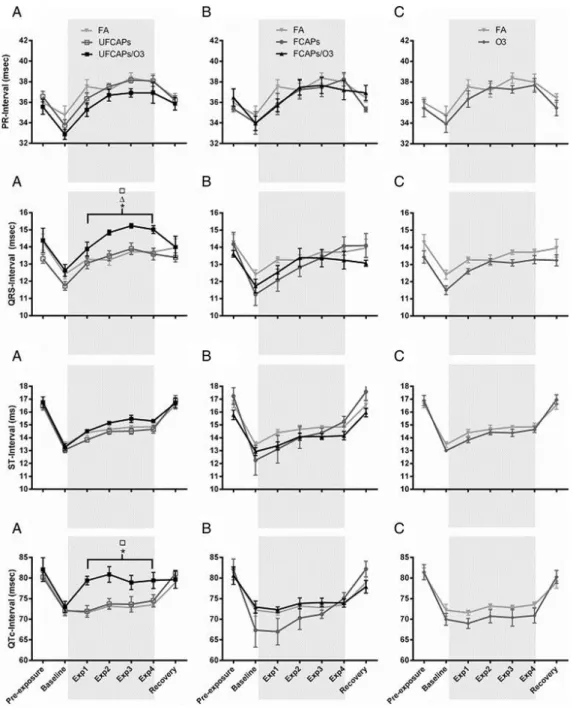
and Heart Rate Variability……….……...56 2.2 Electrocardiogram Effects Before, During and After Exposure
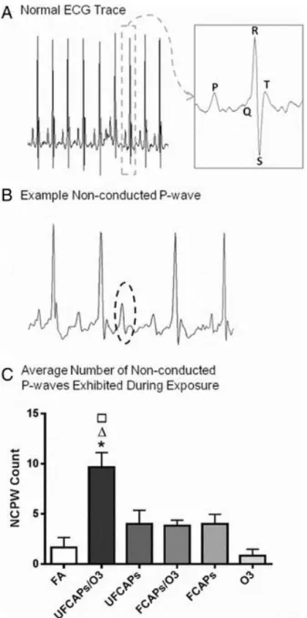
to CAPs Alone or With Ozone……….57 2.3 Typical Mouse Electrocardiogram and Arrhythmia During
Exposure………..…58 2.4 Effects of CAPs Exposure on Left Ventricular Developed
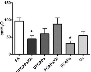
Pressure (LVDP)……….….59 2.5 Effect of CAPs Exposure with and without Ozone on
Rate of Left Ventricular Contractility and Relaxation……….60 2.6 Heart Rate Twenty Minutes After Ischemia-reperfusion……….61 2.7 Post-ischemia Recovery of LVDP……….…...61 3.1 Exposure to Acrolein but not Ozone Increases All Measures
of Heart Rate Variability………...………...85 3.2 Typical Electrocardiogram and Cardiac Arrhythmia in Mice
Exposed to Acrolein and Ozone……….….……….86 3.3 Exposure to Acrolein Increased Cardiac Arrhythmias in
Wild-type Mice………..……..87 3.4 Exposure to Acrolein Increases Left Ventricular Developed
Pressure 24 Hours Post-exposure………...………..87 4.1 TRPA1 KO Mice Demonstrate Altered Cardiac Autonomic
Modulation Compared with Wild-type Mice……….109 4.2 Wild-type and TRPA1 KO Mice Respond Similarly and
Predictably to Pharmacological Agents Affecting Autonomic
4.3 TRPA1 Mediates Acrolein-induced Increases in
Heart Rate Variability and Arrhythmia………..111 4.4 Pharmacological Treatments Affecting Cardiac Autonomic
Modulation Alter the Heart Rate and Heart Rate Variability
Response to Acrolein Exposure……….112 4.5 Pharmacological Treatments Affecting Cardiac Autonomic
Modulation Prevent Acrolein-induced Increases in Heart Rate
Variability and Arrhythmia………113 4.6 TRPA1 KO Mice Demonstrate Altered Ventilatory
Timing Compared to Wild-type Mice……….………...113 4.7 TRPA1 Mediates Acrolein-induced Changes in Ventilatory
Timing………….………...…114 4.8 Pharmacological Treatments Affecting Cardiac Autonomic
Modulation Alters the Ventilatory Response to Acrolein Exposure………….…….115 5.1 Autonomic Imbalance Shifts the Individual Toward Increased
Risk for Adverse Cardiac Events………125 5.2 Cardiac Dysfunction Following a Single Acute Exposure to
Air Pollution is the Result of Altered Autonomic Nervous
LIST OF ABBREVIATIONS ACE – angiotensin converting enzyme
ANOVA – Analysis of Variance ANS – Autonomic Nervous System BAL – Bronchoalveolar Lavage
CAPs – Concentrated Ambient Particles CDC – Centers for Disease Control CK – Creatine Kinase
COPD – Chronic Obstructive Pulmonary Disease CRP – C-reactive protein
dP/dT (-) – Negative Derivative of left ventricular Pressure over Time dP/dT (+) – Positive Derivative of left ventricular Pressure over Time ECG – Electrocardiogram
EPA – United States Environmental Protection Agency FA – Filtered Air
FCAPs – Fine Concentrated Ambient Particles (<2.5um) GTR – glutathione-S-transferase
HAPs – Hazardous Air Pollutants
HBDH - α-hydroxybutyrate dehydrogenase HF – High Frequency
HR – Heart Rate
LDH – Lactate dehydrogenase LF – Low Frequency
LVDP – Left Ventricular Developed Pressure MIA – Microalbumin
NAAQS – National Ambient Air Quality Standards NAG - N-acetyl-b-d-glucosaminidase
NCPW – Non-Conducted P-Wave NTS – Nucleus Tractus Solitarii O3 – Ozone
PM – Particulate Matter
RAR – Rapidly Adapting Receptor RAS – Renin-Angiotensin System
RMSSD – Root Mean Square of the Standard Deviation of subsequent NN intervals RSA – Respiratory Sinus Arrhythmia
SA – Sinoatrial
SAR – Slowly Adapting Receptor
SDNN – Standard Deviation of subsequent NN intervals SEM – Standard Error of the Mean
SOD – Superoxide dismutase SP – Substance P
SSS – Sick Sinus Syndrome
TRPV1 – TRP Subfamily V, Member 1
UFCAPs – Ultrafine Concentrated Ambient Particles (<100nm) VOC – Volatile Organic Compound
CHAPTER I: INTRODUCTION Air Pollution and Public Health
The health impacts of outdoor air pollution largely became apparent following the industrial revolution during the 19th and 20th centuries, when urban centers began to rapidly expand with dense concentrations of both homes and industrial areas, providing ideal conditions for a range of public health concerns including chronic diseases and daily
exposure to a variety of environmental toxicants. During this time coal was widely used for the heating of homes and powering of factories and eventually resulted in some of the most deadly air pollution incidents in modern history. The most widely cited of these is the 1952 London Great Smog episode, where combustion-related emissions combined with a
temperature inversion resulted in the excess deaths of an estimated 12,000 people (Bell & Davis 2001). Pollution levels between December 5th-9th in 1952 London were 50-300% increased compared with previous years (5-19 times current regulatory standards). The smog caused a sharp rise in hospital admission rates for both respiratory and cardiac diseases as well as pneumonia and influenza (Bell & Davis, 2001; Logan, 1956). Two years earlier in 1948 a similar episode had occurred in the U.S., in the small town of Donora, Pennsylvania where 20 of the town’s 14,000 residents died and over 7000 were hospitalized for
The first U.S. federal legislation addressing air pollution was the Air Pollution Control Act of 1955. This act declared that air pollution posed a risk to public health, and authorized the surgeon general to work with state and local air pollution control agencies to recommend research programs and develop methods to reduce or eliminate ambient air pollution. However it maintained that individual states had the responsibility to establish their own air pollution controls. The Air Pollution Control Act primarily provided funds for the federal government to research the sources and effects of air pollution (U.S. Congress 1955). In 1963 the Clean Air Act was passed, putting the onus of air pollution regulation on the federal government. This act required the U.S. Department of Health to create and enforce automobile emissions standards. The Clean Air Act of 1970 further expanded on the federal government’s ability to develop and enforce air pollution standards and controls from both stationary and mobile sources (US EPA 2016). Also in 1970 the Environmental
Protection Agency (EPA) was established for the purpose of consolidating federal research and monitoring programs pertaining to environmental and public heath, as well as developing and enforcing environmental pollution standards. A major component of the 1970 Clean Air Act was the establishment of the National Ambient Air Quality Standards (NAAQS) which today designates six criteria air pollutants that are routinely and broadly monitored by the EPA. These include ambient particulate matter (PM), ozone, carbon monoxide, sulfur dioxide, nitrogen dioxide, and lead (US EPA 2016). In addition to the six criteria air
pollutants the EPA has identified and has regulatory standards for a list of 187 hazardous air pollutants (HAPs) which have the capacity to increase rates of cancer, produce other serious health impacts, or have significant environmental impacts (US EPA). Even with more
of air pollution is an estimated 200,000 premature deaths per year in the U.S (Caiazzo et al. 2013) and 3.7 million premature deaths per year worldwide (WHO, 2012), the vast majority arising from cardiopulmonary injury or disease.
Present day ambient air pollution comprises a diverse mixture of substances including particulate patter (PM), gases, organic compounds, and toxic metals (Brook et al. 2010; Verrier et al. 2002). PM exposure in particular has been linked with a variety of adverse cardiovascular outcomes including myocardial infarction, stroke, arrhythmia, and exacerbation of heart failure. However the 2010 American Heart Association scientific statement pointed out that, “Although PM2.5 mass has rightfully attracted attention as a target for regulation and epidemiological study, more than 98% of the air pollutant mass in the mixture we breathe in urban settings is from gases or vapor phase compounds such as CO, volatile organic carbons (OCs), NO2 , NO [nitric oxide], O3 , and SO2 .” (Brook et al. 2010). Each of these may have independent effects or combined effects with co-pollutants. Aldehydes, a class of volatile organic compounds, have attracted interest since the earliest days of air pollution research due to their ubiquitous and highly irritating nature (Quade R . Stahl 1969).
Acrolein
damage or functional changes which may result in cytotoxicity, oxidative stress and inflammation, as well as react with non-specific binding sites on dermal and airway nociceptive receptor channels. Humans are primarily exposed to acrolein via contact with cigarette smoke, automobile exhaust, structural and wildfire smoke, and some industrial processes including the burning of wood or coal (CDC-ASTDR 2007; Ghilarducci &
Tjeerdema 1995). In 1999 estimated ambient acrolein concentrations ranged from a national average of 0.033 ppm to a high of 0.19 ppm (Woodruff et al. 2007; Walsh 2008). Cigarette smoke contains up to 90 ppm acrolein, and acrolein levels in side-stream tobacco smoke have been measured as high as 10 ppm (Esterbauer et al. 1991). The U.S. EPA currently classifies acrolein as a HAP and determined that it represents the leading non-cancer hazard in ambient air pollution causing adverse respiratory health effects (Ris 2007). Acrolein is known to primarily impact the upper airway, with nasal mucosa representing the most sensitive target of inhalation exposure (Shusterman 2012; CDC-ASTDR 2007), although the effects can extend deeper into the respiratory tract as exposure levels increase (CDC-ASTDR 2007). Inhalation of acrolein induces a variety of respiratory effects including nose and throat irritation (Beauchamp, Andjelkovich, Kligerman, Morgan, & Heck, 1985; Sim & Pattle, 1957), apnea (Lee et al. 1992), pulmonary edema (Hales et al. 1989; Kutzman et al. 1985), asthma like symptoms such as increased bronchial responsiveness and respiratory distress (Ben-Jebria et al. 1994; Bein & Leikauf 2011), and has been shown to exacerbate existing asthma (Leikauf et al. 1989). Acrolein exposure has also been shown to produce
produces short-lived effects including bradycardia (Lee et al. 1992), while chronic exposure to inhaled acrolein can produce dilated cardiomyopathy (Ismahil et al. 2011) and contributes to the development of thromboses (Sithu et al. 2010; O’Toole et al. 2009).
Mechanisms Linking Pulmonary Exposure to Cardiovascular Effects
Historically, airway and pulmonary sites were believed to be the primary target of air pollution. However, now systemic effects including those on the central nervous system, metabolism and cardiovascular systems have been drawing increasing attention over the last 15 to 20 years (Calderón-Garcidueñas et al. 2007; Mills et al. 2009; Kodavanti 2015; Wagner et al. 2014). In fact, a growing body of evidence demonstrates that elevations in ambient air pollution, particularly PM, results in greater risks for cardiovascular related mortality than for any other causes including respiratory disease (Brook et al. 2010).
Biological mechanisms linking air pollution with adverse cardiovascular outcomes involve both the direct effects of pollutants on cardiovascular tissues and indirect effects mediated through more general pathways including oxidative stress and inflammation. Putative mechanism include 1.) Systemic inflammation and oxidative stress, 2.) The direct effect of agents which may readily pass through the pulmonary epithelium into the blood, and 3.) Perturbation of systemic autonomic nervous system balance by air-pollutant
interactions with airway sensory receptors (Simkhovich et al. 2008; Brook et al. 2004; Brook et al. 2010).
cells, as well as vasoactive molecules (Grunig et al. 2014). Additionally, sustained airway inflammation sensitizes airway sensory nerves, provoking the further release of
proinflammatory neurotransmitters and enhancing the proinflammatory state of the airways (Zholos 2015; Lee & Widdicombe 2001). Moreover prolonged periods of oxidative stress can deplete the airway antioxidant capacity, impairing a primary defense mechanism of the airway mucosa and producing further tissue damage (Domej et al. 2014). Over time
profound airway inflammation may contribute to a state of systemic inflammation which can disrupt hemostatic pathways and impair vascular function. In fact, the primary adverse cardiovascular outcome linked with this mechanism is the development of atherosclerosis as well as its sequelae (Park et al. 2010; Gill et al.; Provost et al. 2015).
Constituents of air pollution capable of crossing the pulmonary epithelium include gases, soluble constituents of particulate matter such as organic compounds or metals, and possibly ultra-fine particles (Brook 2008). Since oxygenated blood leaving the lungs makes its first pass through the heart before entering systemic circulation the largest effect of translocated constituents of air pollution would be on the myocardium itself (Nemmar et al. 2002; Nemmar et al. 2004).
In addition, the airways are richly innervated with sensory nerves that react to irritating or noxious stimuli and initiate protective physiological responses (Widdicombe 2001; Lee & Pisarri 2001; Coleridge et al. 1983). These responses are mediated by reflexive changes in autonomic nervous system regulation of cardiopulmonary function which
system balance and cardiovascular function. Under normal physiological conditions, this system ensures that the body maintains its function (e.g. perfusion of tissues, oxygenation, anti-oxidant potential) during changing conditions or even certain extremes. Exposure to an environmental stressor can result in perturbation of internal homeostatic mechanisms
including autonomic nervous system controls such that a subsequent trigger might
significantly increase the risk of adverse cardiovascular events like arrhythmia (Brook et al. 2004). In individuals with underlying cardiovascular or pulmonary disease, the disruption of autonomic balance may actually result in cardiovascular morbidity or mortality given they already have impaired cardiopulmonary compensatory capacity (Goldberg et al. 2001; Routledge et al. 2003; Brook et al. 2004).
Airway Sensory Nerves
worsening of existing disease but also sudden impairment in people who are seemingly healthy.
As such, chemosensory nerve endings in the airway mucosa are one of the first lines of defense against noxious chemicals and stimuli. Activation of these “sensors” trigger crucial protective reflexive responses such as coughing, sneezing and the sensation of irritation and pain (Widdicombe 2001; Bessac & Jordt 2008; Carr & Undem 2003). The respiratory system, from the nose into the deep lung, is innervated by multiple nerve types which have the ability to respond to a variety of stimuli including irritating molecules, cold, changes in osmolality, and changes in lung inflation (Perez et al. 2015; Bessac & Jordt 2008). The cell bodies of airway sensory nerve fibers reside in the jugular and nodose ganglia of the vagus nerve and project to the nucleus tractus solitarius (NTS) in the medulla. Afferent activity arising from airway sensory nerves is transmitted into the central nervous system (i.e. brainstem) where the signal is modulated and then eventually results in efferent signaling via autonomic nerves to the viscera (Carr & Undem 2003; Bonham et al. 2006). As mentioned above, this homeostatic system functions tonically on a breath-by-breath basis affecting respiratory and cardiovascular activity (e.g. respiratory sinus arrhythmia) but is also then immediately altered in the event of exposure.
role of these sensory fibers, as detailed above, is the modulation of HR and cardiac output in response to variation in breathing patterns that are both normal and abnormal (i.e. occurring due to irritation). This includes respiratory sinus arrhythmia (RSA), which is heart rate variability that occurs in synchrony with respiration and causes heart rate to increase during inspiration and decrease during expiration (Perez et al. 2015; Widdicombe 2001). Yet, in terms of air pollution health effects, the most important sensory fibers appear to be the C-fibers, which are characterized by their distinct sensitivity to both exogenous chemical stimuli including sulfur dioxide, acids, cigarette smoke, ozone, acrolein, ammonia and particulate matter, as well as endogenous inflammatory mediators (Kollarik et al. 2010) and which morphologic studies have demonstrated to be seventy-five percent of vagal afferent nerve fibers arising from the respiratory tract (Agostoni et al. 1957).
C-fibers were first identified by Coleridge and Coleridge in 1984 (Coleridge & Coleridge 1984) and since then they have become recognized as important regulators of cardiopulmonary function under both normal and abnormal physiologic conditions. General anatomical and functional features of C-fibers are well conserved across most species in which they have been studied including human, monkey, dog, cat, rabbit, guinea pig, rat, mouse and (Canning & Chou 2009). They are located throughout the respiratory tract
(Coleridge & Coleridge 1984), however more moderate levels of pulmonary fiber discharge, akin to the tonic level of pulmonary C-fiber activation physiologically present without robust exogenous stimulation, produces an increase in respiratory rate and a decrease in tidal
volume (Lee & Pisarri 2001). C-fiber activation also causes reflex bronchoconstriction and airway mucus secretion (Lee & Pisarri 2001). Cardiovascular reflex responses to C-fiber activation include bradycardia along with systemic and pulmonary hypotension (Perez et al. 2015; Lee & Pisarri 2001) all of which are primarily mediated through enhanced vagal modulation through the brainstem (Lee et al. 1992). On the other hand, local effects in the airways are mediated via the release of neuropeptides including substance p, neurokinin A and calcitonin gene related protein neuropeptides (Saria et al. 1988); these include
bronchoconstriction and the development of pulmonary edema, and induction of neuroinflammation (Lee & Pisarri 2001; Kollarik et al. 2010).
2001). Enhanced C-fiber excitability is believed to play a role in the pathophysiology of number of chronic airway diseases including asthma and COPD.
TRPA1
Transient receptor potential ion channels (TRP) are expressed throughout the upper and lower airways on trigeminal and vagal afferent C-fibers. In 1997 successful cloning of the TRP capsaicin, or vanilloid receptor, TRPV1 was reported (Caterina et al. 1997) and immunohistochemical studies confirmed the presence of TRPV1 on the airway sensory fibers in the nasal mucosa, trachea, bronchi and alveoli (Bessac & Jordt 2008). These discoveries started a new wave of chemosensory research with hundreds of studies dedicated to the identification of TRP family members, their pharmacology, structure, function, and
physiological role in chemosensation and nociception in the airways (Eid & Cortright 2009). Initially identified in 1999 as ANKTM1, a theoretical protein expressed in cultured
fibroblasts (Jaquemar et al. 1999), TRP ankyrin 1 (TRPA1) was rediscovered in 2003 as a cold-activated TRP channel found in a subset of nociceptive sensory nerve fibers where it is co-expressed with TRPV1 (Story et al. 2003).
stimuli, but also displays activation by a variety of noxious compounds, many of which evoke cold and pain sensations (Story et al. 2003).
In 2006 a TRPA1 knockout mouse was developed (Kwan et al. 2006). These mice were generated by deleting exons that code for the pore domain of the ion channel, rendering the channels non-functional. Constitutive knockout animals were backcrossed to the
C57BL/6J line for over 10 generations to produce a TRPA1 knockout animal that is essentially in the C57BL/6K genetic background. These mice demonstrate no behavioral responses to topical application of mustard oil, cold (0 C), or punctate stimuli. They do however demonstrate normal startle reflex to loud noise, a normal sense of balance, and normal auditory brainstem response (Kwan et al. 2006). It has also been reported that TRPA1 knockout mice not respond to inhaled oxidants but do respond predictably to the non-TRPA1 specific C-fiber irritant acetic acid (Bessac et al. 2008). Implying that the model’s response to non-TRPA1-activating exposure types is not impaired.
TRPA1 has been investigated thoroughly in its role as a nociceptor in dermal pain pathways, particularly with regard to cold or noxious chemical sensation, and
neuroinflammation (Bautista et al. 2006; Bodkin & Brain 2011; Guimaraes & Jordt 2007). Because of this TRPA1 presents a ready target in the development of anti-inflammatory drugs and analgesics (Brederson et al. 2013; Chen et al. 2011). It is also fairly well
understood as an airway sensor (Bautista et al. 2006) and has been suggested previously as a mediator of cardiovascular effects via reflex modulation of autonomic reflex function (Hooper et al. 2015; Pozsgai et al. 2010). More recently a direct role for TRPA1 in cardiovascular function has been investigated and found that TRPA1 expressed in the vasculature may act as a cold and/or oxidant sensor there as well as contribute to
Due to its presence on pulmonary C-fibers known to initiate autonomic reflex arcs upon activation, its propensity toward activation by a large variety of substances commonly found in ambient air pollution including acrolein, and the availability of a knockout mouse model, TRPA1 represents a prime target for research as a mediator of airway irritant induced cardiovascular dysfunction via the perturbation of autonomic balance and homeostasis.
Autonomic Control of the Heart
The autonomic nervous system is the division of the peripheral nervous system responsible for control of bodily functions not consciously directed, such as breathing, the heartbeat, and digestive processes. It is split into two branches: sympathetic and
parasympathetic. In situations where an organism encounters a stressor the sympathetic nervous system dominates; heart rate and respiration increase, blood pressure increases, and blood is redirected to muscles and organs important to the “fight or flight” response and away from non-essential areas such as the gut. When the stressor is removed and the
organism is returning to a resting state the parasympathetic nervous system dominates; heart rate and respiration slow, and activity in the gut recommences, hence the phrase “rest and digest”. While the actions of the sympathetic and parasympathetic branches of the
autonomic nervous system do oppose each other to some extent, it is not a reciprocal system: when one rises the other does not automatically fall. There can be activation or
co-depression or any degree of activation of either branch simultaneously. Furthermore the actions of either branch of the autonomic nervous system are not systemically uniform; even in a resting state different tissues and organ systems are variably affected by either
primarily parasympathetically dominated while the vasculature is controlled by the
sympathetic branch. Overall the important aspect of the relationship between the actions of the sympathetic and parasympathetic nervous systems is that they are coordinated such that cardiovascular homeostasis is maintained.
Like all vital bodily systems the cardiovascular system is dynamic, reacting moment to moment to both internal and environmental stimuli and adjusting physiological parameters such as heart rate, contractility, and blood pressure to ensure sufficient systemic perfusion and meet the ever changing metabolic demands of the body. The regulation of this complex balancing act is traditionally described in terms of homeostasis. This word comes from the Greek homeo (similar) and stasis (steady) and refers to the body's ability to return toward a relatively stable internal set point, even while continually subjected to external changes. An important aspect of cardiovascular homeostasis is heart rate. Heart rate is normally
(HRV) represents a valuable source of information that can provide insight into the mechanisms of cardiovascular control (Appel et al. 1989).
Heart Rate Variability HRV has become an increasingly popular
endpoint for analysis of autonomic function,
because of its non-invasive nature and because of its applicability in both humans and animal models
including rodents (Farraj, Hazari & Cascio 2011; Rowan et al. 2007; Thireau et al. 2008). Since a simple ECG readout and HR
analysis is all that is required for HRV calculation it is now used commonly as a prognostic indicator in clinical settings (Anon 1996) and in commercially available software used in the fields of psychology and sports medicine. Two broad approaches to calculating HRV are time and frequency domain analysis. Time domain analysis are basic statistical analyses of normalized RR intervals: the standard deviation of subsequent normalized RR intervals (SDNN) and the root mean square of the standard deviation of subsequent normalized RR
Perez et al., 2015. Cardiovasc Toxicol. 2015 Jan;15(1):69-78
intervals (RMSSD). SDNN represents all cyclic components contributing to variability, therefore representing an overall measure of HRV while RMSSD more specifically correlates with parasympathetic modulation (Rowan et al. 2007). HRV can also be calculated via frequency domain analysis whereby high or low frequency bands are assigned and power spectral density analysis is performed using a fast Fourier transformation. This provides basic information regarding the power across a low frequency (LF) vs high frequency (HF) bands. LF power is believed to represent a mixture of sympathetic and parasympathetic components as well as inputs from mechanisms governing blood pressure regulation (Reyes del Paso et al. 2013; Goldstein et al. 2011; Rahman et al. 2011; Moak et al. 2007) while HF power again correlates with parasympathetic modulation but likely also represents inputs from changes in cyclic ventilatory patterns (Billman 2013; Reyes del Paso et al. 2013; Chess et al. 1975; Piccirillo et al. 2009; Heathers 2014).
Changes in HRV have been observed in various stressful circumstances including during exercise (Perini & Veicsteinas 2003; Baumert et al. 2006), in response to fearful stimuli (Liu et al. 2014) and in response to perceived stress (Dishman et al. 2000). Both acute and chronic stress have been shown to induce an overall decrease in HRV whereby some combination of increased sympathetic modulation and vagal withdrawal occurs (Liu et al. 2014; Schubert et al. 2009). With repeated stressful stimulus however habituation is observed, attenuating the intensity and duration of the HRV response (Liu et al. 2014).
responsiveness to both external stimuli and internal control mechanisms (Rahman et al. 2002; Lipsitz & Goldberger 1992; Pincus 1994; Schubert et al. 2009), representing a risk for
adverse cardiovascular events due to reduced compensatory capacity. In fact it has been demonstrated that decreased HRV occurs over a broad range of cardiovascular illnesses and injury (Gang & Malik 2003; Thayer et al. 2010; Tsuji et al. 1996). Reduced HRV has been associated with increased risk of fatal arrhythmias, heart attack and stroke, particularly in populations with existing cardiovascular disease (Ponikowski et al. 1997; Valkama et al. 1995; Kop et al. 2001). Significantly, low HRV has also been associated with adverse cardiovascular events in apparently healthy populations as well (Hillebrand et al. 2013). Although classically low HRV has been accepted as an indicator of heightened
resulting ECG features may be originating in the central nervous system (rather than degeneration of SA node itself).
Ambient air pollution, depending on its physical and chemical composition, has the capacity to act as a potent chemical stressor and has been shown to elicit cardiac autonomic responses (Arjomandi et al. 2015; Gold et al. 2000; Pope et al. 1999; Godleski et al. 2000). While decreased HRV is a commonly accepted indicator of heightened cardiovascular risk following air pollution exposure in humans (Gold et al. 2000), increases HRV have also been demonstrated (Chen & Hwang 2005; Bortkiewicz et al. 2014). For example, increased
RMSSD has been shown to be associated with elevated risk of air pollution-induced arrhythmia (Davoodi et al. 2010) In fact our lab has consistently shown that exposure to different types of air pollutants such as residual oil fly ash (Farraj, Hazari, Haykal-Coates, et al. 2011; Carll et al. 2015) diesel exhaust (Hazari et al. 2011; Carll et al. 2013), ozone (Farraj et al. 2012) and acrolein (Hazari et al. 2014) all lead to an increase in HRV in rodent models. Considering that parasympathetic modulation is a well-characterized reflex response to airway sensory activation (Lee & Pisarri 2001; Alarie 1973; Bessac & Jordt 2010) these results are not surprising.
Interestingly there is also evidence that abnormal variation in heart rate and heart rate variability as seen with enhanced vagal tone (and sick sinus syndrome) has a destabilizing effect on the atrial tissue (Shetty & Scott 2015) which can contribute to the
events but through this mechanism can exacerbate and may also physically worsen their condition.
Taken together it is apparent that any perturbation of homeostatic balance has the potential to increase risk of adverse cardiovascular events if the shift is drastic enough or combined with other perturbation causing stimuli. In that sense, HRV changes in either direction might indicate that sympathetic and parasympathetic nervous systems are not properly coordinating to provide an appropriate heart rate response to environmental stressors or internal cues.
TRPA1: Connecting the Airways and Heart through Central Neural Circuits
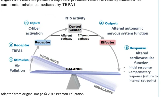
the NTS plays a critical role in the integration of a wide range of peripherally initiated sensory information in addition to airway receptor activation, including baroreflex and chemoreflex receptors activity, as well as the central control of cardiovascular function (Lawrence & Jarrott 1996; Potts). Thus it is reasonable to assert that cardiovascular and respiratory reflex responses to C-fiber activation are similarly mediated via substance P release in the NTS. It is this arc of neural activity (Figure 2.) that may determine the final cardiovascular outcome. The key question as research moves forward in this field is how long do these effects persist and how can the key mediators be identified in those most susceptible? With regards to the latter, I hypothesize that alteration of homeostatic
mechanisms like autonomic control by air pollution exposure, however short-lived, will alter those regulatory processes that employ autonomic efferent signals to maintain a given function. For example, we have already demonstrated that acrolein exposure desensitizes the baroreflex response by blunting the sympathetic outflow to the heart (Hazari et al. 2014). Thus, it may not necessarily be the exposure that directly causes the adverse response but rather some other subsequent event that further “pushes” the body out of balance.
Summary
relationship between high air-pollution days and a significant increase in hospital visits for serious cardiovascular events, particularly in populations with existing cardiovascular
disease, highlight the need for a more complete understanding of the mechanisms underlying this relationship (Colais et al. 2012; Dockery et al. 1993; Pope et al. 2004). On the other hand, when air pollution levels are not exceedingly high, or in the case of acute exposure, it is assumed that the effects are minimal or even non-existent, especially when clinically observable symptoms are lacking. As such, subtle effects may go unnoticed but nonetheless represent a serious risk (Hazari et al. 2011). At these lower and seemingly benign air pollution concentrations or durations, and in the absence of host perception of an effect, it would be suitable to examine the role of airway sensory nerves which not only “sense” the toxicant but also cause internal homeostatic changes that may predispose to cardiopulmonary dysfunction. Airway sensors, particularly TRPA1, located on airway C-fibers have been demonstrated to play a major role in airway reflex responses, and have been suggested as possible mediators of some cardiovascular responses as well (Hazari et al. 2011; Jones et al. 1995; Lee 2010; Wang et al. 2000)(Lee 2010; Wang et al. 2000; Jones et al. 1995; Hazari et al. 2011). The studies presented herein explore TRPA1 as a major sensor of airway irritant pollutants and address the mechanisms through which it produces autonomic imbalance and adverse cardiovascular effects following exposure to air pollution.
Purpose of Research and Global Hypothesis
the risk of cardiac events and arrhythmias increase significantly in the hours and days immediately following air pollution exposure. Yet, epidemiological studies by their nature cannot quantify the physiological response in humans during exposure; this is crucial
information in air pollution toxicology because it can shed light on the early initiating events resulting from exposure which influence and direct subsequent pathways in the hours and days following exposure. Although numerous biological pathways have been proposed by which air pollution may cause cardiovascular events, airway sensory activation may be the most relevant when considering the short-term, reversible, and often latent effects of an acute exposure. Toxic air pollutants, including particulate matter, acrolein, and ozone stimulate sensory nerves in the airways causing several well-described ventilatory and pulmonary effects, however the cardiovascular responses have not been sufficiently characterized.
A subset of these responses are known to be mediated by the transient receptor potential cation A1 (TRPA1) channels present on chemosensitive C-fibers. Thus, TRPA1 represents a likely candidate as an initiator of air-pollution induced cardiovascular
dysfunction. The mechanism through which airway sensory nerve activation perturbs cardiopulmonary function has been broadly described as occurring through modulation of autonomic nervous system function, however specific changes in measures of autonomic modulation have not yet been clearly characterized or linked with changes in
induced cardiotoxicity, which will contribute relevant data to improve assessments of risk, as well as identify potentially sensitive subpopulations and define windows of heightened susceptibility following exposure.
In order to address this hypothesis the following specific aims were established:
Aim 1) Characterize decrements in cardiac mechanical and electrical function observed in the mouse with exposure to air pollution. The aim of this study was to determine the independent effects of fine (FCAPs) and ultrafine (UFCAPs) concentrated ambient particles on cardiac function in C57BL/6 mice, and explore whether ozone (O3) co-exposure would alter the response. These experiments examined a wide range of cardiac endpoints including electrical function and rhythm, which were assessed via
electrocardiogram (ECG) analysis, mechanical properties and post-ischemia recovery, which were measured using a Langendorff technique, as well as biochemical indicators of cardiac
Figure 2. Acute air pollution exposure produces cardiovascular dysfunction via autonomic imbalance mediated by TRPA1
toxicity. This work fills a unique gap in the existing literature as few studies, if any, have examined the effects of simultaneous particulate matter (PM) and O3 exposure on both ECG and mechanical function (e.g. contractility) of the heart. Risk assessments of air pollution health effects have become increasingly challenging given the complexity of present-day air pollution mixtures. Previous data suggests that PM size determines the physiological impact with fine PM causing primarily pulmonary effects and ultrafine PM altering cardiac function. However, air pollution is a mixture of not only PM, but also gaseous irritants, vapors, and biological substances. Thus when examining the effects of a given pollutant, the influence of other components must be considered, and so studies are needed to determine whether the physiological and biochemical responses to multipollutant exposures represent the simple additive effects of the pollutants, their synergism or antagonism. One particularly relevant interaction is that of PM and the ubiquitous gaseous co-pollutant O3. Thus, we hypothesized (1) that inhalation of either fine (FCAPs) or ultrafine CAPs (UFCAPs) would cause cardiac electrical dysfunction, mechanical decrements and arrhythmogenesis in mice; but (2) that UFCAPs, due to its size, would have a greater effect on the heart than FCAPs; and (3) that O3 co-exposure would potentiate the response elicited by both particle sizes.
Aim 2) Determine the role of TRPA1 in acute cardiovascular dysfunction during
exposure to air pollution. Wild-type C57BL/6 (WT) and TRPA1 knockout (KO) mice were exposed to prototypical airway irritants acrolein and ozone to determine the role of TRPA1 in air pollution-induced cardiovascular dysfunction. The cardiovascular response during
interval analysis, heart rate variability (HRV) analysis, and identification and quantification of arrhythmia were the primary endpoints. Potential pollutant-driven alterations in cardiac mechanical function were evaluated post-exposure using a Langendorff cardiac perfusion preparation. The majority of research on TRP channels done thus far has utilized in-vitro methods or isolated organ perfusion systems. While these techniques provided a great deal of insight regarding channel activation and elucidation of the neural pathways potentially involved in the adverse cardiopulmonary response to airway nerve activation, physiological endpoints in the whole organism could only be inferred. Furthermore, the few studies identified which used whole organism, including the TRPA1 knockout mouse, generally anesthetized the animal prior to exposure, potentially confounding a reflex response. Previous studies from this lab have demonstrated that pharmacological inhibition of TRPA1 attenuated the development of diesel exhaust-induced arrhythmia. Thus, this study sought to further establish a role for TRPA1 in air pollution induced cardiac dysfunction and we hypothesized that adverse cardiac electrical and mechanical responses due to acrolein or ozone were mediated by TRPA1.
Aim 3) Characterize baseline autonomic tone and the effects of pharmacological inhibition of sympathetic or parasympathetic neurotransmission in wild-type and TRPA1 knockout mice with exposure to airway irritants.
fully determine the degree of autonomic modulation which results from TRPA1-mediated sensory activation in the airways we need to better understand baseline cardiac autonomic
properties in our model, as well as how these measures change with exposure. Hence, I explored how pharmacological blockade of each autonomic branch affects measures of HRV as well as the cardiac response to air pollution in mice. Wild-type C57BL/6 (WT) and TRPA1 (KO) mice were treated with one of three pharmacological agents producing either sympathetic blockade, parasympathetic blockade, or blockade of both branches of the
autonomic nervous system, before being exposed to filtered air or acrolein. We hypothesized that pharmacological disruption of normal autonomic balance in the mouse would disrupt
basic cardiovascular function as shown by heart rate and heart rate variability as well as blunt
CHAPTER II: OZONE CO-EXPOSURE MODIFIES CARDIAC RESPONSES TO FINE AND ULTRAFINE AMBIENT PARTICULATE MATTER IN MICE: CONCORDANCE OF ELECTROGRAM AND MECHANICAL RESPONSES
Studies have shown a relationship between air pollution and increased risk of cardiovascular morbidity and mortality. Due to the complexity of ambient air pollution composition, recent studies have examined the effects of co-exposure, particularly particulate matter (PM) and gas, to determine whether pollutant interactions alter (e.g. synergistically, antagonistically) the health response. This study examines the independent effects of fine (FCAPs) and ultrafine (UFCAPs) concentrated ambient particles on cardiac function, and determine the impact of ozone (O3) co-exposure on the response. We hypothesized that UFCAPs would cause greater decrement in mechanical function and electrical dysfunction than FCAPs, and that O3 co-exposure would enhance the effects of both particle-types. Conscious/unrestrained radiotelemetered mice were exposed once whole-body to either 190 μg/m3 FCAPs or 140 μg/m3 UFCAPs with/without 0.3 ppm O
3; separate groups were
rate variability when compared to FA but also blocked the decrement in cardiac function. On the other hand, O3 co-exposure with UFCAPs significantly increased QRS-interval, QTc and non-conducted P-wave arrhythmias, and decreased LVDP, rate of contractility and relaxation when compared to controls. These data suggest that particle size and gaseous interactions may play a role in cardiac function decrements one day after exposure. Although FCAPs + O3 only altered autonomic balance, UFCAPs + O3 appeared to be more serious by increasing cardiac arrhythmias and causing mechanical decrements. As such, O3 appears to interact differently with FCAPs and UFCAPs, resulting in varied cardiac changes, which suggests that the cardiovascular effects of particle-gas co-exposures are not simply additive or even generalizable. Additionally, the mode of toxicity underlying this effect may be subtle given none of the exposures described here impaired post-ischemia recovery.
Introduction
Although studies have examined the effects of sequential exposures, for example, ozone (O3) and then PM2.5 causes decreased HRV, systolic blood pressure and heart rate (HR) in rat (Wang et al. 2013), only a few studies have addressed the health effects of simultaneous exposures with distinct pollutants and the effects are still not fully clear. For instance, Brook et al. demonstrated acute arterial vasoconstriction in healthy subjects co-exposed to PM2.5 and O3 (Brook et al. 2002), whereas Urch et al. (Urch et al. 2005) found no significant changes in mean arterial pressure, systolic blood pressure or HR in a similar study population; although constriction was observed with PM2.5 alone. Animal studies also indicate that the effect of combining pollutants does not necessarily yield the expected synergistic response, especially in the case of susceptible models. Wagner et al. recently showed that depression of heart rate and blood pressure during PM2.5 and O3 co-exposure was not as great as either pollutant alone in rats fed a high-fructose diet (Wagner et al. 2013). The respiratory effects of O3 and PM co-exposure are equally conflicting. For example, rats instilled with ozonized DEP had increased inflammatory cells and protein in the lungs (Madden et al. 2000), whereas mice co-exposed to O3 and DEP did not have increased cytotoxicity or inflammation (Farraj et al. 2010). Instead, in this latter study, co-exposed mice had increased bronchoconstriction, which is a measure of lung function. Similar investigations into the effects of simultaneous exposure on cardiac function have not been widely conducted.
responses demonstrated by controlled human and animal PM exposure studies have provided biological plausibility to the health effects of air pollution (Brook et al. 2010; Dockery et al. 1993; Kodavanti et al. 2011; Peters 2005). Some of these are responses observed using ECG and have been shown to be similar in humans and animals (Hazari, Callaway, Winsett, Lamb, Haykal-Coates, et al. 2012; Mills et al. 2007). For instance, some human subjects exposed to PM have decreased heart rate variability (HRV), which is a predictor of increased risk (Tsuji et al. 1996; Tsuji et al. 1994; Gold et al. 2000; Gong et al. 2004), and enhanced arrhythmogenesis (Zareba et al. 2009). Experiments in animals not only show a similar PM-induced decrease in HRV and increased incidence of arrhythmia (Brook et al. 2010), but also functional decrements in the heart such as diminished left ventricular developed pressure (LVDP) and decreased contractility (Chen & Hwang 2005; Gurgueira et al. 2002; Hwang et al. 2008). On the other hand, few studies, if any, have examined the effects of simultaneous PM and O3 exposure on both ECG and mechanical function (e.g. contractility) of the heart.
Materials and Methods
Animals - Ten to twelve-week old female C57BL/6 mice (body weight = 21.6 ± 0.1 g) were used in this study (Jackson Laboratory - Bar Harbor, ME). Mice were initially housed five per cage and maintained on a 12-hr light/dark cycle at approximately 22°C and 50% relative humidity in an AAALAC–approved facility. Food (Prolab RMH 3000; PMI Nutrition International, St. Louis, MO) and water were provided ad libitum. Each mouse implanted with a radiotelemeter was singly housed after surgery. All protocols were approved by the Institutional Animal Care and Use Committee of the U.S. Environmental Protection Agency and are in accordance with the National Institutes of Health Guides for the Care and Use of Laboratory Animals. The animals were treated humanely and with regard for alleviation of suffering.
Experimental Groups - Mice were randomly assigned to one of six exposure groups: (1) fine concentrated ambient particles (FCAPs); (2) ultrafine CAPs (UFCAPs); (3) ozone (O3); (4) FCAPs and O3 co-exposure (FCAPs + O3); (5) UFCAPs and O3 co-exposure (UFCAPs + O3); and (6) filtered air (FA). Each group had n = 6. Separate groups (same as above) of mice were used for Langendorff cardiac perfusion experiments (n = 5-8).
(ETA-F10, Data Sciences International, St Paul, MN); the transmitter was placed under the skin to the right of the midline on the dorsal side. The two electrode leads were then tunneled subcutaneously across the lateral dorsal sides; the distal portions were fixed in positions that approximated those of the lead II of a standard electrocardiogram (ECG). Body heat was maintained both during and immediately after the surgery. Animals were given food and water post-surgery and were housed individually. All animals were allowed 7-10 days to recover from the surgery and reestablish circadian rhythms.
Radiotelemetry data acquisition - Radiotelemetry methodology (Data Sciences
International, Inc., St. Paul, MN) was used to track changes in cardiovascular function by monitoring heart rate (HR), and ECG waveforms immediately following telemeter
implantation, through exposure until 24 hours post-exposure. This methodology provided continuous monitoring and collection of physiologic data from individual mice to a remote receiver. Sixty-second ECG segments were recorded every 5 minutes during the pre- and post-exposure periods and continuously during exposure (baseline and hours 1-4); HR was automatically obtained from the waveforms (Dataquest ART Software, version 3.01, Data Sciences International, St. Paul, MN, USA).
determined for each ECG waveform: PR interval (Pstart-R), QRS complex duration (Qstart-S), ST segment interval (S-Tend) and QT interval (Qstart-Tend). QT interval was corrected for HR using the correction formula for mice QTc = QT/(RR/100)1/2(Mitchell et al. 1998). Figure 3A and B show a typical ECG trace as well as a typical non-conducted p-wave (NCPW)
arrhythmia, which indicates an intermittent atrioventricular block, as observed in mice, respectively.
HRV Analysis - Heart rate variability (HRV) was calculated as the mean of the differences between sequential RRs for the complete set of ECG waveforms using ECGAuto. For each 1-min stream of ECG waveforms, mean time between successive QRS complex peaks (RR interval), mean HR, and mean HRV-analysis–generated time-domain measures were acquired. The time-domain measures included standard deviation of the time between
normal-to-normal beats (SDNN), and root mean squared of successive differences (RMSSD). HRV analysis was also conducted in the frequency domain using a fast-Fourier transform. The spectral power obtained from this transformation represents the total harmonic
variability for the frequency range being analyzed. In this study, the spectrum was divided into low-frequency (LF) and high-frequency (HF) regions. The ratio of these two frequency domains (LF/HF) provides an estimate of the relative balance between sympathetic (LF) and vagal (HF) activity.
containing PM from outside the facility entered the systems and passed through a size
selective inlet removing PM > 2.5 μm so that remaining particles were in the size fractions of interest. The largest source of PM was from mobile sources (≈20%), wood combustion (≈21%), road dust (≈4%) and other minor sources such as brake wear and marine salt; the remaining PM was from secondary sulfates (≈50-55%). Incoming air was then split into two streams and particles were selectively concentrated into either the fine (0.1 to 2.5 μm) or ultrafine mode (<300 nm) and then delivered into two separate chambers. Real time
measurements of number concentration and particle size distribution were performed using a scanning mobility particle sizer (SMPS) and an Aerodynamic Particle Sizer (APS). A
generator was used to produce O3 (0.3 ppm), which was then delivered to a third chamber. Chamber plumbing was altered to allow different configurations of concentrated PM and/or O3 including: FCAPs alone, UFCAPs alone, FCAPs + O3, UFCAPs + O3, O3 alone, or filtered air (FA). Exposure to FCAPs/UFCAPs alone had to be done on separate days from FCAPs/UFCAPs co-exposures with O3 due to limitations in the exposure system (i.e. exposure to CAPs alone and CAPs + O3 could not be done on the same day); day-to-day variations in particle concentrations and composition were expected due to this. The study protocol included two days of animal-to-chamber acclimatization prior to exposure. A
normal four-hour exposure (Exp1 (exposure hour 1), Exp2, Exp3, and Exp4) started with one hour of additional chamber acclimatization (Baseline). All mice were moved back to their home-cages after the exposure (Recovery). The Multiple Pathway Particle Dosimetry
Cassee et al. 2002) for mice and humans; ventilatory parameters were estimated using typical values (Méndez et al. 2010).
Cardiac Perfusion - The procedure for cardiac perfusion has been previously described (Tong et al. 2010). Briefly, 24 hours after exposure, mice were anesthetized with sodium pentobarbital (80 mg/kg, i.p.). Heparin (100 units) was injected intravenously before removal of heart. The hearts were rapidly removed and placed in ice-cold Krebs-Henseleit buffer, after which the aortas were cannulated. Retrograde perfusion via the aorta was performed under constant pressure (100 cmH2O) above the heart. The non-recirculating perfusate was a Krebs-Henseleit buffer containing (in mmol/L) 120 NaCl, 5.9 KCl, 1.2 MgSO4, 1.75 CaCl2, 25 NaHCO3, and 11 glucose. The buffer was aerated with 95% O2—5% CO2 and maintained at pH 7.4 and a temperature of 37°C. For assessment of contractile function, a latex balloon on the tip of a polyethylene catheter was inserted through the left atrium into the left
percentage of the initial pre-ischemic LVDP, was measured at 20, 40 and 60 min of reperfusion after 20 min of ischemia.
Tissue collection and analysis - See Additional file 1 for full details, procedures were performed as previously described (Farraj et al. 2010). Briefly, 24 hrs after exposure, mice were euthanized and blood and lung lavage fluid (BAL) were collected, processed and analyzed. Multiple biochemical markers (e.g. lactate dehydrogenase, protein, etc) were assessed in the BAL, and serum or plasma supernatants were analyzed for creatine kinase, C-reactive protein (CRP), and other markers to assess cardiopulmonary inflammation, injury and oxidative stress.
Statistics - All data are expressed as means ± SEM. Statistical analyses of the data were performed with GraphPad Prism 5 (GraphPad software, San Diego CA). For HR, ECG intervals and HRV, two-way analysis of variance (ANOVA) for repeated-measures and Bonferroni post hoc tests were used to determine statistical differences. A one-way ANOVA was used to analyze arrhythmia counts. For Langendorff cardiac perfusion data, comparisons between groups were performed by one-way ANOVA followed by Bonferroni post hoc test for multiple comparisons. Comparisons were made across all groups taking into account the multiple endpoints, exposure groups and time points as well as any interactions. An oblique principal component cluster analysis and multivariate analysis of variance (MANOVA – GLM procedure and least squares means post hoc test) were performed using SAS version 9.3 software, (SAS Institute Inc, Cary, NC) to determine whether the elements found in the CAPs on their own or in combination with one another had an effect on the cardiac
elements) to a smaller set that still retain the information in the original data set and then examine for effects. Five clusters were revealed and elements belonging to the same cluster had strong correlations. A p-value < 0.05 was considered statistically significant.
Results
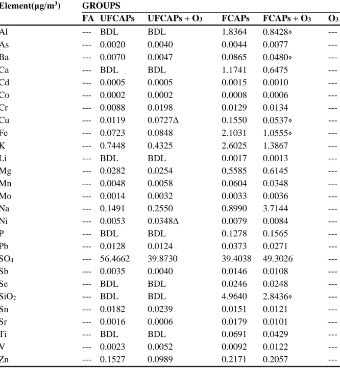
Chamber and exposure characteristics - Table 1 shows the concentration and particle size of CAPs and O3, and chamber characteristics for each exposure group. Table 2 indicates the elemental composition of the particulate matter from each of the exposure groups. Other than iron (Fe), FCAPs and UFCAPs particulate matter were of similar composition with the majority of the elemental fraction composed of SO4.
Estimated particle doses - The following particle doses were calculated for the mice in each of the PM-exposed group: (1) UFCAPs - 0.418 μg (2) FCAPs - 0.426 μg (3) UFCAPs + O3 - 0.264 μg and (4) FCAPs + O3 - 0.446 μg. Using the same model and exposure characteristics the estimated human doses were determined to be: (1) UFCAPs - 103.4 μg (2) FCAPs - 81.3 μg (3) UFCAPs + O3 - 65.8 μg and (4) FCAPs + O3 - 85.0 μg.
Heart Rate - Although all animals experienced an increase in HR while in the exposure chamber before the start of the exposure (Baseline) and a progressive decrease during the 4-hour exposure (Exp1, Exp2, Exp3 and Exp4), there were no significant differences in HR among any of the exposure groups during any time period (Figure 1).
exposure groups pre-, during or post-exposure. There were also no significant differences in the LF/HF between any exposure groups.
Electrocardiogram - Figure 2 shows the electrocardiogram data before, during and after exposure. There were no significant differences in ECG between any of the groups during pre-exposure or recovery. All animals experienced a decrease in PR interval, QRS, ST interval, and QTc during the baseline, which was likely related to the increase in HR. Thereafter, PR interval and ST interval increased in all animals during the exposure; though there were no significant differences. In contrast, QRS and QTc were significantly increased in mice exposed to UFCAPs + O3 when compared to FA. Exposure to O3 alone demonstrated a trend towards decreased QTc when compared with FA.
Cardiac arrhythmia - There was a significant increase in the number of non-conducted P-wave arrhythmias during the 4-hour exposure period to UFCAPs + O3 when compared with FA (Figure 3C). No other significant differences in arrhythmias were observed among any of the exposure groups. Although other types of arrhythmias were noted, they were few in number and not statistically different between any of the groups.
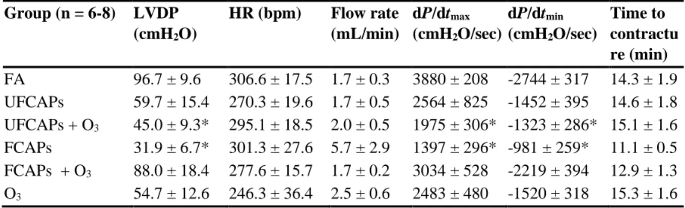
cmH2O/sec), O3 (2483 ± 480 cmH2O/sec) and UFCAPs + O3 (1975 ± 306 cmH2O/sec) when compared to FA (3880 ± 208 cmH2O/sec) and the minimum dP/dt before ischemia was also significantly lower in the UFCAPs (-1452 ± 395 cmH2O/sec), FCAPs (-982 ± 259
cmH2O/sec), O3 (-1520 ± 318 cmH2O/sec) and UFCAPs + O3 (-1323 ± 286 cmH2O/sec) groups when compared to FA (-2744 ± 317 cmH2O/sec) (Figure 5; Table 3). There was no difference in HR, coronary flow rate or ischemic contracture between any exposure groups before ischemia (Table 3).
Multivariate analysis of variance demonstrated that differences in LVDP, maximum dP/dt and minimum dP/dt between the FCAPs alone and FCAPs + O3 groups could be accounted for by the decrease in aluminum (Al), barium (Ba), copper (Cu), iron (Fe) or silicon dioxide (SiO2) compositions (Table 2); these elements clustered together however the analysis could not determine which element specifically was responsible. There were no apparent differences in elemental composition between UFCAPs alone and UFCAP + O3, except nickel (Ni), which were linked to any cardiac response changes, nor were there any other significant linkages with any other cardiac endpoints.
pre-ischemia, there was no significant difference in post-ischemia recovery of LVDP (Figure 7), dP/dtmax, and dP/dtmin between any exposure groups.
Biochemical markers and inflammatory cells in BAL and blood - Exposure to O3 alone or
UFCAPs + O3 caused a significant decrease in glutathione S-transferase (GTR) when compared to controls. There were no other significant differences in any other BAL cells or markers, or any of the serum or plasma markers (Table 4).
Discussion
On the other hand, it is not entirely surprising that on their own FCAPs and UFCAPs did not cause any significant changes in ECG given our previous negative results with a more toxic pollutant (Farraj et al. 2009). Similarly, Campen et al. (Campen et al. 2006) found that Apolipoprotein E (ApoE) -/- mice on a high fat diet, which are assumed to be susceptible to the cardiotoxic effects of inhaled pollutants, did not have any ECG changes when exposed to high concentrations of road dust PM or the vapor phase of gasoline engine exhaust. As far as arrhythmias are concerned, spatial dispersion of cardiac repolarization, which contributes to arrhythmogenesis, is increased in people after co-exposure to CAPs and O3 with each pollutant causing minimal effects on their own (Sivagangabalan et al. 2011). Even in the presence of O3, it is clear from not only our results, but the previously mentioned human data and other humans studies (Fakhri et al. 2009), that relatively low CAPs exposures will likely only cause mild electrical and HRV changes in healthy populations. Thus, a significant ECG effect due to acute exposure may not necessarily be direct evidence of serious cardiovascular morbidity or premature mortality; rather, it may reflect a transient instability that can worsen if exposure continues over a longer period.
prolonged repolarization suggests increased risk of early after-depolarization, which can trigger arrhythmias and potentially myocardial infarction when propagated. Indeed it is not unusual that electrical and mechanical dysfunction were both observed in mice exposed to UFCAPs + O3 given increased arrhythmogenesis has been shown to be associated with changes in myocardial stretch(Milan & MacRae 2005).
dose or even pulmonary deposition given UFCAPs was estimated to be less than FCAPs (pulmonary dose - 0.104 μg vs. 0.135 μg, respectively).
Some of these pathways may lead to subsequent ischemic damage, which has been shown to be increased by PM. Cozzi et al. showed that in mice intra-tracheally instilled with ultrafine PM, infarct size and oxidative stress in the myocardium were significantly increased (Cozzi et al. 2006). This corroborates our previous PM instillation studies which also demonstrated an increase in post-ischemia infarct size and decreased recovery of LVDP (Tong et al. 2010). It appears that the method of exposure significantly impacts the post-ischemia response because even though exposure to FCAPs or UFCAPs + O3 caused significant pre-ischemia functional decrements, there was no change in coronary flow post-pre-ischemia and there appeared to be an improvement of LVDP recovery (Figure 7). These findings are similar to what we observed with inhalation of multipollutant mixtures (McIntosh-Kastrinsky et al. 2013) and may represent activation of some compensatory mechanism post-exposure that actually protects the heart during ischemic injury. Lastly, although infarct size was not measured in our animals, we theorize that there was probably minimum to no increase particularly given we previously observed a decrease in infarct size with multipollutant mixture inhalation (McIntosh-Kastrinsky et al. 2013). Thus, acute inhalation of fine or ultrafine PM alone or in combination with O3 may not be potent enough to cause serious ischemia-related damage and that a higher concentration is necessary to overcome this apparent response threshold.
It is also important to note that although we compare these results to our previous study (Tong et al. 2010), the composition of the current FCAPs and UFCAPs is different. Our CAPs, particularly the UFCAPs, had a higher organic (OC) and total carbon (TC) content; thus possibly explaining the differences in response.
Additionally, O3 may cause epithelial injury and oxidative stress, which facilitate the PM effects (Elder et al. 2000). Adamson et al. (Adamson et al. 1999) showed that O3 and urban particulate co-exposure resulted in greater epithelial injury and interstitial inflammation than for either component alone; not to mention UFP did not have a large biological effect without O3. As such, co-exposures may produce differential responses due to toxicological interactions within the host. Thomson et al. (Thomson et al. 2005) showed that on their own, PM and O3 increased expression of the potent vasoconstrictor endothelin-1 (ET-1) in the lungs and its circulating levels in the plasma, however, together they only caused an upregulation (i.e. without plasma “spill-over”). Although there were no significant changes in inflammatory cells or markers in the blood or lavage, we found that O3 alone and UFCAPs + O3, but not FCAPs or UFCAPs alone, caused significantly decreased serum glutathione S-transferase (GTR) levels, which is indicative of increased oxidative stress; direct measurement of oxidative stress in the myocardium may have revealed a greater involvement as was shown by Cozzi et al. (2006) (Cozzi et al. 2006). Wang et al. previously showed that PM2.5 and O3 increased several markers of inflammation and oxidative stress in rats however their exposure concentrations were significantly higher than those used here (Watkinson et al. 2001). Regardless, synergistic interactions between inhalable PM and O3 can increase the generation of reactive oxygen species due to the porous surface of particles which provides ample surface area for reactivity, but that the potency still depends on particle concentration, size and other factors (Valavanidis et al. 2009; Park et al. 2006).
Tables and Figures Table 1. Chamber and exposure characteristics
GROUPS
FA UFCAPs UFCAPs + O3 FCAPs FCAPs + O3 O3 Temperature
(°C)
22.3 ± 0.1 23.0 ± 0.1 22.6 ± 0.1 22.0 ± 0.1 22.2 ± 0.1 22.5 ± 0.2
Rel. humidity (%)
50.2 ± 0.7 70.5 ± 4.6 56.0 ± 3.6 59.8 ± 3.4 59.0 ± 5.8 52.4 ± 2.2
O3 (ppb) 4.0 ± 0.0 25.7 ± 4.7 298.3 ± 0.7 33.1 ± 2.0 300.0 ± 0.4 299.0 ± 1.1 PM Mass
(ug/m3)
4.9 ± 2.2 138.8 ± 33.1 85.7 ± 6.5 190.9 ± 32.8 211.5 ± 37.3 3.4 ± 1.3
PM Total # (particles/cc)
24.2 ± 1.4 2.1E5 ± 5.6E3 1.6E5 ± 2.0E3 1.0E4 ± 5.2E1 1.1E4 ± 1.6E2 20.2 ± 4.2
Particle size (um)
- 0.076 0.072 0.246 0.235 -
Geo. Std. Dev. - 1.67 1.66 1.96 1.67 -
PM CARBON
TC (μg/m3) 3.4 ± 0.2 67.6 ± 6.5 (48.7%)
46.4 ± 3.4 (54.1%)
53.8 ± 4.4 (28.2%)
47.8 ± 7.3 (22.6%)
4.3 ± 0.4 OC (μg/m3) 3.7 ± 0.2 64.5 ± 5.9
(46.5%)
44.6 ± 3.1 (52.0%)
50.3 ± 4.0 (26.3%)
45.7 ± 6.6 (21.6%)
4.6 ± 0.4 EC (μg/m3) ** 3.1 ±
0.6(2.2%)
1.8 ± 0.3 (2.1%)
3.4 ± 0.5 (1.8%)
2.2 ± 0.7 (1.0%)
**
Reported values are mean ± SEM for each group over all exposure days ** = below detection limit