Copyright © 2003, American Society for Microbiology. All Rights Reserved.
Role of Second-Largest RNA Polymerase I Subunit Zn-Binding
Domain in Enzyme Assembly
Tatyana Naryshkina,
1Adrian Bruning,
1Olivier Gadal,
2and Konstantin Severinov
1,3*
Waksman Institute1and Department of Molecular Biology and Biochemistry,3Rutgers, The State University, Piscataway,
NJ 08854, and Unite´ de Biologie Cellulaire du Noyau, CNRS URA 1773, 75724 Paris cedex 165, France2
Received 18 May 2003/Accepted 26 July 2003
The second-largest subunits of eukaryal RNA polymerases are similar to thesubunits of prokaryal RNA
polymerases throughout much of their lengths. The second-largest subunits from eukaryal RNA polymerases contain a four-cysteine Zn-binding domain at their C termini. The domain is also present in archaeal homologs but is absent from prokaryal homologs. Here, we investigated the role of the C-terminal Zn-binding domain of Rpa135, the second-largest subunit of yeast RNA polymerase I. Analysis of nonfunctional Rpa135 mutants indicated that the Zn-binding domain is required for recruitment of the largest subunit, Rpa190, into the RNA polymerase I complex. Curiously, the essential function of the Rpa135 Zn-binding domain is not related to
Zn2ⴙbinding per se, since replacement of only one of the four cysteine residues with alanine led to the loss of
Rpa135 function. Even more strikingly, replacement of all four cysteines with alanines resulted in functional Rpa135.
Cellular multisubunit RNA polymerases (RNAPs) are re-lated through common ancestry (1, 23), share a common set of core subunits (␣2⬘in prokaryotes [15]), have highly similar
structures (2, 34), and likely have identical mechanisms. All multisubunit RNAPs are metalloenzymes. At least one Mg2⫹ ion is bound in the catalytic center and is essential for catalysis of template-dependent phosphodiester bond formation (33). In addition, all cellular RNAPs contain several tightly bound Zn2⫹ions (24). The number of Zn2⫹ions per RNAP molecule varies between RNAPs from prokaryal and eukaryal lineages, and the functions of the bound Zn2⫹are for the most part not clear.
In eubacterial RNAPs, two Zn2⫹ ions are bound to the largest subunit,⬘. One Zn-binding site is formed by the uni-versally conserved segment A in the N terminus of⬘. Muta-tional data suggest that segment A is involved in transcription termination, possibly through direct interaction of the Zn-binding domain with the nascent RNA (22). Structural data suggest that this Zn-binding site may also contribute to pro-moter recognition (16). The second ⬘ Zn-binding site is present only in prokaryal RNAPs. Functional analysis suggests that Zn2⫹ binding to this site promotes RNAP assembly by bringing two distant parts of⬘together (13).
Sequence analysis of eukaryal RNAP subunits identified multiple putative Zn-binding motifs. Over the years, various amounts of tightly bound Zn2⫹ were reported for different eukaryal RNAPs. The high-resolution structure of a 10-subunit core of RNAP II from yeast revealed eight bound Zn2⫹ions (2, 7). Of these, two Zn2⫹ions are bound in the Rpb220 (⬘) segment A, and one Zn2⫹ion is bound close to the C-terminal end of the second-largest subunit, Rpb150 (). The rest of the Zn2⫹ ions interact with smaller RNAP subunits, which are
either shared by all three nuclear RNAPs or specific for RNAP II. The four-cysteine Zn-binding domain located close to the C terminus of the second-largest RNAP subunit was the focus of this investigation. This domain is a common feature of eu-karyal RNAPs but is absent from proeu-karyal homologs. In the yeast RNAP II structure, this Zn-binding domain forms an independent surface-exposed loop, and the four cysteines to-gether coordinate a Zn2⫹ion, Zn7according to the
nomencla-ture of Cramer et al. (2). The Zn7-binding domain is located at
the bottom of the RNAP II jaw primarily formed by the largest subunit, Rpb220, and is part of the mobile “clamp” module (2, 7). Here, we studied the role of this domain in Rpa135, the second-largest subunit of yeast RNAP I, which is homologous to Rpb150 (8). We took advantage of the fact that RNAP I can be made dispensable for yeast viability (18) and is therefore an attractive model for structural-functional analysis of loss-of-function mutations. The results of our analysis indicate, sur-prisingly, that binding of Zn2⫹is not an essential function of the Zn7loop in RNAP I, because only one of the four Zn7loop
cysteines proved to be essential. Analysis of nonfunctional Zn7
loop mutants shows that this domain is important for recruit-ment of the largest RNAP I subunit, Rpa190 (homologous to eubacterial RNAP⬘) into the RNAP complex.
MATERIALS AND METHODS
Plasmids.Plasmid pNOY302, which harbors the entireRPA135gene fused to multiple hemagglutinin (HA) tags and a His tag, was provided by M. Nomura and is described by Keener et al. (9). Allrpa135mutations were constructed in pNOY302 by site-specific two-step PCR mutagenesis. Single (C1104A, C1107A, C1128A, and C1131A) and double (C1104A C1107A and C1128A C1131A) substitutions were introduced using appropriate mutagenic oligonucleotides and an upstream primer that corresponds to RPA135codons 1007 to 1015 and incorporates a naturalNcoI site that is unique in pNOY302. The 309- to 393-bp PCR products of the first PCR were used as primers for the second PCR, using pNOY302 as a template and an oligonucleotide that annealed to pNOY302 downstream of RPA135 and incorporated anSpeI site which is unique in pNOY302 as a second primer. The products of the second PCR (⬃1.4 kbp) were treated withSpeI andNcoI and cloned into appropriately treated pNOY302. The presence of the desired mutations was confirmed by sequencing of the entire
* Corresponding author. Mailing address: Waksman Institute, 190 Frelinghuysen Rd., Piscataway, NJ 08854. Phone: (732) 445-6095. Fax: (732) 445-5735. E-mail: [email protected].
1046
on September 8, 2020 by guest
http://ec.asm.org/
SpeI-NcoI fragment of recombinant pNOY302. Double mutants substituting RPA135codons C1107A C1128A and C1107A C1131A, triple mutants (C1104A C1128A C1131A and C1107A C1128A C1131A), and a quadruple mutant (C1104A C1107A C1128A C1131A) were constructed by a similar procedure using pNOY302 derivatives containing appropriate single, double, and triple mutations as templates for the second-step PCR.
The following procedure was used to construct Rpa135 deletions and chime-ras. First, aBclI site was introduced afterRPA135codon 1103 of pNOY302 by site-directed mutagenesis as described above to create pNOY302BclI. The oli-gonucleotide incorporating theBclI site also contained a translation termination codon that truncated Rpa135 at position 1103. pNOY302BclI was thus a source ofrpa135⌬. Fragments ofRPB150orRPC128corresponding to codons 1163 to 1224 and 1095 to 1149, respectively, were amplified from yeast genomic DNA using appropriate oligonucleotide primers. In each case, the downstream primer contained anSpeI site after the termination codon of the RNAP gene. PCR-amplified fragments were treated withSpeI and cloned in pNOY302BclI that was first treated withBclI, followed by successive treatments with mung bean nucle-ase andSpeI.
The following multistep procedure was used to create therpa135⌬Zn mutant, lacking the Zn-binding domain. In the first step, two PCRs were performed to amplifyRPA135regions at the left and the right sides of the deletion site. The left-side nonmutagenic PCR primer corresponded toRPA135codons 1007 to 1015 and incorporated anNcoI site. The right-side nonmutagenic primer was complementary to pNOY302 codon positions 2709 to 2723 and incorporated an SpeI site. The mutagenic primers corresponded toRPA135codons 1096 to 1106 and 1170 to 1181 for the left-side PCR and the right-side PCR, respectively. Each mutagenic primer also contained an in-frameNheI site. The products of each PCR were purified and cloned into the pT7blue blunt vector (Novagen). Purified pT7blue-based plasmids harboring the left and right sides of the deletion were treated with SpeI and NheI. The small fragment of pT7blue-based plasmid containing the right side of the deletion ofRPA135was cloned into the based plasmid harboring the left side of the deletion. The resultant pT7blue-based plasmid, pT7blueA135⌬Zn, was treated with SpeI andNcoI, and the rpa135fragment was cloned into appropriately treated pNOY302. The presence of the desired mutation was confirmed by DNA sequencing.
Yeast strains and strain construction.Yeast genetic techniques and growth media have been described previously (4a, 6a). Yeast tester strain NOY408-1a was provided by M. Nomura and has been described by Nogi et al. (18). Yeast cells were transformed using a standard lithium acetate procedure, and trans-formants were selected on appropriate drop-out media. Fob1::GFP was intro-duced in NOY408-1a as described previously (6).
Immunolocalization.Yeast cells were grown in 50-ml cultures of YEPGal (2% yeast extract, 1% peptone, 2% galactose) at 30°C to anA600between 0.5 and 1.
They were fixed with 4% formaldehyde for 30 min at room temperature and processed as described previously (27). Anti-HA staining was done with 1/100-diluted anti-mouse immunoglobulin G Cy3 for 1 h at room temperature. DNA was stained with 4⬘6-diamino-2-phenylindole (DAPI). Samples were examined using a Leica DMRXA fluorescence microscope. Fluorescent signals were col-lected with single band pass filters for excitation of green fluorescent protein (GFP) (GFP; Leica), CY3 (TX2; Leica); and DAPI (A; Leica). Images were acquired with a Hamamatsu C4742-95 cooled charge-coupled device camera controlled by Openlab software (version 2.2.4; Improvision) and processed using Adobe Photoshop software (version 5).
Yeast extract preparation.Yeast cells were grown in 100 ml of YEPGal to an optical density at 600 nm of 1.0. The cells were harvested, resuspended in breakage buffer (150 mM NaCl, 100 mM Tris-HCl, pH 7.5, 1 mM dithiothreitol, 20% glycerol, 1 mM phenylmethylsulfonyl fluoride), and cell extracts were pre-pared using glass beads as previously described (20a) and cleared by centrifuga-tion.
Immunoblotting.Cell extracts (15g of total protein) were loaded on a 6% polyacrylamide sodium dodecyl sulfate (SDS) gel, and the proteins were sepa-rated by electrophoresis and transferred onto nitrocellulose membranes. As primary antibodies, anti-HA.11 mouse antibody from Babco (to detect Rpa135) and an anti-Rpa190 rabbit polyclonal antibody (a generous gift of M. Riva and C. Carles) were used. As secondary antibodies, anti-rabbit or anti-mouse horse-radish peroxidase conjugates were used, followed by detection with chemilumi-nescence (Western Lightning chemilumichemilumi-nescence reagent; NEN Life Science).
IP.Protein A-agarose beads (30l) in 1 ml of immunoprecipitation (IP) buffer (150 mM NaCl, 100 mM Tris-HCl, pH 7.5, 1 mM dithiothreitol, 20% glycerol, 1 mM phenylmethylsulfonyl fluoride, 0.05% Nonidet P-40) were combined with 20 l of normal serum (Sigma) and incubated at 4°C for 1 h. The beads were washed three times with 1 ml of IP buffer, collected by centrifugation, combined with yeast extract, and incubated for 2 h at 4°C with mild agitation. The beads were
collected by low-speed centrifugation and discarded; the supernatants were sup-plemented with a 1:1,000 dilution of Babco anti-HA.11 mouse antibody and incubated for 1 h at 4°C, followed by the addition of protein A-agarose beads, 2 h of incubation at 4°C, and five washes with 1 ml of IP buffer. The immunopre-cipitated proteins were denatured (5 min at 70°C), separated by SDS-polyacryl-amide gel electrophoresis, and revealed by rabbit polyclonal antibodies raised against the RNAP I Rpa190 subunit.
RESULTS
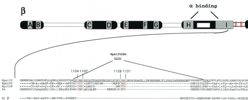
Site-directed mutagenesis ofRPA135.The genetic context of
the Rpa135 C-terminal Zn-binding site is shown in Fig. 1. As can be seen, a four-cysteine motif is inserted in the C-terminal portion of the universally conserved segment I. No such motif is present in prokaryal RNAPsubunits. While the length and the actual sequence of inserted material varies, the presence of two pairs of cysteines (positions 1104 and 1107 and positions 1128 and 1131 in yeast Rpa135) is common to all eukaryal and archaeal-like subunits. The cysteine residues in each pair are separated by two amino acids that are not conserved. Pairs of cysteines separated by two amino acids often serve as Zn-binding nuclei (26). In Rpa135, an additional cysteine is present at position 1127. The corresponding position in the Zn-binding domains of the second-largest subunits from other yeast RNAPs, as well as in the second-largest subunits of RNAP I from other organisms, is occupied by residues other than cysteines (Fig. 1 and data not shown). Because Cys1127is
unlikely to participate in Zn2⫹binding, it was excluded from the analysis that follows.
In vivo analysis of Rpa135 mutants.To check the functional
role of the Zn-binding motif in Rpa135, several site-directed mutations were constructed in theRPA135gene cloned on a multicopy plasmid (9). In Rpa135⌬, Rpa135 residues C-termi-nal of residue 1103 are removed. In Rpa135/Rpb150 and Rpa135/Rpc128, Rpa135 residues downstream of residue 1103 are substituted for the corresponding amino acid sequences of Rpb150 and Rpc128, Rpa135 homologs from RNAP II and RNAP III, respectively. Rpa135⌬Zn models a  subunit of bacterial RNAP; it removes the entire insertion containing the Zn-binding motif (Rpa135 amino acids 1102 to 1174) and creates an uninterrupted conserved segment I sequence. The deletion endpoints were selected based on analysis of yeast RNAP II structure (2) to create minimal perturbations. A 4-amino-acid linker, GASG, that spans the deletion site should cover the⬃10-A˚ distance between the deletion endpoints in the RNAP II structure.
Plasmids expressing mutantrpa135 genes were introduced into the NOY408-1a yeast strain (18). In this strain, the chro-mosomal copy of RPA135 has been disrupted and RPA135 codons 111 to 1104 have been replaced with theLEU2gene. The cells can grow in the presence of galactose, since rRNA is synthesized from a copy of a ribosomal DNA (rDNA) repeat that was cloned on a plasmid and fused to aGAL7promoter, a strong RNAP II promoter induced in the presence of galac-tose. NOY408-1a does not grow on medium containing glu-cose, since theGAL7promoter is repressed under these con-ditions. However, if a functional copy ofRPA135is provided on a plasmid, NOY408-1a can grow on a medium containing glucose, since RNAP I containing plasmid-encoded RPA135 transcribes genomic rDNA in galactose-independent fashion. The results of this in vivo test withrpa135deletion and chimera
VOL. 2, 2003 ZINC AND RNA POLYMERASE FUNCTION 1047
on September 8, 2020 by guest
http://ec.asm.org/
mutations are presented in Fig. 2, top. As can be seen, removal of the Rpa135 Zn-binding domain or the replacement of the Rpa135 Zn-binding domain sequence with corresponding se-quences from RNAP II or RNAP III homologs rendered plas-mid-encoded Rpa135 nonfunctional. Thus, the C-terminal Zn-binding domain is essential, and its function cannot be restored by homologous sequences from other nuclear RNAPs.
Therpa135plasmids carrying the point mutations C1104A, C1107A, C1128A, and C1131A that replaced evolutionarily conserved cysteine residues with alanines were also con-structed and tested in NOY408-1a cells. Of the four substitu-tions, only one, C1107A, did not allow growth on glucose (Fig. 2, top), suggesting that evolutionarily conserved cysteines at positions 1104, 1128, and 1131 are not essential for Rpa135 function, while Cys1107is essential.
Curiously, a quadruple mutation exchanging all four Rpa135 Zn-binding domain cysteines for alanines was functional (Fig. 2, top), indicating that an alanine substitution for Cys1104,
Cys1128, or Cys1131(or a combination of these substitutions)
behaves as a specific intragenic suppressor of the C1107A lethal mutation. To further characterize genetic relationships among Rpa135 Zn-binding domain mutations, additional mul-tiple point mutations were generated. Alanine substitutions involving positions 1104, 1128, and 1131, but excluding position 1107 (i.e., the double substitutions C1128A C1131A and C1104A C1131A and the triple substitution C1104A C1128A C1131A) resulted in functional Rpa135 (Fig. 2, top, and data not shown). In contrast, neither the three double substitutions carrying C1107A nor a triple substitution, C1107A C1128A C1131A, supported growth on glucose, and they were there-fore classified as lethal mutations (Fig. 2, top, and data not shown).
Multiple independent transformants were tested on nonper-missive glucose-containing medium, and in all cases, results identical to those shown in Fig. 2, top, were obtained, thus excluding marker rescue as the reason for the growth of NOY408-1a cells harboring plasmid-encoded viable rpa135 mutations on nonpermissive medium. Based on these results, we have reached the following unexpected conclusions: (i) Zn binding by the evolutionarily conserved Rpa135 Zn-binding domain is not required for Rpa135 activity; (ii) replacement of cysteine at position 1107 creates a nonfunctional Rpa135; (iii) the defect created by the C1107A substitution can be tolerated only if no other cysteine is present in the motif.
To ensure that the absence of complementation by nonfunc-tionalrpa135 mutations was not due to defects in stability or levels of expression of plasmid-encoded mutant Rpa135 sub-units, the amounts of Rpa135 in extracts of NOY408-1a cells transformed with plasmids carrying functional and nonfunc-tionalrpa135mutations were determined by Western blotting with anti-HA antibody (an N-terminal HA tag is present in all plasmid-encoded Rpa135 subunits used here). A representa-tive result is shown in Fig. 2, bottom. As can be seen, while all of the mutant subunits were present in amounts that were lower than the amounts of wild-type Rpa135 produced from the pNOY302 plasmid, cells expressing both functional and nonfunctional derivatives of RPA135 produced similar amounts of Rpa135. We therefore conclude that the lethal phenotype ofrpa135mutations is not a simple consequence of decreased amounts of mutant subunits present in the cell.
Nonfunctional Rpa135 subunits do not localize to the
nu-cleolus.The inability of some of the Rpa135 mutants to
sup-port cell growth can be due to (i) lower levels of mutant RNAP I activity, (ii) inability of mutant RNAP I to localize to the FIG. 1. Genetic context of the Zn-binding domain in Rpa135. At the top, the second-largest (-like) subunit of a multisubunit DNA-dependent RNAP is schematically presented. The lettered boxes represent evolutionarily conserved segments, and the open boxes represent evolutionarily variable segments. Sites involved in interaction with␣-like subunits are indicated. Below, a portion of conserved segment I sequence of yeast Rpa135 is presented in a single-letter code. The sequence is aligned with corresponding sequences from yeast RNAP II (Rpb150) and RNAP III (Rpc128),Pyrococcus abyssiRNAP (Pa), andE. coliRNAP(Ec). The dots indicate identity, and the hyphens indicate gaps. The cysteine residues involved in Zn binding are shown in red (and in the schematic by red lines), and their numbers (in Rpa135) are indicated above the alignment. The sequence of the Rpa135⌬mutant that lacks amino acids beyond Val1103is shown at the bottom of the alignment. The⌬Zn mutation
that removes the Zn-binding domain and introduces a 4-amino-acid linker, GASG, is shown above the Rpa135 sequence.
on September 8, 2020 by guest
http://ec.asm.org/
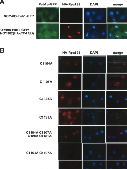
nucleolus, or (iii) inability of mutant Rpa135 to assemble into the RNAP I complex. To test which of these possibilities is realized, we first determined the cellular localization of HA-tagged, plasmid-encoded Rpa135 and its mutants using indi-rect immunofluorescence microscopy.
To follow the position of the nucleolus, a derivative of NOY408-1a harboring a chromosomal gene encoding a C-terminal fusion of GFP to Fob1, an rDNA binding protein that can be used as a nucleolar marker (3), was constructed (see Materials and Methods). As expected, the new strain, NOY408-Fob1-GFP, was dependent on galactose for growth (data not shown). Microscopic examination of NOY408-Fob1-GFP cells revealed weak Fob1p-NOY408-Fob1-GFP signal with a large do-main of the nucleoplasm stained with DAPI, as observed pre-viously for strains lacking functional RNAP I (Fig. 3A, top row) (20, 25).
We next transformed NOY408-Fob1-GFP with a plasmid expressing wild-type HA-tagged RPA135, grew cells in the presence of galactose, and examined the localizations of Fob1 and Rpa135 (Fig. 3A, bottom row). The Fob1-GFP was con-centrated in prominent crescent-like structures typical of yeast nucleoli. A specific signal from epitope-tagged Rpa135 was also detected. This signal appeared adjacent to DAPI staining and colocalized with the signal from Fob1p-GFP. No HA-Rpa135-specific signal was detected in NOY408-Fob1-GFP cells, as expected (Fig. 3A, top row). We therefore conclude that plasmid encoded HA-tagged Rpa135 and, presumably, RNAP I is localized in nucleoli (12).
The cellular-localization experiment was next repeated with NOY408-1a cells transformed with plasmids expressing mutant rpa135 genes (Fig. 3B and data not shown). The results indi-cated that for all plasmids that supported growth on glucose, mutant Rpa135 subunits, and presumably RNAP I containing mutant Rpa135, localized to nucleoli. In contrast, cells harbor-ing plasmids that were unable to support growth on glucose revealed no nucleolar localization of Rpa135, suggesting either that the mutant subunit was unable to assemble in RNAP or that the mutant RNAP was unable to enter the nucleolus. It should be noted that for many functional mutants the amount of Rpa135 localized to the nucleolus was lower than in the case of wild-type Rpa135. However, the smaller amounts of nucle-olar Rpa135 and, by extension, nuclenucle-olar RNAP I were appar-ently sufficient to allow growth in the presence of glucose at all temperatures tested (25, 30, and 37°C; data not shown).
Nonfunctional Rpa135 mutants do not form a stable RNAP
I core enzyme complex. The results of the experiments
pre-sented above suggest that nonfunctional Rpa135 mutants fail to localize to the nucleolus, the site of RNAP I function. The defect could be caused by the inability of RNAP I harboring mutant Rpa135 subunits to be transported to the nucleolus or it could be due to the inability of mutant Rpa135 to assemble in RNAP I core enzyme.
Based on what we know about RNAP assembly in pro-karyotes (10, 17, 29), yeast RNAP II (11), and yeast RNAP I (17), it is reasonable to assume that Rpa135 first interacts, through strictly conserved residues in segment H and the N-FIG. 2. Analysis of Rpa135 mutants in vivo. (Top) Results of growth of NOY408-1a yeast transformed with plasmids carrying the indicated alleles ofRPA135in the presence of glucose (YEPGlu) or galactose (YEPGal). (Bottom) Yeast cells transformed with plasmids carrying the indicated alleles ofRPA135were collected from YEPD plates, and cell extracts were prepared. Cell extract aliquots containing equal amounts of protein were loaded on an SDS gel, and after electrophoresis and blotting, Rpa135 in the extracts was revealed with anti-HA antibody.
VOL. 2, 2003 ZINC AND RNA POLYMERASE FUNCTION 1049
on September 8, 2020 by guest
http://ec.asm.org/
FIG. 3. Immunolocalization of mutant Rpa135 subunits. (A) Plasmid-encoded Rpa135 is located in the nucleolus. Immunofluorescence microscopy was performed as described in Materials and Methods. The yeast strain used was a NOY408-1a derivative transformed with Fob1-GFP and a multicopy plasmid with (bottom row) or without (top row) HA-tagged wild-typeRPA135. The results of localization of HA-Rpa135 (red), rDNA (visualized using Fob1-GFP; green), and nucleoplasm DAPI (blue) are shown. (B) In vivo localization of mutant Rpa135 subunits. The localization of the indicated Rpa135 mutants (red) is presented and compared to DAPI staining (blue). The functional alleles C1104A, C1128A, and C1131A and the quadruple mutant (C1104A C1107A C1128A C1131) are concentrated in the nucleolus and colocalized with Fob1-GFP (not shown). Note that the lethal alleles C1107A, C1104A C1107A, and⌬Zn are not concentrated in the nucleolus.
on September 8, 2020 by guest
http://ec.asm.org/
terminal portion of segment I, with the␣-like subunit AC40. The␣-like subassembly should next recruit the largest RNAP I subunit, Rpa190, to the complex. To determine whether Rpa135 harboring nonfunctional mutations in the Zn-binding domain is able to recruit Rpa190, we asked whether Rpa190 can be coimmunoprecipitated with anti-HA tag antibodies, which specifically react with the N-terminal epitope tag of plasmid-encoded Rpa135. The results of coimmunoprecipita-tion experiments using extracts prepared from NOY408-1a cells harboring plasmids expressing two functional RPA135 alleles (wild-typeRPA135andrpa135harboring the quadruple mutation C1105A C1107A C1128A C1131A) and a nonfunc-tional allele, C1107A, are presented in Fig. 4. Similar results were obtained with other mutants (data not shown). As pected, Rpa190 coimmunoprecipitated with Rpa135 from ex-tracts prepared from cells harboring a plasmid expressing wild-type Rpa135 (Fig. 4, lane 6). No Rpa190 was precipitated from extracts prepared from cells harboring a plasmid expressing the C1107A allele (Fig. 4, lane 7). In contrast, Rpa190 was coimmunoprecipitated from extracts expressing the quadruple mutation, though the amounts were significantly lower than in the wild-type control (Fig. 4, compare lanes 8 and 6). Probing with an anti-Rpa190 antibody of proteins in total-cell extracts revealed that Rpa190 was present in roughly the same amounts in cells harboring both active and inactive plasmid-encoded Rpa135 (Fig. 4, lanes 2 to 4). We therefore conclude that C-terminal Zn-binding motif mutations that affect essential in vivo Rpa135 function either prevent RNAP I assembly or de-stabilize the RNAP I complex.
DISCUSSION
Several previous investigations reported mutational analyses of Zn-binding domains of eukaryal RNAPs. The results indi-cate that Zn-binding domains are not involved in RNAP func-tion per se but contribute to the assembly and/or stability of these important enzymes (4, 5, 19, 21, 27, 30, 31) or are
in-volved in interactions with RNAP-binding proteins (14, 28, 32). In this study, we report the results of structural-functional analysis of a four-cysteine Zn-binding domain in the C-termi-nal portion of Rpa135. The domain is present in the second-largest subunits of all eukaryal and archeal RNAPs, and its function(s) has not been studied. Our data suggest that the Zn-binding domain is involved in RNAP I assembly and helps recruit Rpa190 into the RNAP I core enzyme complex. This result is in agreement with studies performed withEscherichia coliRNAP, in which it was demonstrated that a portion of the evolutionarily conserved segment I that is close to the -unit C terminus is required for recruitment of the largest sub-unit, ⬘, into the RNAP core enzyme complex (29). A site-specific deletion of the Zn-binding domain that creates Rpa135 with segment I sequence corresponding to the bacterial seg-ment I is not functional and fails to recruit Rpa190. The ad-ditional assembly requirement in the form of a Zn-binding domain may reflect weaker affinity of the largest RNAP I subunits for each other compared to the largestE. coliRNAP subunits. Rpa135 Zn-binding domain amino acids other than the four cysteines are evolutionarily conserved in RNAP I homologs, suggesting that Zn-binding domain interaction with Rpa190 is specific. However, the Zn-binding domain alone is not sufficient for recruitment of the largest subunit into the RNAP complex, since the Rpa135/Rpb150 and Rpa135/ Rpc128 chimeras studied here failed to recruit the largest subunits of RNAP II and RNAP III into the complex (data not shown).
In yeast RNAP II structure, the Rpb150 C-terminal Zn-binding domain is located very close to two Zn-Zn-binding sites in the N-terminal portion of the largest subunit, Rpb220. The interaction between Zn-binding domains of the largest subunit may therefore be important for RNAP II assembly. The Zn-binding domains of the largest subunits of RNAP I also appear to interact, based on genetic-suppression data of Yano and Nomura (31). These authors showed that a ts-5 allele that causes a point substitution in the N-terminal Zn-binding do-main of Rpb190 is suppressed by an rpa135 mutation that introduces a replacement of residue 1127 in the Rpa135 Zn-binding domain. Thets-5allele apparently destabilizes RNAP I, and the rpa135 suppressor mutation restores the complex stability (31). The interaction affected by ts-5 must be very specific, since none of therpa135mutations studied here sup-pressed the ts-5 phenotype (T. Naryshkina and K. Severinov, unpublished observations).
Curiously, the essential function of the Rpa135 Zn-binding domain is not related to Zn2⫹binding per se. Systematic ala-nine mutagenesis of four Rpa135 Zn-binding domain cysteine residues demonstrates that replacement of only one residue, Cys1107, leads to a lethal phenotype. A similar result was
ob-tained by Rubbi et al. (21), who studied a small subunit com-mon to all three yeast RNAPs, ABC10␣, and showed that only one cysteine of the four-cysteine Zn-binding motif of ABC10␣
is essential. The results of our analysis suggest that Rpa135 Cys1107is essential or that in the context of C1107A
substitu-tion the three remaining Cys residues are responsible for the lethal phenotype. Structural analysis of the homologous Zn-binding domain of Rpb150 and the Rpa135 Cys1107homolog
Rpb150 Cys1166 fails to explain the apparent singularity of
Cys1107. Surprisingly, replacement of all four Rpa135
Zn-bind-FIG. 4. Nonfunctional variants of Rpa135 fail to recruit Rpa190 in RNAP I complex. Coimmunoprecipitations were done as described in Materials and Methods. At the left, the amounts of Rpa190 in cell extracts prepared from yeast cells containing the indicated RPA135
plasmids were determined by separating proteins on an SDS gel and blotting them with an anti-RPA190 antibody. On the right, material precipitated from the extracts shown on the right with an anti-HA antibody recognizing Rpa135 was resolved by SDS-polyacrylamide gel electrophoresis, and probed with an anti-RPA190 antibody. M, marker lane containing purified yeast RNAP I.
VOL. 2, 2003 ZINC AND RNA POLYMERASE FUNCTION 1051
on September 8, 2020 by guest
http://ec.asm.org/
ing domain cysteines, including Cys1107, allows RNAP I
con-taining mutant Rpa135 to serve as the only source of rRNA. Thus, the C1107A mutation is suppressed by a triple alanine substitution for Cys1104, Cys1128, and Cys1131. While the
mo-lecular basis of this unprecedented suppression is not known, the result is consistent with the indirect effect of C1107A sub-stitution on cell viability. One possibility is that in the wild-type Rpa135 context, a Zn2⫹ion stabilizes a particular conforma-tion required for RNAP I assembly and stability. Replace-ments of Cys1104, Cys1128, and Cys1131decrease and/or abolish
Zn2⫹binding and affect the conformation of the Zn-binding domain, but the effect is not sufficient to decrease RNAP I assembly below the threshold required for viability. In contrast, replacements of Cys1107 that leave other Zn-binding domain
cysteines intact not only affect the conformation of the Zn-binding domain but also create an unproductive interaction(s) between the remaining cysteines and other sites within the RNAP I subassembly(s) containing mutant Rpa135. Replace-ment of all four cysteines removes this unproductive interac-tion and is therefore viable.
ACKNOWLEDGMENTS
We are grateful to M. Riva and C. Carles for generously providing anti-RPA190 antibodies and to M. Nomura for gifts of strains and plasmids.
This work was supported by a March of Dimes Birth Defect Foun-dation Research Grant and NIH grant RO1 GM59295 to K.S. T.N. was partially supported by a Charles and Johanna Busch Foundation Post-doctoral Fellowship. O.G. is an Institut Pasteur Bourse Roux Fellow and thanks Ulf Nehrbass for his kind support.
REFERENCES
1. Allison, L. A., M. Moyle, M. Shales, and C. J. Ingles.1985. Extensive homology among the largest subunits of eukaryotic and prokaryotic RNA polymerases. Cell42:599–610.
2. Cramer, P., D. A. Bushnell, and R. D. Kornberg.2001. Structural basis of transcription: RNA polymerase II at 2.8 angstrom resolution. Science292: 1863–1876.
3. Defossez, P. A., R. Prusty, M. Kaeberlein, S. J. Lin, P. Ferrigno, P. A. Silver, R. L. Keil, and L. Guarente.1999. Elimination of replication block protein Fob1 extends the life span of yeast mother cells. Mol. Cell3:447–455. 4. Donaldson, I. M., and J. D. Friesen.2000. Zinc stoichiometry of yeast RNA
polymerase II and characterization of mutations in the zinc-binding domain of the largest subunit. J. Biol. Chem.275:13780–13788.
4a.Flores, A, J. F. Briand, O. Gadal, J. C. Andrau, L. Rubbi, V. Van Mullem, C. Boschiero, M. Goussot, C. Marck, C. Carles, P. Thuriaux, A. Sentenac, and M. Werner.1999. A protein-protein interaction map of yeast RNA polymer-ase III. Proc. Natl. Acad. Sci. USA96:7815–7820.
5. Gadal, O., G. V. Shpakovski, and P. Thuriaux.1999. Mutants in ABC10, a conserved subunit shared by all three yeast RNA polymerases, specifically affect RNA polymerase I assembly. J. Biol. Chem.274:8421–8427. 6. Gadal, O., S. Labarre, C. Boschiero, and P. Thuriaux.2002. Hmo1, an
HMG-box protein, belongs to the yeast ribosomal DNA transcription sys-tem. EMBO J.21:5498–5507.
6a.Gadal, O., S. Mariotte-Labarre, S. Chedin, E. Quemeneur, C. Carles, A. Sen-tenac, and P. Thuriaux.1997. A34.5, a nonessential component of yeast RNA polymerase I, cooperates with subunit A14 and DNA topoisomerase I to pro-duce a functional rRNA synthesis machine. Mol. Cell. Biol.17:1787–1795. 7. Gnatt, A. L., P. Cramer, J. Fu, D. A. Bushnell, and R. D. Kornberg.2001.
Structural basis of transcription: an RNA polymerase II elongation complex at 3.3 A˚ resolution. Science292:1876–1882.
8. Ishihama, A., M. Kimura, and H. Mitsuzawa.1998. Subunits of yeast RNA polymerases: structure and function. Curr. Opin. Microbiol.1:190–196. 9. Keener, J., C. A. Josaitis, J. A. Dodd, and M. Nomura.1998. Reconstitution
of yeast RNA polymerase I transcription in vitro from purified components. TATA-binding protein is not required for basal transcription. J. Biol. Chem. 273:33795–33802.
10. Kimura, M., and A. Ishihama.1995. Functional map of the alpha subunit of Escherichia coliRNA polymerase: amino acid substitution within the amino-terminal assembly domain. J. Mol. Biol.254:342–349.
11. Kolodziej, P. A., and R. A. Young.1991. Mutations in the three largest
subunits of yeast RNA polymerase II that affect enzyme assembly. Mol. Cell. Biol.11:4669–4678.
12. Leger-Silvestre, I., S. Trumtel, J. Noaillac-Depeyre, and N. Gas.1999. Func-tional compartmentalization of the nucleus in the budding yeast Saccharo-myces cerevisiae. Chromosoma108:103–113.
13. Markov, D., T. Naryshkina, A. Mustaev, and K. Severinov.1999. A zinc binding site in the largest subunit of DNA-dependent RNA polymerase is involved in enzyme assembly. Genes Dev.13:2439–2448.
14. McCusker, J. H., M. Yamagishi, J. M. Kolb, and M. Nomura.1991. Sup-pressor analysis of temperature-sensitive RNA polymerase I mutations in Saccharomyces cerevisiae: suppression of mutations in a zinc-binding motif by transposed mutant genes. Mol. Cell. Biol.11:746–753.
15. Minakhin, L., S. Bhagat, A. Brunning, E. A. Campbell, S. A. Darst, R. H. Ebright, and K. Severinov.2001. Bacterial RNA polymerase subunitand eukaryotic RNA polymerase subunit RPB6 are sequence, structural, and functional homologs and promote RNA polymerase assembly. Proc. Natl. Acad. Sci. USA98:892–897.
16. Murakami, K. S., S. Masuda, E. A. Campbell, O. Muzzin, and S. A. Darst. 2002. Structural basis of transcription initiation: an RNA polymerase ho-loenzyme-DNA complex. Science296:1285–1290.
17. Naryshkina, T., D. Rogulja, L. Golub, and K. Severinov.2000. Inter- and intrasubunit interactions during the formation of RNA polymerase assembly intermediate. J. Biol. Chem.275:31183–31190.
18. Nogi, Y., R. Yano, and M. Nomura.1991. Synthesis of large rRNAs by RNA polymerase II in mutants ofSaccharomyces cerevisiaedefective in RNA polymerase I. Proc. Natl. Acad. Sci. USA88:3962–3966.
19. Nogi, Y., R. Yano, J. Dodd, C. Carles, and M. Nomura.1993. Gene RRN4 in Saccharomyces cerevisiaeencodes the A12.2 subunit of RNA polymerase I and is essential only at high temperatures. Mol. Cell. Biol.13:114–122. 20. Oakes, M., Y. Nogi, M. W. Clark, and M. Nomura.1993. Structural
alter-ations of the nucleolus in mutants ofSaccharomyces cerevisiaedefective in RNA polymerase I. Mol. Cell. Biol.13:2441–2455.
20a.Riva, M., J. M. Buhler, A. Sentenac, P. Fromageot, and D. C. Hawthorne. 1982. Natural variation in yeast RNA polymerase A. Formation of a mosaic RNA polymerase A in a meiotic segregant from an interspecific hybrid. J. Biol. Chem.257:4570–4577.
21. Rubbi, L., S. Labarre-Mariotte, S. Chedin, and P. Thuriaux.1999. Func-tional characterization of ABC10␣, an essential polypeptide shared by all three forms of eukaryotic DNA-dependent RNA polymerases. J. Biol. Chem.274:31485–31492.
22. Sen, R., R. A. King, N. Mzhavia, P. L. Madsen, and R. A. Weisberg.2002. Sequence-specific interaction of nascent antiterminator RNA with the zinc-finger motif ofEscherichia coliRNA polymerase. Mol. Microbiol.46:215–222. 23. Sweetser, D., M. Nonet, and R. A. Young.1987. Prokaryotic and eukaryotic RNA polymerases have homologous core subunits. Proc. Natl. Acad. Sci. USA84:1192–1196.
24. Treich, I., M. Riva, and A. Sentenac.1991. Zinc-binding subunits of yeast RNA polymerases. J. Biol. Chem.266:21971–21976.
25. Trumtel, S., I. Leger-Silvestre, P. E. Gleizes, F. Teulieres, and N. Gas.2000. Assembly and functional organization of the nucleolus: ultrastructural anal-ysis ofSaccharomyces cerevisiaemutants. Mol. Biol. Cell.11:2175–2189. 26. Vallee, B. L., and D. S. Auld.1990. Zinc coordination, function, and structure
of zinc enzymes and other proteins. Biochemistry29:5647–5659. 27. Van Mullem, V., E. Landrieux, J. Vandenhaute, and P. Thuriaux.2002.
Rpa12p, a conserved RNA polymerase I subunit with two functional do-mains. Mol. Microbiol.43:1105–1113.
28. Van Mullem, V., M. Wery, M. Werner, J. Vandenhaute, and P. Thuriaux. 2002. The Rpb9 subunit of RNA polymerase II binds transcription factor TFIIE and interferes with the SAGA and elongator histone acetyltrans-ferases. J. Biol. Chem.277:10220–10225.
29. Wang, Y., K. Severinov, N. Loizos, D. Fenyo¨, E. Heyduk, T. Heyduk, B. T. Chait, and S. A. Darst.1997. Determinants forEscherichia coliRNA poly-merase assembly within thesubunit. J. Mol. Biol.270:648–662. 30. Werner, M., S. Hermann-Le Denmat, I. Treich, A. Sentenac, and P.
Thuri-aux.1992. Effect of mutations in a zinc-binding domain of yeast RNA polymerase C (III) on enzyme function and subunit association. Mol. Cell. Biol.12:1087–1095.
31. Yano, R., and M. Nomura.1991. Suppressor analysis of temperature-sensi-tive mutations of the largest subunit of RNA polymerase I inSaccharomyces cerevisiae: a suppressor gene encodes the second-largest subunit of RNA polymerase I. Mol. Cell. Biol.11:754–764.
32. Yano, R., M. Oakes, M. Yamaghishi, J. A. Dodd, and M. Nomura.1992. Cloning and characterization of SRP1, a suppressor of temperature-sensitive RNA polymerase I mutations, inSaccharomyces cerevisiae. Mol. Cell. Biol. 12:5640–5651.
33. Zaychikov, E., E. Martin, L. Denissova, M. Kozlov, V. Markovtsov, M. Kashlev, H. Heumann, V. Nikiforov, A. Goldfarb, and A. Mustaev.1996. Mapping of catalytic residues in the RNA polymerase active center. Science 273:107–109.
34. Zhang, G., E. A. Campbell, L. Minakhin, C. Richter, K. Severinov, and S. A. Darst.1999. Crystal structure ofThermus aquaticuscore RNA polymerase at 3.3 A˚ resolution. Cell98:811–824.