Dynamic Changes of Gut Microbial Communities of Bumble
Bee Queens through Important Life Stages
Liuhao Wang,
a,bJie Wu,
aKai Li,
aBen M. Sadd,
eYulong Guo,
aDaohua Zhuang,
fZhengyi Zhang,
aYanping Chen,
gJay D. Evans,
gJun Guo,
hZhigang Zhang,
c,dJilian Li
aaKey Laboratory of Pollinating Insect Biology of the Ministry of Agriculture, Institute of Apicultural Research, Chinese Academy of Agricultural Science, Beijing, China bCollege of Resources and Environmental Sciences, Henan Institute of Science and Technology, Xinxiang, Henan, China
cState Key Laboratory of Genetic Resources and Evolution, Laboratory of Evolutionary & Functional Genomics, Kunming Institute of Zoology, Chinese Academy of
Sciences, Kunming, Yunnan, China
dState Key Laboratory for Conservation and Utilization of Bio-Resources in Yunnan, Yunnan University, Kunming, Yunnan, China eSchool of Biological Sciences, Illinois State University, Normal, Illinois, USA
fShenzhen Digital Life Institute, Shenzhen, China
gUnited States Department of Agriculture (USDA)—Agricultural Research Service (ARS) Bee Research Laboratory, Beltsville, Maryland, USA hFaculty of Life Science and Technology, Kunming University of Science and Technology, Kunming, Yunnan, China
ABSTRACT
Bumble bees are important pollinators in natural and agricultural
eco-systems. Their social colonies are founded by individual queens, which, as the
pre-dominant reproductive females of colonies, contribute to colony function through
worker production and fitness through male and new queen production. Therefore,
queen health is paramount, but even though there has been an increasing emphasis
on the role of gut microbiota for animal health, there is limited information on the
gut microbial dynamics of bumble bee queens. Employing 16S rRNA amplicon
se-quencing and quantitative PCR, we investigate how the adult life stage and
physio-logical state influence a queen’s gut bacterial community diversity and composition
in unmated, mated, and ovipositing queens of
Bombus lantschouensis
. We found
sig-nificant shifts in total gut microbe abundance and microbiota composition across
queen states. There are specific compositional signatures associated with different
stages, with unmated and ovipositing queens showing the greatest similarity in
composition and mated queens being distinct. The bacterial genera
Gilliamella
,
Snodgrassella
, and
Lactobacillus
were relatively dominant in unmated and ovipositing
queens, with
Bifidobacterium
dominant in ovipositing queens only.
Bacillus
,
Lactococ-cus
, and
Pseudomonas
increased following queen mating. Intriguingly, however,
fur-ther analysis of unmated queens matching the mated queens in age showed that
changes are independent of the act of mating. Our study is the first to explore the
gut microbiome of bumble bee queens across key life stages from adult eclosion to
egg laying and provides useful information for future studies of the function of gut
bacteria in queen development and colony performance.
IMPORTANCE
Bumble bee queens undergo a number of biological changes as they
transition through adult emergence, mating, overwintering, foraging, and colony
ini-tiation including egg laying. Therefore, they represent an important system to
un-derstand the link between physiological, behavioral, and environmental changes and
host-associated microbiota. It is plausible that the bumble bee queen gut bacteria
play a role in shaping the ability of the queen to survive environmental extremes
and reproduce, due to long-established coevolutionary relationships between the
host and microbiome members.
KEYWORDS
bumble bees, 16S rRNA locus, queens, physiological states, gut
microbiome
CitationWang L, Wu J, Li K, Sadd BM, Guo Y, Zhuang D, Zhang Z, Chen Y, Evans JD, Guo J, Zhang Z, Li J. 2019. Dynamic changes of gut microbial communities of bumble bee queens through important life stages. mSystems 4:
e00631-19.https://doi.org/10.1128/mSystems
.00631-19.
EditorJanet K. Jansson, Pacific Northwest National Laboratory
Copyright© 2019 Wang et al. This is an open-access article distributed under the terms of theCreative Commons Attribution 4.0 International license.
Address correspondence to Zhigang Zhang, [email protected], or Jilian Li, [email protected].
Liuhao Wang, Jie Wu, and Kai Li contributed equally to this work. Author order was determined by drawing straws.
Received8 October 2019
Accepted21 November 2019
Published
RESEARCH ARTICLE
Host-Microbe Biology
10 December 2019
on September 8, 2020 by guest
http://msystems.asm.org/
O
ur results show that there is a significant difference in diversity and composition
of the gut microbial communities in queens of
Bombus lantschouensis
across
different states. This study provides insight into the relationship between the bacterial
community and the physiological states of bumble bee queens and lays the foundation
for further studies of the functioning of the gut microbiota in the health and
repro-ductive success of bumble bee queens.
Symbiotic bacteria play important roles in physiology, behavior, and ultimately
fitness of their animal hosts, including insects (1, 2). They can supply the necessary
nutrition for their host (3, 4), improve the host’s development and fecundity (5, 6),
modulate their metabolism (7), and induce insects’ aggregations (8, 9) as well as
determine kin recognition and mate choice (10–12). Conversely, host physiology and
developmental stage can influence the composition of host-associated microbiota (13,
14). Bumble bees are social insects with annual colony cycles, and queens undergo a
number of physiological and developmental changes as they progress through mating
and diapause to subsequent oviposition and colony production (15, 16). However, there
is limited information on the association between these changes and the critical gut
microbiota of bumble bees.
Compared with the gut microbiota of many other animals, adult workers of social
apid bees (bumble bees and honey bees) harbor a relatively simple yet specialized gut
microbiota dominated by several recently described bacterial species, including
Gillia-mella apicola
,
Snodgrassella alvi
, and specialized species of
Lactobacillus
(17–22). A
number of beneficial interactions among these microbes and the honey and bumble
bees have been reported, including increased metabolic functionality, protection from
invading pathogens through facilitation of the immune response (23, 24), or
exclusion-ary effects (25–27).
In contrast to worker bees, few studies have examined microbial communities that
are associated with honey bee and bumble bee queens, even though their health and
proper function are central to the productivity of their colonies. Parmentier et al. (28)
found that typical core gut microbial communities in adults are absent in the larvae of
wild bumble bees, which suggested that the compositions of microbial communities
are different among different developmental stages or castes of bumble bees. The
microbiota has also been shown to change during the developmental trajectory of
honey bee queens (29) and the hibernation of bumble bee queens (30). Tarpy et al. (29)
suggested that mainly enteric bacteria are present in honey bee queens at an early
stage, with the predominant gut bacteria being
Alphaproteobacteria
at maturity, and
yet the size and composition of workers’ symbiotic bacteria are relatively stable across
ages. Likewise, through an isolated queen experiment, Powell et al. (31) found fewer of
the primary honey bee microbiota in honey bee queen guts, and the species of bacteria
were different from those of workers. Queens of honey bees and bumble bees have
distinct biology. The latter goes through diapause and has a solitary founding stage,
neither of which occurs in honey bees. These differences and the fact that founding
bumble bee queens are the source of certain components of the gut microbiota of
workers in the subsequent colony (32) make understanding the dynamics of the gut
microbiota across key life stages of bumble bee queens crucial. In bumble bee queens,
Bosmans et al. (30) revealed that the bacterial community composition during
hiber-nation is richer, including some psychrophilic and psychrotrophic taxa, than in
nonhi-bernating active queens. These studies indicate that changes of gut microbiota of
honey bee and bumble bee queens may be associated with the physiological variation
and developmental stage. However, temporal dynamics of bumble bee queen gut
microbiota remain underexplored during sexual maturity, and patterns may provide
novel insights into the interplay between queen development and physiology and the
queen’s microbiota, the change to which may offer feedback on microbiota functioning
in queen hosts.
Bumble bees are important pollinators of many flowering plant species in temperate
to subarctic and alpine zones (33). In recent decades, many bumble bee species have
been identified as declining, particularly in Europe and North America (34). Many
on September 8, 2020 by guest
http://msystems.asm.org/
factors have been suggested to be responsible for these declines (35). With ongoing
land use and climate changes, some bumble bee species have also been predicted to
become critically endangered and vulnerable in central mainland China and
northeast-ern Asia in the future (36). At the same time, techniques for artificial hibnortheast-ernation and
large-scale propagation have been developed that enable the commercial production
of bumble bee colonies in the hundreds of thousands annually (37). Population
declines and the agricultural importance of bumble bees necessitate a greater
under-standing of factors implicated in bumble bee health, such as the gut microbiota and
their health-related functions.
Temperate bumble bees have an annual eusocial life cycle, with a solitary queen
phase between mating and the foundation of new colonies in spring following the
emergence of queens from hibernation (16). Toward the end of the colony cycle in late
summer, sexuals (virgin queens and males) are produced. Young queens mate, usually
with a single male for many species; hibernate; and subsequently emerge to produce
the next generation (Fig. 1) (38, 39). The queen is critical to the development of the
microbiota of individuals within the colony, with vertical transmission of certain core
FIG 1 Bumble bee life cycle. In the wild,Bombus lantschouensisqueens emerge from hibernation in spring, forage (A), and find a nesting location in which to lay eggs and initiate a new colony (B). For the initiation of colonies to produce experimental queens in the laboratory, spring queens were collected from the field (first generation). The worker population grows, and toward the end of the colony cycle in late summer, sexuals (virgin queens and males) are produced (C). Young queens mate with only one male (D) and subsequently hibernate to produce the next generation (E, second generation B). In this study, we assessed three stages ofBombus lantschouensisqueens: unmated queens (UQs; virgin queens) following queen eclosion to adulthood in colonies (C), mated queens (MQs; mating successfully with drone, D), and ovipositing queens (OQs; queens actively laying eggs, second generation B).Gut Microbial Dynamics of Bumble Bee Queens
on September 8, 2020 by guest
http://msystems.asm.org/
gut microbes occurring from mother to offspring (32). The composition of the queen’s
microbiota and its contribution to health are also critically important given that the
queen is the principal reproductive female in a colony, playing a crucial role in colony
development and longevity (40–42).
A better understanding of the composition of gut microbiota in different
physio-logical states of bumble bee queens would shed light on the complex interplay
between the microbiota and queen health. To this end, using an amplicon sequencing
approach, we assessed microbiota composition in three queen types of the bumble bee
Bombus lantschouensis
: unmated queens (UQs; virgin queens) 7 days after adult
eclo-sion; mated queens (MQs), mated at 7 days posteclosion and sampled 7 days later; and
posthibernation ovipositing queens (OQs; queens actively laying eggs). These queens
were offspring from laboratory-reared colonies (Fig. 1) and fed a controlled diet. To
confirm differences, targeted quantification PCR (qPCR) was carried out on dominant
bacterial genera identified in the three groups. This analysis included a temporal
analysis of abundances comparing unmated (1 to 7 days posteclosion) and mated (1 to
7 days postmating) queens. To further distinguish intrinsic changes from those
associ-ated with mating itself, abundances were also assessed in another group of unmassoci-ated
queens sampled every 2 days between 1 and 15 days posteclosion. Our study is the first
to explore dynamic changes of the gut microbiota across important life stages of
bumble bee queens, from adult eclosion to colony foundation and egg laying. It shows
dynamic diversity and variation of gut bacterial communities and improves our
under-standing of possible relationships between the gut microbial communities and
differ-ent developmdiffer-ental and physiological states of bumble bee queens.
RESULTS
16S rRNA gene sequencing analysis and taxa generated.
We processed and
filtered sequences, clustered them into operational taxonomic units (OTUs) with 97%
minimum identity, and excluded plastids, singletons, and OTUs restricted to single
samples. A total of 2,107,642 sequences were obtained in 86 samples including
unmated queens (UQs;
n
⫽
30), mated queens (MQs;
n
⫽
27), and ovipositing queens
(OQs;
n
⫽
29), which range from 9,915 to 44,451 (24,507
⫾
961, mean
⫾
standard error
[SE]) per sample. These sequences were clustered into 390 OTUs, with a range of 17 to
138 per sample (67
⫾
4, mean
⫾
SE). The core OTUs comprised approximately 17.94%
of the total candidates, while 282, 186, and 175 OTUs were identified uniquely in the
UQ, MQ, and OQ groups, respectively (see Fig. S1 in the supplemental material).
To better understand the differences in the microbiome between the three queen
stages, OTU sequences were blasted against the annotated SILVA 16S rRNA reference
database (
https://www.arb-silva.de
). Twenty phyla were detected across all samples.
However, four bacterial phyla accounted for more than 99% of all sequences. Ranked
by relative abundance, these phyla were
Proteobacteria
(66%),
Firmicutes
(26%),
Acti-nobacteria
(6%), and
Bacteroidetes
(1%). Unclassified bacteria at the phylum level were
rare and represented less than 1% of all sequences (Fig. S2).
Estimates and variations of microbial local diversity among samples from three
stages of queens.
We employed three species-richness measures of richness, Chao1,
and abundance-based coverage estimator (ACE) to investigate the number of different
OTUs (i.e., species richness) between queen groups. All measures gave qualitatively
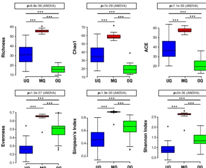
similar results, with the richness significantly affected by group identity (Fig. 2). Gut
microbial communities from the MQ state had the highest richness, followed by UQs,
and OQs with the lowest (
P
⬍
0.0001). Evenness was also calculated for the microbial
communities, to investigate how the queen status influenced the equality of
distribu-tion of different microbes within each gut. The highest species evenness was observed
for the MQ state, indicating that abundances of the diverse gut microbes associated
with the MQ state were the most evenly distributed (Fig. 2). Unlike richness, the
evenness of communities of OQs was greater than those of UQs. Finally, Simpson and
Shannon diversity indices were calculated incorporating richness and evenness. The
results for overall diversity mirrored those for evenness, with MQs having the greatest
on September 8, 2020 by guest
http://msystems.asm.org/
diversity, followed by OQs, and UQs having the lowest (Fig. 2). The evenness and
diversity results indicate that the gut microbial community of the unmated queen is
dominated by only a few species. Indeed, the bacterial genera
Gilliamella
and
Snodgras-sella
were the two most dominant gut microbiota taxa, accounting for 82.5%
⫾
3.49%
(mean
⫾
SE) of all total sequence reads in unmated queens. In brief, our results suggest
variation in the gut microbial community structure of the three queen stages (UQ, MQ,
and OQ).
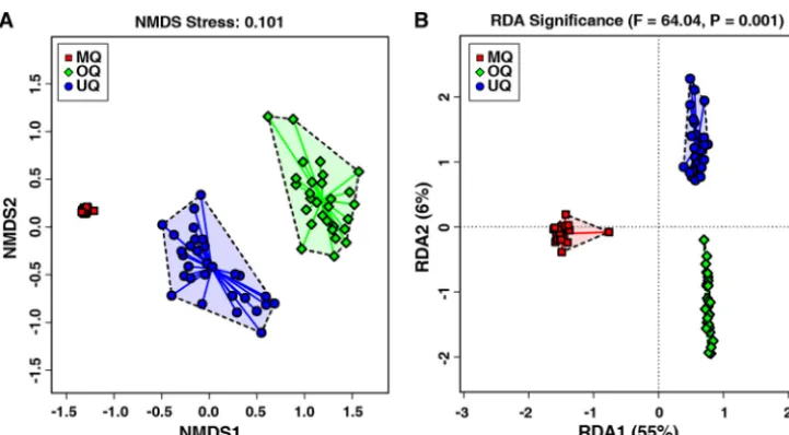
To test the above inference, we conducted beta-diversity analysis of the microbial
communities among UQ, MQ, and OQ states using both unsupervised and supervised
methods. The unsupervised nonmetric multidimensional scaling (NMDS) plots
(Bray-Curtis distance matrix) revealed a clear separation of samples according to the different
bumble bee queen stages (stress
⫽
0.101 provides a good representation in reduced
dimensions) (Fig. 3A). The supervised redundancy analysis (RDA) further indicated that
as the queens progress through the different physiological states, these can
signifi-cantly influence the gut microbial community composition of the host (
P
⫽
0.001,
Fig. 3B). These results obtained by two independent methods to assess beta-diversity
consistently suggested that there were significant structural separations of the gut
microbiota among UQs, MQs, and OQs. The microbial community structure associated
with the MQ state was significantly distinguished from UQ and OQ states (implicated by
RDA1 with 55% of variation in Fig. 3B). Enterotype analysis of the genus-level table for
the microbial communities of all samples also resulted in an optimal number of
enterotypes (clusters of similar communities) of three (Fig. S3). This finding indicates
FIG 2 The microbial community diversity of different queen states. Box plots show OTU measures of raw richness, ACE, Chao1, evenness, and Simpson and Shannon diversity indices.***indicates significant differences among groups (P⬍0.0001). UQ, unmated queen; MQ, mated queen; OQ, ovipositing queen.
Gut Microbial Dynamics of Bumble Bee Queens
on September 8, 2020 by guest
http://msystems.asm.org/
that the gut microbial community structure of mated queens is unique and may be a
consequence of physiological changes associated with mating itself or the
develop-ment of the microbiota as the queen ages or moves toward diapause. However, the
greater similarity between UQs and OQs suggests that the shift is more likely to be
associated with changes during the time or process of mating.
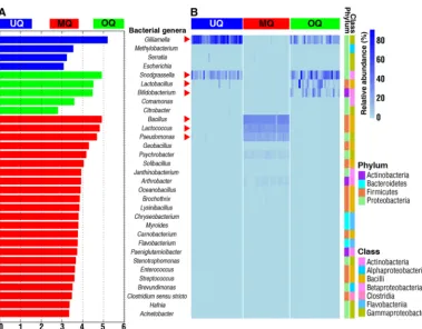
The discovery of gut microbial biomarkers associated with different queen
states.
Specific gut microbiota features can reflect specific disease or normal
physio-logical conditions of the host (43). Hence, we analyzed the relative abundance of four
phyla and their distribution in the guts of three stages of queens (Fig. S2).
Proteobac-teria
ware the most predominant gut bacterial phylum in both UQs (96.4%
⫾
0.77%,
mean
⫾
SE) and OQs (72.2%
⫾
4.42%, mean
⫾
SE). In contrast,
Firmicutes
account for
65.5%
⫾
1.16% (mean
⫾
SE) of total reads in MQs. These results indicate potential
broad-scale gut microbial markers unique to the three queen stages. Using LEfSe
analysis, we identified gut microbiota features specific to the three queen groups
(Fig. 4A). Among them, there were four bacterial genera in UQs, five genera in OQs, and
23 genera in MQs (Fig. 4A). Combined with a heatmap view of the relative abundance
of gut bacterial genera, our results show that seven bacterial genera can be used as
signatures of the three queen states (Fig. 4B). These genera showed significant
differ-ences in relative abundance between queen states (
P
⬍
0.05, Kruskal-Wallis test).
Gilliamella
and
Snodgrassella
were abundant bacterial genera associated with both UQs
and OQs, with high abundances of
Lactobacillus
and
Bifidobacterium
also associated
with OQs. The main bacterial genera in mated queen (MQ) guts were
Bacillus
,
Lacto-coccus
, and
Pseudomonas
. Besides the identified highly abundant bacterial phylotypes
for the MQ state, there were also low-abundance bacterial genera associated with MQs,
including
Proteobacteria
(
Psychrobacter
,
Methylobacterium
,
Serratia
,
Escherichia
,
Coma-monas
,
Citrobacter
,
Janthinobacterium
,
Stenotrophomonas
,
Brevundimonas
,
Hafnia
, and
Acinetobacter
),
Firmicutes
(
Brochothrix
,
Oceanobacillus
,
Geobacillus
,
Solibacillus
,
Lysinibacil-lus
,
Carnobacterium
,
Enterococcus
,
Streptococcus
, and
Clostridium sensu stricto
),
Actinobac-teria
(
Arthrobacter
and
Paeniglutamicibacter
), and
Bacteroidetes
(
Myroides
,
Chryseobac-terium
, and
Flavobacterium
) (Fig. 4). The relatively diverse compositions in MQs were
consistent with the finding of the highest alpha-diversity being in MQs, as presented in
Fig. 2. These results demonstrate specific gut microbial features associated with queens
at the postmating stage.
FIG 3 The similarity and variation of gut microbial community structures across the three queen groups of unmated queens (UQ), mated queens (MQ), and ovipositing queens (OQ). (A) Unsupervised NMDS plot of beta-diversity (Bray-Curtis dissimilarity) showing clustering of the gut microbiota from individual samples (n⫽86). Distances between individual samples reflect the extent of the similarity of gut microbiota. (B) Supervised RDA of the relationship between queen states and the relative abundance of bacterial genera. ThePvalue above the figure indicates that the variable (UQ, MQ, and OQ) significantly explains variation in sample distances.
on September 8, 2020 by guest
http://msystems.asm.org/
Copy number validation of differentially abundant bacterial genera in the
three queen stages.
To verify the accuracy of culture-independent analysis of bacterial
genera described by Fig. 4, we used 16S rRNA gene-targeted group-specific primers for
real-time PCR analysis of seven identified predominant bacterial genera (
Gilliamella
,
Snodgrassella
,
Lactobacillus
,
Bifidobacterium
,
Bacillus
,
Lactococcus
, and
Pseudomonas
) in
unmated queens, mated queens, and ovipositing queens. The mean absolute number
(
⫾
SD) of the overall bacterial rRNA genes of each queen stage and age was estimated
to vary from 1.47
⫻
10
8(
⫾
[4.5
⫻
10
7]) to 6.35
⫻
10
8(
⫾
[1.1
⫻
10
8]) copies per gut, with
each of the predominant bacterial genera differentially contributing to this total (Fig. 5
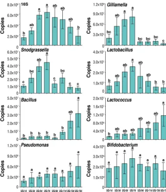
and 6).
This targeted approach confirmed the patterns seen in Fig. 4 for UQs 7 days
posteclosion, MQs 7 days after mating, and OQs (Fig. 5). In addition, we investigated the
temporal dynamics of changes within UQ, OQ, and MQ groups.
Gilliamella
,
Snodgras-sella
, and
Lactobacillus
increased with age posteclosion (1 to 7 days) in UQs and
declined significantly following mating at 7 days across the MQ stage (1 to 7 days
postmating) but rebounded to the peak seen in UQs 7 days posteclosion by the time
of queen egg laying (OQ). This initial increase, decline, and rebound were also present
in the total 16S rRNA copies (Fig. 5), indicating changes in total bacterial abundance.
However, the decline from 7 days posteclosion was not as pronounced due to increases
in other genera.
Bacillus
,
Lactococcus
, and
Pseudomonas
increased postmating from low
levels in the UQ state, returning to UQ state abundances in OQs. Uniquely, the bacterial
genus
Bifidobacterium
was found at higher abundances only in OQs.
To further understand the relationship of microbiota composition with queen state
and age, the same bacterial genera and total bacterial abundance were quantified in
FIG 4 Comparison of the predominant bacterial genera among the three queen groupings of unmated queens (UQ), mated queens (MQ), and ovipositing queens (OQ). (A) LEfSe analysis indicates significantly different abundances of bacterial genera for each group. LDA score value is 2. (B) Heatmap depicts the relative abundances of the identified gut bacterial genera across the different queen groupings. Relative abundance of the gut bacterial genera Gilliamella, Snodgrassella,Lactobacillus,Bifidobacterium,Bacillus,Pseudomonas, andLactococcusamong three groups with significant differences (P⬍0.05, Kruskal-Wallis tests). qPCR analyses were used to validate relative abundances of the seven differentially abundant genera marked by red arrowheads.
Gut Microbial Dynamics of Bumble Bee Queens
on September 8, 2020 by guest
http://msystems.asm.org/
unmated queens at eight time points from 1 to 15 days posteclosion (Fig. 6).
Interest-ingly, the results for all genera qualitatively mirrored those across UQ and MQ states
shown in Fig. 5, even though these queens remained unmated throughout the time
period. This indicates that the changes in microbiota composition in mated queens are
not entirely the result of mating
per se
but rather are the consequence of age- or
physiology-related changes as queens reach maturity and approach hibernation.
DISCUSSION
Our study demonstrates temporal dynamics in the gut microbiota of bumble bee
queens at 1 to 15 days post-adult eclosion and posthibernation, comparing three
physiological states: unmated, mated, and ovipositing. Over the first week following
eclosion, we see increases in three well-known apid bee symbionts,
Gilliamella
,
Snodgrassella
, and
Lactobacillus
, which have previously been reported in worker
bum-ble bees (44) and also show such a similar temporal increase in honey bee workers (45).
The prehibernation peak of these three bacterial genera is consistent with sexual
maturity in bumble bee queens, indicating their potential to functionally contribute to
the promotion of physiological development of virgin bumble bee queens. Surprisingly,
independent of mating status, after the 7-day peak, these early-abundant three
bac-terial genera were dramatically replaced by other bacteria, such as those members
FIG 5 The change of copy numbers of overall bacteria (16S rRNA copies) and bacterial genera at different time points of the three queen groupings. UQ, unmated queen; MQ, mated queens; OQ, ovipositing queens. Days (d) for UQ represent days posteclosion and for MQ represent days postmating, with mating occurring at 7 days posteclosion. Bars represent means⫾SEM. Different letters above bars within plots represent significant differences (pairwisettests,P⬍0.05).on September 8, 2020 by guest
http://msystems.asm.org/
belonging to the
Bacillus
genus. The independence of this change from actual mating
suggests that the shift may be the consequence of age- or physiology-related changes
as queens reach maturity and approach hibernation. This change is particularly
inter-esting given that the predominance of
Gilliamella
,
Snodgrassella
, and
Lactobacillus
was
restored in posthibernation ovipositing queens. Additionally, an increase in
Bifidobac-terium
is unique to the ovipositing queen group.
Bifidobacterium
increases during late
pregnancy in humans, with a potential beneficial role (46, 47). Overall, the dynamic
nature of the microbiota of bumble bee queens, including of some core and
function-ally important taxa, suggests links to the specific biology of bumble bee queens.
Whether gut microbiotas influence sexual maturation of their animal hosts remains
little explored. Our results imply a connection between a change in the microbiota of
bumble bee queens and the period of sexual maturity, but causation cannot be
inferred. It has been shown that the mouse microbiota is required for sex-specific
diurnal rhythms of gene expression and metabolism, showing that it plays a key role in
ensuring proper sexual maturation and growth hormone secretion (48). In addition,
studies have found that gut symbionts have potential effects on reproductive behaviors
in insects. For example, in
Drosophila melanogaster
commensal bacteria play a role in
mating preferences (10) and alteration of female microbiota counteracts a default male
outbreeding strategy by inhibiting female sexual signaling (49). The gut microbiota has
also been shown to modify olfactory sense-guided microbial preferences and foraging
FIG 6 The change of copy numbers of overall bacteria (16S rRNA copies) and bacterial genera at different time points for UQs (unmated queens). Days (d) represent days posteclosion. Bars represent means⫾SEM. Different letters above bars within plots represent significant differences (pairwisettests, P⬍0.05).Gut Microbial Dynamics of Bumble Bee Queens
on September 8, 2020 by guest
http://msystems.asm.org/
decisions in
Drosophila
, indicating a role of animal microbiota in shaping host
fitness-related behavior through their chemosensory responses (50). Moreover, the gut
bac-terium
Lactobacillus brevis
has been found to modulate locomotor activity in
D.
melanogaster
, which is mediated by the level of a sugar and the activity of neurons that
produce the molecule octopamine (51). While the changes in the microbiota around
the time of mating that we have uncovered in our study are intriguing, further work
would be required to elucidate if the microbiota influences bumble bee queen mating
behavior and chemical communication required for copulation, such as queen sex
pheromone production (52).
Disruption of the gut microbiota of primary termite reproductives has been shown
to have negative consequences for reproduction (53). A similar connection has been
made between microbiota presence and parthenogenetic reproduction in
Daphnia
water fleas (54). Unlike these experimentally induced disturbances of the microbiota,
our results show a significant but naturally occurring change in the bumble bee queen
microbiota. State-related changes have been shown for humans, with a shift in the gut
microbiota during pregnancy (55–57), but our study is one of the first to report such a
dramatic shift in an insect. These alterations could be adaptive, with positive effects on
physiological development and behavior, or simply a side effect of intrinsic
physiolog-ical changes ongoing within queens as they mature or of interactions with diet (58). Of
importance for understanding the causes and consequences of these dynamic changes
is the demonstration that while changes occurred around the time of mating, they are
not the result of mating itself. However, further investigations are required to infer
causation and any consequences of the decrease of earlier core bacteria and
enrich-ment of
Bacillus
,
Pseudomonas
, and
Lactococcus
in the mature bumble bee queen.
There is a potential for a direct active role of these enriched genera. Sabaté et al. (59)
showed that
Bacillus subtilis
strains isolated from honey bee gut produced surfactins
and fungicides that can inhibit the important honey bee pathogens
Paenibacillus larvae
and
Ascosphaera apis
. Also,
Bacillus
species can produce amylase that helps in the
processing of flower nectar into honey in honey bees (60). In rotifers,
Lactococcus
was
found to serve as a probiotic to enhance growth and immunity (61, 62).
Pseudomonas
species in insects have been shown to be involved in detoxification (63) and digestion
through amylolytic, xylanolytic, and diazotrophic activities that could contribute to the
nutritional supplement and nitrogen balance (64, 65).
A particularly intriguing finding is that the changes seen in the microbiota after 7
days posteclosion in both mated and unmated queens are reversed in ovipositing
queens posthibernation. This restoration of the dominance of core gut microbes
including
Gilliamella
,
Snodgrassella
, and
Lactobacillus
, in addition to an increase in
Bifidobacterium
, is likely critical for colony success, given the key role that many of these
taxa play in bees. Hibernation constitutes a period of considerable environmental and
physiological changes, yet relatively little is known about the relationship between gut
microbiota and hibernation. However, hibernation has been shown to be associated
with changes in the microbiota of some organisms (66–68). For example, Sommer et al.
(68) showed that the microbiota and serum metabolites in brown bears differ
season-ally between hibernating and active phases and that transplants of the specific
micro-biota into mice transferred some of the seasonal metabolic features seen in bears. For
temperate bumble bees, hibernation is usual after mating and before oviposition as an
adaptation to challenges imposed by winter. This period of diapause is associated with
many changes in metabolism and physiology in general, alongside the environmental
alterations (15). A difference in the microbiota between queens before and during
hibernation has been observed (30), which could be associated with the period of
hibernation itself. However, our observations indicate that significant changes to the
gut microbial community of bumble bee queens occur prior to entrance into
hiberna-tion and are largely reset in queens following hibernahiberna-tion, when they are egg laying.
Bumble bee queens utilize storage of energy, such as lipids and glycogen, to survive
low winter temperatures (69). Similar to the work of Bosmans et al. (30), we detected
some cold-loving and cold-tolerant bacterial genera, such as
Acinetobacter
,
on September 8, 2020 by guest
http://msystems.asm.org/
bacterium
,
Hafnia
,
Psychrobacter
, and
Pseudomonas
, in samples of mated queens and
also older unmated queens prior to hibernation. Their presence, even prior to the
initiation of abiotic environmental changes associated with hibernation, could support
the queens during the environmental transition during hibernation. The contribution of
this distinct microbial community to hibernation success, relative to earlier and later
microbial community compositions, is thus important to investigate further.
The microbiotas associated with organisms may be closely linked with physiological
and behavioral changes in their hosts, either responding to these changes indirectly or
directly being involved in them. Bumble bee queens undergo a number of biological
changes as they transition through adult emergence, mating, overwintering, foraging,
and colony initiation including egg laying. Therefore, they represent an important
system to understand the link between physiological, behavioral, and environmental
changes and host-associated microbiota. It is plausible that the bumble bee queen gut
bacteria play a role in shaping the ability of the queen to survive environmental
extremes and reproduce, due to long-established coevolutionary relationships between
the host and microbiome members. Our results show that there is a significant
difference in diversity and composition of the gut bacterial species between bumble
bee queens at different ages and physiological stages. This provides a critical insight
into the relationship between the bacterial community and queen status in bumble
bees and establishes the basis for further work to determine if the microbiota changes
identified are causal in the health and success of critically important bumble bee
queens.
MATERIALS AND METHODS
Overview of sampling.An approach using Illumina amplicon sequencing of the V3-V4 region of bacterial 16S rRNA was used to investigate differences in the microbiomes of queens at the different developmental stages of virgin unmated queens (UQs; 7 days post-adult eclosion,n⫽30), mated queens (MQs; mated at 7 days posteclosion and sampled 7 days later,n⫽27), and ovipositing queens after diapause (OQs;n⫽29). A targeted approach focusing on bacterial genera to verify these results by qPCR and to additionally assess temporal changes was carried out in UQs at 1, 3, 5, and 7 days post-adult eclosion and in MQs at 1, 3, 5, and 7 days postmating, taking place 7 days posteclosion (n⫽5 per time point). Furthermore, to investigate changes independent of mating, a final temporal assessment of UQs was carried out covering the period when MQs were sampled (1, 3, 5, 7, 9, 11, 13, and 15 days posteclosion,n⫽5 per time point).
Queen stages and sample collection.Queens ofB. lantschouensiswere collected in the spring of 2016 from natural populations in Gansu and Ningxia provinces of China and identified by morphology and molecular methods (70, 71). Since the animals investigated in this study are neither vertebrates nor regulated invertebrates, ethical approval was not required, and the bees were sampled on property in Gansu and Ningxia provinces with consent of the manager of the Botanical Garden. Collected queens were reared in small plastic cages in the dark at a temperature of 27⫾1°C and relative humidity of 50 to 60%. Sugar water (1:1, vol/vol) and apricot pollen were provided ad libitum to 100 colonies subsequently produced until males and gynes (new queens) emerged. Sampling for UQs, MQs, and OQs was carried out as outlined above. For matings, at 7 days post-queen eclosion, queens and males were kept at a ratio of 1:2, respectively, in a 4-m by 3-m by 2-m (length by width by height) net enclosure to ensure that one queen would mate with one male. For sampling of postdiapause OQs, mated queens were reared in a small wooden box until they became less active; they were then transferred to 4°C for diapause. After 4 months, they were revived and fed in the dark under the rearing conditions described above. Queens that had laid eggs and whose first batch of workers had emerged were collected for the OQ group. All collected samples were snap-frozen in liquid nitrogen and then stored at⫺80°C until the subsequent molecular analyses.
Extraction of the gut DNA.Before removing the whole gut from queens, including crop, midgut, ileum, and rectum, the sample surface was sterilized individually with 70% and 90% ethanol solution for 1 min, respectively, followed by multiple washes using double-distilled water. The abdomen was dissected with sterilized scissors and tweezers, and the whole alimentary canal was removed and transferred into a 1.5-ml microcentrifuge tube filled with 100l double-distilled water and ceramic beads (0.1 mm) for the subsequent DNA extraction.
Gut samples were homogenized in a tissue lyser (Qiagen, Hilden, Germany) followed by genomic DNA isolation using the Wizard Genomic DNA purification kit (Promega; A1120) according to the manufacturer’s instructions, with DNA suspended in 30l nuclease-free water. The concentration and quality of extracted DNA were assessed using a Qubit fluorometer (Invitrogen, Carlsbad, CA, USA) and 2% agarose gel electrophoresis, respectively. Extracted DNA was stored at⫺20°C until further processing.
Illumina sequencing and bioinformatics analysis.The hypervariable V3-V4 region of the bacterial 16S rRNA gene was amplified with the primers 341F (5=-CCTAYGGGRBGCASCAG-3=) and 806R (5=-GGA CTACNNGGGTATCTAAT-3=) (72). Twenty-microliter PCR mixtures were set up with 4l 5⫻FastPfu buffer,
Gut Microbial Dynamics of Bumble Bee Queens
on September 8, 2020 by guest
http://msystems.asm.org/
2l deoxynucleoside triphosphates (dNTPs) (2.5 mM), 0.8l each primer, 0.4l FastPfu polymerase, and template DNA (10 ng). Reactions proceeded in a GeneAmp 9700 (ABI) thermocycler with 95°C for 5 min; 27 cycles of denaturation at 95°C for 30 s, annealing at 55°C for 30 s, and elongation at 72°C for 45 s, followed by an additional elongation at 72°C for 10 min; and a dissociation stage at the end of the run. PCR products were detected by 2% agarose gel electrophoresis and purified using the QIAquick gel extraction kit (Qiagen). Library pools were constructed with equal amounts of each PCR product by using the TruSeq Nano DNA LT sample prep kit (Illumina), which was amplified through paired-end sequence on the Illumina MiSeq platform.
Raw Illumina sequence reads were modified by filtration, merging, and quality control, and barcode and primer sequences were removed, leaving library-specific tags. The fastq-join method was used to merge sequences using QIIME software (73), with an overlap length larger than 10 bp and mismatch ratio lower than 20%. Operational taxonomic unit (OTU) analysis was performed using the Uparse package (version 7.0.1001) with a 97% sequence identity on the basis of the effective tags (74). Each OTU was taxonomically assigned based on the SILVA 16S rRNA reference database using the assign_taxonomy.py program (http://qiime.org/scripts/assign_taxonomy.html). OTUs were processed by removing chloroplast sequences, mitochondrial sequences, and unclassified sequences and then obtaining species annotation information (confident threshold value,⬎0.8) (75, 76). Statistical differences in relative abundances of OTUs in different samples were analyzed by a nonparametric Kruskal-Wallis test, with analyses carried out using SPSS (version 17). The OTUs with relative abundance values of⬎0.001% (above three tags per sample) in at least one sample were retained.
The bacterial community diversities of gut samples were calculated and analyzed using the online software Calypso (http://cgenome.net/wiki/index.php/Calypso) with square root-based normalization of relative abundance. After samples were rarefied to even read depth, alpha-diversity measures of richness (Chao1 and ACE), evenness, and diversity indices (Simpson and Shannon) were compared between unmated, mated, and ovipositing queens by ANOVA. To determine if there are significant difference of gut microbial community structures among the three bumble bee queen states, the unsupervised nonmetric multidimensional scaling (NMDS) analysis of beta-diversity (Bray-Curtis distance matrix) was first conducted (77). The supervised redundancy analysis (RDA) was used to further validate complex associations between community composition and multiple explanatory variables (i.e., unmated, mated, and ovipositing queens in our study) (78). ThePvalue reported indicates if each explanatory variable is significantly associated with variation in gut microbial composition. Gut bacterial genera associated with different physiological conditions of bumble bee queens were further identified using the linear discriminant analysis (LDA) effect size method (LEfSe) with default parameters (79). Enterotype analysis was carried out as in a previous study (44).
Genus-specific primer design and PCR amplification.16S rRNA gene sequences of key bacterial genera were retrieved from the GenBank database. The software DNAMAN was used to align and analyze sequences and identify highly conserved regions for designing primer pairs that were unique for each genus, using Primer Premier, version 5.0. Primer sequences ofBacillus,Pseudomonas, andLactococcus were BacF (GATGCGTAGCCGACCTGAGA) and BacR (GGCGTTGCTCCGTCAGACTT), PseF (CCGTAACTGGTC TGAGAGGATG) and PseR (GCATGGCTGGATCAGGCTTT), and LactF (GCGATGATACATAGCCGACCTG) and LactR (AGTTAGCCGTCCCTTTCTGGTT), respectively. Primers forGilliamella,Snodgrassella,Lactobacillus, andBifidobacteriumwere from the previous study (80–82), as were the universal 16S rRNA primers to determine overall bacterial load in each queen gut sample (83, 84). To confirm the specificity of each bacterial primer set, 20-l PCR amplification was performed in a reaction mixture containing 10l of SYBR PremixEx TaqII (Tli RNase H Plus) (2⫻), 0.8l of the forward primer (10M), 0.8l of the reverse primer (10M), 1l of DNA sample, and 7.4l of double-distilled water. The PCR cycling conditions were as follows: predenaturation at 95°C for 30 s, followed by 40 cycles of denaturation at 95°C for 5 s and annealing at 60°C for 30 s, with a subsequent melt curve to check the specificity of the amplified fragments. The product sizes of PCR amplification were confirmed by 2% agarose gel electrophoresis.
Absolute qPCR assay.Single-band PCR products were purified using the EasyPure PCR purification kit and then inserted into the T vector using the pEASY-T1 simple cloning kit. The recombinant plasmid DNA was transformed into competent cells. Mixtures were uniformly smeared on Luria broth (LB) agar plates and cultured overnight at 37°C. The positive bacterial clones were selected and used for plasmid extraction according to the manufacturer’s instructions for the AxyPrep plasmid DNA minikit (Axygen; APMNP50). The concentration and quality of recombinant plasmid were measured by spectrophotom-etry (NanoDrop 2000, ThermoFisher) and visualized through 2% agarose gel electrophoresis, respectively. The recombinant plasmid DNA was stored at⫺80°C until use.
Based on the concentration of recombinant plasmids and the formula developed by Dhanasekaran et al. (85), copy numbers of the recombinant plasmid DNA were determined. The stock solution was 10-fold serially diluted to achieve different concentrations (from 108to 103copies/l) to generate a
standard curve.
Absolute quantitative PCR was performed in parallel with samples and corresponding serially diluted standards. The reaction mixture and thermocycler conditions of the PCR were the same as described above. Template DNA was diluted 10-fold before use. Each sample was run in triplicate. The actual copy numbers of specific bacterial 16S rRNA genes in samples were calculated by the threshold cycle (CT) value
relative to the relevant standard curve (86). Each standard curve was constructed by a liner regression of the logarithmic values of the estimated copy number of diluted standards (x axis) against the correspondingCTvalues (yaxis). The amplification efficiency (E) was related to the slope according to the
formulaE⫽10(⫺1/slope)⫺1 (87). The analyses of genus-specific bacterial 16S rRNA gene copy numbers
in different samples were performed in SPSS (version 17). Values were normalized with log
on September 8, 2020 by guest
http://msystems.asm.org/
tion. The significant differences in the copy numbers of bacteria at different time points were determined by one-way ANOVAs and least significant difference tests on the log-transformed values.
Data availability.The raw sequence data reported in this paper have been deposited in the Genome Sequence Archive (88) in the BIG Data Center (89), Beijing Institute of Genomics (BIG), Chinese Academy of Sciences, under accession number CRA001462 and are publicly accessible athttp://bigd.big.ac.cn/gsa.
SUPPLEMENTAL MATERIAL
Supplemental material for this article may be found at
https://doi.org/10.1128/
mSystems.00631-19
.
FIG S1
, PDF file, 0.05 MB.
FIG S2
, PDF file, 0.03 MB.
FIG S3
, PDF file, 0.3 MB.
ACKNOWLEDGMENTS
This study was supported by the Chinese National Natural Science Foundation (no.
31572338), the Agricultural Science and Technology Innovation Program
(CAAS–ASTIP-2016 –IAR) and China Agriculture Research System (CARS-45), the Key Research
Pro-gram of the Chinese Academy of Sciences (no. KFZD-SW-219), and the National Key
Research and Development Program of China (no. 2018YFC2000500). The contribution
of B.M.S. was supported by the National Institutes of Health (grant 1R15GM129681-01).
We appreciate help from a major biopharmaceutical technology company in
Shang-hai, allowing us to use the Illumina MiSeq platform for 16S amplicon sequencing. We
thank Jiaxing Huang, Shan Liu, and Jun Guo for help and advice in the laboratory.
Conceived and designed the experiments: Jilian Li, Zhigang Zhang, Jie Wu.
Per-formed the experiments: Liuhao Wang, Kai Li, Yulong Guo. Analyzed the data: Liuhao
Wang, Kai Li, Daohua Zhuang, Yulong Guo, Jilian Li, Zhigang Zhang. Contributed
reagents/materials/analysis tools: Liuhao Wang, Kai Li, Daohua Zhuang, Yulong Guo,
Jun Guo, Zhengyi Zhang, Ben M. Sadd, Jilian Li, Zhigang Zhang. Wrote the paper:
Liuhao Wang, Jilian Li, Zhigang Zhang, Ben M. Sadd, Yanping Chen, Jay D. Evans. All
authors contributed to and approved the final version.
We declare that we have no competing interests.
REFERENCES
1. Douglas AE. 2015. Multiorganismal insects: diversity and function of resident microorganisms. Annu Rev Entomol 60:17–34.https://doi.org/ 10.1146/annurev-ento-010814-020822.
2. Onchuru TO, Martinez AJ, Ingham CS, Kaltenpoth M. 2018. Transmission of mutualistic bacteria in social and gregarious insects. Curr Opin Insect Sci 28:50 –58.https://doi.org/10.1016/j.cois.2018.05.002.
3. Baumann P, Moran NA, Baumann L. 1997. The evolution and genetics of aphid endosymbionts. Bioscience 47:12–20.https://doi.org/10.2307/ 1313002.
4. Hansen AK, Moran NA. 2014. The impact of microbial symbionts on host plant utilization by herbivorous insects. Mol Ecol 23:1473–1496.https:// doi.org/10.1111/mec.12421.
5. Shin SC, Kim SH, You H, Kim B, Kim AC, Lee KA, Yoon JH, Ryu JH, Lee WJ. 2011.Drosophilamicrobiome modulates host developmental and met-abolic homeostasis via insulin signaling. Science 334:670 – 674.https:// doi.org/10.1126/science.1212782.
6. Lee JB, Park KE, Lee SA, Jang SH, Eo HJ, Jang HA, Kim CH, Ohbayashi T, Matsuura Y, Kikuchi Y, Futahashi R, Fukatsu T, Lee BL. 2017. Gut symbi-otic bacteria stimulate insect growth and egg production by modulating hexamerinandvitellogeningene expression. Dev Comp Immunol 69: 12–22.https://doi.org/10.1016/j.dci.2016.11.019.
7. Wong A-N, Dobson AJ, Douglas AE. 2014. Gut microbiota dictates the metabolic response ofDrosophilato diet. J Exp Biol 217:1894 –1901. https://doi.org/10.1242/jeb.101725.
8. Wada-Katsumata A, Zurek L, Nalyanya G, Roelofs WL, Zhang AJ, Schal C. 2015. Gut bacteria mediate aggregation in the German cockroach. Proc Natl Acad Sci U S A 112:15678 –15683. https://doi.org/10.1073/pnas .1504031112.
9. Dillon R, Vennard C, Charnley A. 2002. A note: gut bacteria produce components of a locus cohesion pheromone. J Appl Microbiol 92: 759 –763.https://doi.org/10.1046/j.1365-2672.2002.01581.x.
10. Sharon G, Segal D, Ringo JM, Hefetz A, Zilber-Rosenberg L, Rosenberg E. 2010. Commensal bacteria play a role in mating preference ofDrosophila melanogaster. Proc Natl Acad Sci U S A 107:20051–20056. https://doi .org/10.1073/pnas.1009906107.
11. Lize A, McKay R, Lewis Z. 2014. Kin recognition in Drosophila: the importance of ecology and gut microbiota. ISME J 8:469 – 477.https:// doi.org/10.1038/ismej.2013.157.
12. Walsh BS, Heys C, Lewis Z. 2017. Gut microbiota influences female choice and fecundity in the nuptial gift-giving species,Drosophila subobscura (Diptera: Drosophilidae). Eur J Entomol 114:439 – 445.https://doi.org/10 .14411/eje.2017.056.
13. Berlanga M, Paster BJ, Guerrero R. 2009. The taxophysiological paradox: changes in the intestinal microbiota of the xylophagous cockroach Cryptocercus punctulatusdepending on the physiological state of the host. Int Microbiol 12:227–236.https://doi.org/10.2436/20.1501.01.102. 14. Chen B, Teh B-S, Sun C, Hu S, Lu X, Boland W, Shao Y. 2016. Biodiversity
and activity of the gut microbiota across the life history of the insect herbivoreSpodoptera littoralis. Sci Rep 6:29505.https://doi.org/10.1038/ srep29505.
15. Amsalem E, Galbraith DA, Cnaani J, Teal PA, Grozinger CM. 2015. Con-servation and modification of genetic and physiological toolkits under-pinning diapause in bumble bee queens. Mol Ecol 24:5596 –5615. https://doi.org/10.1111/mec.13410.
16. Amsalem E, Grozinger CM, Padilla M, Hefetz A. 2015. The physiological and genomic bases of bumble bee social behaviour. Adv Insect Physiol 18:37–93.https://doi.org/10.1016/bs.aiip.2015.01.001.
17. Moran NA. 2015. Genomics of the honey bee microbiome. Curr Opin Insect Sci 10:22–28.https://doi.org/10.1016/j.cois.2015.04.003. 18. Kwong WK, Moran NA. 2016. Gut microbial communities of social bees.
Nat Rev Microbiol 14:374 –384.https://doi.org/10.1038/nrmicro.2016.43. 19. Koch H, Schmid-Hempel P. 2011. Bacterial communities in central Euro-Gut Microbial Dynamics of Bumble Bee Queens
on September 8, 2020 by guest
http://msystems.asm.org/
pean bumblebees: low diversity and high specificity. Microb Ecol 62: 121–133.https://doi.org/10.1007/s00248-011-9854-3.
20. Martinson VG, Danforth BN, Minckley RL, Rueppell O, Tingek S, Moran NA. 2011. A simple and distinctive microbiota associated with honey bees and bumble bees. Mol Ecol 20:619 – 628.https://doi.org/10.1111/j .1365-294X.2010.04959.x.
21. Kwong WK, Engel P, Koch H, Moran NA. 2014. Genomics and host special-ization of honey bee and bumble bee gut symbionts. Proc Natl Acad Sci U S A 111:11509 –11514.https://doi.org/10.1073/pnas.1405838111. 22. Meeus I, Parmentier L, Billiet A, Maebe K, Nieuwerburgh FV, Deforce D,
Wäckers F, Vandamme P, Smagghe G. 2015. 16S rRNA amplicon sequencing demonstrates that indoor-reared bumblebees (Bombus terrestris) harbor a core subset of bacteria normally associated with the wild host. PLoS One 10:e0125152.https://doi.org/10.1371/journal.pone.0125152.
23. Kwong WK, Mancenido AL, Moran NA. 2017. Immune system stimulation by the native gut microbiota of honey bees. R Soc Open Sci 4:170003. https://doi.org/10.1098/rsos.170003.
24. Emery O, Schmidt K, Engel P. 2017. Immune system stimulation by the gut symbiontFrischella perrarain the honey bee (Apis mellifera). Mol Ecol 26:2576 –2590.https://doi.org/10.1111/mec.14058.
25. Engel P, Martinson VG, Moran NA. 2012. Functional diversity within the simple gut microbiota of the honey bee. Proc Natl Acad Sci U S A 109:11002–11007.https://doi.org/10.1073/pnas.1202970109.
26. Koch H, Schmid-Hempel P. 2011. Socially transmitted gut microbiota pro-tect bumble bees against an intestinal parasite. Proc Natl Acad Sci U S A 108:19288 –19292.https://doi.org/10.1073/pnas.1110474108.
27. Forsgren E, Olofsson TC, Vásquez A, Fries I. 2010. Novel lactic acid bacteria inhibitingPaenibacillus larvaein honey bee larvae. Apidologie 41:99 –108.https://doi.org/10.1051/apido/2009065.
28. Parmentier A, Meeus I, Nieuwerburgh FV, Deforce D, Vandamme P, Smagghe G. 2018. A different gut microbial community between larvae and adults of a wild bumblebee nest (Bombus pascuorum). Insect Sci 25:66 –74.https://doi.org/10.1111/1744-7917.12381.
29. Tarpy DR, Mattila HR, Newton I. 2015. Development of the honey bee gut microbiome throughout the queen-rearing process. Appl Environ Microbiol 81:3182–3191.https://doi.org/10.1128/AEM.00307-15. 30. Bosmans L, Pozo MI, Verreth C, Crauwels S, Wäckers F, Jacquemyn H,
Lievens B. 2018. Hibernation leads to altered gut communities in bum-blebee queens (Bombus terrestris). Insects 9:E188. https://doi.org/10 .3390/insects9040188.
31. Powell JE, Eiri D, Moran NA, Rangel J. 2018. Modulation of the honey bee queen microbiota: effects of early social contact. PLoS One 13:e0200527. https://doi.org/10.1371/journal.pone.0200527.
32. Koch H, Abrol DP, Li JL, Schmid-Hempel P. 2013. Diversity and evolu-tionary patterns of bacterial gut associates of corbiculate bees. Mol Ecol 22:2028 –2044.https://doi.org/10.1111/mec.12209.
33. Tomono T, Sota T. 1997. The life and pollination ecology of bumblebees in the alpine zone of central Japan. Jpn J Entomol 65:237–255. 34. Cameron SA, Lozier JD, Strange JP, Koch JB, Cordes N, Solter LF, Griswold
TL. 2011. Patterns of widespread decline in North American bumble bees. Proc Natl Acad Sci U S A 108:662– 667.https://doi.org/10.1073/ pnas.1014743108.
35. Goulson D, Nicholls E, Botías C, Rotheray EL. 2015. Bee declines driven by combined stress from parasites, pesticides, and lack of flowers. Science 347:1255957.https://doi.org/10.1126/science.1255957.
36. Naeem M, Liu MJ, Huang JX, Ding GL, Potapov G, Jung CL, An JD. 2019. Vulnerability of East Asian bumblebee species to future climate and land cover changes. Agric Ecosyst Environ 277:11–20. https://doi.org/10 .1016/j.agee.2019.03.002.
37. Velthuis HHW, Van Doorn A. 2006. A century of advances in bumblebee domestication and the economic and environmental aspects of its commercialization for pollination. Apidologie 37:421– 451. https://doi .org/10.1051/apido:2006019.
38. Alford DV. 1969. A study of the hibernation of bumble bees (Hymenoptera: Bombidae) in southern England. J Anim Ecol 38:149 –170. https://doi.org/10.2307/2743.
39. Schmid-Hempel R, Schmid-Hempel P. 2000. Female mating frequencies in Bombus spp. from central Europe. Insect Soc 47:36 – 41.https://doi .org/10.1007/s000400050006.
40. Michener CD. 1974. The social behavior of the bees: a comparative study. The Belknap Press of Harvard University Press, Cambridge MA. 41. Beekman M, Van Stratum P. 2000. Does the diapause experience of
bumblebee queensBombus terrestrisaffect colony characteristics? Ecol Entomol 25:1– 6.https://doi.org/10.1046/j.1365-2311.2000.00235.x.
42. Evans E, Burns I, Spivak M. 2007. Befriending bumble bees: a practical guide to raising local bumble bees. University of Minnesota Extension, Saint Paul, MN.
43. Bäckhed F, Fraser CM, Ringel Y, Sanders ME, Sartor RB, Sherman PM, Versalovic J, Young V, Finlay BB. 2012. Defining a healthy human gut microbiome: current concepts, future directions, and clinical applica-tions. Cell Host Microbe 12:611– 622. https://doi.org/10.1016/j.chom .2012.10.012.
44. Li JL, Powell JE, Guo J, Evans JD, Wu J, Williams P, Lin Q, Moran NA, Zhang ZG. 2015. Two gut community enterotypes recur in diverse bumblebee species. Curr Biol 25:R652–R653. https://doi.org/10.1016/j .cub.2015.06.031.
45. Powell JE, Martinson VG, Urban-Mead K, Moran NA. 2014. Routes of acquisition of the gut microbiota of the honey beeApis mellifera. Appl Environ Microbiol 80:7378 –7387.https://doi.org/10.1128/AEM.01861-14. 46. Nuriel-Ohayon M, Neuman H, Ziv O, Belogolovski A, Barsheshet Y, Bloch N, Uzan A, Lahav R, Peretz A, Frishman S, Hod M, Hadar E, Louzoun Y, Avni O, Koren O. 2019. Progesterone increasesBifidobacteriumrelative abundance during late pregnancy. Cell Rep 27:730 –736.https://doi.org/ 10.1016/j.celrep.2019.03.075.
47. Dahl C, Stanislawski M, Iszatt N, Mandal S, Lozupone C, Clemente JC, Knight R, Stigum H, Eggesbø M. 2017. Gut microbiome of mothers delivering prematurely shows reduced diversity and lower relative abun-dance of Bifidobacterium and Streptococcus. PLoS One 12:e0184336. https://doi.org/10.1371/journal.pone.0184336.
48. Weger BD, Gobet C, Yeung J, Martin E, Jimenez S, Betrisey B, Foata F, Berger B, Balvay A, Foussier A, Charpagne A, Boizet-Bonhoure B, Chou CJ, Naef F, Gachon F. 2019. The mouse microbiome is required for sex-specific diurnal rhythms of gene expression and metabolism. Cell Metab 29:362–382.https://doi.org/10.1016/j.cmet.2018.09.023. 49. Heys C, Lize A, Colinet H, Price TAR, Prescott M, Ingleby F, Lewis Z. 2018.
Evidence that the microbiota counteracts male out breeding strategy by inhibiting sexual signaling in females. Front Ecol Evol 6:29.https://doi .org/10.3389/fevo.2018.00029.
50. Wong CAN, Wang QP, Morimoto J, Senior AM, Lihoreau M, Neely GG, Simpson SJ, Ponton F. 2017. Gut microbiota modifies olfactory-guided microbial preferences and foraging decisions inDrosophila. Curr Biol 27:2397–2404.https://doi.org/10.1016/j.cub.2017.07.022.
51. Schretter CE, Vielmetter J, Bartos I, Marka Z, Marka S, Argade S, Mazmanian SK. 2018. A gut microbial factor modulates locomotor behaviour in Dro-sophila. Nature 563:402– 406.https://doi.org/10.1038/s41586-018-0634-9. 52. Krieger GM, Duchateau MJ, Van Doorn A, Ibarra F, Francke W, Ayasse M.
2006. Identification of queen sex pheromone components of the bum-blebeeBombus terrestris. J Chem Ecol 32:453– 471.https://doi.org/10 .1007/s10886-005-9013-8.
53. Rosengaus RB, Zecher CN, Schultheis KF, Brucker RM, Bordenstein SR. 2011. Disruption of the termite gut microbiota and its prolonged con-sequences for fitness. Appl Environ Microbiol 77:4303– 4312.https://doi .org/10.1128/AEM.01886-10.
54. Sison-Mangus MP, Mushegian AA, Ebert D. 2015. Water fleas require microbiota for survival, growth and reproduction. ISME J 9:59 – 67. https://doi.org/10.1038/ismej.2014.116.
55. Koren O, Goodrich JK, Cullender TC, Spor A, Laitinen K, Bäckhed HK, Gonzalez A, Werner JJ, Angenent LT, Knight R, Bäckhed F, Isolauri E, Salminen S, Ley RE. 2012. Host remodeling of the gut microbiome and metabolic changes during pregnancy. Cell 150:470 – 480.https://doi.org/ 10.1016/j.cell.2012.07.008.
56. Santacruz A, Collado MC, García-Valdés L, Segura MT, Martín-Lagos JA, Anjos T, Martí-Romero M, Lopez RM, Florido J, Campoy C, Sanz Y. 2010. Gut microbiota composition is associated with body weight, weight gain and biochemical parameters in pregnant women. Br J Nutr 104:83–92. https://doi.org/10.1017/S0007114510000176.
57. DiGiulio DB, Callahan BJ, McMurdie PJ, Costello EK, Lyell DJ, Robacze-wska A, Sun CL, Goltsman DSA, Wong RJ, Shaw G, Stevenson DK, Holmes SP, Relman DA. 2015. Temporal and spatial variation of the human microbiota during pregnancy. Proc Natl Acad Sci U S A 112: 11060 –11065.https://doi.org/10.1073/pnas.1502875112.
58. Billiet A, Meeus I, Van Nieuwerburgh F, Deforce D, Wäckers F, Smagghe G. 2016. Impact of sugar syrup and pollen diet on the bacterial diversity in the gut of indoor-reared bumblebees (Bombus terrestris). Apidologie 47:548 –560.https://doi.org/10.1007/s13592-015-0399-1.
59. Sabaté DC, Carrillo L, Audisio MC. 2009. Inhibition ofPaenibacillus larvae andAscosphaera apisbyBacillus subtilisisolated from honeybee gut and