The cervical myodural bridge, a review of
literature and clinical implications
Dennis E. Enix,
DC, MBA1Frank Scali,
DC2Matthew E. Pontell,
BSc31 Associate Professor, Division of Research, Logan University 2 American University of the Caribbean, School of Medicine
3 Department of Anatomical Sciences, St. George’s University, School of Medicine Corresponding author’s contact information:
Dennis E. Enix DC, MBA E-mail: [email protected]
Address: Research Division, Logan University 1851 Schoettler Rd., Chesterfield, MO, 63017 U.S.A. Phone (636-230-1951)
©JCCA 2014
Institutions where work was performed: Cadaveric tissue samples were obtained at the Department of Anatomical Sciences at Logan University and the Department of Anatomical Sciences at St. George’s University, School of Medicine. Histological analysis of tissues was performed at the Research Microscopy Core in the Department of Pathology at Saint Louis University School of Medicine.
Declaration: The authors have no financial associations, commercial associations or other possible conflicts of interest relevant to this study to disclose. Dr. Enix receives salary support from Logan University and partial funding support from HRSA grant # R18HP15125 and a grant from Standard Process corp.
The role of posterior cervical musculature in sensorimotor control, cervicocephalic pain, and stabilization of the spinal cord has been recently described. Anatomical soft tissue connections which cross the cervical epidural space link suboccipital muscle fascia and dura. These myodural bridges provide passive and active anchoring of the spinal cord. They may also be involved in a dural tension monitoring system to prevent dural infolding, and maintain patency of the spinal cord. Modulation of dural tension may be initiated via a sensory reflex to muscular contractile
Le rôle de la musculature cervicale postérieure dans le contrôle sensorimoteur, la douleur cervico-céphalique et la stabilisation de la moelle épinière n’a que
Introduction
The cervical spine is a complex anatomical structure that is of interest to anatomists, biomechanists, and clin-icians. Soft tissue communications linking suboccipital muscle fascia and the dura and its role in cervical neuro-muscular control have been examined recently.1-8 These
myodural bridges have been associated with the etiology of cervicocephalic headaches, and cervicocephalic pain syndromes.3,9 These epidural connections may also be
in-volved passively as a dural anchor and as an active stabil-izer of the spinal cord.4,6,10
Recent studies have described myodural communi-cations bridging the epidural spaces between the rectus capitis posterior minor (RCPmi), rectus capitis poster-ior major (RCPma), and obliquus capitis inferposter-ior (OCI) suboccipital muscles and the dura mater of the cervical spine.1-8 Anatomical studies by Khan, Hack, Scali,
Pon-tell, and others have reported on the presence of myodural bridges linking sub-occipital muscles with the dura mater of the cervical spine.1,2,4,6 Additional studies by
Shin-omiya, Humphreys, Nash, Zumpano and Tagil confirmed these findings.13,10-13 Connections between the
suboccipi-tal musculature fascia and cervical dura mater have impli-cations in cervicocephalic pain syndromes, sensorimotor function, and postural control.14-17 The clinical relevance
of these cervical epidural membranes and their relation-ship to cervicogenic and tension headache syndromes has been discussed by multiple authors including Bates,
Schoenen, Haldeman, Fernandez-De-Las-Penas and others.9,14-17 Changes in cervical proprioception, balance,
sympathetic tone, conversion of muscle type, and dural enfolding secondary to cervical spine injuries have been described by several authors including Palmgren, Rix, Uhlig, Cailliet, Lusczyk.18-25,
Anatomical research and reviews of existing literature concerning the fascial connections between the suboccipi-tal muscles and the cervical dura have been reported re-cently.1-8 In 1992 a fascial connection between was briefly
mentioned by Kahn et al., in a report of the posterior intervertebral spaces of the cervical craniovertebral joint.1
It was noted however, that a true membrane connecting the posterior arch of the atlas to the laminae of the axis did not exist, but rather two fibrous planes that transected this space.1 Hack et al, reported on a myodural bridge
be-tween the fascia of the rectus capitis posterior minor (RC-Pmi) muscles in 1995.2,26 An examination of the posterior
atlantooccipital interspace showed a continuous band of tissue with fibers oriented primarily perpendicular to the muscle and dura.2,26 A animal model study of 43
speci-mens in 1996 by Shinomiya et al. examined the ligament-ous attachments within the cervical posterior epidural space.10 They confirmed the presence of abundant
pos-terior epidural ligaments attaching to the pospos-terior dura mater.10 Dissections of seven human cadaveric specimens
by Rutten et al. in 1997, agreed with previous findings of an epidural connection between the RCPmi and the pos-tissues. Unanticipated movements such as hyperflexion
extension injuries stimulate deep suboccipital muscles and transmit tensile forces through the bridge to the cervical dura. Due to its larger cross sectional area, the rectus capitis posterior major myodural bridge may exert greater mechanical traction on the dura than the rectus capitis posterior minor. University ethics committee approval and anatomical donor consent was obtained for this study.
(JCCA 2014;58(2):184-192)
k e y w o r d s: rectus capitis posterior major, obliquus
capitis inferior, myodural bridge, dura mater
comme les blessures résultant d’une hyperflexion-extension stimulent les muscles sous-occipitaux profonds et transmettent des efforts de traction par le pont sur la dure-mère cervicale. En raison de sa plus grande section transversale, le pont myodural grand droit postérieur peut exercer une plus grande traction mécanique sur la dure-mère que le muscle petit droit postérieur. L’approbation du comité d’éthique de l’université et le consentement du donneur anatomique ont été obtenus pour la présente étude.
(JCCA 2014;58(2):184-192)
m o t s c l é s : grand droit postérieur, oblique inférieur
terior wall of the spinal cord dura mater.24 Alix and Bates
reported on previous anatomical findings and suggested that the ligamentous connection between muscle and the pain sensitive dura mater may provide an anatomic and physiological basis for cervicogenic headaches.9 After a
gross anatomical dissection of 30 cadaveric specimens in 2003, followed by magnetic resonance imaging (MRI) of 4 specimens, Humphreys’ et al, also confirmed the pres-ence of a fibrous connection bridging the epidural space linking the dura and RCPmi.13 In a case report describing
an anatomical variation of a bifurcated RCPmi discovered during a routine dissection in 2005, Tagil confirmed the presence of dense connective tissue linking suboccipital muscle fascia to the cervical dura mater.3 Nash et al,
em-ployed sheet plastinations and confocal microscopy in an examination of the posterior atlanto-occipital interspace.12
They noted the presence of connective tissue attaching the RCPmi fascia and the spinal dura mater in the posterior cranio-cervical region in adult human cadavers.12 A larger
study of seventy five cadavers by Zumpano et al. in 2006, examined variations in prevalence, tissue-type, gender in the soft-tissue bridge between RCPmi and dura mater.11
They reported similar findings in the structure of the RC-Pmi myodural bridge as previous authors.27 According
to Kahkeshani and Ward, the myodural bridge has been underreported due the time necessary to properly dissect this region.27 To encourage further study; they describe a
method for the deep dissection of the suboccipital triangle which preserves the RCPmi and its attachments for fur-ther inspection.27
We recently reported on two additional epidural con-nections to the cervical dura.4-7 Myodural bridges
ex-tending from the anterior fascia of the rectus capitis pos-terior major (RCPma) and obliquus capitis inferior (OCI) muscles, attaching on the cervical dura mater was docu-mented recently by Scali, and Pontell in 2011 and 2013 (fig1).4-7 In an anatomical study by Scali et al., 13
em-balmed human cadaveric specimens showed broad fascial connections traversing the cervical epidural space from the RCPma muscle fascia, anterio-inferiorly to the poster-ior dural surface (fig 2).4 RCPma myodural tissues from
11 specimens stained with hematoxylin and eosin indicat-ed that the fascial connection insertindicat-ed directly into the fa-scia surrounding the RCPma and attached to the posterior surface of the dura (fig 3).5 Immunohistochemical
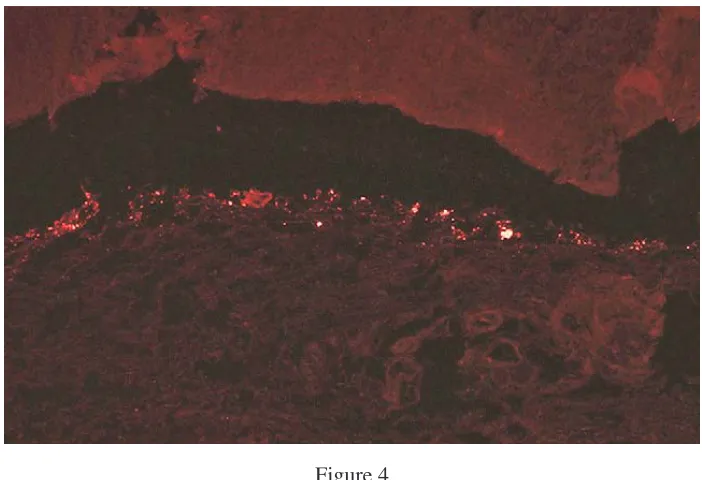
analy-sis using anti-neurofilament protein fluorescent antibody
staining showed a pattern of nerve distribution through-out one tissue sample (fig 4).5 Pontell et al. dissected nine
human cadaveric specimens, examining 14 OCI muscles and surrounding tissue.6 We documented a continuous
fi-brous tissue originating at the anterior fascia of the OCI muscle belly and projecting anteriorly across the atlanto-axial interspace and attaching to the posterolateral aspect of the cervical dura mater between the first and second cervical vertebrae.6 Histological analysis was performed
on 12 OCI suboccipital muscles, connective tissue, and dura mater from human cadavers between the ages of 49 to 81.7 Microscopic examination of OCI myodural
tissues stained with hematoxylin and eosin showed the connective tissue emanating from the ventral OCI mus-cular fascia and inserted directly into the posterolateral aspect of the cervical dura mater.7 A single OCI myodural
connection stained for immuno-peroxidase using Dako’s neurofilament protein monoclonal antibodies revealed fascicles traveling perpendicular, and parallel with the OCI myodural bridge.7 Due to proximity, the RCPma
and OCI muscles appear to form a single atlantoaxial myodural bridge, they are however, separate structures.7
We were unable to find a similar connection between the obliquus capitis superior and the dura mater.7 To examine
the prevalence of these structures, T-2 weighted magnetic resonance imaging (MRI) of the atlanto-axial interspace of 240 individuals was performed in 2012 by Scali et, al.8
Sixty four percent of the MRI’s reviewed demonstrated a posterior concavity of the cervical dura mater consistent with a ligament attachment site.8 Of this group, 24% also
had oblique, linear hypointense fibers which appeared to attach to the cervical dura mater.8 The breadth of studies
which have examined these soft tissue dural connections, precludes them being considered variations of normal anatomy.1-8,11-13,27
In this article, we focus on the anatomical, functional significance, and clinical relevance of the RCPma and OCI myodural bridge. Consideration of its function as a passive and active stabilizer of the cervical spinal cord is discussed.
Anatomy of the suboccipital muscles
Figure 1
Illustration of a dissection of the deep suboccipital region of the cervical spine. The rectus capitis posterior minor (RCPmi), rectus capitis posterior major (RCPma), and the obliquis capitis inferior (OCI) muscle fascia have communications with the dura mater via soft tissue. The encircled illustration (right) depicts a midsagittal dissection revealing the RCPma, RCPmi, and OCI muscles. The cervical myodural bridge (a) traverses the epidural space between the posterior elements of the C1 and C2 vertebrae. Both myodural structures link the suboccipital muscle fascia in to the cervical dura mater (*).Used with permission from: Magnetic resonance imaging investigation of the atlanto-axial interspace. Clin Anat. 2013 May; 26(4):444-9. Scali et al.
(Original anatomical artwork by Frank Scali, D.C., and Danny Quirk)
Figure 2
Figure 4
Sagittal section of the myodural bridge between the rectus capitis posterior major and the cervical dura mater depicting positive fluorescence after staining with antineurofilament protein antibodies. Used with permission from:
Histological Analysis of the Connection between the Rectus Capitis Posterior Major’s myodural bridge, The Spine Journal 13 (2013) 558-563., Scali F, Pontell M, Enix D, Marshall E.
Figure 3
Hematoxylin and eosin stained tissues of left side sagittal section showing the soft tissue communication (a) between the RCPma (b) and the cervical dura mater (c) in a male cadaveric specimen. The magnified area shows the soft tissue
communication at the point of contact with the dura mater (d). Used with permission from: Histological Analysis of the Connection between the Rectus Capitis Posterior Major’s myodural bridge, The Spine Journal 13 (2013) 558-563.,
the cervical spine.3,4,22 Cervical suboccipital muscles are
richly innervated and contain relatively high muscle spin-dle content.18,21 Muscle spindle fibers found in the
RC-Pma and OCI muscles are a source of primary afferents, representing major contributors to cervical spine neuro-muscular control.3,18,19,28,29 Consistent with their function
of complex coordination and organization, high muscle spindle concentrations are typically found in smaller muscle groups responsible for fine motor skills.18,29,30
Kulkarni et al. documented the density of muscle spindles in fetal tissue per gram of muscle tissue.18 A large
num-ber of muscle spindles were noted in both the OCI and RCPma suboccipital muscles, with 242 and 98 spindles per gram of muscle tissue, compared to the trapezius and latissimus dorsi postural muscles with 2.2 and 1.4 muscle spindles per gram.18 Muscle spindles are typically more
concentrated in regions richest in slow fibers, and while the OCI muscles have been reported as a blend of un-evenly dispersed type I and II fibers, their muscle spindles are distributed disproportionately in the deep areas rich-est in slow twitch fibers.18,29 This unique configuration of
muscle types suggests that these muscles may serve mul-tiple functions including monitoring kinesthetic changes, maintaining constant force for eccentric head posture, and creating fast phasic movements when needed.18,29,31
An electromyographic examination showed the RCPmi to be under active contraction while the head is in an upright neutral position, with muscle activity increasing signifi-cantly during cervical retraction.32 The cervical muscles
are subject to conversion from slow twitch to fast twitch muscles with injury.20,21 Alteration in muscle type, to a
more glycogenic morphology creates a muscle prone to facilitation.21,31 This change can alter the discharge of
pri-mary afferents to the central nervous system effecting cer-vical neuromuscular control.19-21,31 Additionally, a loss of
proprioceptive inhibition of nociceptors at the dorsal horn of the spinal cord can result in chronic pain.3,25
Clinical implications
The spinal canals’ midsagittal diameter of 10 mm which increases with flexion and decreases with extension, can impact the patency of the cervical cord.22,25 In a study
of 19 cadaveric specimens by Hong et. al., showed sig-nificant differences in dural thickness between different levels in the thoracic and lumbar spines, with the dura slightly thicker in men than women.33 Buckling of
cervic-al ligaments and dura mater have been reported with cer-vical spine extension.22-25,34 Cervical extension can create
infolding of the ligamentum flavuum, which encroaches upon the cord.30 It is the elastin fibers found in the in the
ligamentum flavuum that function to inhibit this inward buckling of the ligament into the spinal cord.22,24,34,35 The
dura mater, which is densely populated with nociceptors, also contains elastin fibers which are oriented in such a way as to resist the load placed on them.34,35 The strain
on the posterior side being greater than on the anterior side.25,34,35 Our recent histological analysis of cervical
dura elastin fibers in eight cadaveric specimens, con-firms that elastin density changes from caudal to cranial as well as the orientation of fibers which run parallel and perpendicular to each other. Cervical extension may therefore also cause inward buckling of the dura itself, compromising the dorsal subarachnoid space.31,34,36 In a
prospective analysis of fifty patients receiving cervical injections under myelography; six percent demonstrat-ed dural infolding on cervical extension, narrowing the posterior subarachnoid space.36 Traumatic and iatrogenic
tears to the cervical dura can effect up to 36% of cervical spine injuries.23 Inflammation of the meninges, subdural
hematomas, epidural infections, and nerve root compres-sion and postural headaches are all the sequel of cerebral spinal fluid leaks with dural tears.23,25 Meningeal
vascu-lar irritation can also cause hypertonicity of the posterior neck muscles, resulting in permanent tension on the dura, stimulating nociceptive dural fibers.23,26,37 Considering the
close proximity of the leptomeninges to the dura mater, a system to maintain the integrity of the subarachnoid space and, cerebrospinal fluid flow may exist.14,15,25,28
Simi-lar to the denticulate ligaments which secure the spinal cord within the subarachnoid space, the myodural bridge crosses the epidural space to anchor the spinal dura during head and neck motion.4,6,10,23,25,28
Neuromuscular stabilization of the spinal cord Contraction of the RCPma, RCPmi, and OCI sub-occipi-tal muscles which puts the myodural bridge under tension, transmitting forces across it to place the dura under ten-sion and stabilize the spinal cord.4,6,26,38,39 In addition to
ac-tive contraction, the suboccipital muscles respond reflex-ively to involuntary and unanticipated movements of the head and neck.20,25 Modulation of dural tension may also
tissues.5,7 Central nuclei that exert control over the deep
suboccipital muscles, including the RCPma, RCPmi and OCI muscles could respond reflexively as a feedback con-trol of dural tension.5,7,25,28 Like many systems involving a
feedforward or feedback mechanism of control, dural ten-sion regulated through muscular contraction of suboccipi-tal muscles is dependent on internal and external factors affecting those systems.4,6,28,31 The regulation of tension
across the myodural bridge as a spinal cord stabilizer may prevent dural infolding, reducing stimulation of nocicep-tive pain mechanisms.5,7,39 Hypertrophy of suboccipital
muscles or a failure of this system to maintain constant tension, may result in clinical manifestations arising from increased dural tension.1,4,6,9,20
Discussion
Functional anatomy of the myodural bridges
Many authors have speculated on the functional signifi-cance of the myodural bridge, generally attributing a mechanical advantage to it in stabilizing the spinal cord from dural enfolding.1-13 The reflexive myotatic response
of suboccipital muscles has been proposed by several authors as a likely mechanism to place the dura under tension.4-9,38,39 In a study of 20 cadavers, Nakagawa
re-ported that cervical cephalocaudal stresses may be due to the parallel orientation of elastin fibers in the dura and concluded its function was to resist hyperextension and compressive infolding of the dura.34 Hack suggested that
the purpose of the RCPmi myodural bridge might be to assist in resisting dural infolding, previously noted by Burt.2,36 Shinomiya et. al., concluded that the role of the
posterior cervical epidural ligaments is to provide an anchor to stabilize the dura mater from anterior transla-tion during flexion.10 Without a posterior epidural
attach-ment, the dural canal can shift anteriorly compressing the spinal cord causing flexion myelopathy.10 Rutten reported
on high muscle spindle content in the RCPmi, postulat-ing that the myodural bridges function may to monitor stresses on the cervical dura mater, reflexively preventing infolding.24 They reported that tissue injury from cervical
whiplash could affect mechanoreceptive properties, caus-ing the monitorcaus-ing system that maintains dural tension to fail.21,24,31 Alix & Bates also discussed the duras tendency
to fold inward on the spinal cord, and the myodural bridg-es ability to rbridg-esist this movement.9 McPartland and
Bro-deur proposed that the RCPmi plays a role in preventing dural crimping when the head is extended or moved backward, inhibiting normal circulation of cerebrospinal fluid.38 Humphries reported on previous studies
describ-ing the primary mechanical function of the RCPmi to re-sist dural buckling during cervical extension, preventing damage to the spinal cord.13 In a case report describing
an anatomical variation of the suboccipital muscles, Tagil also noted that the spinal cord is believed to be protected by the dense connective tissue that links the suboccipital muscles to the cervical dura mater.3 Kahkeshani and Ward
indicated that a direct connection linking the musculo-skeletal system to the dura mater provides a mechanical explanation for the efficacy of cervical massage and ma-nipulative treatment for headaches.27
Along with the description of the RCPma myodural bridge by Scali et al., we proposed that modulation of cervical dural tension may include factors other than a myotatic reflex.4,5 Myodural biofeedback may play a role
in maintaining the integrity of the subarachnoid space.4,5
We noted during dissection that manual traction applied to the RCPma caused movement of the spinal root within several levels.4 Due to its larger cross sectional area, the
rectus capitis posterior major myodural bridge may exert greater mechanical traction on the dura than the rectus capitis posterior minor.4 We described another soft tissue
connection traversing the epidural space between the OCI muscle fascia and the posterior sleeve of the dura mater in a paper by Pontell et al.6 It was reported that the OCI
myodural connections function dynamically to prevent dural infolding during cervical extension, similar to the RCPmi and RCPma bridges.6,7
We agree with previous authors who describe a stabil-izing function of the RCPmi muscle myodural bridge and propose a similar role for the RCPma and OCI muscle myodural bridges. In addition to the passive anchoring of the dura described by Nakagawa, Hack, Humphries, and Shinomiya, a myo-reflexive response described by Rutten provides an active stabilizing component.2,7,10,13,19,34,28,34
Besides the reflexive myotatic response of the suboccipital muscles, the presence of neuronal fibers in these tissues, may suggest functions other than the passive anchoring of these muscles to the posterolateral dura mater.5,7
make this an area of great interest.9,13-17,20,39 The
anatom-ical and histologanatom-ical evidence of these soft tissue com-munications bridging the epidural space from the RCPma and OCI muscles to the dura mater offers insight into the cervical spines’ complex function of neuromuscular con-trol.5,7,18,39 Further examination of the tensile forces in the
myodural bridge is needed. Biomechanical testing to con-firm the tensile forces on these tissues currently in prog-ress.
Conclusion
Anatomical soft tissue bridges which cross the cervical epidural space, connecting suboccipital muscle fascia and dura have passive and active functions to anchor the spinal cord. These myodural bridges may be involved in a dural tension monitoring system to prevent dural infolding and maintain patency of the spinal cord. Failure of this system could result in altered cerebral spinal fluid flow, changes in sensorimotor function, cervicocephalic headaches, and dural related pathologies.
Acknowledgements:
The authors would like to thank Logan University gradu-ate students Patrick Battaglia, and Robbyn Keating for assistance with cadaveric dissections and manuscript re-view and Dr. Jan Ryerse at the Research Microscopy Core in the Department of Pathology at Saint Louis University school of Medicine for preparation and interpretation of histological data and Danny Quirk for original illustra-tions.
References
1. Kahn JL, Sick H, Kortiké JG. Les espaces intervertébraux postérieurs de la jointure crânio-rachidienne. Acta Anat. 1992;144:65-70.
2. Hack GD, Kortizer RT, Robinson WL. Anatomic relation between the rectus capitis posterior minor muscle and the dura mater. Spine. 1995;20:2484-6.
3. Tagil SM, Ozçakar L, Bozkurt MC. Insight into understanding the anatomical and clinical aspects of supernumerary rectus capitis posterior muscles. Clin Anat. 2005;18:373-375.
4. Scali F, Marsili ES, Pontell ME. Anatomical connection between the rectus capitis posterior major and the dura mater. Spine. 2011;36:E1612-4.
5. Scali F, Pontell ME, Enix DE, Marshall E. Histological analysis of the rectus capitis posterior major’s myodural bridge. The Spine Journal. 2013;13(5):558-563.
6. Pontell ME, Scali F, Marshall E, Enix DE. The obliquus capitius, inferior myodural bridge. Clin Anat. 2013;26(4):450-4.
7. Pontell M, Scali F, Enix DE, Marshall E. Histological examination of the human obliquus capitis inferior myodural bridge. Annals Anatomy. 2013;195(6):522-26. 8. Scali F, Pontell ME, Welk AB, Malmstrom TK, Marshall E,
Kettner NW. Magnetic resonance imaging investigation of the atlanto-axial interspace. Clin Anat. 2013;26(4):444-9. 9. Alix ME, Bates DK. A proposed etiology of cervicogenic
headache: the neurophysiologic basis and anatomic relationship between the dura mater and the rectus capitis posterior minor muscle. J Man Manip Ther. 1999;22:534-9.
10. Shinomiya K, Dawson J, Spengler DM, Konrad P, Blumenkopf B. An analysis of the posterior epidural ligament role on the cervical spinal cord. Spine. 1996;21:2081-8.
11. Zumpano MP, Hartwell S, Jagos C. Soft tissue connection between rectus capitis posterior minor and the posterior atlanto-occipital membrane. Clin Anat. 2006; 19:522-527. 12. Nash L, Nicholson H, Lee ASJ, Johnson G M, Zhang M.
Configuration in the posterior atlanto-occipital interspace. Spine. 2005;30:1359-1366.
13. Humphreys BK, Kenin S, Hubbard B, Cramer G. Investigation of connective tissue attachments to the cervical spinal dura mater. Clin Anat. 2003; 16:484-493. 14. Bogduk N. Cervicogenic Headache: anatomic basis and
pathophysiologic Mechanisms. Current Pain and Headache Reports. 2001; 5:382-386.
15. Haldeman S, Dagenais S. Cervicogenic headaches: a critical review. Spine J. 2001;1(1):31-46.
16. Fernandez-De-Las-Penas C. Clinical evaluation of cervicogenic headache: a clinical perspective. J Man Manip Ther. 2008;16:81.
17. Schoenen J, Fumal A. Tension-type headache: current research and clinical management. Lancet Neurology. 2008; 7(1):70-83.
18. Kulkarni V, Chandy MJ, Babu KS. Quantitative study of muscle spindles in suboccipital muscles of human fetuses. Neurol India. 2001;49(4):355-9.
19. Palmgren PJ, Andreasson D, Eriksson M, Hägglund H. Cervicocephalic kinesthetic sensibility and postural balance in patients with nontraumatic chronic neck pain – a pilot study. Chiropractic & Osteopathy. 2009;17:6. 20. Rix GD, Bagust J. Cervicocephalic kinesthetic sensibility
in patients with chronic, nontraumatic cervical spine pain. Arch Phys Med Rehabil. 2001;82:911-9.
21. Uhlig Y, Weber BR, Grob D, Muntener M, Fiber composition and fiber transformations in neck muscles with dysfunction of the cervical spine. J Orthop Res. 1995;13(2):240-9.
23. Lusczyk MJ, Blaisdell GY, Wiater B P, Bellabarba C, Chapman JR, Agel JA, Bransford RJ. Traumatic Dural Tears: what do we know and are they a problem? The Spine Journal. 2014;14(1):49-56.
24. Rutten HP, Szpak K, van Mameren H, et al. Letter to Editor; response to Anatomic relation between the rectus capitis posterior minor muscle and the dura mater. Spine. 1997; 22:924-8.
25. Cramer GD, Darby SA. Clinical Anatomy of the Spine, Spinal Cord and ANS, 2e. Mosby Publishers; 2 edition. ISBN-10: 0323079547.
26. Hack GD, Hallgren RC. Chronic headache relief after section of suboccipital muscle dural connections: a case report. Headache. 2004;44:84-9.
27. Kahkeshani K, Ward PJ. Connection between the spinal dura mater and suboccipital musculature: Evidence for the myodural bridge and a route for its dissection – A review. Clin Anat. 2012;25(4):415-22.
28. Siegel A, Sapru HN. The spinal cord, In: Essential Neuroscience. Ed. Betty Sun. 2nd ed. Philadelphia: Lippincott Williams & Wilkins, 2011, 150-60. 29. Richmond FJR, Singh K, Corneil BD. Marked
non-uniformity of fiber-type composition in the primate suboccipital muscle obliquus capitis inferior. Exp Brain Res. 1999;125:14-18.
30. Farshadmanesh F, Chang P, Wang H, et al. Neck muscle synergies during stimulation and inactivation of the Interstitial Nucleus of Cajal (INC). J Neurophysiol. 2008;100:1677-1685.
31. Korr IM, The physiological basis of osteopathic medicine. New York (Insight Publishing) 1982.
32. Hallgren RC, Pierce SJ, Prokop LL, Rowan JJ, Lee AS. Electromyographic activity of rectus capitis posterior minor muscles associated with voluntary retraction of the head. Spine J. 2014;14(1):104-12.
33. Hong J, Suh SW, Park SY, Modi HN, Rhyu IJ, Kwon S, Yu H, Byun J. Analysis of dural sac thickness in human spine – cadaver study with confocal infrared laser microscope. The Spine Journal. 2011;11(12):1121-1127.
34. Nakagawa H, Mikawa Y, Watanabe R. Elastin in the human posterior longitudinal ligament and spinal dura: A histologic and biochemical study. Spine. 1994;19(19):2164-2169.
35. Mazgajczyk M, Scigala K, Czy M. Mechanical properties of cervical dura mater. Acta Bioeng Biomech. 2013;14;1. 36. Burt TB, Seeger JF, Carmody RF, Yang PJ. Dural infolding
during C1-2 myelography. Radiology. 1986;158(2):546-7. 37. Hu JW, Vernon H, Tatourian I, Changes in neck
electromyography associated with meningeal noxious stimulation, In: J Manipulative Physiol Ther. 1995 Nov-Dec; 18(9):577-81.