ScholarlyCommons
Publicly Accessible Penn Dissertations
1-1-2015
Context- Dependent Gene Expression Programs
Promote Lymphocyte Development and Function
and Suppress Transformation
Amy Demicco
University of Pennsylvania, [email protected]
Follow this and additional works at:
http://repository.upenn.edu/edissertations
Part of the
Allergy and Immunology Commons
,
Cell Biology Commons
,
Immunology and
Infectious Disease Commons
,
Medical Immunology Commons
, and the
Molecular Biology
Commons
This paper is posted at ScholarlyCommons.http://repository.upenn.edu/edissertations/1682 For more information, please [email protected].
Recommended Citation
Demicco, Amy, "Context- Dependent Gene Expression Programs Promote Lymphocyte Development and Function and Suppress Transformation" (2015).Publicly Accessible Penn Dissertations. 1682.
Development and Function and Suppress Transformation
Abstract
Coordinated orchestration of gene expression programs at the transcriptional, transcriptional, and post-translational levels is essential for development and function of all cells, including lymphocytes. Normal tissue function also demands that the genome be faithfully passed from mother to daughter cell during the many rounds of cell division required to generate a mammalian organism. Genome integrity is maintained in part by integration of DNA damage signaling with cell cycle control. These mechanisms are especially critical for lymphocytes following V(D)J recombination, since V(D)J recombination involves genetic cutting and pasting of germline gene segments to form antigen receptors (AgRs). Using conditional deletion of the p53 tumor suppressor in mice, I found that p53 promotes genome stability in developmental stage-specific ways. Inactivation of p53 beginning in hematopoietic stem cells yields thymic tumors with aneuploidy; whereas deletion of p53 at the beginning of thymocyte development results in tumors bearing T cell AgR
translocations. I also show that downregulation of the G1 phase cyclin D3 occurs in immature B and T cells in response to exogenously-induced DNA breaks through lineage-specific mechanisms. Further, this
downregulation of D3 may be important for delaying S phase entry in response to DNA breaks, providing an additional mechanism to promote genome stability during lymphocyte development. Finally, I discovered novel roles of the HuR RNA-binding protein in regulating B cell function. Specifically, HuR is largely dispensable for B cell development and in vitro B
cell function; however, it is crucial for the in vivo T cell-dependent immune response in mice, likely by facilitating the ability of B cells to interact with other immune cells in the follicular milieu. This work provides new insight into the lineage- and developmental stage-specific ways in which complex gene expression programs contribute to the normal development and function of B and T lymphocytes, while suppressing malignant transformation.
Degree Type
Dissertation
Degree Name
Doctor of Philosophy (PhD)
Graduate Group
Cell & Molecular Biology
First Advisor
Craig H. Bassing
Keywords
cell cycle checkpoints, cyclin D3, genome stability, HuR, lymphocyte development, p53
Molecular Biology
LYMPHOCYTE DEVELOPMENT AND FUNCTION AND SUPPRESS
TRANSFORMATION
Amy DeMicco
A DISSERTATION
in
Cell and Molecular Biology
Presented to the Faculties of the University of Pennsylvania
in
Partial Fulfillment of the Requirements for the
Degree of Doctor of Philosophy
2015
Supervisor of Dissertation
_______________________
Craig H. Bassing, Ph.D.
Associate Professor of Pathology and Laboratory Medicine
Graduate Group Chairperson
_______________________
Daniel S. Kessler
Associate Professor of Cell and Developmental Biology
Dissertation Committee
Kristen Lynch, Ph.D. (chair), Professor of Biochemistry and Biophysics
Michael Atchison, Ph.D., Professor of Biochemistry
David Roth, M.D., Ph.D., Simon Flexner Professor of Pathology and Laboratory Medicine
CONTEXT- DEPENDENT GENE EXPRESSION PROGRAMS PROMOTE
LYMPHOCYTE DEVELOPMENT AND FUNCTION AND SUPPRESS
TRANSFORMATION
COPYRIGHT
2015
Amy C. DeMicco
This work is licensed under the Creative Commons Attribution- NonCommercial-ShareAlike 3.0 License
To view a copy of this license, visit
iii
ACKNOWLEDGMENTS
Many thanks go out to my advisor, Craig Bassing, for his unflagging support during my thesis
work. I gratefully acknowledge the time, effort, and advice of my thesis committee members,
Kristen Lynch (chair), Michael Atchison, David Roth, and Matthew Weitzman, without whom this
thesis would not have been possible. I am also appreciative of several current and former
Bassing lab members, including Katherine, Natalie, Julie, Meg, and Sarah for their camaraderie
during my years in the lab. Claire O’leary offered much scientific advice, particularly on analysis
of the proteomics dataset described in the appendix. Tyler Reich, formerly of the Bassing lab,
contributed data characterizing the LckCre HuRflox/flox mice. I especially want to acknowledge
that several of the experiments described in Chapter IV were performed in whole or part by Martin
Naradikian from the laboratory of Michael Cancro. To this end, I also want to thank Mike and
Martin for providing valuable advice and reagents to aid in the completion of work that is very
much outside of the normal expertise of the Bassing lab. This work was financially supported by
the Cell and Molecular Biology training grant (GM 072290), NCI F31 pre-doctoral Fellowship
iv
ABSTRACT
CONTEXT- DEPENDENT GENE EXPRESSION PROGRAMS PROMOTE
LYMPHOCYTE DEVELOPMENT AND FUNCTION AND SUPPRESS
TRANSFORMATION
Amy DeMicco
Craig H. Bassing
Coordinated orchestration of gene expression programs at the transcriptional,
post-transcriptional, and post-translational levels is essential for development and function of
all cells, including lymphocytes. Normal tissue function also demands that the genome
be faithfully passed from mother to daughter cell during the many rounds of cell division
required to generate a mammalian organism. Genome integrity is maintained in part by
integration of DNA damage signaling with cell cycle control. These mechanisms are
especially critical for lymphocytes following V(D)J recombination, since VDJ
recombination involves genetic cutting and pasting of germline gene segments to form
antigen receptors (AgRs). Using conditional deletion of the p53 tumor suppressor in
mice, I found that p53 promotes genome stability in developmental stage-specific ways.
Inactivation of p53 beginning in hematopoietic stem cells yields thymic tumors with
aneuploidy; whereas deletion of p53 at the beginning of thymocyte development results
in tumors bearing T cell AgR translocations. I also show that downregulation of the G1
phase cyclin D3 occurs in immature B and T cells in response to exogenously-induced
DNA breaks through lineage-specific mechanisms. Further, this downregulation of D3
may be important for delaying S phase entry in response to DNA breaks, providing an
additional mechanism to promote genome stability during lymphocyte development.
v
function. Specifically, HuR is largely dispensable for B cell development and in vitro B
cell function; however, it is crucial for the in vivo T cell-dependent immune response in
mice, likely by facilitating the ability of B cells to interact with other immune cells in the
follicular milieu. This work provides new insight into the lineage- and developmental
stage-specific ways in which complex gene expression programs contribute to the
normal development and function of B and T lymphocytes, while suppressing malignant
vi
TABLE OF CONTENTS
ACKNOWLEDGMENTS
... VIIII
LIST OF TABLES
... VIII
LIST OF ILLUSTRATIONS
... IX
CHAPTER I: INTRODUCTION
... 1
CHAPTER II: SOMATIC INACTIVATION OF TP53 IN HEMATOPOIETIC STEM
CELLS OR THYMOCYTES PREDISPOSES MICE TO THYMIC LYMPHOMAS
WITH CLONAL TRANSLOCATIONS
... 15
ABSTRACT ... 15
INTRODUCTION ... 16
RESULTS ... 20
DISCUSSION ... 28
FIGURES ... 31
CHAPTER III: LINEAGE- AND DEVELOPMENTAL STAGE-SPECIFIC
MECHANISMS REGULATE CYCLIN D3 EXPRESSION IN RESPONSE TO
IONIZING RADIATION
... 44
ABSTRACT ... 44
INTRODUCTION ... 45
RESULTS ... 49
DISCUSSION ... 58
FIGURES ... 62
vii
ABSTRACT ... 70
INTRODUCTION ... 71
RESULTS ... 74
DISCUSSION ... 86
FIGURES ... 92
CHAPTER V: DISCUSSION
... 105
METHODS
... 114
APPENDIX
... 127
viii
LIST OF TABLES
Chapter II
Table 1. Summary of VP tumor cohort. 35
Table 2. Summary of LP tumor cohort. 36
Methods
Table 3. Primer sequences. 124
Table 4. Antibodies used. 125
Appendix
Table A-1. List of proteins potentially misexpressed in HuR-deficient B cells
ix
LIST OF ILLUSTRATIONS
Chapter I
Figure I-1. Cell cycle progression. 1
Figure I-2. p53 tumor suppressor: inputs and outputs. 4
Figure I-3. V(D)J recombination and G1/S checkpoint control. 6
Figure I-4. T cell-dependent B cell activation. 10
Chapter II
Figure 1. Mice with conditional inactivation of p53 initiating in HSCs or DN
thymocytes reproducibly succumb to thymic lymphomas. 31
Figure 2. Mice with conditional inactivation of p53 initiating in HSCs or DN
thymocytes develop clonal thymic lymphomas. 32
Figure 3. Mice with conditional inactivation of p53 initiating in HSCs also develop
clonal B lineage lymphomas. 33
Figure 4. Mice with conditional inactivation of p53 initiating in HSCs or DN thymocytes develop lymphomas with oncogenic antigen receptor locus
translocations. 34
Supplementary figure 1. Cre-mediated deletion of floxed Tp53 exons is robust
and limited to expected tissues. 37
Supplementary figure 2. Lymphocyte development is normal in young LP and
VP mice. 38
Supplementary figure 3. Flow cytometry analysis of VP and LP thymic
lymphomas 40
Supplementary figure 4. Spectral karyotyping analysis of VP and LP
x
Chapter III
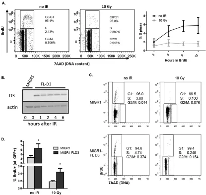
Figure 1. Irradiation of primary mouse pre-B cells induces loss of D3 protein in an ATM-, p53-, and Dicer-dependent manner, but does not alter D3 protein
stability 62
Figure 2. Irradiation of mouse pre-B cells induces an ATM-dependent loss of
D3 transcription. 63
Figure 3. Overexpression of non-regulated D3 causes an increased number of pre-B cells to fail cell cycle arrest after IR. 64
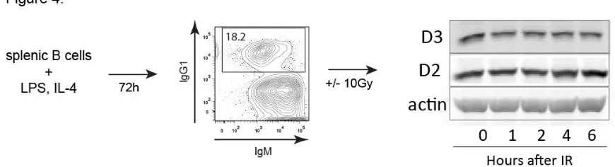
Figure 4. Mature B cells underoing CSR in vitro do not downregulate D3 upon
IR treatment 65
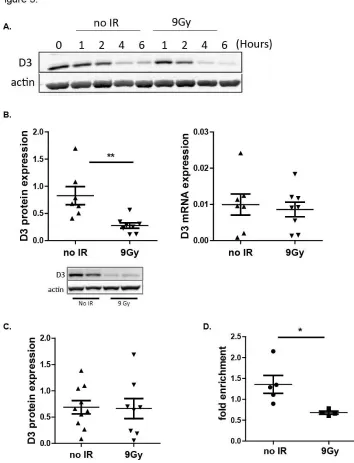
Figure 5. Thymocytes from irradiated mice show reduced D3 protein but not
mRNA. 66
Figure 6. Thymocyte-specific deletion of HuR impairs DN to DP development
and reduces D3 expression 67
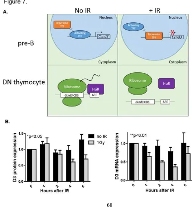
Figure 7. Model of D3 regulation in response to IR-induced breaks in developing lymphocytes and extension to physiologic DSB levels. 68
Chapter IV
Figure 1. B lineage-specific deletion of HuR leads to decreased numbers of
immature and mature B cells. 92
Figure 2. HuR protects immature B cells from p53-dependent elimination. 94
Figure 3. In vitro stimulated HuRΔ/Δ B cells exhibit a mild proliferation defect, enhanced survival, and normal IgH isotype switching. 96
Figure 4. HuR is required for antibody production and numbers of peritoneal B1 cells but dispensable for in vitro functions 98
Figure 5. HuR is required in vivo for generation of GC B cells and high-affinity
xi
Figure 6. HuR is required for in vivo formation of germinal centers in context. 101
Supplemental figure 1. Characterization of HuRΔ/Δ and Mb1Cre+ mice. 103
Supplemental figure 2. Characterization of HuRΔ/Δ and Mb1Cre+ B cell
function. 104
Appendix:
Figure A-1. Proteins identified by proteomic analysis of WT and HuRΔ/Δ B
cells stimulated in vitro for 48h with α-IgM and α-CD40. 127
Figure A-2. HuR-insufficient cells have increased expression of cyclin D3 but
1
Chapter I: Introduction
Maintenance of genome stability during proliferation
The error-free transmission of genetic material from mother cell to daughter cell
is critical for cell survival as well as for organismal survival. Although mutations can and
do occur in quiescent cells, the DNA is particularly vulnerable during its replication and
distribution between dividing cells. Thus, the cell cycle machinery is intimately
coordinated with the DNA damage response (DDR). Each round of cell division begins in
G1 phase when internal or external pro-proliferative signals cause upregulation of one of
three D-type cyclins (reviewed in (Malumbres and Barbacid 2009, Musgrove et al.
2011)). D-cyclins pair with one of their kinase partners, cyclin-dependent kinase 4 or 6
(CDK4/6). Active
D-CDK4/6
complexes
phosphorylate
many target
proteins, leading
to the inactivation
of Rb family
proteins and
inducing the
expression of
cyclin E. Cyclin E- CDK2 complexes phosphorylate targets to initiate the replication of
DNA, which takes place in S phase. S phase is followed by G2, where the cell readies
2
Once the cell reaches late G1, at a point termed the “restriction point”, the cell is
committed to a full round of proliferation, and will carry out the necessary processes in
chronological order with no external stimuli required (Pardee 1989). However, this
sequence of events can be interrupted or terminated at distinct points when problems
arise. The ability of cells to respond to problems with the DNA during the cell cycle is
crucial for preserving the correct DNA sequence, as evidenced by the frequent mutation
of checkpoint factors in many types of human tumors (Kastan and Bartek 2004,
Squatrito et al. 2010).
DNA is vulnerable to breakage and misrepair during DNA replication. Attempting
to replicate broken DNA increases the likelihood of large-scale genetic amplification,
loss, or translocation, in which pieces of distinct chromosomes become aberrantly joined
(Alt et al. 2013). Thus, the G1/S checkpoint prevents the initiation of replication in
response to DSBs sensed during G1 phase. DSBs are first bound by the MRN complex,
consisting of the proteins Mre11, Rad50, and Nbs1 (Lee and Paull 2005). The MRN
complex recruits and activates ATM, which in turn phosphorylates the histone H2A
variant H2AX. Phosphorylated H2AX (γ-H2AX) augments the DSB signal and recruits
other DDR factors such as MDC1 (Stucki et al. 2005, Savic et al. 2009). ATM
phosphorylates many additional targets at S/TQ motifs, including Chk2 and p53 (Banin
et al. 1998, Matsuoka et al. 2000, Matsuoka et al. 2007). Chk2 also phosphorylates p53,
further contributing to stabilization of the p53 protein and promoting its activity (Hirao et
al. 2000). Activated p53 transcriptionally and post-transcriptionally activates gene targets
that promote cell cycle arrest (Beckerman and Prives 2010, Marcel et al. 2015).
Within S phase, replication stress occurs when the helicase that unwinds double
3
stress can be caused by factors including inadequate supply of nucleotides, structural
obstacles in the DNA that stall the polymerase, DNA lesions, and co-occurring gene
transcription (Mazouzi et al. 2014). When the helicase and polymerase become
uncoupled, the resulting long stretch of single-stranded DNA is bound by RPA proteins,
which signal the recruitment and activation of response factors, including the kinase ATR
(Zou and Elledge 2003, Mazouzi et al. 2014). Activated ATR phosphorylates and
activates Chk1 in order to induce an intra-S phase checkpoint, characterized by
inhibition of new origin firing (Toledo et al. 2013). If replication stress is severe or
persistent, the replication fork may collapse, generating a DSB, which then recruits ATM
and the related kinase DNA-PK (Liu et al. 2012, Toledo et al. 2013). Collapsed
replication forks are often able to appropriately re-start (Petermann and Helleday 2010,
Mazouzi et al. 2014); however, failure to initiate the intra-S phase checkpoint promotes
genomic instability (Lopez-Contreras and Fernandez-Capetillo 2010).
Normal cellular function depends on correct gene dosage. Gene dosage can be
disturbed by small mutations and deletions as well as whole chromosomal gains and
losses. Aberrant chromosome numbers, known as aneuploidy, results from defective
chromosome segregation within M phase (Veitia and Potier 2015). Normal mitosis
involves a highly ordered set of processes beginning with the condensation of each
chromosome, pairing and alignment of sister chromatids at the midzone between the two
centrosomes (Nath et al. 2015). The spindle assembly checkpoint (SAC) pauses mitosis
until each chromosome is successfully captured by microtubules. Once the SAC is
alleviated by proper chromosome capture and alignment, chromosomes are pulled to
opposite poles, and then cytokinesis occurs to complete cell division. Aneuploidy often
4
chromosomes are properly positioned and captured (Holland and Cleveland 2012).
Aneuploidy is common in human tumors (Mitelman 2015).
Because the average cell experiences many breaks and nucleotide lesions per
day (Lodish 2004), even the existence of redundant and multi-faceted checkpoint and
repair pathways is insufficient to prevent errors. Therefore, mechanisms for sensing and
eliminating cells with defective DNA are critical for tissue fitness and suppression of
tumorigenesis. Apoptosis is the process by which damaged cells initiate their own death
and undergo orderly disassembly of DNA and cellular components. Perhaps the
best-studied inducer of apoptosis is the tumor suppressor p53 (Meek 2009). In cases of
severely damaged DNA, activated p53 induces pro-apoptotic factors like Bax, which are
able to turn on caspases to degrade DNA and other cellular material (Meek 2009).
Consistent with its role in promoting genome stability, p53 is the most prevalently
mutated tumor suppressor gene across all human tumors, and its inactivation is
associated with
increased genome
instability (Cheung et
al. 2009, Dicker et al.
2009). However, in
some contexts, the
activation of p53 does
not lead to apoptosis
(Figure I-2). For
example, oncogene
5
et al. 2012, Cisowski et al. 2015), and transient p53 activation in response to DSBs
induces transcription of CDK-inhibitors like p21 (Beckerman and Prives 2010). Additional
evidence suggests that the cellular context within which p53 inactivation occurs
influences the phenotype of consequent tumors (Rowh et al. 2011, DeMicco et al. 2013).
No matter the specific biologic outcome, the role of p53 is to minimize genome instability
and prevent the propagation of defective cells.
Coordination of programmed DNA breaks, proliferation, and differentiation during
lymphocyte development
B and T lymphocytes provide a useful model in which to study the integration of
DNA damage and cell cycle control. Adaptive immunity in mice and humans requires the
development and function of B and T lymphocytes, each with a unique and specific
antigen receptor (AgR). The diversity of B and T cell receptors (BCRs and TCRs) is
accomplished by genetic cutting and pasting of germline encoded variable (V), diversity
(D), and joining (J) segments through the process of V(D)J recombination (reviewed in
(Bassing et al. 2002, Alt et al. 2013)). B cell receptors are composed of an
immunoglobulin heavy chain (IgH) protein paired with either an Ig kappa or Ig lambda
light chain. The majority of T cell receptors are comprised of a TCRα and TCRβ chain;
however, some portion of T cells carry TCRγ and TCRδ receptors (von Boehmer 2004,
Xiong and Raulet 2007). Each of these proteins contains a variable exon that is
assembled by V(D)J recombination, as well as a constant region. V(D)J recombination
begins when a complex containing Rag1 and Rag2 proteins recognizes recombination
6
recombined and nicks the DNA at each (McBlane et al. 1995). Transesterification results
in two DSBs with one side of each break containing a closed hairpin structure (McBlane
et al. 1995). The Artemis nuclease opens the hairpin (Ma et al. 2002), and the
non-homologous (NHEJ) machinery repairs the broken DNA, resulting in an
extra-chromosomal signal join and the protein-encoding coding join. Further diversity in the
coding join is accomplished by stochastic non-templated addition of nucleotides at
junctions by the enzyme terminal deoxynucleotide transferase (Benedict et al. 2000).
Proliferation of lymphocytes at specific times within their lifespan is critical for
generation of normal numbers of naïve B and T cells. However, many of these periods of
proliferation closely follow programmed DSBs in developmental time (Bednarski and
7
DSBs is by restricting V(D)J recombination to G1 phase. This is achieved in part by
G1-specific expression of Rag2 protein (Li et al. 1996). In addition, breaks initiated by Rag
activate a set of DDR and checkpoint genes that largely overlaps with those induced by
exogenous breaks (Bredemeyer et al. 2008), consistent with the idea that canonical
checkpoint pathways are also important for controlling genome stability during V(D)J
recombination (Figure I-3).
Lymphocyte development can fail at many stages. For example, due to the
stoachastic addition or deletion of nucleotides in the V(D)J join (Benedict et al. 2000,
Boboila et al. 2012), the recombined sequence has a one in three chance of being
in-frame as relates to translation. Although cells with out of in-frame rearrangements usually
attempt further rearrangements (Schatz and Ji 2011), some cells ultimately fail to
generate a join capable of encoding protein. Additionally, Rag-induced breaks are
mis-repaired at some frequency, leading to copy number variations and/or translocations
(Nussenzweig and Nussenzweig 2010). Further, some rearrangements produce
receptors that react to self-antigens, and thus should be eliminated from the repertoire.
Since millions of immature lymphocytes are generated in the mouse per day (Opstelten
and Osmond 1983), even occasional escape of defective cells could cause tumors or
autoimmunity.
Quality control for AgRs on developing B and T lymphocytes occurs at the
pre-BCR and β-selection checkpoints, respectively (Morris and Allen 2012, Rickert 2013).
Pro-B cells undergo stepwise rearrangement of the IgH locus, in which D segments are
joined to J segments before the V segment is recombined with the D-J join. If the coding
join yields in an in-frame sequence capable of generating a functional IgH chain, this
8
2013). Pre-BCR signaling inhibits further recombination at the IgH locus (Grawunder et
al. 1995), and upregulates pro-survival factors, largely through NFκB-dependent
pathways (Saijo et al. 2003), ensuring that only B cells with a functional IgH chain can
proceed through development. Pre-BCR signaling also upregulates cyclin D3, driving
cells through several rounds of division (Cooper et al. 2006). The pre-BCR checkpoint
may also reduce autoimmune specificities by promoting allelic exclusion (Rickert 2013).
Pre-B cells that pass this checkpoint then re-arrange an Ig light chain, attempting Igκ first
followed by Igλ (Clark et al. 2014). Generation of an IgL that is able to pair with the cell’s
unique IgH chain permits further survival and differentiation (Bednarski and Sleckman
2012). Immature B cells expressing a complete BCR leave the bone marrow for
secondary lymphoid organs, where they complete their maturation into mature naïve B
cells (Allman et al. 1993).
Analogous to the pre-BCR checkpoint, α/β T cells undergo positive and negative
selection during their development in the thymus (von Boehmer 2004, Morris and Allen
2012). Double negative (DN) thymocytes characterized by expression of neither CD4 nor
CD8 undergo TCRβ recombination. TCRβ pairs with pre-Tα to form the pre-TCR
(Guidos 2006). TCRs are selected such that they can recognize self-major
histocompatibility (MHC) peptides with low affinity, thereby eliminating cells that cannot
“see” MHC, but also eliminating strongly auto-reactive T cells (Morris and Allen 2012).
Cells that pass this checkpoint differentiate into CD4+CD8+ (double positive, DP)
thymocytes, which then recombine the TCRα locus (Bassing et al. 2002). DP cells that
successfully generate a mature TCR differentiate into either CD4+ or CD8+ (single
positive, SP) T cells. Newly minted mature T cells exit the thymus and migrate to
9
B cell-mediated immune responses
Mature B cells are broadly characterized as B1 or B2 cells. The majority of B1
cells reside in the peritoneal cavity where they secrete antibodies to repetitive
pathogenic antigens (Zhang 2013). B2 B cells predominate in the spleen and LN where
they participate in pathogen-specific adaptive immune responses (Garraud et al. 2012).
B2 B cells can be further classified as either marginal zone (MZ) or follicular (Fo) B cells.
Within the spleen, MZ B cells are positioned between the red pulp, containing dendritic
cells, macrophages, and other cell types, and the lymphocyte-rich white pulp, (Pillai and
Cariappa 2009). MZ B cells bridge the gap between innate- and adaptive- immune
responses, more often encoding polyvalent BCRs that recognize antigenic patterns
(Cerutti et al. 2013). MZ B cells mount rapid, relatively low-affinity antibody responses, in
a largely T cell-independent manner (Cerutti et al. 2013). In contrast, Fo B cells, so
named because they occupy B cell follicles within the splenic white pulp, are most
important in the response to T cell-dependent antigen encounters (Victora and
Nussenzweig 2012).
Fo B cells constantly circulate through the spleen, waiting to encounter the
cognate antigen that fits their unique BCR. B cells most often receive antigens via
professional antigen-presenting cells such as dendritic cells (DCs). Antigen binding to
the BCR activates the B cell, resulting in broad changes in gene expression and cell
activity (Klein and Dalla-Favera 2008). To undergo a T cell-dependent response, these
activated B cells must encounter and engage an activated CD4+ T cell that shares the
10
encounter with the cognate antigen, usually also from a DC (Vinuesa and Cyster 2011).
When these B-T cell encounters occur productively at the outer edge of the B cell follicle,
both cells undergo further activation and differentiation and move into the follicle to
initiate a germinal center (Pereira et al. 2010, Victora and Nussenzweig 2012). Germinal
centers are transient structures consisting predominantly of B cells undergoing rapid
proliferation, class switch recombination (CSR) and somatic hypermutation (SHM).
Germinal centers are thus required for long-lasting memory of high-affinity, class
switched antibody responses.
The GC can be histologically divided into the light zone (LZ) and the dark zone
(DZ) (Victora and Nussenzweig 2012). DZ B cells are highly proliferative. In contrast,
CSR and SHM take place in the LZ. The IgH locus is unique among AgR loci in that it
contains several constant regions, which can be utilized following CSR. Activated B cells
express the activation-induced deamination (AID) enzyme, which deaminates cytosine
residues within regulatory regions upstream of IgH constant regions (Muramatsu et al.
2000, Honjo et al. 2002). Processing of these deaminated residues results in a DSB,
11
different constant regions imbues the resulting BCR and antibody with distinct properties
that are important for the humoral immune response on an organismal level (Longerich
et al. 2006). The AID enzyme also initiates the process of SHM (Muramatsu et al. 2000,
Honjo et al. 2002). SHM introduces point mutations into the antigen-specific variable
region of the IgH gene causing variations in affinity of the receptor for antigen. Many of
these mutated BCRs have poor affinity for antigen and ultimately die; however, clones
with improved antigen affinity survive and may differentiate into antibody secreting cells
called plasma cells or long-lived memory B cells (Victora and Nussenzweig 2012).
Further, the mutagenic activity of AID in the GC reaction makes mature B cells
vulnerable not only to IgH translocations, but also to point mutations incurred during
SHM (Alt et al. 2013).
Although the majority of cells in the germinal center are B cells, other cell types
play important roles. T follicular helper (Tfh) cells are a specialized subset of CD4+ T
cells and are required for the initiation and maintenance of GCs (Victora and
Nussenzweig 2012, Cubas et al. 2013). Tfh cells promote the GC response in part by
secretion of IL-6 and IL-21 (Eto et al. 2011). T cells also present the ligand for the CD40
receptor expressed on B cells, thereby providing critical survival and differentiation
signals to GC B cells (Foy et al. 1994, Linterman and Vinuesa 2010). Conversely,
differentiation and maintenance of mature GC Tfhs requires B cells to express
antigen-loaded MHCII molecules (Goenka et al. 2011, Baumjohann et al. 2013), the inducible
co-stimulator T cell-ligand (ICOSL) (Nurieva et al. 2008), and likely other co-co-stimulatory
molecules (Akiba et al. 1999, Groth et al. 2002). Thus, GC B cells and GC Tfh cells are
12
Regulation of gene expression at multiple levels throughout lymphocyte development
and activation
Throughout lymphocyte development and function gene expression changes are
tightly regulated, and many overlapping layers of regulatory mechanisms precisely tune
the biologic response. Gene expression can be regulated at the levels of transcription,
splicing, mRNA decay, mRNA translation, protein turnover, and protein activity. Many
examples of each of these mechanisms are observed in lymphocyte-specific
phenomena and play critical roles in the development and function of lymphocytes.
At the level of transcription, combinatorial binding of transcription factors
activates or represses transcription at promoter and enhancer elements upstream of
coding sequences. In turn, these factors are influenced by epigenetic marks placed on
chromatin encasing the genes themselves, as well as by the three-dimensional
architecture of the chromatin within the nucleus. V(D)J recombination provides ample
evidence of this type of gene regulation. Recombination of the correct AgR loci in each
lineage, as well as the ordering of recombination within each locus is tightly regulated,
largely at the level of transcription and chromatin accessibility (reviewed in (Schatz and
Ji 2011)).
Beyond transcription, alternative splicing generates multiple transcripts
emanating from the same gene. The transcription factor XBP1 is required for
differentiation of GC B cells into plasma cells (Reimold et al. 2001). However, XBP1 is
only activated upon alternative splicing of the message by the upstream nuclease IRE1
13
mRNA in human T cells results in expression of different protein isoforms, which in turn
influence TCR signaling (McKenney et al. 1995, Tong et al. 2005).
Once a mature mRNA leaves the nucleus, it can be stabilized or de-stabilized by
miRNAs or RNA-binding proteins (RBPs), and also can also be variably translated into
protein. RBPs are now appreciated to play a prominent role in post-transcriptional gene
regulation in lymphocytes (Turner and Hodson 2012). Many cytokine mRNAs contain
3’UTR elements that are targets of RBPs. For example, the HuR RBP stabilizes cytokine
mRNAs, including those encoding TNFα and IL-4 (Dean et al. 2001, Atasoy et al. 2003,
Yarovinsky et al. 2006). HuR also regulates the stability of cyclin D3 mRNA in response
to nutrient stress in human T cells (Rodriguez et al. 2010), thereby regulating cell cycle
progression.
Proteins are often post-translationally modified, affecting their stability,
subcellular localization, and/or activity. For example, nuclear versus cytoplasmic
localization of Rag proteins may contribute to regulation of Rag activity. Genotoxic stress
causes inducible ATM-dependent Rag2 export from the nucleus (Rodgers et al. 2015).
Rag2 is also de-stabilized by CDK-dependent phosphorylation at the Rag2 C-terminus,
ensuring that the Rag complex is only active during G1 phase (Li et al. 1996). Cell cycle
control is also regulated in part by targeted protein degradation. Cyclin D3 is
proteolytically degraded following its phosphorylation by GSK3β in response to cyclic
AMP signaling in mature B cells (Naderi et al. 2004). Localization and activity of the HuR
RBP is regulated by phosphorylation of several sites by upstream regulators including
14
In summary, lymphocyte development and function require carefully coordinated
gene expression programs that promote normal lymphocyte development and function,
while suppressing malignant transformation. Further, as shown in Chapters II-IV, these
processes are carried out in ways that are often specific to the developmental stage and
lineage of the cell in question. These findings shed new light on mechanisms controlling
diverse cellular processes that in sum maintain genome stability and promote adaptive
15
Chapter II: Somatic inactivation of Tp53 in hematopoietic stem cells
or thymocytes predisposes mice to thymic lymphomas with clonal
translocations
ABSTRACT
TP53 protects cells from transformation by responding to stresses including aneuploidy
and DNA double-strand breaks (DSBs). TP53 induces apoptosis of lymphocytes with
persistent DSBs at antigen receptor loci and other genomic loci to prevent these lesions
from generating oncogenic translocations. Despite this critical function of TP53, germline
Tp53−/− mice succumb to immature T-cell (thymic) lymphomas that exhibit aneuploidy
and lack clonal translocations. However, Tp53−/− mice occasionally develop B lineage
lymphomas and Tp53 deletion in pro-B cells causes lymphomas with oncogenic
immunoglobulin (Ig) locus translocations. In addition, human lymphoid cancers with
somatic TP53 inactivation often harbor oncogenic IG or T-cell receptor (TCR) locus
translocations. To determine whether somatic Tp53 inactivation unmasks translocations
or alters the frequency of B lineage tumors in mice, we generated and analyzed mice
with conditional Tp53 deletion initiating in hematopoietic stem cells (HSCs) or in
lineage-committed thymocytes. Median tumor-free survival of each strain was similar to the
lifespan of Tp53−/− mice. Mice with HSC deletion of Tp53 predominantly succumbed to
thymic lymphomas with clonal translocations not involving Tcr loci; however, these mice
occasionally developed mature B-cell lymphomas that harbored clonal Ig translocations.
Deletion of Tp53 in thymocytes caused thymic lymphomas with aneuploidy and/or clonal
translocations, including oncogenic Tcr locus translocations. These data demonstrate
16
malignancies in mice where somatic deletion of Tp53 initiating in thymocytes is sufficient
to cause thymic lymphomas with oncogenic translocations.
INTRODUCTION
The TP53 tumor suppressor maintains cellular homeostasis in response to a
wide range of stresses including aneuploidy, DSBs, genomic instability, and oncogene
activation (Meek 2009, Reinhardt and Schumacher 2012). These stresses stabilize and
activate TP53, leading to changes in expression of target genes, including those
involved in cell cycle control and apoptosis (Meek 2009, Reinhardt and Schumacher
2012). TP53 is the most frequently inactivated tumor suppressor gene, with mutation or
deletion occurring in over 60% of all human cancers (Cheung et al. 2009, Meek 2009),
indicating that TP53 prevents malignant transformation of multiple cell types. Although
TP53 inactivation occurs less frequently in lymphoid malignancies than in solid tumors,
TP53 loss is more common in aggressive lymphoma subtypes and correlates with
increased tumor grade, treatment resistance, and poor patient survival (Bhatia et al.
1992, Stilgenbauer et al. 2002, Hof et al. 2011).
Lymphocyte development involves cellular proliferation and antigen receptor
gene assembly. Bone marrow HSCs differentiate into early progenitor B cells that remain
in the bone marrow or into early thymic progenitors that migrate to the thymus. These
cells proliferate and differentiate into pro-B or pro-T cells, respectively, which induce
expression of the RAG1/RAG2 (RAG) endonuclease (Chi et al. 2009, Ramirez et al.
2010). RAG catalyzes the assembly of TCR and Ig genes in G1 phase cells through
induction of DSBs at variable (V), diversity (D), and joining (J) gene segments (Schatz
17
DSBs to generate V(D)J coding joins that encode the first exons of TCR and Ig genes
(Lieber 2010, Alt et al. 2013). Assembly of TCRβ, TCRγ, and TCRδ genes occurs in
CD4−CD8− “double-negative” (DN) pro-T cells (Krangel et al. 2004, Jung et al. 2006).
Expression of functional TCRγ and TCRδ genes signals differentiation into mature γδ T
cells (Xiong and Raulet 2007). In contrast, expression of functional TCRβ genes triggers
proliferation as cells differentiate into CD4+CD8+ “double positive” (DP) thymocytes (Bell
and Bhandoola 2008). In DP thymocytes, TCRα gene assembly followed by αβ TCR
selection permit differentiation into CD4+ or CD8+ "single positive" (SP) thymocytes that
exit the thymus as naive αβ T cells (Krangel et al. 2004, von Boehmer and Melchers
2010). Assembly and expression of IgH genes in pro-B cells drives proliferation as cells
differentiate into pre-B cells, which must recombine either Igκ or Igλ genes to
differentiate into immature B cells that exit the bone marrow and migrate to the spleen as
they mature (Jung et al. 2006, Nemazee 2006, Kuo and Schlissel 2009). In response to
antigen, mature B cells proliferate and undergo IgH class switch recombination (CSR)
through DSB intermediates (Longerich et al. 2006, Keim et al. 2013) (Bassing et al.
2003, Boboila et al. 2012). In addition to programmed DSBs in antigen receptor loci,
lymphocytes experience spontaneous DSBs that arise from errors in DNA replication
during periods of proliferation (Bassing and Alt 2004).
TP53 inactivation occurs in human B and T lineage lymphomas containing
aneuploidy as well as in those exhibiting genomic instability (Cheung et al. 2009, Dicker
et al. 2009), suggesting that functions of TP53 in response to chromosome
missegregation and DSBs are each important to suppress transformation of
differentiating lymphocytes. Aberrant segregation of chromosomes during cellular
18
(Thompson and Compton 2010). Induction of DSBs stabilizes and activates TP53, which
promotes temporary cell cycle arrest to provide cells time to repair these lesions or
induce apoptosis if they cannot be repaired (Reinhardt and Schumacher 2012).
Germline Tp53 inactivation in mice leads to aneuploidy and genomic instability in
differentiating and mature lymphocytes (Fukasawa et al. 1997), and enables pro-T and
pro-B cells with un-repaired RAG-induced Tcrδ and IgH locus DSBs to survive, progress
into S phase, and generate translocations (Nacht et al. 1996, Difilippantonio et al. 2002,
Zhu et al. 2002, Gladdy et al. 2003, Dujka et al. 2010). Despite roles of TP53 in
response to both chromosome missegregation and DSBs, most Tp53-/- mice succumb to
aneuploid TCRβ+ thymic lymphomas that lack Tcr translocations, though a small fraction
succumb to B cell lymphomas that have not been assayed for translocations
(Donehower et al. 1992, Jacks et al. 1994, Liao et al. 1998, Ward et al. 1999, Celeste et
al. 2003, Jacobs et al. 2011). However, mice with combined germline inactivation of
Tp53 and NHEJ factors reproducibly succumb to pro-B cell lymphomas with
RAG-dependent IgH translocations that amplify the c-Myc oncogene (Guidos et al. 1996,
Difilippantonio et al. 2000, Zhu et al. 2002), and occasionally develop TCRβ− thymic
lymphomas with Tcrδ translocations (Rooney et al. 2004). In addition, Tp53-/- mice with
germline inactivation of the H2ax DSB repair factor predominantly succumb to TCRβ−
thymic lymphomas with clonal translocations not involving Tcr loci, but occasionally
develop TCRβ− lymphomas with Tcrα/δ translocations or pro-B cell lymphomas with
Igh;c-myc translocations (Bassing et al. 2003, Celeste et al. 2003). Furthermore, on a
genetic background with a block in αβ T cell development at the DN stage, germline
Tp53 deficiency causes TCRβ− thymic lymphomas with RAG-dependent translocations
19
that TP53 functions in response to chromosome missegregation and DSBs are critical to
prevent transformation of differentiating lymphocytes. However, they concluded that
Tp53 only suppresses oncogenic translocations in cells with DSB repair or differentiation
impaired.
Cancers develop through somatic acquisition and selection of mutations such as
TP53 inactivation and other oncogenic lesions (Aparicio and Caldas 2013). We
previously showed, that while germline H2ax-/-Tp53-/- mice succumb to TCRβ- thymic
lymphomas with clonal translocations, conditional deletion of H2ax and Tp53 in mouse
DN thymocytes prolongs lifespan and leads to TCRβ+ thymic lymphomas (Bassing et al.
2003, Celeste et al. 2003, Yin et al. 2011). That somatic inactivation of H2ax and Tp53
causes more mature thymic lymphomas with longer latency compared to germline
inactivation indicates that oncogenic lesions prior to T cell commitment drive
transformation of thymocytes. We recently showed that conditional deletion of Tp53 in
pro-B cells predisposes mice to B lineage lymphomas with oncogenic translocations,
including Igh;c-myc and other Ig translocations (Rowh et al. 2011). The tumor-free
survival of these mice is similar to that of Tp53-/- mice, suggesting that development of
thymic lymphomas from aneuploidy prevents B cell lymphomas from oncogenic Ig
translocations in Tp53-/-mice. To determine whether somatic inactivation of Tp53
unmasks translocations or alters the frequency of B lineage lymphomas in mice, we
generated mice with conditional Tp53 deletion initiating in HSCs or DN thymocytes.
These strains each succumbed at similar ages to thymic lymphomas, although HSC
deletion occasionally caused B lineage lymphomas. HSC deletion of Tp53 led to clonal
translocations not involving Tcr loci in thymic lymphomas and Igh translocations in B
20
and/or clonal translocations including Tcrα/δ locus translocations. Our data demonstrate
that the developmental stage of Tp53 inactivation affects karyotypes of lymphoid
cancers in mice where somatic deletion of Tp53 initiating in thymocytes is sufficient to
cause thymic lymphomas with oncogenic translocations.
RESULTS
Conditional deletion of Tp53 in HSCs or thymocytes predisposes mice to thymic
lymphomas.
To determine whether Tp53 inactivation initiating in HSCs or thymocytes
predisposes mice to lymphoma, we established and characterized Vav-cre+/−p53flox/flox
(VP) and Lck-cre+/−p53flox/flox
(LP) mice. Vav-cre and Lck-cre induce deletion of "floxed"
genes in HSCs and DN thymocytes, respectively (Lee et al. 2001, Georgiades et al. 2002).
We detected nearly complete deletion of Tp53 in bone marrow cells and thymocytes of
VP mice and in thymocytes of LP mice, but no Tp53 deletion in LP bone marrow cells
(Supplementary figure 1), confirming the expected developmental timing of Tp53
inactivation in VP and LP mice. We also found grossly normal T and B cell development
in VP and LP mice as compared to age-matched wild-type controls (Supplementary figure
2), consistent with the phenotype of Tp53−/− mice (Lowe et al. 1993). We generated and
aged cohorts of 22 VP and 20 LP mice to evaluate their spontaneous predisposition to
cancer. We observed that cohort VP mice survived cancer-free between 91-365 days with
median age of mortality of 144.5 days, whereas cohort LP mice survived cancer-free
between 70-365 days with a median age of mortality of 119 days (Figure 1). The median
ages of cancer-free survival of cohort VP and LP mice were not statistically significant
21
al. 1992, Jacks et al. 1994). All VP and LP mice succumbed to lymphoma, except for one
LP mouse that was euthanized due to development of a sarcoma and one mouse of each
genotype that died without lymphoma (Table 1, 2). Most VP mice and LP mice succumbed
to lymphomas that were only visible in the thymus (Table 1, 2). However, three VP mice
(nos. 202, 228, and 623) and two LP mice (nos. 119 and 983) had lymphoma cells in their
spleens, while one VP mouse (no. 602) succumbed to a disseminated lymphoma found in
the thymus, spleen, and multiple lymph nodes (Table 1, 2). In addition, two VP mice (nos.
421 and 426) succumbed to lymphomas that were located in the spleen and multiple
lymph nodes, but not visible in the thymus. We did not characterize cohorts of
Vav-cre+/−, Lck-cre+/−, or p53flox/flox mice since none of these mice exhibits increased tumor
predisposition (Jonkers et al. 2001, Lee et al. 2001, Georgiades et al. 2002). Consistent
with this notion, none of the Vav-cre+/−, Lck-cre+/−, or p53flox/flox mice we used for breeding
until one year of age developed cancer.
Tp53 inactivation in HSCs or thymocytes causes clonal immature T cell
lymphomas.
To determine the lymphocyte lineages and developmental stages to which
inactivation of Tp53 in HSCs or DN thymocytes causes cellular transformation, we first
analyzed VP and LP lymphomas by flow cytometry using antibodies that recognize cell
surface markers of specific lymphocyte lineages and developmental stages. For
lymphomas found within the thymus, we assessed cell surface expression of TCRδ,
TCRβ, CD4, CD8, and the CD3ε molecule through which αβ TCRs signal (Bell and
Bhandoola 2008). For lymphomas found in lymph nodes, we assessed cell surface
22
staining observed for some tumors, we simplified our classification by denoting a
lymphoma as positive for an epitope if more than half of the cells fell within a positive
gate established from flow cytometry of non-malignant lymphocytes. Of the 17 VP thymic
lymphomas assayed, most were TCRβ+CD3+CD4+CD8+, but a substantial number were
also TCRβ−CD3− with or without CD4 and CD8 expression (Figure 1B; Table 1;
Supplementary figure 3). Lymphoma cells in the spleens of the two VP mice (nos. 421
and 426) that lacked thymic lymphomas were B220+IgM+ (Figure 1B; Table 1;
Supplementary figure 3). Lymphoma no. 421 contained Igκ+ and Igκ− cells, while
lymphoma no. 426 contained mostly Igκ− cells (Figure 1B; Table 1; Supplementary figure
3). Of the 15 LP thymic lymphomas assayed, 10 were TCRβ+CD3+CD4+CD8+ only one
was TCRβ−CD3− (Figure 1B; Table 2; Supplementary figure 3). Many VP and LP thymic
lymphomas displayed subpopulations with different expression patterns of TCRβ, CD3,
CD4, and/or CD8, indicating that they represent either oligoclonal lymphomas arising
from distinct initiating cells or clonal lymphomas with subpopulations that have
differentially silenced and/or re-expressed genes. VP thymic lymphomas more often
showed TCRβ, CD4, CD8, and CD3 expression characteristic of immature T cell
developmental stages than LP thymic lymphomas. Collectively, our flow cytometry
analysis of VP and LP lymphomas indicates that deletion of Tp53 in mouse HSCs or DN
thymocytes causes predominantly immature T cell lymphomas, with HSC deletion
leading to a higher percentage of tumors from an earlier T cell developmental stage and
occasional mature B cell lymphomas.
To determine the lymphocyte lineage of the four TCRβ−CD3−CD4−CD8−VP
thymic lymphomas and to distinguish between oligoclonal and clonal immature T cell
23
LP lymphomas. For DN thymocytes to survive and differentiate, Tcrβ rearrangements
must occur on one allele.(Jiang et al. 1996) To characterize Tcrβ rearrangements, we
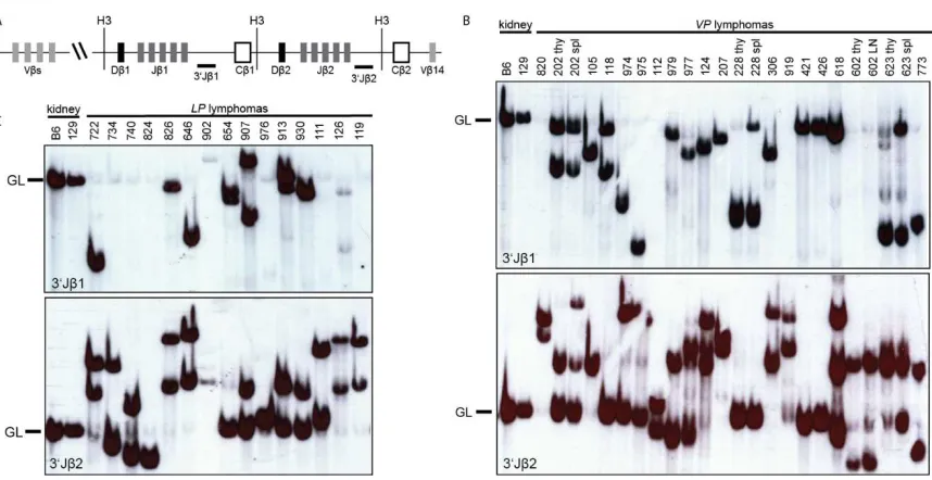
conducted Southern blotting on HindIII-digested genomic DNA of VP and LP lymphomas
with 3'Jβ1 and 3'Jβ2 probes (Figure 2A-C). We isolated DNA from the tumor-containing
organ without further purification, under assumption that most cells were malignant. This
was confirmed by flow cytometry. Further, this approach does not detect unique
rearrangements in single cells, such as those occurring in normal lymphocytes. Thus,
only germline configurations in pro-lymphocytes or in non-lymphoid cells will be detected
as background. We detected Tcrβ rearrangements in the TCRβ−CD3−CD4−CD8−VP
thymic lymphomas (nos. 228, 602, 618, and 773) (Figure 2B), demonstrating that these
malignancies are immature T cell lymphomas. We also found that 16 of 18 VP and all 15
LP thymic lymphomas analyzed contained one or two rearranged Tcrβ alleles and
therefore arose from the expansion of a single cancer-initiating cell (Figure 2B,C). These
data indicate that the diverse expression of surface epitopes observed within some VP
and LP lymphomas represents tumor subpopulations that have differentially silenced
and/or re-expressed these genes. The remaining two VP thymic lymphomas (nos. 124
and 618) contained three Tcrβ rearrangements (Figure 2B), indicating that these cancers
either arose from the expansion of two cancer-initiating cells or one cancer-initiating cell
that continued to assemble Tcrβ genes after malignant transformation. We also
performed Southern analysis of Tcrβ rearrangements on VP lymphomas found in the
spleen or lymph nodes of mice. We found that three of these four VP lymphomas
analyzed contained the same Tcrβ rearrangements as the thymic lymphomas from the
same animals (Figure 2B), demonstrating that these mice succumbed to a disseminated
24
rearrangement that was not present in the thymic lymphoma of this mouse (Figure 2B),
suggesting that this splenic lymphoma developed from the thymic lymphoma in
association with ongoing Tcrβ rearrangement. Our Southern analysis of Tcrβ
rearrangements in VP and LP lymphomas indicates that inactivation of Tp53 in mouse
HSCs or DN thymocytes causes mainly clonal immature T cell malignancies.
To determine whether the two B lineage lymphomas that arose in VP cohort mice
were clonal and whether they arose from developing or mature B cells, we analyzed Igh
and Igκ rearrangements in these tumors. To characterize Igh rearrangements, we
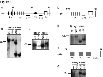
conducted Southern blot analysis of EcoRI-digested genomic DNA of VP lymphomas
nos. 421 and 426 with 3'JH and 3'Sµ probes (Figure 3A-C). We found that these tumors
contained one (no. 426) or two (no. 421) JH rearrangements and therefore arose from
the clonal expansion of a single cancer-initiating cell (Figure 3B). We also detected Sµ
recombination in lymphoma no. 421 (Figure 3C), suggesting that this tumor may have
developed from a B cell that had attempted CSR. Southern blot analysis of BamH
I-digested genomic DNA from VP lymphomas nos. 421 and 426 with the 3'Jκ probe
showed that each of these tumors contained Jκ rearrangements (Figure 3D,E). This
suggests that IgM+Igκ−VP lymphoma no. 426 developed from a B lymphocyte that had
developed at least to the pre-B cell stage. Our Southern analysis of these two VP B
lineage lymphomas indicates that deletion of Tp53 in mouse HSCs can cause clonal B
lineage lymphomas.
Conditional inactivation of Tp53 in HSCs or thymocytes causes lymphomas with
25
To determine whether conditional deletion of Tp53 initiating in HSCs or DN
thymocytes causes lymphomas with chromosomal translocations, we conducted
Spectral Karyotyping (SKY) on seven VP and eight LP lymphomas. SKY is a molecular
cytogenetic approach to visualize all chromosomes in a single metaphase spread to
identify translocations and fusions between chromosomes (Liyanage et al. 1996). We
defined clonal translocations as those found in greater than half of the metaphases
analyzed for a given tumor. SKY revealed that two of the five VP thymic lymphomas
(nos. 118 and 820) analyzed had clonal translocations, with lymphoma no. 773
containing three different clonal chromosome fusions (Figure 4A; Table 1;
Supplementary figure 4). One VP thymic lymphomas (no. 975) contained two non-clonal
chromosome fusions (Table 2; Supplementary figure 4). The remaining VP thymic
lymphoma (no. 207) lacked translocations but exhibited aneuploidy (Table 1;
Supplementary figure 4). None of the clonal translocations in VP thymic lymphomas
involved chromosomes on which Tcr or Ig loci reside. In contrast, SKY revealed that
both VP B lymphomas contained clonal translocations involving chromosomes on which
the Igh (chromosome 12 in no. 421) or Igκ (chromosome 6 in no. 426) locus resides
(Figure 4B; Table 1; Supplementary figure 4). SKY also demonstrated that three of the
eight LP thymic lymphomas (nos. 826, 902, and 976) analyzed harbored clonal
translocations, with lymphoma no. 902 also containing a clonal chromosome fusion
(Figure 4A; Table 2; Supplementary figure 4). Notably, two of these tumors (no. 902 and
976) contained clonal translocations involving chromosome 14 which carries the Tcrα/δ
locus. The other five LP thymic lymphomas lacked translocations but exhibited
26
in HSCs or thymocytes causes lymphomas with clonal translocations, with thymocyte
deletion also causing aneuploid lymphomas.
To determine whether conditional deletion of Tp53 initiating in HSCs or DN
thymocytes causes lymphomas with clonal Ig or Tcr translocations, respectively, we
conducted FISH on the four tumors with potential clonal Igh, Igκ, or Tcrα/δ
translocations. For this purpose, we hybridized 5' and 3' Ig or Tcr locus probes and
identified Ig or Tcr translocations by detection of probe signals on different
chromosomes (Figure 4B,C). Since the c-myc oncogene on chromosome 15 is activated
by Igh or Tcrα/δ translocations in mouse lymphomas (Rooney et al. 2004, Rowh et al.
2011), we also used a c-myc probe to identify potential Igh;c-myc and Tcrα/δ;c-myc
translocations in VP lymphoma no. 421 and LP lymphoma no. 902. FISH revealed
co-localization of 3'Igh and c-myc probe signals on one chromosome derivative in VP B
lineage lymphoma no. 421 (Figure 4B), indicating that the clonal t(12;15) translocation in
this tumor is an Igh;c-myc translocation. Unfortunately, we were unable to determine
potential involvement of the Igκ locus in the clonal t(6;4) translocation of VP B lineage
lymphoma no. 426 due to insufficient numbers of metaphases from this tumor. FISH
revealed splitting of 5’ and 3’ probes on the clonal t(16;14) translocation in metaphases
from LP thymic lymphoma no. 976 (Supplementary figure 4 and not shown), indicating
that this translocation tumor involves the Tcrα/δ locus. Finally, FISH showed
co-localization of multiple copies each of5'Tcrα/δ and c-myc probe signals on one
chromosome derivative in metaphases from LP thymic lymphoma no. 902 (Figure 4C
and not shown), indicating that the clonal t(14;15;4) translocation in this tumor is a
Tcrα/δ;c-myc translocation with amplification of the c-myc oncogene and Tcrα
27
demonstrates that deletion of Tp53 initiating in HSCs or DN thymocytes can cause,
respectively, B lineage lymphomas with oncogenic Igh translocations or thymic
28
DISCUSSION
Our study demonstrates that the context of Tp53 inactivation influences
lymphoma predisposition, and that inactivation of Tp53 in HSCs or in thymocytes
predisposes mice to thymic lymphomas with clonal translocations including those
involving the Tcrα/δlocus. We generated mice with conditional deletion of Tp53 initiating
in HSCs or in DN thymocytes and compared their tumor predisposition to the
well-characterized cancer phenotype of germline Tp53-deficient mice. We found that HSC
initiation of Tp53 inactivation predisposed mice to predominantly thymic lymphomas with
clonal translocations not involving antigen receptor loci and occasionally to peripheral B
cell lymphomas with clonal Ig translocations. Inactivation of Tp53 in DN thymocytes
predisposed mice to thymic lymphomas that exhibited aneuploidy or contained clonal
translocations frequently involving Tcrα/δ loci. In contrast, germline inactivation of Tp53
predisposes mice to aneuploid thymic lymphomas lacking clonal translocations
(Donehower et al. 1992, Jacks et al. 1994, Celeste et al. 2003). Other than the timing of
Tp53 inactivation, the only difference between VP and LP mice and Tp53−/− mice is
constitutive expression of Cre. Constitutive Cre expression causes genomic instability, at
least in mouse embryonic cells cultured in vitro (Loonstra et al. 2001, Silver and
Livingston 2001), suggesting that the translocations found in VP and LP lymphomas
could be Cre-induced lesions. Since the lymphoma predisposition of Vav-cre:Tp53−/−
mice has not been reported, we cannot conclude whether the clonal translocations and
chromosome fusions found in VP thymic lymphomas arise independently of Vav-cre
expression. Yet, considering that Vav-cre mice are not predisposed to cancer
(Georgiades et al. 2002), our findings demonstrate that Tp53 serves important functions
29
genomic instability. Notably, constitutive Cre expression from Lck-cre initiating in DN
thymocytes of Tp53−/− mice does not alter onset or karyotype of thymic lymphomas that
arise in these mice (Cheung et al. 2002). Therefore, we conclude from the cancer
predisposition of LP mice that Tp53 serves critical functions in suppressing generation
and/or oncogenic potential of Tcrα/δ translocations during αβ T cell development.
The objective of our study was to determine whether oncogenic lesions arising
during embryogenesis and/or in HSCs precludes development of thymic lymphomas with
clonal translocations including Tcr translocations. VP and LP mice succumb to tumors at
similar ages as Tp53−/−,Lck-cre:Tp53−/− mice, and Mb1-cre:Tp53flox/floxmice (Donehower
et al. 1992, Jacks et al. 1994, Celeste et al. 2003, Rowh et al. 2011). In contrast to the
aneuploid thymic lymphomas that arise in Tp53−/− mice, we found that VP and LP mice
developed lymphomas with aneuploidy or clonal translocations, including Ig or Tcrα/δ
translocations. The distinct cancer phenotypes of these mice indicate that loss of Tp53
during embryogenesis, in cells before lymphocyte commitment, and/or in thymocytes
masks development of lymphomas with oncogenic translocations in germline Tp53
-deficient mice. In addition, the development of LP thymic lymphomas with aneuploidy or
clonal translocations indicates that functions of Tp53 in response to both chromosome
missegregation and DSBs are each critical for preventing malignant transformation of
thymocytes.
T-cell acute lymphoblastic leukemia (T-ALL) remains a significant cause of
cancer morbidity and mortality in both children and adults (Smith et al. 2010, Maloney et
al. 2012). Advances have been made in treatment of patients with T-ALL, however
drug-resistance and relapse are common causes of treatment failure and most patients with
30
using genotoxic drugs that can cause serious health issues through effects on normal
cells, demonstrating a need to develop more specific and less toxic therapies (Bhatia
2012). T-ALLs have heterogeneous karyotypes, with about half being aneuploid and the
remainder containing translocations including oncogenic Tcrα/δ translocations (Graux et
al. 2006, Mrozek et al. 2009, Le Noir et al. 2012). Although inactivating TP53 mutations
are not common in T-ALL, these genetic lesions are often associated with drug
resistance, rapid disease progression, and poor survival (Cheung et al. 2009).
Therefore, LP mice may provide a useful pre-clinical model to evaluate the potential
31
FIGURES
Figure 1. Mice with conditional inactivation of Tp53 initiating in HSCs or DN thymocytes reproducibly succumb to thymic lymphomas. (A) Kaplan–Meier curves comparing tumor-free survival of 22 VP and 20LP mice. All tumors were thymic lymphomas other than two VP mice that succumbed to B lineage lymphomas (indicated by asterisks).
32
33
Figure 3. Mice with conditional inactivation of Tp53 initiating in HSCs also develop clonal B lineage lymphomas. (A) Schematic of the mouse IgH locus showing relative locations of representative DH segments, the four JH segments, the Sμ region, and the
first CH exon, Cμ. The positions of the EcoRI restriction sites (RI) and 3’JH and 3’Sμ
probes used for Southern blots are also shown. (B and C) Southern blot analysis of EcoRI-digested DNA isolated from VP lymphomas no. 421 or 426 or from kidneys of C57BL/6 (B6) or 129/SvEv (129) control mice using the (B) 3’JH and (C) 3’Sμ probes.
Germline (GL) bands for each probe are indicated. (D) Schematic of the mouse Igκ locus showing relative locations of the five Jκ segments and the Cκ exon. The positions of the BamHI restriction sites (BHI) and 3Jκ probe used for Southern blots are also shown. (E) Southern blot analysis of BamHI-digested DNA isolated from VP lymphomas no. 421 or 426 or from kidneys of C57BL/6 (B6) or 129/SvEv (129) control mice using the 3’Jκ probe. Germline (GL) band for the 3’Jκ probe is indicated. (F) Schematic of the mouse c-Myc locus showing relative locations of the three c-c-Myc exons, and of the EcoRI
34
Figure 4. Mice with conditional inactivation of Tp53 initiating in HSCs or DN thymocytes develop lymphomas with oncogenic antigen receptor locus translocations. (A)
Cytogenetic analysis of a metaphase from VP lymphoma no. 820 with the clonal t(2;17) translocation circled or isolated. (A, i) Spectral image. (A, ii) DAPI image. (A, iii)
Karyotype table. (B) Cytogenetic analyses of a metaphase or chromosome from VP lymphoma no. 421 with the clonal t(12;15) translocation circled. (B, i) SKY image. (B, ii) DAPI image. (B, iii) Karyotype table. (B, iv). SKY (left) or FISH image (right) of the t(12;15) translocation hybridized with 3’IgH (green) and c-Myc (red) probes. (C)
35
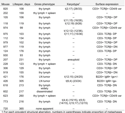
Table 1. Summary of VP tumor cohort.
Mouse Lifespan, days Gross phenotype Karyotype1 Surface expression
820 108 thy lymph t(2;17) (28/33) CD3+ TCRβ+ CD4/8 var
202 159 thy lymph + spleen -- --
105 106 thy lymph -- CD3+ TCRβ+ DP
118 119 thy lymph
t(11;15) (16/26),
t(12;19) (9/26) CD3+ TCRβ+ DP 974 99 thy lymph -- CD3+ TCRβ+ CD4/8 var
975 103 thy lymph
t(12;12) (12/36),
t(11;11) (15/36) CD3- TCRβ- DP
112 134 thy lymph -- CD3- TCRβ- DP
979 102 thy lymph -- CD3+ TCRβ+ DP
977 119 thy lymph -- CD3+ TCRβ+ DN
124 176 thy lymph -- CD3- TCRβ- DP
930 93 thy lymph -- --
207 231 thy lymph aneuploid CD3+ TCRβ+ DP 228 123 thy lymph + spleen -- CD3- TCRβ- DN
306 129 thy lymph -- CD3+ TCRβ+ DP
919 155 thy lymph -- CD3+ TCRβ+ DN
421 176 LN tumor t(12;15) (24/25) B220+ IgM+ Igκ+/- 426 305 LN tumor t(6;4) (23/24) B220+ IgM+ Igκ-
618 213 thy lymph -- CD3- TCRβ- DN
602 217
widely
disseminated -- CD3- TCRβ- DN 623 260 thy lymph + spleen -- CD3+ TCRβ+ CD4/8 var
773 216 thy lymph (14/15), t(15;17) (12/15) t(4;4) (15/15), t(5;5) CD3- TCRβ- DN
720 365 none apparent -- --
1 For each prevalent structural aberration, numbers in parentheses indicate proportion of metaphases analyzed carrying that aberration.
-- Dashes indicate parameters not assessed.
Abbreviations:
thy lymph: thymic lymphoma
CD4/8 var: variable levels of CD4 and/or CD8 expression DP: CD4+CD8+ double positive
DN: CD4-CD8- double negative
36
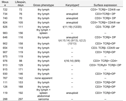
Table 2. Summary of LP tumor cohort.
Mous e
Lifespan,
days Gross phenotype Karyotype† Surface expression 722 72 thy lymph -- CD3+ TCRβ+ CD4/8 var 734 76 thy lymph aneuploid CD3+TCRβ+ DP 740 70 thy lymph aneuploid CD3+ TCRβ+ DP 824 105 thy lymph aneuploid CD3+ TCRβ+ CD4/8 var 826 111 thy lymph t(11;19) (12/20) CD3- TCRβ- DP
983 156
thy lymph +
spleen -- --
646 116 thy lymph aneuploid CD3+ TCRβ+ DP
902 158 thy lymph
t(4;15;14) (9/13), t(2;2)
(10/13) CD3+ TCRβ+DP 654 118 thy lymph -- CD3- TCRβ- CD4/8 var
907 119 thy lymph -- CD3+ TCRβ+DP
975 119 thy lymph -- --
976 98 thy lymph t(16;14) (8/9) CD3+ TCRβ+ CD4+ 913 129 thy lymph -- CD3+ TCRγδ+ TCRβ+ DP
915 171 thy lymph -- --
930 146 thy lymph -- CD3+ TCRβ+DP
767 142 none apparent -- --
111 153 thy lymph -- CD3+ TCRβ+DP
126 168 thy lymph -- CD3+ TCRβ+DP
119 192
thy lymph +
spleen aneuploid CD3+ TCRβ+DP
288 297
abdominal
sarcoma -- --