_____________________________________________________________________________________________________
26(1): 1-13, 2019; Article no.IJBCRR.49398
ISSN: 2231-086X, NLM ID: 101654445
Field Fungal Diversity in Freshly Harvested Maize
Xiang Dong Sun
1,2*, Hong Shan
1,2, Lili Li
1,2, Ping Su
1,2, Jing Lan
1,2, Lin Zhao
1,2and Huan Chun Yang
1,21Quality and Safety Institute of Agricultural Products, Heilongjiang Academy of Agricultural Sciences,
Harbin, China. 2
Laboratory of Quality and Safety Risk Assessment for Agro-products (Harbin), Ministry of Agriculture, Harbin, China.
Authors’ contributions
This work was carried out in collaboration among all authors. Author XDS conceived and designed the experiments. Authors HS, LL, PS and JL performed the experiments. Authors XDS and HS analyzed the data. Authors LZ and HCY contributed analysis tools. Authors XDS and HS prepared the manuscript. All authors read and approved the final manuscript.
Article Information
DOI: 10.9734/IJBCRR/2019/v26i130088 Editor(s): (1) Dr. Fidanka Trajkova, Assistant Professor, Faculty of Agriculture, Goce Delcev University of Stip, Macedonia. Reviewers: (1) Joseph Gweyi-Onyango, Kenyatta University, Kenya. (2)Julia Pérez Ramos, Universidad Autónoma Metropolitana-Xochimilco, Mexico. Complete Peer review History:http://www.sdiarticle3.com/review-history/49398
Received 25 March 2019 Accepted 05 June 2019 Published 11 June 2019
ABSTRACT
Maize is a major crop in China and maize production in Heilongjiang Province ranks No.1 in the country in annual maize production in the whole country. Maize is prone to invasion by fungi and mycotoxins produced by these fungi are proven to be serious threats to animals as well as human
health. Through high through-put sequencing we detected the dominant phylum to be Ascomycota;
Dothideomycetes, Sordariomycetes, Eurotiomycetes and Tremellomycetes, Saccharomycetes were
the dominant classes; Hypocreales, Eurotiales, Capnodiales, Saccharomycetales, Tremellales, and
Pleosporales were the main orders; Nectriaceae, Trichocomaceae, Cladosporiaceae, Debaryomycetaceae, Tremellaceae, and Pleosporaceae were major families; Gibberella, Cladosporium, Papiliotrema, Penicillium, Scheffersomyces, Talaromyces, and Epicoccum were the
most abundant phylotypes at the genus level. Epicoccum_nigrum, Gibberella_zeae,
Papiliotrema_flavescens, and Scheffersomyces_shehatae were the dominant fungal species. Great fungal diversity was observed in the maize samples harvested in the five major maize-growing regions in Heilongjiang Province. Maize-1 in Nenjiang County was observed to have the greatest
fungal diversity and abundance among the five regions. Since some of the fungal species are mycotoxin producing, it is necessary to take precautions to ensure the maize is stored under safe conditions to prevent the occurrence of mycotoxins and the growth and reproduction of other fungi which results in deterioration in the quality of maize.
Keywords: Fungi; diversity; maize; high through-put sequencing.
1. INTRODUCTION
Maize (Zea mays L.) is an important foodstuff,
feed, and raw material in China. Heilongjiang province produced 37 million tons of maize in 2017, ranks No.1 in total annual maize production in China, and accounts for 14.3 percent of the total maize production in the country [1]. In 2018, the total yield of maize in Heilongjiang province reached 39.82 million tons [2]. However, due to the shorter frost-free period in Heilongjiang province, maize may not be completely dry when harvested in October. The average moisture content in these kernels was recorded to be as high as 26.88% [3], the moisture content in most maize varieties often determined between 30-40% in this province [4]. The maize produced in this province frequently suffers from early frost and low temperature inhibits moisture evaporation from the maize kernels. Maize is prone to invasion by fungi and contamination by the mycotoxins secreted. Consequently, maize harvested in Heilongjiang Province under such high moisture contents are highly probable to be infected by various fungi, especially without drying treatment after harvest and storage under improper conditions. In addition, most small scale farmers dry their maize on polyethylene sheets or mats while others spread it on the bare ground in their yards or along countryside roads (give citation). These drying methods are slow and cause growth and development of fungi which in turn increases the potential for mycotoxin production. Some farmers also store or heap maize on the ground or in simple maize shelters, which leaves the maize vulnerable to rain or snow.
Fungi plays a key role in maize safety, and understanding the fungi community structure is of great importance in order to take appropriate measures to ensure maize safety. Fungi infects maize crops early in the field and may produce mycotoxins during this period. Consumer are concerned about this issue and consequently it is necessary to investigate the state of fungi contamination of maize in Heilongjiang province (add reference). Maize is widely cultivated in China under different climatic conditions and
extensively contaminated by various fungi. However, little information is currently available on the fungal diversity of field fungi, especially aflatoxigenic fungal contamination of maize in the main growing region of Heilongjiang province. The objective of this study are to investigate fungal diversity of freshly harvested maize in the five main growing regions of Heilongjiang province through high-throughput sequencing and FUNGuild and try to find the abundance difference of dominant fungi among the five regions in the freshly harvested maize samples.
2. MATERIALS AND METHODS
2.1 Sample Preparation
Five maize samples were harvested from five regions in the major maize growing areas in Heilongjiang as indicated in Fig. 1. Daxijiang Farm in Nenjiang County (maize-1), Rongjun Farm in Nenjiang County (maize-2), Heitai town in Mishan County (maize-3), Wanfa town in Bayan County (maize-4), and Jiangwan town in Yilan County (maize-5). Around 10 maize cobs were cracked from maize fields, placed into plastic bags, and immediately tightly sealed during 20-22 October 2017. After arriving at the
lab, the maize cobs were hand-threshed from
each maize sample and three 50 g maize kernels were weighed from each sample into 1000 ml Erlenmeyer flasks with 500 mL PBS buffer
(KH2PO4 0.27 g, NA2HPO4 1.42 g, NaCl 8 g, KCl
ice transportation conditions to perform high through-put sequencing of the PCR products. The obtained data was assembled into sequenc tags and subject to BLAST in GenBank for classification of the microbes, followed by OTU, diversity, and inter-sample comparative analyses.
2.2 Protocol
2.2.1 DNA extraction and PCR amplification
Microbial DNA was extracted from stool samples using the E.Z.N.A. stool DNA Kit (Omega Bio Norcross, GA, U.S.) according to manufacturer’s protocols. The ITS region of the Eukaryotic ribosomal RNA gene was amplified by PCR (95°C for 2 min, followed by 27 cycles at 98°C for 10 s, 62°C for 30 s, and 68°C for 30 s and a final extension at 68°C for 10 min) using primers ITS3_KYO2F 5’- GATGAAGAACGYAGYRAA and ITS4R 5’- TCCTCCGCTTATTGATATGC where the barcode is an eight-base sequence
Fig. 1. Distribution of samples collecting locations in
Illustration of the geographical location of Heilongjiang province in China. b. Distribution of samples collecting locations in Heilongjiang province
Fig. 2. P. R. China
Sun et al.; IJBCRR, 26(1): 1-13, 2019; Article no.IJBCRR.49398
ice transportation conditions to perform high put sequencing of the PCR products. The obtained data was assembled into sequence tags and subject to BLAST in GenBank for classification of the microbes, followed by OTU, sample comparative analyses.
2.2.1 DNA extraction and PCR amplification
Microbial DNA was extracted from stool samples the E.Z.N.A. stool DNA Kit (Omega Bio-Tek, Norcross, GA, U.S.) according to manufacturer’s protocols. The ITS region of the Eukaryotic A gene was amplified by PCR in, followed by 27 cycles at 98°C for 0 s and a final °C for 10 min) using primers GATGAAGAACGYAGYRAA -3’ TCCTCCGCTTATTGATATGC-3’, base sequence
unique to each sample. PCR reactions were performed in triplicate 50 μL mixture containing 5 μL of 10 × KOD Buffer, 5 μL of 2.5 mM dNTPs, 1.5 μL of each primer (5 μM), 1 μL of KOD Polymerase, and 100 ng of template DNA.
2.2.2 Illumina Hiseq2500 sequencing
Amplicons were extracted from 2% agarose gels and purified using the AxyPrep DNA Gel Extraction Kit (Axygen Biosciences, Union City, CA, U.S.) according to the manufacturer’s instructions and quantified using QuantiFluor™ ST (Promega, U.S.). Purified amplicons were pooled in equimolar and paired-end sequenced (2 × 250) on an Illumina platform according to the standard protocols.
2.2.3 Bioinformatics analysis
The Bioinformatics analysis procedure was conducted according to Fig. 2.
* b
Jiangwan Daxijiang
Rongjun
Heitai Wanfa
Fig. 1. Distribution of samples collecting locations in Heilongjiang province of China. a. Illustration of the geographical location of Heilongjiang province in China. b. Distribution of
samples collecting locations in Heilongjiang province
Fig. 2. ITS analysis flow chart
; Article no.IJBCRR.49398
unique to each sample. PCR reactions were L mixture containing 5 μL of 2.5 mM dNTPs, 1.5 μL of each primer (5 μM), 1 μL of KOD Polymerase, and 100 ng of template DNA.
sequencing
Amplicons were extracted from 2% agarose gels AxyPrep DNA Gel Extraction Kit (Axygen Biosciences, Union City, CA, U.S.) according to the manufacturer’s instructions and quantified using QuantiFluor™-ST (Promega, U.S.). Purified amplicons were
end sequenced an Illumina platform according to the
The Bioinformatics analysis procedure was
Heitai
2.2.4 Quality control and reads assembly
Reads filtering: Raw data containing adapters or low-quality reads would affect the following assembly and analysis. Therefore, to get high-quality clean reads, raw reads were further filtered according to the following rules:
1) Removing reads containing more than 10% of unknown nucleotides (N);
2) Removing reads containing less than 80%
of bases with quality (Q-value)>20.
Reads assembly: Paired-end clean reads were merged as raw tags using FLSAH [5] (v 1.2.11) with a minimum overlap of 10 bp and mismatch error rates of 2%.
Raw tag filtering: Noisy sequences of raw tags were filtered by QIIME [6] (V1.9.1) pipeline under specific filtering conditions [7] to obtain high-quality clean tags.
Chimera checking and removal: Clean tags were searched against the reference database (http://drive5.com/uchime/uchime_download.html)
to perform Reference-based chimera
checking using UCHIME algorithm (http://www. drive5.com/usearch/manual/uchime_algo.html). All chimeric tags were removed and finally obtained effective tags for further analysis.
2.2.5 OTU cluster
The effective tags were clustered into operational taxonomic units (OTUs) with greater than 97% similarity using UPARSE [8] pipeline. The tag sequence with the highest abundance was selected as a reprehensive sequence within each cluster. Between groups, Venn analysis was performed in R to identify unique and common OTUs.
2.2.6 Taxonomy classification
The representative sequences were classified into organisms by a naive Bayesian model using a RDP classifier [9] (Version 2.2) based on UNITE [10] Database (https://unite.ut.ee/). The abundance statistics of each taxonomy and a phylogenetic tree was constructed in a Perl script and visualized using SVG. Biomarker features in each group were screened by Metastats and LEfSe software.
2.2.7 Alpha diversity analysis
Chao1, Simpson and all other alpha diversity
indices were calculated in QIIME. OTU
rarefaction curve and Rank abundance curves were plotted in QIIME. Statistics of between-group Alpha index comparison was calculated by a Welch's t-test and a Wilcoxon rank test in R. Alpha index comparisons among groups was computed by a Tukey’s HSD test and a Kruskal-Wallis H test in R.
2.2.8 Beta diversity analysis
The weighted and unweighted unifrac distance matrix was generated by QIIME. Multivariate statistical techniques including PCA, principal coordinates analysis (PCoA) and NMDS of (Un)weighted unifrac distances were calculated and plotted in R. Statistics of Welch's t-test, Wilcoxon rank test Tukey’s HSD test, Kruskal-Wallis H test, Adonis (also called Permanova) and Anosim test was calculated using R.
2.2.9 Functional prediction
The functional group (guild) of the OTUs was inferred using FUNGuild [11] (v1.0).
3. RESULTS AND DISCUSSION
3.1 Fungal Diversity and Richness in
Single Maize Samples and
Comparison of These Indexes among the Five Samples
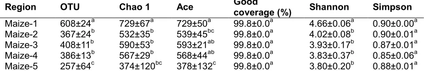
Total fungal ITS tags (103283, 89719, 96884, 92409, and 129746) were recovered from 5 samples (maize-1, maize-2, maize-3, maize-4, and maize-5, respectively). The library samples were then clustered into fungal Operational Taxonomic Units (OTUs) at 97% similarity (Table 1).
Maize-1 was determined to have the highest number of OTUs out of the the five samples (regions), while Maize-5 obtained the minimum value. No significant difference was found in OTU values among Maize-2, Maize-3, and Maize-4. An OTU is usually recognized as a genus of microorganism. Consequently, a total of 608, 367, 408, 386, and 257 fungal genera were observed in the 5 maize samples, respectively.
Sun et al.; IJBCRR, 26(1): 1-13, 2019; Article no.IJBCRR.49398
Table 1. Community richness, diversity and coverage indexes in the five samples*
Region OTU Chao 1 Ace Good
coverage (%) Shannon Simpson
Maize-1 608±24a 729±67a 729±50a 99.8±0.0a 4.66±0.06a 0.90±0.00a
Maize-2 367±24b 532±35b 539±45bc 99.8±0.0a 4.02±0.08b 0.90±0.01a
Maize-3 408±11b 590±53b 593±21ab 99.8±0.0a 3.93±0.17b 0.87±0.01a
Maize-4 386±13b 567±29b 568±44ab 99.8±0.0a 3.83±0.37b 0.85±0.06a
Maize-5 257±64c 374±120bc 378±132c 99.8±0.0a 3.80±0.20b 0.88±0.01a
The data represents mean ± SD. Data followed by the same superscript letter in the same column is not
significantly different
difference was found in the values of Good coverage. Maize-1 and Maize-2 were collected in Nenjiang County which is located to the north of the other three regions and whose temperature ranges between -4°C and 9°C in October. In comparison, the southern location of the other three areas result in a warmer climate and recorded temperatures of 12°C, 1-12°C, and 0-12°C in October in the regions of Mazie-3 (in Mishan County), Maize-4 (in Bayan County), and Maize-5 (in Yilan County), respectively. It is reasonable to assume that the numbers of fungal genera in a warmer area should be greater than that of in a cooler one. However, the rain from the day before sample collection in Nenjiang County made the maize cobs wet and likely resulted in greater numbers of fungal genera, and consequently higher numbers of OTU. Other factors such as air humidity, fungal diseases, birds, insects, wind, etc. can comprehensively affect the fungal community in maize cobs.
To compare fungal diversity and richness among the five maize samples (regions), data was statistically analyzed and presented in Table 1. The Chao 1 and ACE are abundance indexes; greater values of Chao 1 (richness estimate) and ACE indicate more community richness. As seen in Table 1, the greatest Chao 1 and ACE values in Maize-1 indicate the highest fungal abundance, whereas the smallest Chao 1 and ACE values in Maize-5 represent the lowest fungal abundance.
The Shannon and Simpson are diversity indexes. The Shannon index assesses the general biodiversity, accounting for both the abundance and evenness of the fungal species present, while the Simpson diversity index is a measure of diversity which also takes into account both richness and evenness of the fungal community. A greater Shannon and Simpson value indicates greater community diversity. However, the Shannon index emphasizes richness whereas Simpson pays more attention to dominant species. The greatest Shannon value of Maize-1
(4.66) indicates the greatest community diversity while the Shannon values of the other four maize samples are not significantly different. A significant difference exists between Maize-1 and the other four maize samples. No significant difference was statistically observed for Simpson values in the five samples (regions). This is inconsistent with the result of Shannon values, likely due to the different equations applied for the two indices.
As stated above, Maize-1 and Maize-2 were collected in Nenjiang County which is located to the north of the other three regions and whose temperature range was lower in comparison with the other three regions in October. Consequently, it is reasonable to expect a relative smaller community diversity. It is most likely the rain from the day before sample collection in Nenjiang County made the maize cobs wet and probably resulted in greater numbers of fungal genera thus the greatest community diversity in Maize-1 and Maize-2.
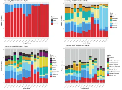
3.2 Fungal Community Composition
For the 5 maize samples, fungal community compositions were determined at seven levels: Domain, Phylum, Class, Order, Family, Genus, and Species. Fig. 3 demonstrates the taxonomy stack distribution of the Phylum, Order, Genus, and Species of the identified fungi. Of the classifiable sequences, two Phyla were identified
as seen in Fig. 3a: Ascomycota and
Basidiomycota, in which Ascomycota is absolutely dominant. At the Class level,
Dothideomycetes, Sordariomycetes,
Tremellomycetes, Eurotiomycetes, and
Saccharomycetes were identified, where
Dothideomycetes and Sordariomycetes are absolutely dominant. At the Order level,
Capnodiales, Pleosporales, Hypocreales, Tremellales, Sporidiobolales, and Eurotiales
were determined, in which Capnodiales,
3b). At the Family level, Cladosporiaceae, Pleosporaceae, Nectriaceae, Tremellaceae, Debaryomycetaceae, Cordycipitaceae, Saccharomycodaceae, and Bulleraceae were
detected. Cladosporiaceae, Trichocomaceae, as
well as Nectriaceae, are the dominant families
(data not shown in Fig. 3). As seen in Fig. 3c,
Cladosporium Epicoccum, Alternaria, Gibberella, Penicillium, Papiliotrema, Talaromyces, Meyerozyma, Aspergillus, and Scheffersomyces
were detected at the genus level in these maize samples. From Fig. 3d, it can be observed that unclassified species account for large portions of the whole bars. Due to a limitation of the UNITE Database, large amounts of species were unable
to be classified. Papiliotrema_flavescens,
Gibberella_zeae, Scheffersomyces_shehatae, and Penicillium_polonicum are the dominant fungal species found in the determined samples.
A relatively high percentage of
Meyerozyma_guilliermondii and
Penicillium_polonicum was detected in two
samples (Maize-2 and Maize-5).
Fusarium_proliferatum,Penicillium_adametzioide
s, and Gibberella_fujikuroi were also detected at
a relatively low percentage.
Gibberella zeae, usually known by the name of
its anamorph Fusarium graminearum, is
identified as a plant pathogen which causes
Fusarium head blight and can produce toxins,
particularly deoxynivalenol (DON). Fusarium spp.
produces a number of diverse secondary metabolites, including some fatal mycotoxins [12] and they are attributed to the most important toxigenic fungi in the Northern temperate areas
[13]. F. proliferatum is a fungus distributed
worldwide that has been associated with many diseases in a number of economic plants including maize, and produce mycotoxins such
as fumonisin B1, moniliformin, beauvericin, and
fusaproliferin [14].
Through a naive Bayesian model using RDP classifier based on UNITE Database analysis of the assembled sequences, three
species of yeasts, Papiliotrema_flavescens,
Scheffersomyces _shehatae, and
Meyerozyma_guilliermondii were found in the
maize samples. In addition,
Penicillium_adametzioides was reported to
be a potential biocontrol agent for
ochratoxin - producing fungus in grapes [15].
Sun et al.; IJBCRR, 26(1): 1-13, 2019; Article no.IJBCRR.49398
Penicillium_polonicum is an endophytic fungus that produces secondary metabolites including bioactive compounds which can be used as
antibiotics [16,17]. Gibberella fujikuroi is a fungal
plant pathogen that produces a wide variety of secondary metabolites including gibberellins,
carotenes, bikaverins, and hydroxylated
anthraquinones (mainly pigments). Epicoccum
nigrum is a plant pathogen and endophyte which produces metabolites such as colored pigments that can be used as antioxidant, anticancer, antibacterial and antifungal agents against other pathogenic fungi [18,19].
As seen in Table 2, for the five samples (regions), the dominant fungi at the Genus level are
different. For Maize-1 Cladosporium,
Papiliotrema, Gibberella, and Epicoccum are the
four dominant genera, for Maize-2 Cladosporium,
Meyerozyma, Papiliotrema, and
Scheffersomyces are the dominant genera, for
Maize-3 Papiliotrema, Cladosporium, Gibberella,
and Epicoccum are the dominant genera, for
Maize-4 Gibberella, Scheffersomyces,
Cladosporium, and Epicoccum are the dominant
genera, and for Maize-5 Talaromyces,
Penicillium, Gibberella, and Aspergillus are the dominant genera.
Cladosporium is recognized as a psychrophile hence it is more adaptable to cool temperature condition, it is the most abundant genus in Maize-1 and Maize-2, since the growing regions of Maize-1 and Maize-2 were Nenjiang County located around 49 degrees north latitude, whereas the growing areas of Maize-3, Maize-4, and Maize-5 are located south of 46.5 degrees north latitude. Consequently, the temperatures in Mishan, Bayan, and Yilan Counties are a little bit
warmer than that of in Nenjiang County.
Cladosporium has proven to be a mycotoxin-producing fungus and potentially pathogenic one frequently occurring in outdoor environments [20].
Talaromyces is a genus of fungi in the family
Trichocomaceae, it is identified as a sexual state of Penicillium that produces soft walled ascomata covered with interwoven hyphae [21]. Some of the Talaromyces species are medically important that produce mycotoxins such as Rubratoxin which is a potential hepatotoxin in food products
[22]. Talaromyces marneffei is an emerging
fungal pathogen resulting in a fatal mycosis in especially immunocompromised persons from
Asian countries such as China [23]. Talaromyces
islandicus is one of the most harmful and destructive fungi that result in the yellowing of rice during storage [24,25] and also produces
mycotoxins including cyclochlorotine,
erythroskyrine, islanditoxin, as well as luteoskyrin which exhibit hepatotoxic and carcinogenic characteristics [21].
In addition, the proportion of Talaromyces
(27.80%), Penicillium (22.54%), and Aspergillus
(12.07%) in Maize-5 were significantly greater than those of in the other four maize samples (regions). It seems like fungal diversity in warmer regions would be richer than those of in cooler ones.
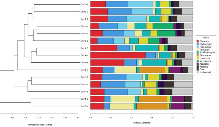
3.3 Cluster Analysis of the 5 Maize Samples
As seen in Fig. 4, the fungi of the 15 maize treatments (three replicates for each sample) from Heilongjiang province were cluster analyzed.
Table 2. Fungal diversity and abundance (%) of maize samples at genus and species levels collected from the five regions*
Levels Fungal strains Maize-1 Daxijiang Maize-2 Rongjun Maize-3 Heitai Maize-4 Wanfa Maize-5 Jiangwan Genus Gibberella Cladosporium 11.64±1.79a 23.51±0.55a 8.940±2.56a 18.25±2.56b 16.35±1.28a 21.10±2.86a 37.64±14.39b 11.29±1.28b 13.02±0.81a 7.14±3.35b
Papiliotrema 14.60±1.67a 10.36±0.74b 21.63±2.67c 2.51±0.46d 0.97±0.73d
Penicillium Scheffersomyces Talaromyces Epicoccum Meyerozyma Aspergillus 2.18±0.51a 2.62±0.62a 0.46±0.03a 10.18±0.78a 2.40±0.58a 0.74±0.05a 6.28±1.45b 9.38±1.57a 0.26±0.28a 7.23±1.74a 16.64±4.56b 2.12±0.65a 2.38±0.09a 6.93±3.57a 0.50±0.69a 8.13±1.70a 1.62±0.57a 0.38±0.08a 2.46±0.52a 19.33±7.00b 1.37±0.13a 3.68±0.76b 2.29±0.89a 0.44±0.06a 22.54±1.41c 2.89±0.41a 27.80±2.39b 2.63±0.53b 1.43±0.53a 12.07±1.20b
Alternaria 4.49±0.77a 3.71±0.64a 1.63±0.27b 1.33±0.24b 1.05±0.68b
Unclassified 19.12±0.82a 13.78±9.41a 13.34±2.34a 11.34±4.12a 2.36±1.80b
Three treatments of Maize2, 3, and Maize-4 can be clustered into three groups, respectively. Three treatments of Maize-1 can be clustered into one group. Only Maize-5 cannot be clustered into one group. This is probably a result of near geographical proximity among the 5 spots where the maize samples were collected leading them to have a similar fungal community. Maize fields in one region may vary in soil types, water resources, types of fertilization, maize seed varieties, and other environmental factors. These might increase the possibility of fungal diversity and make it difficult to cluster the fungi in maize samples from the same region into one group. Nevertheless, most of the fungi in maize samples from the same region are able to be clustered into the same group.
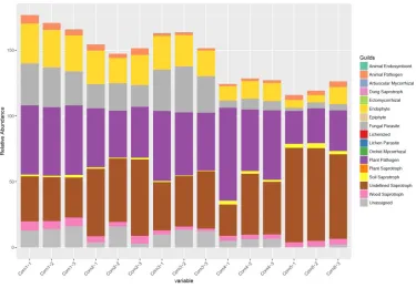
3.4 Fungal Communities and Functional Guilds Analysis
Fungal communities and functional guilds of the 5 maize samples are shown in Fig. 5. As seen in this Figure, an open environment enables the maize to be a plant host to a wide range of
environmental fungi. The most abundant
phylotypes are seen to be plant pathogens and undefined saprotrophs, followed by fungal parasites and endophytes as well as animal
pathogens, soil saprotrophs, and wood
saprotrophs. For all the maize samples, plant pathogens and undefined saprotrophs are absolutely dominant. For the mycotoxigenic fungi species, they are in the category of a plant pathogen.
It is estimated that around 70% of all major crop
diseases were induced by fungal plant
pathogens. In addition, through yield losses and
mycotoxin contamination, 15% of global
agricultural production was destroyed [26]. By far, plant pathogens and especially mycotoxigenic fungi are considered to be the most harmful class of plant pathogens. As a cosmopolitan genus of
filamentous ascomycete fungi, Fusarium includes
a number of toxin-producing plant pathogens of
agricultural importance [27]. Gibberella zeae is
plant pathogen which causes Fusarium head
blight and produces toxins, deoxynivalenol (DON) in particular. For the maize freshly harvested in
Heilongjiang province, the Fusarium_proliferatum
identified probably include mycotoxigenic species. Other fungal genera and species such as
Talaromyces, Penicillium, and Aspergillus, etc., possibly pose a potential threat to maize quality and safety through quality destruction and mycotoxin production, although a fungitoxicity test has not been conducted yet.
Sun et al.; IJBCRR, 26(1): 1-13, 2019; Article no.IJBCRR.49398
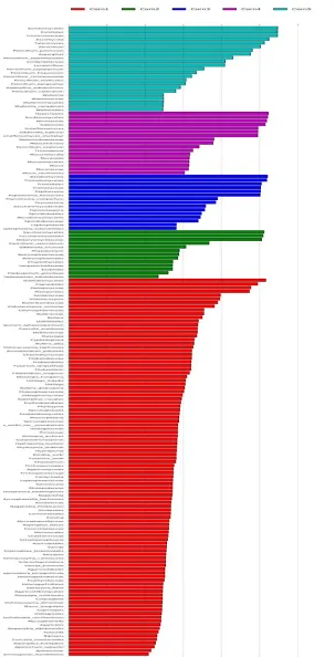
Fig. 6. Cladogram, LDA score and relative abundance of fungi of corn samples
Sun et al.; IJBCRR, 26(1): 1-13, 2019; Article no.IJBCRR.49398
3.5 LEfSe Analysis
Key phylotypes of maize fungi microbiota representing the five samples (regions) identified using linear discriminant analysis (LDA) effect size (LEfSe) are shown in Fig. 6. As seen in Fig 6a, the Cladogram indicates that the numbers of 44 fungal orders are significantly different from those of the corresponding orders or classes in the other four maize samples (regions). There are 24, 8, 4, 0, and 8 orders in Maize-1, Maize-2,
Maize-3, Mazie-4, and Maize-5 samples,
respectively. The fungi count of these orders or classes are significantly different from one sample to the other four samples. Their LDA scores are greater than 2 (Fig. 6b) and they might be considered as specific fungi associated with different regions, especially those with the highest LDA scores in each maize sample (region) (around 13 orders or classes in total).
For example, Dothideomycetes and Capnodiales
for Maize-1, Saccharomycetes and
Saccharomycetales for Maize-2, Basidiomycota
and Tremellomycetes for Maize-3, Hypocreales
and Sordariomycetes for Maize-4, and
Eurotiomycetes and Eurotiales for Maize-5. Consequently, it might be possible to develop biomarkers through using the above fungi to distinguish maize from various regions.
4. CONCLUSION
To explore filed fungal diversity and the potential of fungal mycotoxin production in maize, freshly harvested maize samples in Heilongjiang province were collected and detected via high through-put sequencing. Our results indicate that the five major fungal genera in maize are
Cladosporium, Gibberella, Papiliotrema, Penicillium, and Talaromyces. However, the dominant genera in different samples are different probably due to various climatic conditions, fertilizing manner, seed varieties, and
etc. Epicoccum_nigrum, Fusarium_proliferatum,
and Gibberella_zeae are identified harmful fungi
species, in which Fusarium_proliferatum has the
potential to produce mycotoxins such as
fumonisin. Maize planted in these regions has the potential to produce Fumonisin, DON, and NIV. In addition, 44 fungal orders or classes have been identified as the specific fungal orders or classes which distinguish maize planted in one region from the other four regions, in which around 13 orders or classes in total are especially suitable for the development of biomarkers. Consequently, it is necessary to take adequate measures to prevent fungi growth and
reproduction, especially mycotoxins production, as well as related damage induced by
non-mycotoxins-producing fungi growth and
reproduction.
ACKNOWLEDGEMENT
Financial support from the Foundation for Excellent Academic Leaders of Harbin (2013RFXYJ049 and 2016RAXYJ085) and National Quality and Safety Risk Assessment Project (GJFP2019042) are gratefully acknowledged.
COMPETING INTERESTS
Authors have declared that no competing interests exist.
REFERENCES
1. National Bureau of Statistics of China
(NBSC).
Available:http://data.stats.gov.cn/index.htm. (Accessed on Jan.9, 2019)
2. Northeast Net (NEN).
Available:https://baijiahao.baidu.com/s?id= 1621055268166434387&wfr=spider&for=p c.
(Accessed on Jan.8, 2019)
3. Chen P. Evaluation of grain direct harvest
adaptability and effect of dehydration regulation on maize varieties in cold region.
Master Thesis, Heilongjiang Bayi
Agricultural University, Daqing, China (In Chinese); 2018.
4. Wang ZH, Zhang ZC, Chang H, Zhang JY,
Wang LF. Analysis of physiological mature stage and kernel naturally dry down rate in 38 corn inbred lines in Heilongjiang. Journal of Maize Sciences. 2001;9(2):53-55 (In Chinese).
5. Magoč T, Salzberg SL. FLASH: Fast length
adjustment of short reads to improve genome assemblies. Bioinformatics. 2011; 27(21):2957-2963.
6. Caporaso JG, Kuczynski J, Stombaugh J,
sequencing data. Nature Methods. 2010; 7(5):335-336.
7. Bokulich NA, Subramanian S, Faith JJ,
Gevers D, Gordon JI, Knight R, Mills DA,
Caporaso JG. Quality-filtering vastly
improves diversity estimates from Illumina amplicon sequencing. Nature Methods. 2013;10(1):57-59.
8. Edgar RC. UPARSE: Highly accurate OTU
sequences from microbial amplicon reads. Nature Methods. 2013;10(10):996-998.
9. Wang Q, Garrity GM, Tiedje JM, Cole JR.
Naive Bayesian classifier for rapid
assignment of rRNA sequences into the new bacterial taxonomy. Applied and Environmental Microbiology. 2007;73(16): 5261-5267.
10. Kõljalg U, Larsson KH, Abarenkov K,
Nilsson RH, Alexander IJ, Eberhardt U, Erland S, Høiland K, Kjøller R, Larsson E, Pennanen T, Sen R, Taylor AFS, Tedersoo L, Vrålstad T. UNITE: A database providing
web‐based methods for the molecular
identification of ectomycorrhizal fungi. New Phytologist. 2005;166(3):1063-1068.
11. Nguyen NH, Song ZW, Scott T, Branco BS,
Tedersoo L, Menke J, Schilling JS,
Kennedy PG. FUNGuild: An open
annotation tool for parsing fungal
community datasets by ecological guild. Fungal Ecology. 2016;20:241-248.
12. Desjardins AE, Manhanadhar HK, Plattner
RD, Manandhar GG, Poling SM, Maragos CM. Fusarium species from Nepalese Maize and production of mycotoxins and gibberellic acid by selected species. Applied and Environmental Microbiology. 2000;66:1020-1025.
13. Gutleb AC, Morrison E, Murk AJ.
Cytotoxicity assays for mycotoxins
produced by Fusarium strains: A review.
Environmental Toxicology and
Pharmacology. 2002;11:309-320.
14. Logrieco A, Mulè G, Moretti A, Bottalico
A. Toxigenic Fusarium species and
mycotoxins associated with maize ear rot in Europe. European Journal of Plant Pathology. 2002;108:597-609.
15. Ahmed H, Strub C, Hilaire F,
Schorr-Galindo S. First report: Penicillium
adametzioides, a potential biocontrol agent for ochratoxin-producing fungus in grapes,
resulting from natural product pre-
harvest treatment. Food Control.
2015;51:23-30.
16. Bai J, Zhang P, Bao G, Gu JG, Han L,
Zhang LW, Xu YQ. Imaging mass
spectrometry-guided fast identification of antifungal secondary metabolites from
Penicillium polonicum. Applied
Microbiology and Biotechnology. 2018;102:
8493-8500.
17. Ding GZ, Liu J, Wang JM, Fang L, Yu SS.
Secondary metabolites from the
endophytic fungi Penicillium polonicum and
Aspergillus fumigatus. Journal of Asian
Natural Products Research. 2013;15(5):
446-452.
18. Dzoyem JP, Melong R, Tsamo AT, Maffo T,
Kapche DGWF, Ngadjui BT, McGaw LJ, Eloff JN. Cytotoxicity, antioxidant and antibacterial activity of four compounds
produced by an endophytic fungus
Epicoccum nigrum associated with Entada
abyssinica. Revista Brasileira de
Farmacognosia. 2017;27:251-253.
19. Perveen R, Raza MA, Iqbal T, Naz I, Sehar
S, Ahmed S. Isolation of anticancer and antimicrobial metabolites from Epicoccum nigrum; endophyte of Ferula sumbul. Microbial Pathogenesis. 2017;110:214-224.
20. Alwatban MA, Hadi S, Moslem MA.
Mycotoxin production in Cladosporium species influenced by temperature regimes. Journal of Pure & Applied Microbiology. 2014;8(5):4061-4069.
21. Yilmaz N, Visagie CM, Houbraken J,
Frisvad JC, Samson RA. Polyphasic taxonomy of the genus Talaromyces. Studies in Mycology. 2014;78:175-341.
22. Engelhardt JA, Carlton WW. Rubratoxins.
In: Mycotoxins and phytoallexins (Sharma, RP, Salunkhe DK, eds). CRC Press, Boca Raton, Florida. 1991;259–289.
23. Deng ZL, Ribas JL, Gibson DW, Connor
DH. Infections caused by Penicillium
marneffei in China and Southeast Asia. Review of eighteen cases and report of four more Chinese cases. Reviews of Infectious Diseases. 1988;10:640-652.
24. Sakai A, Tanaka H, Konishi Y, Hanazawa
R, Ota T, Nakahara Y, Sekiguchi S, Oshida E, Takino M, Ichinoe M, Yoshikawa K, Yoshizawa T, Takatori K. Mycological examination of domestic unpolished rice and mycotoxin production by isolated Penicillium islandicum. Journal of the Food Hygienic Society of Japan. 2005;46:205– 212.
25. Oh JY, Kim EN, Ryoo MI, Kim KD.
Morphological and molecular identification of Penicillium islandicum isolate KU101
from stored rice. Plant Pathology Journal.
Sun et al.; IJBCRR, 26(1): 1-13, 2019; Article no.IJBCRR.49398
26. Prado S, Nay Bm Kunz C.
Paraconiothyrium variabile, an ascomycete
endophyte, suppresses mycotoxin
production in the plant pathogen Fusarium
oxysporum. Journal de Mycologie Médicale. 2015;25(2):e96-e97.
27. Ma LJ, Geiser DM, Proctor RH, Rooney
AP, O'Donnell K, Trail F, Gardiner DM,
Manners JM, Kazan K. Fusarium
pathogenomics. Annual Review of
Microbiology. 2013;67:399-416.
_________________________________________________________________________________
© 2019 Sun et al.; This is an Open Access article distributed under the terms of the Creative Commons Attribution License (http://creativecommons.org/licenses/by/4.0), which permits unrestricted use, distribution, and reproduction in any medium, provided the original work is properly cited.
Peer-review history: