R E S E A R C H A R T I C L E
Open Access
Efficiency of platelet-rich plasma therapy in
knee osteoarthritis does not depend on
level of cartilage damage
Rene Burchard
1,2,3*, Hanno Huflage
1, Christian Soost
4, Oliver Richter
5, Bertil Bouillon
1,6and Jan Adriaan Graw
7,8Abstract
Objectives:Osteoarthritis of the knee is common and often leads to significant physical disability. While classic conservative therapeutic approaches aim for symptoms like pain and inflammation, procedures like the intraarticular application of hyaluronic acids (HA) or platelet-rich plasma (PRP) are thought to stimulate the endogenous HA production, stop catabolism of cartilage tissue, and promote tissue regeneration. To analyse whether the positive effects of PRP injections are associated with the level of cartilage damage, patient satisfaction with the treatment was correlated with the level of knee joint osteoarthritis quantified by MRI.
Methods:PRP was performed with a low-leukocyte autologous conditioned plasma (ACP) system in 59 patients. A pre-treatment MRI was performed and a Whole-Organ MRI Score (WORMS) was used to score the level of knee osteoarthritis by 14 features: integrity of the cartilage, affection of the bone marrow, subcortical cysts, bone attrition, osteophytes, integrity of the menisci and ligaments, presence of synovitis, loose bodies, and periarticular cysts. A multivariate analysis with ordinary least squares regressions was used.
Results:Although pain symptoms and severity of clinical osteoarthritis symptoms decreased, regression analysis could not detect a correlation between the degree of cartilage damage measured by the WORMS score and a positive response to PRP therapy.
Conclusion:This study suggests that intraarticular injection of PRP might improve osteoarthritis symptoms and reduces the pain in patients suffering from osteoarthritis of the knee joint independent from the level of cartilage damages quantified by the whole-organ MRI scoring method WORMS.
Keywords:Osteoarthritis, Knee, Platelet-rich plasma, Cartilage damage
Introduction
Osteoarthritis of the knee has a prevalence of 10–18% and without treatment often leads to significant physical dis-ability [1–3]. Conservative therapies to relieve pain and inactivity include treatment with nonsteroidal anti-inflammatory drugs (NSAIDs), physical activity, and braces. In addition, invasive therapeutic approaches in-clude injections of steroids, hyaluronic acids (HA), or platelet-rich plasma (PRP) [4]. When conservative treat-ment approaches become insufficient, surgical treattreat-ment options include arthroscopic debridement and lavage,
osteotomy, and unicompartimental or total knee arthro-plasty [5]. However, surgical treatment of knee osteoarth-ritis should only be performed when conservative treatment options fail. This strategy is particularly relevant when cartilage damages are only mild to moderate [6].
Mechanical or biochemical non-physiological stimuli and a loss of bone-cartilage homeostasis were identified as triggering factors for the development of osteoarth-ritis [7]. While classic conservative methods like treat-ment with NSAIDs mainly target symptoms like pain and inflammation, alternative procedures like intraarti-cular application of HA or PRP to stimulate the endogenous HA production, stop catabolism of cartilage tissue and promote cellular metabolism and tissue re-generation [8, 9]. Many studies have compared the
© The Author(s). 2019Open AccessThis article is distributed under the terms of the Creative Commons Attribution 4.0 International License (http://creativecommons.org/licenses/by/4.0/), which permits unrestricted use, distribution, and reproduction in any medium, provided you give appropriate credit to the original author(s) and the source, provide a link to the Creative Commons license, and indicate if changes were made. The Creative Commons Public Domain Dedication waiver (http://creativecommons.org/publicdomain/zero/1.0/) applies to the data made available in this article, unless otherwise stated.
* Correspondence:[email protected]
1Department of Health, University of Witten/Herdecke, Witten, Germany 2Department of Trauma- and Orthopaedic Surgery, Kreisklinikum Siegen,
Siegen, Germany
clinical outcome of these competitive techniques with scoring systems such as the visual analogue scale (VAS) to quantify the severity of pain or the Western Ontario and McMaster Universities Osteoarthritis Index (WOMAC) the most widely used assessment to evaluate pain, stiffness, and physical functions in arthritis re-search [8,10–17]. Although results are inhomogeneous, PRP therapy appears to have a positive effect on bio-logical cartilage repair [9,15].
While conventional radiography provides high-resolution images of bone tissue, a direct visualisation of cartilage is not possible. In addition, bone marrow changes like bone marrow edema are not detected by conventional radiography. In contrast, magnetic reson-ance imaging (MRI) is more suitable for detailed investi-gations of osteoarthritic joints with mild to severe cartilage damages [18–20]. Because MRI can discrimin-ate all articular tissues, the current study used MRI as the ideal suited diagnostic method to evaluate the cartil-age status.
The main objective of this study was to analyse whether patient satisfaction with PRP therapy was associated with the degree of cartilage damage quantified with MRI.
Methods
The Medical Ethics Committee of the Medical Council Westphalia-Lippe approved this study (number of ethical approval: 2015-685-f-S). Written informed consent was obtained from all study participants before participation.
All patients > 18 years with MRI-proven knee osteoarth-ritis, walking ability, and indication for PRP treatment were enrolled in the study at our centre in 2016 after in-formed consent was obtained. Patients with an age under 18 years, MRI-imaging without signs of knee osteoarth-ritis, limitation of motion range for flexion < 90° and ex-tension > 20°, arthroscopic or open surgery within the past 3 months, fibromyalgia, chronic fatigue syndrome, coagu-lation disorder, thrombocytopenia (< 150.000 platelets per mm3), patients who received intra-articular injection of steroids, anaesthetics, or viscosupplementation within the last 12 months, pregnant patients and patients with chronic diseases, such as rheumatoid arthritis, or signifi-cant cardiovascular comorbidities, current infections, can-cer, or diabetes, and patients with severe damage of the homolateral hip or ankle were excluded from this study. Patients were not treated with anti-inflammatory drugs during the period of PRP treatment. The Western Ontario and McMaster Universities Osteoarthritis Index (WOMAC) and the visual analogue scale (VAS) were sur-veyed before the first injection and after a mean follow-up of 24 weeks [16,17].
To evaluate the level of osteoarthritis, a pre-treatment MRI was performed and analysed by a senior consultant radiologist with the whole-organ MRI scoring method
(WORMS) according to Peterfy and colleagues [21]. The WORMS method scores the level of knee osteoarthritis by 14 features, such as integrity of the cartilage (cartilage damage), affection of the bone marrow, subcortical cysts, bone attrition, osteophytes, integrity of the menisci and ligaments, presence of synovitis, loose bodies, and peri-articular cysts [21]. Furthermore, the cartilage damage was used for regression analysis as a dependent variable (WORMS Cartilage Score). A 1.5-T whole-body scanner with a circumferential knee coil (Philips Ingenia® 1.5 T, Philips GmbH, Hamburg, Germany) was used for im-aging. Scanner settings and sequences were adjusted according to Peterfy and colleagues [21].
PRP was performed with a low-leukocyte autologous conditioned plasma (ACP) system by drawing 15 ml of blood with the ACP Double Syringe® (Arthrex, Naples, USA). Blood was centrifuged at 1500 rpm for 5 min in a Rotofix® 32 A centrifuge (Andreas Hettich GmbH, Tut-tlingen, Germany). The ACP Double Syringe® provides a closed mechanism to separate the plasma from the cell pool. Five millilitres of leukocyte-low and platelet-rich plasma was extracted. The obtained plasma was injected into the knee joint within 5 min after extraction. Patients were treated once a week for three times.
Statistics
A power analysis was performed with the software pack-age G*Power® to compute the a priori required sample size for the mean comparisons (n = 32) and the regres-sion analysis (n= 50) [22]. Data were analysed with stat-istical software package SPSS® Version 25 (IBM, Armonk, North Castle, New York, USA). The mean dif-ferences were analysed with the Wilcoxon signed-rank test. For the multivariate analysis, OLS (ordinary least squares) regressions were used. OLS regression (multiple linear regression model) is suitable for the exact quanti-fication of causal relationships with metric independent variables. Data are presented as mean values with stand-ard error of the mean (SEM). Apvalue of less than 0.05 was considered significant.
Results
Fifty-nine patients (52.5% male) with a mean age of 58.78 ± 1.54 years and a body mass index (BMI) of 26.11 ± 0.50 underwent PRP therapy for osteoarthritis of the knee. According to the pre-treatment WORMS Cartilage Score, the severity of osteoarthritis was quantified as mild in 12 (20.3%), moderate in 33 (55.9%), and severe in 14 (23.7%) of the cases.
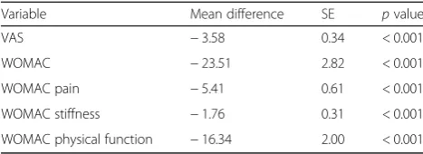
WOMAC-Score decreased by 23.51 ± 2.82 points (p< 0.001, Fig.1). Detailed changes of the WOMAC-Score were collected for pain −5.41 ± 0.61 points (p< 0.001), stiffness −1.76 ± 0.31 points (p< 0.001), and physical function −16.34 ± 2.00 points (p< 0.001). No significant differences were found for the level of osteoarthritis.
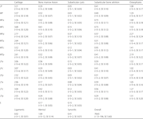
To quantify cartilage damage in the osteoarthritic knee joints, MRI imaging was performed. MRI-based quantifica-tion of cartilage damage by the WORMS score showed a mean selective cartilage damage of 3.00 ± 0.15 points. The overall WORMS score had a mean value of 86.66 ± 5.75 points. Details of this analysis are shown in Table2[21].
To examine whether a positive response to PRP therapy was associated with the degree of cartilage damage or osteoarthritis, regression analysis was performed. There was no correlation between the degree of osteoarthritis (overall WORMS score;p= 0.647) or the isolated degree of cartilage damage (WORMS Cartilage Score;p= 0.805) and a positive response to PRP therapy (Table 3).
However, according to the measured VAS and WOMAC scores, female participants showed a greater WOMAC-Score reduction of osteoarthritis after PRP therapy com-pared to male participants (p= 0.004 depending on overall
WORMS score and p= 0.003 depending on WORMS
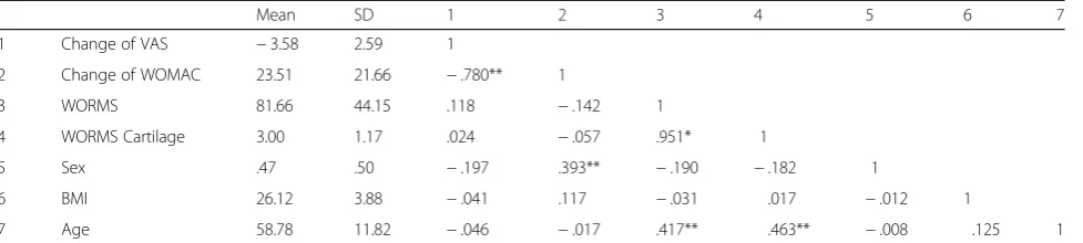
Cartilage Score) (Table 3). In addition, a Bravais-Pearson pair-wise correlation was performed to check the validity of the regression model (Table4).
Discussion
To analyse whether the positive effects of PRP injections are associated with the level of cartilage damage, the sat-isfaction of the patients with their treatment was corre-lated with the level of knee joint osteoarthritis quantified by MRI. Regression analysis could not detect a correl-ation between the level of cartilage damage or the level of osteoarthritis and a positive response to PRP therapy in the studied patient population.
A fair number of studies with different injection treat-ments for patients suffering from osteoarthritis of the knee have been completed so far [8,10–15]. For mild to moder-ate cartilage damages, Filardo and colleagues found a simi-lar clinical outcome after PRP therapy compared to therapy with HA [8]. However, they only used radiographic images to determine the degenerative changes (Kellgren-Lawrence-Score 1–3) and excluded cases with severe osteoarthritis. Similar results were reported by other study groups [10–12]. Frequently, an advantage of PRP therapy com-pared to HA application was seen for the treatment of mild-moderate knee osteoarthritis in younger patients and in male patients [13–15, 23–25]. Nevertheless, all of these studies either used a different PRP preparation protocol or did not provide detailed information to reproduce the treat-ment algorithm [26]. For depletion of leukocytes, Milants Table 1Post-treatment changes of pain, stiffness, and physical
function
Variable Mean difference SE pvalue VAS −3.58 0.34 < 0.001 WOMAC −23.51 2.82 < 0.001 WOMAC pain −5.41 0.61 < 0.001 WOMAC stiffness −1.76 0.31 < 0.001 WOMAC physical function −16.34 2.00 < 0.001
Changes of the visual analogue scale (VAS) and Western Ontario and McMaster Universities Osteoarthritis Index (WOMAC) scores after treatment with platelet-rich plasma (PRP); mean difference describes the change of the mean score after therapy with PRP compared to the mean score before PRP therapy; statistical comparisons are made with non-parametric Wilcoxon signed-rank test;SEstandard error
Table 2MRI-based WORMS analysis
Cartilage Bone marrow lesion Subarticular cysts Subarticular bone attrition Osteophytes LP 3.21
(r0–6; SE 0.19)
0.29
(r0–2; SE 0.08)
0.05
(r0–1; SE 0.03)
0.41
(r0–3; SE 0.10)
1.08
(r0–5; SE 0.17) MP 3.37
(r0–6; SE 0.18)
0.32
(r0–2; SE 0.07)
0.03
(r0–1; SE 0.02)
0.49
(r0–3; SE 0.09)
1.29
(r0–6; SE 0.17) MFa 3.26
(r0–6; SE 0.21)
0.42
(r0–2; SE 0.08)
0.15
(r0–2; SE 0.05)
0.73
(r0–3; SE 0.11)
1.68
(r0–5; SE 0.18) MFc 3.68
(r0–6; SE 0.20)
0.61
(r0–3; SE 0.10)
0.15
(r0–2; SE 0.06)
0.73
(r0–3; SE 0.12)
1.53
(r0–5; SE 0.18) MFp 2.77
(r0–6; SE 0.24)
0.27
(r0–2; SE 0.07)
0.37
(r0–3; SE 0.10)
0.37
(r0–2; SE 0.08)
2.27
(r0–6; SE 0.24) MTa 2.64
(r0–6; SE 0.21)
0.22
(r0–2; SE 0.06)
0.02
(r0–1; SE 0.02)
0.31
(r0–2; SE 0.08)
1.63
(r0–4; SE 0.17) MTc 3.52
(r0–6; SE 0.20)
0.51
(r0–3; SE 0.10)
0.12
(r0–1; SE 0.04)
0.95
(r0–3; SE 0.12)
1.41
(r0–5; SE 0.17) MTp 2.61
(r0–5; SE 0.19)
0.32
(r0–2; SE 0.08)
0.25
(r0–2; SE 0.07)
0.41
(r0–2; SE 0.08)
2.19
(r0–5; SE 0.22) LFa 3.06
(r0–6; SE 0.22)
0.36
(r0–3; SE 0.09)
0.15
(r0–2; SE 0.05)
0.59
(r0–2; SE 0.10)
1.32
(r0–5; SE 0.17) LFc 3.07
(r0–6; SE 0.17)
0.29
(r0–3; SE 0.09)
0.03
(r0–1; SE 0.02)
0.41
(r0–2; SE 0.10)
1.02
(r0–4; SE 0.15) LFp 2.32
(r0–5; SE 0.22)
0.10
(r0–2; SE 0.05)
0.03
(r0–1; SE 0.02)
0.31
(r0–2; SE 0.07)
1.37
(r0–4; SE 0.18) LTa 2.96
(r0–6; SE 0.19)
0.17
(r0–2; SE 0.07)
0.08
(r0–2; SE 0.04)
0.39
(r0–2; SE 0.08)
1.05
(r0–6; SE 0.16) LTc 3.04
(r0–6; SE 0.22)
0.42
(r0–3; SE 0.11)
0.12
(r0–2; SE 0.05)
0.56
(r0–3; SE 0.11)
1.27
(r0–5; SE 0.17) LTp 2.55
(r0–6; SE 0.20)
0.29
(r0–2; SE 0.08)
0.10
(r0–2; SE 0.05)
0.29
(r0–2; SE 0.08)
1.31
(r0–5; SE 0.20)
S 0.17
(r0–1; SE 0.05)
0.15
(r0–2; SE 0.05)
Ligaments Menisci Synovitis Overall 0.02
(r0–1; SE 0.01)
1.00
(r0–12; SE 0.14)
0.41
(r0–2; SE 0.07)
78.65
(r13–196; SE 5.60)
Detailed results of the magnetic resonance imaging (MRI)-based Whole-Organ MRI Score. The analysis includes common MRI-findings for osteoarthritis in 15 anatomical sections of the knee joint;LPlateral patella,MPmedial patella,MFmedial femur,MTmedial tibia,LFlateral femur,LTlateral tibia,aanterior,ccentral,
pposterior,Ssubspinous
Table 3Post-treatment changes of pain and activity level—regression analysis
Dependent variable
Change of VAS depending on WORMS overall
Change of VAS depending on WORMS Cartilage
Change of WOMAC depending on WORMS overall
Change of WOMAC depending on WORMS Cartilage
Constant −2.036 (.464) −1.900 (.498) 1.012 (.963) −.224 (.992) WORMS .007 (.410) −.032 (.647)
WORMS Cartilage .026 (.940) .659 (.805) Sex −.895 (.204) −1.004 (.157) 16.442** (.004) 17.235** (.003) BMI −.018 (.838) −.025 (.780) .670 (.343) .709 (.314) Age −.021 (.520) −.011 (.753) −.004 (.989) −.084 (.747)
n 59 59 59 59
and colleagues suggested a method with only one spinning cycle during the centrifugation step for preparation of the PRP [27]. Moreover, they reported a platelet concentration of fivefold lower than the baseline [27]. For the current study, the Arthrex® ACP protocol according to Marlovits and colleagues was used [28].
Our results suggest that the level of cartilage damage after PRP therapy in knee osteoarthritis is not associated with the clinical outcome measured by the VAS and WOMAC scoring systems. Similar to findings of Jubert and colleagues, our data suggest that late-stage osteo-arthritis does not seem to be an exclusion criterion for PRP therapy because clinical outcome did not differ be-tween patients with mild to moderate and severe disease stages [29]. While restorative effects on the cartilage by PRP therapy have been discussed controversially, anti-inflammatory effects, down-regulation of cytokine levels, and joint homeostasis might explain favourable effects in patients with severe osteoarthritis [30, 31]. However, currently, there is a lack of evidence to support the the-ory of a regeneration of substantial or irreversible cartil-age damcartil-ages by PRP therapy [32]. Furthermore, like other study groups, we could not observe a superior ef-fect of PRP therapy when treating younger men [11,33].
Most of the above-mentioned studies compared results of PRP therapy to the disease progress of osteoarthritis using weight-bearing radiography to stage the cartilage damage level according to the Kellgren-Lawrence score, which is based on radiographic images exclusively [8]. However, detectable radiographic changes appear mainly in the later stages of the disease [19]. MRI technology provides robust acquisition protocols to study progress and level of osteoarthritis in the knee [20]. Therefore, in the current study, the complex and high detailed MRI-based WORMS protocol was used to stage the level of cartilage damages [21].
Besides the strengths of this study such as the use of detailed examination techniques with MRI and inclusion of patients with severe osteoarthritis stages, this study has several limitations. Besides the single-centre design and the low number of cases, the lack of a placebo
control group, and a rather short-term follow-up limit the conclusions. Furthermore, the population in the pre-sented study is older compared to other investigations in the literature [34–36].
Conclusion
The findings of the current study suggest that positive effects of intraarticular injections of PRP might improve quality of life and reduce the pain of patients suffering from osteoarthritis of the knee joint independent from the level of cartilage damage.
Abbreviations
ACP:Autologous conditioned plasma; MRI: Magnetic resonance imaging; PRP: Platelet-rich plasma; VAS: Visual analogue scale; WOMAC: Western Ontario and McMaster Universities Osteoarthritis Index; WORMS: Whole-organ MRI scoring method
Acknowledgements
Dr. Graw is participant in the BIH-Charité Clinician Scientist Program funded by the Charité–Universitätsmedizin Berlin and the Berlin Institute of Health.
Authors’contributions
RB, HH, and BB conceived and designed the experiments. RB, HH, and OR performed the experiments. RB, HH, OR, and JAG analysed the data. CS performed the statistical analysis. RB, CS, and JAG wrote the paper. All authors read and approved the final version of the manuscript.
Funding
No funding was received for this study.
Availability of data and materials
Data are available via the corresponding author.
Ethics approval and consent to participate
The Medical Ethics Committee of the Medical Council Westphalia-Lippe approved this study (number of ethical approval: 2015-685-f-S). Written informed consent was obtained from all study participants before participation.
Consent for publication
Not applicable.
Competing interests
The authors declare that they have no competing interests.
Author details
1Department of Health, University of Witten/Herdecke, Witten, Germany. 2
Department of Trauma- and Orthopaedic Surgery, Kreisklinikum Siegen, Siegen, Germany.3School of Science and Technology, University of Siegen,
Siegen, Germany.4Department of Statistics an Econometrics, University of
Siegen, Siegen, Germany.5Institute of Diagnostic Radiology at Kreisklinikum
Table 4Post-treatment changes of pain and activity level—descriptive statistics
Mean SD 1 2 3 4 5 6 7
1 Change of VAS −3.58 2.59 1
2 Change of WOMAC 23.51 21.66 −.780** 1
3 WORMS 81.66 44.15 .118 −.142 1
4 WORMS Cartilage 3.00 1.17 .024 −.057 .951* 1
5 Sex .47 .50 −.197 .393** −.190 −.182 1
6 BMI 26.12 3.88 −.041 .117 −.031 .017 −.012 1
7 Age 58.78 11.82 −.046 −.017 .417** .463** −.008 .125 1
Siegen, Siegen, Germany.6Department of Orthopaedics, Trauma Surgery and
Sports Traumatology, Kliniken der Stadt Köln, Köln, Germany.7Department of
Anaesthesiology and Operative Intensive Care Medicine (CCM, CVK) Charité, Universitätsmedizin Berlin, Berlin, Germany.8Berlin Institute of Health (BIH), Berlin, Germany.
Received: 23 November 2018 Accepted: 17 May 2019
References
1. WHO Department of Chronic Diseases and Health Promotion (2017).http:// www.who.int/chp/topics/rheumatic/en. Accessed 30 Sep 2017.
2. Covinsky KE, Lindquist K, Dunlop DD, Gill TM, Yelin E. Effect of arthritis in middle age on older-age functioning. J Am Geriatr Soc. 2008;56(1):23–8. 3. McDonough CM, Jette AM. The contribution of osteoarthritis to functional
limitations and disability. Clin Geriatr Med. 2010;26(3):387–99. 4. Michael JW, Schlüter-Brust KU, Eysel P. The epidemiology, etiology,
diagnosis, and treatment of osteoarthritis of the knee. Dtsch Arztebl Int. 2010;107(9):152–62.https://doi.org/10.3238/arztebl.2010.0152. 5. Sutton PM, Holloway ES. The young osteoarthritic knee: dilemmas in
management. BMC Med. 2013.https://doi.org/10.1186/1741-7015-11-14. 6. Kirkley A, Birmingham TB, Litchfield RB, Giffin JR, Willits KR, Wong CJ, Feagan
BG, Donner A, Griffin SH, D'Ascanio LM, Pope JE, Fowler PJ. A randomized trial of arthroscopic surgery for osteoarthritis of the knee. N Engl J Med. 2008;359(11):1097–107.https://doi.org/10.1056/NEJMoa0708333.
7. Anitua E, Sánchez M, Orive G, Padilla S. A biological therapy to osteoarthritis treatment using platelet-rich plasma. Expert Opin Biol Ther. 2013;13(8):1161– 72.https://doi.org/10.1517/14712598.2013.801450.
8. Filardo G, Kon E, Di Martino A, Di Matteo B, Merli ML, Cenacchi A, Fornasari PM, Marcacci M. Platelet-rich plasma vs hyaluronic acid to treat knee degenerative pathology: study design and preliminary results of a randomized controlled trial. BMC Musculoskelet Disord. 2012;13:229.https:// doi.org/10.1186/1471-2474-13-229.
9. Sundman EA, Cole BJ, Karas V, Della Valle C, Tetreault MW, Mohammed HO, Fortier LA. The anti-inflammatory and matrix restorative mechanisms of platelet-rich plasma in osteoarthritis. Am J Sports Med. 2014;42(1):35–41.
https://doi.org/10.1177/0363546513507766.
10. Cole BJ, Karas V, Hussey K, Pilz K, Fortier LA. Hyaluronic acid versus platelet-rich plasma: a prospective, double-blind randomized controlled trial comparing clinical outcomes and effects on intra-articular biology for the treatment of knee osteoarthritis. Am J Sports Med. 2017;45(2):339–46.
https://doi.org/10.1177/0363546516665809.
11. Cerza F, Carnì S, Carcangiu A, Di Vavo I, Schiavilla V, Pecora A, De Biasi G, Ciuffreda M. Comparison between hyaluronic acid and platelet-rich plasma, intra-articular infiltration in the treatment of gonarthrosis. Am J Sports Med. 2012;40(12):2822–7.https://doi.org/10.1177/0363546512461902.
12. Filardo G, Di Matteo B, Di Martino A, Merli ML, Cenacchi A, Fornasari P, Marcacci M, Kon E. Platelet-rich plasma intra-articular knee injections show no superiority versus viscosupplementation: a randomized controlled trial. Am J Sports Med. 2015;43(7):1575–82.https://doi.org/10.1177/
0363546515582027.
13. Duymus TM, Mutlu S, Dernek B, Komur B, Aydogmus S, Kesiktas FN. Choice of intra-articular injection in treatment of knee osteoarthritis: platelet-rich plasma, hyaluronic acid or ozone options. Knee Surg Sports Traumatol Arthrosc. 2017;25(2):485–92.https://doi.org/10.1007/s00167-016-4110-5. 14. Kon E, Mandelbaum B, Buda R, Filardo G, Delcogliano M, Timoncini A, Fornasari
PM, Giannini S, Marcacci M. Platelet-rich plasma intra-articular injection versus hyaluronic acid viscosupplementation as treatments for cartilage pathology: from early degeneration to osteoarthritis. Arthroscopy. 2011;27(11):1490–501.
https://doi.org/10.1016/j.arthro.2011.05.011Epub 2011 Aug 10. 15. Liu J, Song W, Yuan T, Xu Z, Jia W, Zhang C. A comparison between
platelet-rich plasma (PRP) and hyaluronate acid on the healing of cartilage defects. PLoS One. 2014;9(5):e97293.https://doi.org/10.1371/journal.pone. 0097293.
16. Bellamy N, Buchanan WW, Goldsmith CH, Campbell J, Stitt LW. Validation study of WOMAC: a health status instrument for measuring clinically important patient relevant outcomes to antirheumatic drug therapy in patients with osteoarthritis of the hip or knee. J Rheumatol. 1988;15(12):1833–40.
17. Scott J, Huskisson EC. Graphic representation of pain. Pain. 1976;2:175–84. 18. von Engelhardt LV, Raddatz M, Bouillon B, Spahn G, Dàvid A, Haage P, Lichtinger TK.
How reliable is MRI in diagnosing cartilaginous lesions in patients with first and
recurrent lateral patellar dislocations? BMC Musculoskelet Disord. 2010;11:149.https:// doi.org/10.1186/1471-2474-11-149.
19. Cromer MS, Bourne RM, Fransen M, Fulton R, Wang SC. Responsiveness of quantitative cartilage measures over one year in knee osteoarthritis: comparison of radiography and MRI assessments. J Magn Reson Imaging. 2014;39(1):103–9.https:// doi.org/10.1002/jmri.24141.
20. Eckstein F, Cicuttini F, Raynauld JP, Waterton JC, Peterfy C. Magnetic resonance imaging (MRI) of articular cartilage in knee osteoarthritis (OA): morphological assessment. Osteoarthritis Cartilage. 2006;14(Suppl A):A46–75.
21. Peterfy CG, Guermazi A, Zaim S, Tirman PF, Miaux Y, White D, Kothari M, Lu Y, Fye K, Zhao S, Genant HK. Whole-organ magnetic resonance imaging score (WORMS) of the knee in osteoarthritis. Osteoarthr Cartil. 2004;12(3):177–90.
22. Faul F, Erdfelder E, Lang AG, Buchner A. G*power 3: a flexible statistical power analysis program for the social, behavioral, and biomedical sciences. Behav Res Methods. 2007;39:175–91.
23. Meheux CJ, McCulloch PC, Lintner DM, Varner KE, Harris JD. Efficacy of intra-articular platelet-rich plasma injections in knee osteoarthritis: a systematic review. Arthroscopy. 2016;32(3):495–505.https://doi.org/10.1016/j.arthro.2015.08.005. 24. Chang KV, Hung CY, Aliwarga F, Wang TG, Han DS, Chen WS. Comparativ
effectiveness of platelet-rich plasma injections for treating knee joint cartilage degenerative pathology: a systematic review and meta-analysis. Arch Phys Med Rehabil. 2014;95(3):562–75.https://doi.org/10.1016/j.apmr.2013.11.006. 25. Filardo G, Kon E, Buda R, Timoncini A, Di Martino A, Cenacchi A, Fornasari
PM, Giannini S, Marcacci M. Platelet-rich plasma intra-articular knee injections for the treatment of degenerative cartilage lesions and osteoarthritis. Knee Surg Sports Traumatol Arthrosc. 2011;19(4):528–35.
https://doi.org/10.1007/s00167-010-1238-6.
26. Chahla J, Cinque ME, Piuzzi NS, Mannava S, Geeslin AG, Murray IR, Dornan GJ, Muschler GF, LaPrade RF. A call for standardization in platelet-rich plasma preparation protocols and composition reporting: a systematic review of the clinical orthopaedic literature. J Bone Joint Surg Am. 2017;99(20):1769–79.https://doi.org/10. 2106/JBJS.16.01374.
27. Milants C, Bruyère O, Kaux JF. Responders to platelet-rich plasma in osteoarthritis: a technical analysis. Biomed Res Int. 2017;2017:7538604.https://doi.org/10.1155/2017/ 7538604.
28. Marlovits S, Mousavi M, Gäbler C, Erdös J, Vécsei V. A new simplified technique for producing platelet-rich plasma: a short technical note. Eur Spine J. 2004;13(Suppl 1): S102–6.
29. Joshi Jubert N, Rodríguez L, Reverté-Vinaixa MM, Navarro A. Platelet-rich plasma injections for advanced knee osteoarthritis: a prospective, randomized, double-blinded clinical trial. Orthop J Sports Med. 2017;5(2).https://doi.org/10.1177/ 2325967116689386.
30. Kon E, Buda R, Filardo G, Di Martino A, Timoncini A, Cenacchi A, Fornasari PM, Giannini S, Marcacci M. Platelet-rich plasma: intra-articular knee injections produced favorable results on degenerative cartilage lesions. Knee Surg Sports Traumatol Arthrosc. 2010;18(4):472–9.https://doi.org/10.1007/s00167-009-0940-8.
31. Marmotti A, Rossi R, Castoldi F, Roveda E, Michielon G, Peretti GM. PRP and articular cartilage: a clinical update. Biomed Res Int. 2015;2015:542502.https://doi.org/10. 1155/2015/542502.
32. Ayhan E, Kesmezacar H, Akgun I. Intraarticular injections (corticosteroid, hyaluronic acid, platelet rich plasma) for the knee osteoarthritis. World J Orthop. 2014;5(3):351– 61.https://doi.org/10.5312/wjo.v5.i3.351.
33. Patel S, Dhillon MS, Aggarwal S, Marwaha N, Jain A. Treatment with platelet-rich plasma is more effective than placebo for knee osteoarthritis: a prospective, double-blind, randomized trial. Am J Sports Med. 2013;41(2):356–64.https://doi.org/10.1177/ 0363546512471299.
34. Treatment of Osteoarthritis by Intra-articular Injection of Bone Marrow Mesenchymal Stem Cells With Platelet Rich Plasma (CMM-PRGF/ART).https://clinicaltrials.gov/ct2/ show/NCT02365142. Accessed 10 Mar 2019.
35. PRP vs HA Intra-articular Knee Injections for Cartilage Defects.https://clinicaltrials.gov/ ct2/show/NCT02012530. Accessed 10 Mar 2019.
36. Mesenchymal Stem Cells Enhanced With PRP Versus PRP In OA Knee (MSCPRPOAK).https://clinicaltrials.gov/ct2/show/NCT01985633. Accessed 10 Mar 2019.
Publisher’s Note