Pattern Visual Evoked Potentials:
Comparison of Onset, Reversal and Offset Components.
Fatima Suham Shawkat
A thesis submitted for the degree of
Doctor of Philosophy
Institute of Child Health
University College London Medical School
ProQuest Number: 10017335
All rights reserved
INFORMATION TO ALL USERS
The quality of this reproduction is dependent upon the quality of the copy submitted.
In the unlikely event that the author did not send a complete manuscript and there are missing pages, these will be noted. Also, if material had to be removed,
a note will indicate the deletion.
uest.
ProQuest 10017335
Published by ProQuest LLC(2016). Copyright of the Dissertation is held by the Author.
All rights reserved.
This work is protected against unauthorized copying under Title 17, United States Code. Microform Edition © ProQuest LLC.
ProQuest LLC
789 East Eisenhower Parkway P.O. Box 1346
Abstract
Pattern stimulation has proved itself to be an effective technique of studying the visual system in health and disease. However, there are differences in the property of VEPs elicited to different forms of pattern stimulation, namely, reversal, onset and offset. Responses to these three stimuli have usually been studied independently and the relationship between the response components is uncertain. This thesis is concerned with assessing properties and interrelationships between reversal, onset and offset pattern VEPs in controls and in a clinical population of amblyopes.
The three stimuli were delivered sequentially in a single recording epoch so that a direct comparison could be made for virtually identical subject and recording conditions. Half field stimulation was adopted to separate macular and paramacular contributions. The effects of checksize, scotomata and contrast were assessed, and interocular interaction was investigated. The relationship between the three YEP modes was studied by manipulating contrast and spatial phase so that components could be traced from onset/offset modes to the reversal mode. A total of 56 normal and 18 amblyopic subjects were studied.
Ipsilateral reversal (N80, P I 00 and N145), onset (ipsilateral CII and contralateral P I 05) and, to a lesser extent ipsilateral offset components were enhanced by using small checksizes. They were also susceptible to central scotomata; degraded when contrast change was low; and showed the greatest extent of interocular interaction. These features indicate that they are predominantly of macular origin. Contralateral reversal and offset potentials, and ipsilateral onset Cl, were enhanced by large checks and were relatively unaffected by central scotomata, suggesting predominant contributions from paramacular activity. Onset contralateral P I 05 waveform was sharply defined with macular stimulation but became broad and bifid with paramacular stimulation. These findings were confirmed in amblyopes, in whom macular vision is compromised. Contrast change studies indicate offset and reversal components are closely related, and suggest similar physiological origins. Onset Cl and CE could be traced through to reversal PlOO and N145, respectively. When small checks (12') were used, onset Co could be traced through to reversal N80 component.
Acknowledgements
My sincere thanks to Dr. Tony Kriss, my supervisor, for his invaluable ideas, guidance and support throughout the years.
I would also like to thank Dr. Martin Halliday for his advice and inspiration. Dr. Steven Jones for loan of equipment, and Mr. David Taylor for his support and for allowing me to carry out my studies in the Ophthalmology Department at Great Ormond Street, and for permission to use his patients as subjects.
I am very grateful to Mr. Jack Pitman for his technical help and maintenance of equipment, to Dr. Mike Hayward for writing the computer software used in data analysis and presentation, and to Mr. Peter West for his assistance with computer software.
Contents
Page
Abstract 3
Chapter 1: Introduction 14
1.1 Evoked potentials: Historical background 14
1.2 The visual evoked potential 14
1.2.1 The flash and patterned flash VEP 15
1.2.2 Pattem-onset VEP 17
1.2.3 Pattem-reversal VEP 23
1.2.4 Pattern-offset VEP 29
1.2.5 Comparison of pattem-onset, -reversal and -offset VEPs 30 1.2.6 Pattern, motion and contrast contributions to the VEP 34
1.3 Basic physiological aspects and the VEP 37
1.3.1 The retina 38
1.3.2 The lateral geniculate nucleus 41
1.3.3 The cortex 42
1.2.4 Generators of the VEP 51
1.4 Aims 56
Chapter 2: Sequential pattem-onset, -reversal and -offset VEPs: Rationale and
mediodology
2.1 Introduction 58
2.2 Recording technique and equipment 60
2.3 Stimulus 62
2.4 VEP measurements 64
Chapter 3: Effects of checksize
3.1 Introduction 66
3.2 Subjects and methodology 68
3.3 Results 68
3.3.2 Pattem-reversal VEPs 73
3.3.3 Pattern-offset VEPs 75
3.4 Discussion 77
3.5 Conclusions 85
Chapter 4: Effects of experimental scotomata
4.1 Introduction 87
4.2 Methodology 91
4.3 Results 93
4.4 Discussion 100
4.5 Conclusions 104
Chapter 5: Effects of contrast change: A means of studying the transition between onset, reversal and offset components
5.1 Introduction 105
5.2 Methodology 110
5.3 Results 114
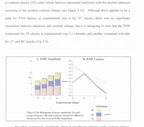
5.3.1 Effects of contrast change on pattern onset/offset VEPs 114 5.3.2 Effects of contrast change on pattern reversal VEPs 118 5.3.3 Observations on the transition between pattem-onset/offset stimulation to
reversal stimulation 122
5.4 Discussion 129
4.5 Conclusions 136
Chapter 6: Comparison of pattem-onset, -reversal and -offset VEPs in amblyopic children
6.1 Introduction 138
6.1.1 Pattern VEPs in children 138
6.1.2 Amblyopia 142
6.1.3 Neuroanatomical and physiological basis of amblyopia 144
6.1.4 Pattern VEPs in amblyopia 148
6.2 M ethodology 152
6.2.2 Technique 6.3 Results
6.3.1 Effects of age - comparison of normal children and adults 6.3.2 Comparison of amblyopic with fellow eyes in amblyopic children
6.3.3 Comparison of amblyopes and normally sighted subjects 6.4 Discussion
6.4.1 Effects of Age 6.4.2 Effects of amblyopia 6.5 Conclusions
153 153 153 157 160 162 162 166 170
Chapter 7: Interocular interaction reflected in pattem-onset, -reversal and -offset VEPs in normal and amblyopic subjects
7.1 Introduction 172
7.2 Methodology 173
7.3 Results 173
7.3.1 Binocular / monocular amplitude ratios 173 7.3.2 Binocular-monocular latency differences 176
7.4 Discussion 180
7.5 Conclusions 186
Chapter 8: General discussion and conclusions
8.1 Comparison of pattem-onset, -reversal and -offset VEPs 8.2 Physiological basis of VEP components
8.3 Proposals for future studies: Clinical and experimental applications
187 197 206
References 211
List of Figures
Chapter 1: Introduction
Figure 1.1: Diagram of a coronal section through the left LGN of macaque. 41 Figure 1.2: Diagram showing the mapping of the visual field on the primary visual cortex.44 Figure 1.3 Diagram of human and macaque brains showing topography of presumptive
cortical visual areas. 46
Figure 1.4: Summary diagram of functional segregation in the primate visual system. 50 Figure 1.4: Diagram showing projections from retina to cortical and subcortical structures. 51
Chapter 2: Sequential pattem-onset, -reversal and -offset VEPs: Rationale and methodology
Figure 2.1: Diagram showing 7 channel montage and typical pattem-onset -reversal and -
offset responses. 61
Figure 2.2: Photographs of experimental set up showing subject seated in front of the two
oscilloscopes and mirror stereoscope. 63
Figure 2.3: Left half-field ipsilateral and contralateral pattem-onset, -reversal and -offset
components as recorded from channels 2 and 4. 65
Chapter 3: Effects of checksize
Figure 3.1: Typical full and half-field VEP responses as recorded from the midline channel
and two lateral channels. 69
Figure 3.2: Group average ipsilateral (channel 2) and contralateral (channel 4) responses for
10 checksizes. 70
Figure 3.3: Graph showing mean onset component amplitude with checksize. 72 Figure 3.4:: Graph showing mean onset component latency with checksize 72 Figure 3.5: Graph showing mean reversal component amplitude with checksize 74 Figure 3.6: Graph showing mean reversal component latency with checksize 74 Figure 3.7: Graph showing mean offset component amplitude with checksize 75 Figure 3.8: Graph showing mean offset component latency with checksize 75
Chapter 4: Effects of experimental scotomata
hemi-scotomata used in the experiments. 92 Figure 4.2: Group average responses of ipsilateral VEPs across five different checksizes, without and with the four scotomata of increasing size. 94 Figure 4.3 Group average responses of contralateral VEPs across five different checksizes, without and with the four scotomata of increasing size. 95 Figure 4.4: Stacked histograms of mean VEP component amplitudes for increasing scotomata
size and checksize. 98
Figure 4.5: Stacked histograms of mean VEP component latencies for increasing scotomata
size and checksize. 99
Chapters: Effects of contrast change: A means of studying the relationship between
pattem-onset, -reversal and -offset components.
Figure 5.1: Diagrams showing the transition of the checkerboard from experimental steps 1 (low step contrast change) through to step 8 (pure pattem-reversal). 111 Figure 5.2: Schematic graphs showing luminance change (A) and contrast change (B) with
each experimental step. 112
Figure 5.3: Diagram showing the two VEP responses obtained over the 500ms recording
epoch, for experimental steps 1, 4 and 8. 113
5 to 8, for high to low and low to high contrast change responses. 121 Figure 5.13 Group average ipsilateral VEPs of experimental steps 1 to 8 showing the transition from low contrast pattem-onset/offset through to pure pattem-reversal mode of
stimulation for different checksizes. 124
Figure 5.14: Group average contralateral VEPs of experimental steps 1 to 8 showing the transition from low contrast pattem-onset/offset through to pure pattem-reversal mode of
stimulation for different checksizes. 125
Figure 5.15: VEPs to the largest 80' checks of experimental steps 1 to 8 from a subject showing the apparent amalgamation of pattem-onset CII and GUI and their evolution to form
a bifid reversal PlOO positivity. 127
Figure 5.16: VEPs to the smallest 12' checks of experimental steps 1 to 8 showing Co pattem- onset component which subsequently evolved into the reversal N80 component. 128
Chapter 6: Comparison of pattem-onset, -reversal and -offset VEPs in amblyopic children.
Figure 6,1: Developmental changes in the human visual system as reflected in the latency of pattem reversal VEPs (From Moskowitz and Sokol, 1983). 141 Figure 6.2: Scatter plots of significant onset (A) and offset (B)components with age. 154 Figure 6.3: Histogram of mean onset ipsilateral CIII and contralateral P I 05 amplitudes across
age groups. 155
Figure 6.4: Histogram of offset ipsilateral component amplitudes across age groups. 156 Figure 6.5: Histogram of reversal N145 and offset N165 latencies across age groups. 156 Figure 6.6: Histogram of onset component latencies across age groups. 157 Figure 6.7: Binocular and monocular ipsilateral and contralateral responses to different
checksizes from an amblyopic subject. 158
Figure 6.8: Group average ipsilateral and contralateral VEPs (n=18) of amblyopic and
non-amblyopic eyes across the 4 checksizes. 159
Figure 6.9: Onset ipsilateral CH (A) and contralateral P105 (B) amplitudes for amblyopic and
Figure 6.10: Variation of ipsilateral reversal component latencies with checksize.for amblyopic
and fellow eyes. 160
Figure 6.11: Reversal PlOO amplitude (A) and latency (B) variation with checksize for the
amblyopic and fellow eyes. 160
Chapter 7: Interocular interaction of pattem-onset, -reversal and -offset VEPs in normal and
amblyopic subjects.
Figure 7.1: Binocular and monocular ipsilateral and contralateral responses to different
checksizes from a control subject. 174
Figure 7.2: Mean PlOO ratio (+/- S.E. mean) for controls and amblyopes. 176
Figure 7.3: Group average binocular VEPs superimposed upon monocular left eye VEPs of
normal subjects (n= 31). 177
Figure 7.4: Mean binocular-monocular latency differences (4-/- S.E.mean) for reversal N80
and PlOO components. 179
Chapter 8: General discussion and conclusions.
Figure 8.1 : Diagram summarising the proposed link between the VEP components of the three
modes of pattem stimulation. 194
Figure 8.2: Simplified schematic diagram showing the concurrent processing magnocellular
List of Tables
Chapter 6: Comparison of pattem-onset, -reversal and -offset VEPs in amblyopic children.
Table 6.1: Table showing means and standard deviations of VEP component amplitudes that significantly differed between amblyopic and control eyes (p<0.05). 161 Table 6.2: Table showing means and standard deviations of VEP component latencies that significantly differed between amblyopic and control eyes (p<0.05) 162
Chapter 8: General discussion and conclusions.
Table 8.1: Summary of macular and paramacular VEP differences 188
Table 8.2: Overview or results 190
Abbreviations
' : minute of arc ® : degree
c/deg : cycles per degree dB: decibels
deg: degree
EEG: electroencephalography Hz: Hertz
ms: millisecond
MRI: magnetic resonance imaging NPN : negative-positive-negative PET: positron emission tomography PNP: positive-negative-positive sec: second
SE mean : standard error of mean Std Dev : standard deviation pV: microvolt
1 .Introduction
1.1 Evoked Potentials: Historical Background
Evoked potentials are voltage fluctuations generated by the brain and spinal cord in response to stimulation of a sensory modality. They are elicited following auditory, visual, somatosensory and gustatory stimulation.
Dawson (1947) first demonstrated that stimulus-specific, cortical evoked potentials could be recorded at the scalp. He showed by using photographic superimposition that the evoked potential could be discerned from the background EEG. In subsequent years, Dawson described signal averaging using an electro-mechanical machine which sampled and added individual responses (Dawson 1951, 1954). Barlow (1957) devised an electronic averager, based on an analog cross-correlator system. Subsequently, Clark et al (1961) described a digital computer (the Average Response Computer) called the "Laboratory Instrument Computer", which is acknowledged as the forerunner of modem laboratory digital computers for signal averaging.
1.2 The Visual Evoked Potential
1.2.1 The Flash and Patterned Flash VEP
Early work employed unstructured flash stimulus presented at slow rates (less than 4/sec). The flash VEP is a polyphasic complex of components. Cobb and Dawson (1960) first studied the flash VEP and reported four early components starting with an initial positive peak at 20-25 ms after the stimulus. Different authors have used a variety of ways to classify the VEP components (letters, numbers, etc.). Cigànek (1961), identified components by Roman numerals and divided them into three time groups: the earliest group was labelled 'primary' (components I, II and IE), the next group was called 'secondary' (IV, V, Va, VI and VI) and these were followed by the 'after discharge'. The 'primary' components were postulated to be generated by Brodmann's area 17. Gastaut and Regis (1965) compared VEP waveforms described by different laboratories (e.g. Cobb and Dawson, 1960; Cigànek, 1961; Schwartz and Shagass, 1964). Although there was some variation in waveform and in the number of components, there did appear to be a consistent positive component with onset around 80ms and culminating in a peak around 100-150ms. Inter-laboratory variation can be partly accounted for by different stimulus conditions, different electrode montages and stimulus parameters (e.g., intensity, colour and frequency). Intra-individual variability, poor sensitivity to pathological conditions and poor correlation with visual acuity, rendered it unreliable in clinical testing.
followed by another negativity at 170-210ms and a positivity around 200 to 235ms. Amplitude reached a maximum for checksizes 10'-20', independent of the intensity (check sizes tested ranged from 4deg. to 3'). It was also found that if field size was increased beyond 4 degrees, there was no further increase in amplitudes to either structured or unstructured flash stimulation. Harter and White (1968, 1969) studied the effects of defocus on contour sharpness and check size, and found that two components were particularly sensitive: a) a negativity at around 90-200ms and, b) a positivity around 180-200ms. VEP amplitude was greatest for sharply focused patterns using small 10' to 20' checks, however, if the pattem was defocused (by ±1 to ±5 dioptres) progressively larger checks were needed to elicit the largest VEP.
An important problem in interpreting response changes to patterned flash stimulation is that they represent a combination of both luminance and pattem contributions. Jeffreys (1968) sought to overcome this problem by subtracting VEP luminance changes from those to flashed pattem.. Luminance and pattem responses show different waveforms, and have different topographic distributions thus suggesting different origins. Duffy et al (1967) and Lambroso et al (1969) showed that the pattem specific responses were reduced or absent in amblyopic patients, thus strongly suggesting that these responses were associated with stimulation of the central region of the retina.
stimuli are sufficiently far apart in time (more than 200ms.).
1.2.2 Pattem-Onset VEP
Jeffreys and Axford (1972 a,b) identified components of the pattem-onset response: The first positive component Cl (with a latency range of 65-80ms), the negative component CII (latency range 90-110 ms) and a later positivity CHI (latency range 150-200ms). C l and C n were both greatly influenced by the retinal location stimulated. Their studies showed that corresponding peaks, C l and CII, inverted in polarity following stimulation of the upper and lower half-fields. For the left and right half-field responses, the transverse distribution of Cl, but not CII, showed a lateral polarity reversal across the occipital midline. They concluded that C l and CII have spatially separate generator sources. The longitudinal distribution of CII appears to conform with that of a simple dipole model related to the retinotopic representation at the extrastriate cortex (areas 18 and 19). Extrastriate areas also probably generate CIII. They hypothesised that Cl reflected dipole-like activity in the striate cortex (area 17) within and bordering the calcarine fissure. The pattem-onset VEP recorded from a midline electrode following lower half-field stimulation, had a similar waveform in most subjects (12 out of 15). The main differences when comparing the subjects were in the relative amplitude of the three components, and in the peak latencies.
Spekreijse et al (1973) studied effects of contrast on onset VEP amplitude. They found that onset VEPs saturate at a relatively low contrast level, but at pre-saturation levels there was an almost linear relationship between VEP amplitude and stimulus contrast.
contrast dependence. However, at spatial frequencies higher than 2c/deg, onset VEPs tended to be larger and persisted at lower contrast levels than did the offset VEPs. At low stimulus contrast and high spatial frequency (above 8 c/deg), onset responses only were detected. Vassilev and Strashimirov ascribed their findings to preferred sensitivity of transient channels to low spatial frequency, and to predominance of sustained channels at higher spatial frequencies. However, there is a wide spatial frequency range (at least from 2 to 8 c/deg.) where suprathreshold gratings effectively stimulate both types of channels.
The latencies and the saturation amplitudes of the onset components are closely related to changes in stimulus parameters. For example Jeffreys (1977) showed that stimulus outlines have higher thresholds, but similar saturation amplitudes, to those obtained to a corresponding pattem of solid squares. In addition, VEPs obtained to different pattem shapes had similar thresholds and latencies, but different saturation amplitudes. The component amplitude thresholds and latencies were found to be dependent on contrast, duration, overall luminance and on the spatial density and dimensions of the pattem elements, but were not greatly influenced by the actual stmctural details of the pattem elements. By contrast, the saturation amplitudes, especially of CII and CIII were less dependent on contrast and overall luminance but were dependent on the stmctural details of the pattem.
Jeffreys (1977) reported a more rapid rate of attenuation of CII and CIII compared with Cl, as the resting contrast level is increased from zero (- i.e. blank field). Thus, this data also indicates an association between the behaviour of CII and CHI and the subjective definition of the outlines of the pattem elements. It seems that once the resting contrast level is raised just above threshold, CII and CHI are relatively more attenuated but the C l amplitude remains fairly large, and the stimulus appears as a contrast change only.
The amplitudes of these pattern-related components, especially CH and CHI are dependent on the structural details of the stimulus pattem (contour), but seem independent of the polarity of the contrast of the pattem - that is, similar onset VEPs are obtained to corresponding pattems whose elements are either lighter or darker than the background. The most striking feature of this stmcture dependency is the consistent increase in amplitude obtained when pattems of continuous contours are broken up into discontinuous elements.
The effects of pattem pre-exposure on these pattem onset VEPs show further differences in the properties of the individual components (James and Jeffreys, 1975; Jeffreys, 1977). Comparatively short pre-exposure periods have been found to produce a marked attenuation of CII and CHI, but not of CL This selective attenuation to adaptation of CII and c m can occur even for the successive presentations of the stimulus pattem in a conventional averaging mn if relatively long pattem durations are used. Experiments by Jeffreys, using independently controlled adapting and test pattems, have shown that, following pre-exposure, there is an increase of the component thresholds and latencies in addition to the eventual attenuation of C n and CHI. These adaptation effects appear to be both orientation and dimension specific.
77-93 ms, (probably analogous to Cl) which decreased in amplitude with spatial frequency, whereas the following negative peak at 97-114 ms, (CII) increased in amplitude with spatial frequency. Stationary pattem adaptation to a grating of the same spatial frequency as the test grating significantly reduced the CII amplitude at 4, 2 and 1 c/deg. Cl, however, was unaffected by stationary pattem adaptation at all combinations of test and adapting spatial frequencies. Since CII, and not Cl, was significantly attenuated following adaptation and testing at 1 c/deg, Hudnell et al concluded that the neurones generating these components are functionally distinct. The use of a common adaptation grating discounted the possibility that CII, but not Cl, was affected due to a difference in the rates of retinal image modulation caused by eye movements made while viewing adaptation gratings of different spatial frequencies. It was postulated that the neurones generating CII were adapted at a lower rate of retinal image modulation than that apparently required for adaptation of the neurones generating Cl, which suggests a difference between these neurones in the rate of stimulus modulation necessary to activation.
The pattem onset components have been found to be selectively influenced by gradual de-focusing of the stimulus pattem. Cl being more resistant to de-focusing than either CII or CIII, especially for large size pattem elements (Jeffreys, 1977). Whereas lenses of 3 dioptres produce a substantial attenuation of CII and CIII to different checksize pattem onset, progressively stronger lenses are needed to produce a comparable attenuation of C l for increases in checksize. Subjectively when there is sufficient defocusing to make the outlines of the pattem elements indistinct, CII and CHI are invariably attenuated. By contrast. C l can remain relatively unattenuated in conditions where the large square pattem appears as an array of very blurred contrast patches.
that the alignment of the steady contours and the test pattem is not necessary for this attenuation, but that their relative linear dimensions seem important, especially for CH. For example, CII and GDI are attenuated by the steady pattem of short bars or squares even when displaced, but CE seems little affected by an aligned steady grid pattem. In all cases there is a significant attenuation of CEI. This effect seems to differ from those described by Spekreijse et al., (1973), who found that the pattem reversal VEPs to low contrast checkerboard pattems were almost completely suppressed by the presence of high contrast, fine grid pattems, and this effect required the accurate alignment of the grids with the edges of the squares. Such interaction effects between the steady and test pattem stimuli also exist when these elements are not overlapping and are in fact spatially separated.
The marked differences in the properties of Cl compared with CE and CEI, indicate that there are two types of cortical processes. One is a contrast-specific mechanism, that contributes to Cl and the other is a contour specific mechanism that contributes to CII and CEI mainly, but also to a minor extent to CL The contrast-specific process responds predominantly to the onset and/or offset of the stimulus pattem and is relatively unadaptive. The contour-specific process responds to the onset of the stimulus and is highly adaptive, ceasing to respond to prolonged static pattem stimulation and is both orientation and dimension specific.
specific VEP sources are sensitive to stimulation within localized areas of the visual field. In one of the animal studies designed to investigate the contrast dependency of single units, Maffei and Fiorentini (1973), found that simple cells in the cat consistently showed a linear relationship between the magnitude of their response and the logarithm of the contrast of a sinusoidal grating pattem. Only a small proportion of complex cells in the striate cortex showed such a linear relationship and the response of these cells tended to saturate at relatively low contrast levels. As simple cells are exclusively present in the striate cortex, and as they show partial summation within the 'on' and 'o ff regions of their location-specific receptive fields, this suggests their possible involvement in the contrast-specific contributions to CL Maffei and Fiorentini also found that the subjective estimate of pattem contrast was closely correlated with the logarithm of stimulus contrast, and that a similar relationship was found for simple cells, and for the amplitude of VEPs obtained to the high frequency altemation of a sinusoidal grating pattem (Campbell and Maffei, 1970). The properties of the contour-specific mechanisms however, make it unlikely that they are closely associated with simple cells, and, their sensitivity to discontinuous contours suggests a possible involvement of hypercomplex-type neurones (Hubei and Wiesel 1965).
Kulikowski (1977) reported an earlier negative component of the onset response at 70- 120ms. This component was also studied by Drasdo (1980), who referred to it as Co'. He found that the maximum response occurred with checks 3' to 4.5', or with bars 2' to 3' for equal square-wave gratings, when using a foveal field of 2.5deg diameter. He estimated that the optimal bar width at the fovea is about 2', which corresponds to the receptive field characteristics that have been reported by Poggio et al. (1977) for the striate area of the monkey. Drasdo (1980) found that with a small 2.5deg field. Cl was reduced in the midline electrode, but was often present in lateral electrodes, whereas conversely, Co was maximal in the midline but decreased rapidly away from the midline. Interestingly, depending on spatial frequency, the peak of Cl varied in latency: Drasdo suggested that there were relative latency differences in the behaviour of Cl and CII, and that Co emerged when the leading edges of C l and CII changed their relationship with different spatial frequencies. This provided further evidence on the different origins of Cl and CII. These findings for the foveally elicited Co, and the fact that it is small and not readily identifiable in all subjects, may explain the wide range of latencies reported for Co.
1.2.3 Pattem-Reversal VEP
Campbell and Maffei (1970) found that when the grating stimulus is reversed in contrast at a rate of 8Hz, and when the recording band-width is restricted from 8Hz to 25Hz, the VEP waveform is almost sinusoidal. When grating contrast is reduced, the VEP amplitude decreases proportionately and a linear relation was found between amplitude and the logarithm of the grating contrast, which held for a wide range of spatial frequencies from 3.5 to 18c/deg.
Plant, Zimmerman and Durden (1983) found that the first major negative wave (N l) and the first major positivity (PI) were easily identifiable. This N l-P l complex to gratings below 1 c/deg was similar for pattem reversal and onset modes of stimulation. The N l became more prominent with spatial frequency above 1 c/deg. This effect was more pronounced in the case of pattem onset and the response to pattem reversal diminished in amplitude at a lower spatial frequency than did the response to pattem onset. The late positivity (P2) was most prominent in pattem onset recordings and at intermediate spatial frequencies.
change.
Checkerboard reversal stimuli are preferred and more commonly used for clinical testing and experimental studies as they tend to produce more clearly defined components than grating stimuli (Halliday, 1982). The response to checkerboard reversal stimulation has a positive component with a latency of about 100ms, which can be seen in virtually all healthy subjects. The amplitude of this component in healthy subjects is little affected by changes in the mean intensity (about 15% reduction in amplitude per log unit decrease in luminance). For latency, a decrease in luminance of one log unit produces about 15 millisecond increase (Halliday 1982).
The full-field pattern reversal stimulation elicits a characteristic triphasic complex, with a negativity at 75ms, a positivity at 100ms and a negativity at 145ms. (N80, PlOO, N145). Left and right half-field stimulation (extending beyond the macular area, 0-2°) will elicit triphasic complexes that appear to have opposite polarities over the left and right sides of the occipital cortex. An N80-P100-N145 complex is recorded over the occipital scalp, ipsilateral to the stimulated half-field, whereas on the contralateral side, a complex with a P75, N105, P I 35 is commonly recorded (Blumhardt et al, 1978). These ipsilateral and contralateral complexes depend to a considerable extent on stimulation of different parts of the visual field. Studies by Blumhardt et al. (1978, 1989) and Halliday et al (1979), in which the macular area of the stimulus field are masked off, and studies involving the progressive construction of the visual fields, have shown that the ipsilateral NPN complex is mainly associated with the macular area of the visual field and that the contralateral PNP complex is mainly associated with the stimulation of the paramacular areas.
component appeared to be generated by cortical areas subserving the paramacular area (4-8 degree radius). Lower field stimulation gave a positivity, also at around 100ms, but this was maximal at 2.5 to 5cm above the inion, and, predominantly associated with the macular field.
W hen the distribution of the PlOO latency is plotted for a healthy population using a standardized stimulus, high contrast, large field, the range is relatively narrow, the majority (>95%) of values falling within a 20ms interval. Amplitude, on the other hand, is more variable. This makes it more difficult to use amplitude information clinically. However, the difference in the amplitude of the responses when comparing each eye is very small in healthy individuals and a ratio of the amplitude of one eye to the other should approximate 1. Halliday et al. (1977) reported that the mean ratio of left/right eye amplitudes for 30 individuals was 1.03 (std dev: 0.15).
In about 5% of normals, the main positivity at around 100ms (PlOO) consists of two sub-peaks approximately of equal amplitude and separated by 10-20 ms. This has been termed the W - waveform (Picton, 1979; Shahrokhi et al, 1978). Piéton estimated the latency of the PlOO as a mid-point between the two peaks. Stockard et al (1979) indicated that the W waveforms are seen mainly in the occipital-vertex derivations, and that the initial positivity is the real PlOO, whereas the second deflection which appears as a 'positivity' is in fact a negativity picked up by the reference electrode and thus appears inverted.
eyes, respectively, p<0.0001). It has been suggested that smaller head size, thinner skull thickness and the higher body temperatures in women may account for their shorter latencies and larger amplitudes (Stockard, 1979; Christie and McCrearty, 1977).
The latency of the PlOO component is not much affected by habituation, fatigue or level of attention. Meienberg et al (1979) measured 20 pattern reversal VEPs obtained consecutively in one recording session. They found that the PlOO latency varied less than ±5% , whereas the PlOO amplitude varied upto ±15%. The variations appeared to be random. However, Shahrokhi et al., (1978) noted that PlOO amplitude tended to decrease with a decreased level of attention. It is probable that changes in attention affect fixation accuracy, level of accommodation and alterations of EEC alpha activity.
The checks that produce the clearest patterns subjectively, also produce the largest amplitude VEPs and decrease or increase of that ideal checksize produces progressively smaller VEPs (Harter and White, 1970). The check size that produces the highest amplitude VEP is progressively greater as more peripheral retina is stimulated. This is in good agreement with the fact that visual acuity deteriorates progressively in the peripheral retina (20/50 at 1.5-4.5 deg; 20/100 at 4.0-11.5 deg and 20/200 at more than 9deg - Harter, 1970). Behrman, Nissim and Arden (1972) observed that for 9' checks, the maximum response was obtained with 1.49 degree diameter fields, for 16' checks, the maximum was obtained with 6 degree fields, whereas for 35' checks, the maximum was obtained with 18 degree fields. The relative insensitivity of the periphery to small checks is likely to be due to the decreased density of cones and greater convergence of peripheral cones into ganglion cells at the periphery (and progressive increase in receptive field size) (Hirsch and Hylton, 1984). Peripheral retina is accordingly also represented by relatively smaller cortical areas.
checksize that can just produce a detectable response (Harter et al, 1968).
Latency of the PlOO component is not as significantly affected by checksize as amplitude. Van Lith et al (1978) studied the variance of PlOO latency with checksize and noticed no significant differences between 20', 40' and 80' checks, with 26 degree field size. Spekreijse et al., (1972) reported that steady state VEPs to pattern reversal has a maximal amplitude for 11' checks. In an independent study of a group of five subjects, checksizes ranging from 11' to 18' elicited the largest amplitude VEPs. There is, of course an ambiguity in the interpretation of such studies of the effect of checksize, since when checksize is increased, the number of squares/edges falling on the fovea is decreased, thus decreased stimulation which may account for part of the observed attenuation of the VEP amplitude. From the point of routine clinical testing, moderate sized checks are preferable, (50'), as moderate defocusing or poor refraction tends to have relatively little effect on the PlOO latency. However, amplitude tends to decrease in parallel with the reduction in the acuity level.
Collins et al (1979) showed that a foveal checkerboard stimulus of 12' checks is very sensitive to refractive errors. Harding and Wright (1986) compared the effects of optical blurring on pattem-onset, reversal and flash VEPs. No effect was found for the flash VEP, however, pattem-onset and reversal showed increases in latency with optical blurring which were more pronounced for 14' checks compared with 56' checks. Interestingly pattem-onset CII component showed a much greater shift in latency than the reversal PlOO, even with relatively low amounts of defocusing (e.g. +1D).
than 2,5 degrees, the response became clearly checksize dependent. The VEPs which were not affected by checksize (1.25 and 2.5 degrees) were thought to be predominantly luminance dependent.
1.2.4 Pattern-Offset YEP
The visual evoked potentials to pattem onset and reversal give a relatively stereotype waveform. However, a variety of waveforms have been reported for pattern-offset. Harter (1971), used the cessation of a train of perceptually fused stroboscope flashes, back illuminating a pattem as stimulus. The main component was a negative peak at around 100ms, in one case followed by a positive peak at about 200ms. Spekreijse et al (1973), who recorded pattem-offset responses, reported a single sharp positive deflection around 120 ms. followed in some subjects by a negative peak at 180 ms.
Jeffreys (1977) compared pattem onset, offset and reversal responses elicited using a tachistoscope. Both the offset response and the reversal response had a positive peak around 120ms, though preceded in some cases by a weak negative deflection. Jeffreys used a short duration (25ms) presentation and the offset pattem VEPs contained pattem-onset contributions due to overlap with the onset related components, particularly CII and GUI. By increasing stimulus duration it was found that the most consistent features of these offset related potentials are positive and negative peaks at about 110 and 150 ms, respectively.
1.2.5 Comparison of Pattem-Onset, -Reversal and -Offset VEPs
Kriss and Halliday, (1980), and Ochs and Aminoff, (1980) used similar slide projector stimulus systems and large stimulus fields of 50' checks to elicit pattem-reversal and onset/offset VEPs. Kriss and Halliday concluded that the pattem reversal and pattem offset components were essentially of similar morphology, whereas Ochs and Aminoff put forward the view that the pattem reversal waveform could be produced by the effect of pattem adaptation on the pattem onset response: When pattem onset VEPs were recorded immediately after adaptation to the stimulating pattem, the main negativity (CII) was lost and the latency of the main positivity (Cl) was increased by 14ms, causing it to appear similar to the pattem-reversal PlOO component both in terms of morphology and latency. Ochs and Aminoff thus suggested that adaptation to the pattem, as found in the conventional reversal VEP paradigm, causes the waveform differences between pattem reversal and pattem-onset.
to be due to its resistance to adaptation compared to CII, and, the presence of residual contrast. It was also reported that the latency of the offset Cl was very close to the value to which the onset C l and reversal PlOO converged.
Plant et al's (1983) finding that the amplitude of pattem onset is larger at a higher spatial frequency than pattem reversal, has not been reported before, though it may be related to Kulikowski's suggestion that at lower spatial frequencies (< 3c/deg), the reversal VEP is dominated by movement components and the onset VEP by pattem related components. Hence, these may correspond to the activation of Y and X systems respectively, as it is known that the receptive fields of X cells are smaller than those of Y cells.
There are but a few topographical studies of the offset response. Kriss and Halliday (1980) compared the scalp distributions of onset, offset and reversal responses elicited by a half-field (0-16 degrees radius) with 50 minute checks and found that pattem offset responses had a distribution that was almost identical to that evoked by reversal, for lateral and altitudinal half-field stimulation. However, the onset response distribution, was found to be different. Left and right half-field stimulation gave an ipsilateral offset and reversal distribution of the first prominent positive component (PlOO), relative to the half-field stimulated, and a triphasic (positive-negative-positive -PNP) complex with a conspicuous negative peak (NlOO) from the contralateral channels. Pattem onset however, produced ipsilaterally, a positivity that was significantly earlier (P90) and on the contralateral side and a broad positivity (P I05) which was maximal at about 5 cms lateral from the midline. Kriss and Halliday found that for upper field stimulation, both pattem offset and reversal gave a prominent negativity at 80 ms with little or no polarity reversal below the inion and a positivity at 100 ms for lower field stimulation.
especially in males, but that onset Cl (referred to as P75) and CII (NlOO) were larger in the elderly. It was found that all offset and reversal potentials, and onset CH (NlOO) and CIII (P170) showed an inverted 'U' relationship between checksize and amplitude, with 18 minute checks giving the largest responses, whereas onset Cl (P75) showed a progressive increase in size from smallest to largest checks. In half of the subjects, stimulation with the smallest checks (4.5 minutes), caused the onset CII to become bifid but offset and reversal were unchanged. There was also a significant tendency, for all reversal and offset components and onset c m , to give shorter latencies with larger checksizes, whereas C l and CII showed no such relationship between latency and checksize.
decrease responses of the pattem onset/offset stimuli. For extrafoveal stimulation, however, the algebraic sum of the increase and decrease responses compares quite well with the pattem reversal response. This evidence suggests that contrast increase and decrease responses differ widely in dynamic behaviour and the dependence on stimulus parameters is thought to portray a different cortical representation for the two responses.
There are marked differences between pattem onset, reversal and offset responses in the rate at which latency is increased with logarithmic decreases in stimulus luminance. Van der Tweel et al (1979) reported that the CII component of the onset response, elicited by either 40' or 60' checks, increases in latency by some 30ms for each log-unit luminance decrease. They also reported that if stimulus contrast at each step of luminance decrease was adjusted so that it was a fixed multiple of its threshold value, the response waveforms and amplitudes remained very similar despite the change in overall luminance. The pattem reversal response has a rate of latency increase associated with luminance decrease that is about half that of the onset response. Halliday (1982) showed that the reversal PlOO potential showed a latency increase of some 15ms and a 15% decrease in amplitude for each log unit of luminance decrease.
Kriss et al (1984) looked at the effect of stimulus luminance decrease on onset, reversal and offset, elicited by 18' checks in a lOdeg diameter field, in healthy subjects. The three responses were averaged in a single sweep lasting one second, using a TV monitor screen. The results showed that the latency increase for later components was largest for each mode of pattem stimulus. This effect tended to broaden and degrade the responses at low luminance levels. There was a tendency for the offset responses to show the greatest rate of latency and amplitude change with decreasing luminance, followed by onset and then reversal responses.
between black and white checks to the sum of their individual luminances. Spekreijse and his team have extensively studied the effects of contrast using 20 minute or smaller checks in a field less than 4 degrees in diameter (1972, 1973). The amplitude of all pattem responses saturate at relatively low contrast of 10-20% (i.e. amplitude reaches a maximum despite further increases in contrast). For onset responses, checks of 20' or more tend to saturate at lower contrasts than smaller checks. Spekreijse and Estevez (1972) looked at two contrast levels : 3.8% and 15% , and found that onset responses were largest for 15'-20' checks at both contrast levels; offset responses were largest for 15'-20' checks at 3.8% contrast and for 5'-7.5' checks at 15% contrast. Spekreijse et al (1973) showed that the onset responses were more resilient than offset responses to decreases in stimulus luminance at low contrast levels (10%, 5% and 3%).
1.2.6 Pattem, Motion and Contrast Contributions to the VEP
Estevez and Spekreijse (1974) and Kriss and Halliday (1980), showed a close resemblance between pattem reversal and pattem offset. However, several workers have suggested that the pattern reversal VEP is likely to contain contributions from motion-specific and pattem-specific mechanisms and thus pattem reversal itself cannot fully be attributed to pattem onset/offset alone.
for this suggestion stems from the fact that the reversal VEPs were abolished when the sharply focused edged of the checks were occluded by fine lines, but this occurred only when the checks were small. Kulikowski (1977) based his work on the psychophysical evidence that a reversing pattem has two distinguishable thresholds: one for detection of motion or flicker, the other for the detection of the pattem. He considered VEPs to a grating of low spatial frequency to be related to a transient (i.e. ac-coupled) neural mechanism as such a VEP depends only on the change in contrast and not on the initial or final value. Kulikowski argued that the criterion for transient related VEPs is that the onset and offset waveforms are similar and that the two constituents sum linearly so that the pattem related constituents of the VEP could be obtained by subtracting the offset responses from the onset responses. These dichotomies are similar to those proposed by Jeffrey's (1977) where a contrast specific mechanism contributes predominantly to onset Cl, and a contour specific mechanism contributes to CII and CIII, and to a lesser extent to CL
Kubova (1992) described motion-onset VEPs in terms of a positive-negative-positive (PNP) complex with the negative N2 peak at a latency of about 160-200ms as the most prominent component. Kuba and Kubova (1992) believe that the major positive peak for motion onset VEPs reported by the Amsterdam groups is primarily pattem generated and is a variant of pattem offset VEPs, caused by pattem disappearance effect at the onset of motion with a high temporal frequency (>6Hz). Such P^ dominated VEPs also occur mainly when the stimulus is limited to the macular area (central 6 degrees). Kuba and Kubova (1992), however, have shown that the N2 dominated motion-onset VEPs are recordable as far out as 50 degrees of eccentricity with amplitudes being significantly larger for extramacular compared with macular stimulation. They state that their observed lateralization of this major negativity supports their hypothesis of its motion specificity and the extrastriate origin of the visual perception of motion.
motion are used, are proposed as being more appropriate in eliciting motion specific responses. These stimuli are perceived as motion only, and they appear to evoke motion responses without other confounding factors such as change in position of contrast (Manning and Mazzucchelli, 1992; Probst et al, 1993; Snowden et al., 1995).
In summary, studies have shown that components related to pattem-onset, -reversal and -offset VEPs behave differently depending on the stimulus characteristics. Pattem- reversal and -offset VEPs appear to be of similar morphology: a triphasic negative-positive- negative complex. If the stimulus is confined to half-the visual field then this complex becomes lateralised over the ipsilateral side of the scalp, and a VEP complex of opposite polarity (positive-negative-positive) is often recorded on the contralateral side of the scalp. The main ipsilateral positivity has been well studied and behaves as if macular in origin. The contralateral main negativity however, appears to be of paramacular origin. Pattem-onset components have been labelled consecutively as Cl (positive polarity), CII and CHI. These components also appear to lateralised ipsilaterally on half-field stimulation, with a positive component (P105), often present on the contralateral side of the scalp. Experiments have shown that contrast-specific mechanisms contribute to Cl and contour-specific mechanisms contribute to CII and CIII.
1.3 Basic Physiological Aspects and the VEP
that respond to the particular stimulus used. Even though a VEP is generated by cortical cells, some of its properties may be pre-determined at the retinal level. As described above, pattem VEP amplitude and latency are influenced by a number of factors such as contrast, spatial frequency, orientation and luminance. The following sections will outline the relevant neurophysiology and neuroanatomy of the visual pathway as related to mechanisms thought to generate the VEP. These will predominantly involve the retino-geniculo-cortical pathway, though the subcortical visual pathway will be briefly described.
1.3.1 The Retina
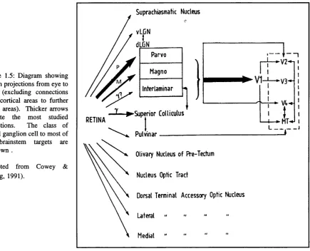
Signals from a large number of photoreceptors (rods and cones) converge on bipolar neurones which in turn synapse with retinal ganglion cells. The ganglion cell axons, which form the optic nerve, synapse at the lateral geniculate nucleus (LGN) and these in turn synapse at the visual cortex reaching it via the optic tract. A few axons are involved in the control of pupillary diameter and connect to the Edinger-Westphal nucleus and accessory optic tract and some other axons connect to the superior colliculus.
and direction selectivities of cortical cells, however, Schiller et al (1986) suggested that it may be thus organised so as to provide fast transfer of information regarding light increments and decrements with equal sensitivity.
The concept of the 'receptive field' of individual neurones (Fischer, 1973) is important for understanding why spatial contrast influences VEP studies. Neurophysiologists have thought of the visual system in terms of neurons that are driven by stimulation within a small discrete area of the visual field. These receptive fields have been mapped by flashing or moving stimuli such as spots or bars. Signals from photoreceptors of a portion of stimulated (illuminated) retina converge onto two separate pools of the receptive field, these pools are known as centre and surround (Enroth-Cugell and Robson, 1966; Rodieck and Stone, 1965). The ganglion cell response recorded from its axon is a fluctuation (increase or decrease) from the mean background firing rate. When both centre and surround organisation of the ganglion cell receptive fields are illuminated, the modulated response decreases compared to if the illumination is o f the centre alone. As a result of this antagonistic organization, the size of centre relative to the surround establishes the spatial selectivity of individual neurones. Because o f the antagonistic organisation of separate pools for centre and surround portions of the receptive fields of bipolar neurones and ganglion cells, the retinal output does not simply represent the illumination of a patch of retina. Instead the response of bipolar and ganglion cells is determined by spatial contrast, that is a difference in illumination between adjacent retinal areas, rather than by the sum of their separate illuminations.
stimulus or grating will have a similar effect in the retinal and LGN but not so in the cortex (Hubei and Wiesel, 1968).
Ganglion cells differ in the size of their receptive fields. Generally, the closer a neurone to the anatomical fovea, the smaller its receptive field. However, each region of the retina is subserved by a range of ganglion cells with different receptive field sizes. The population of parafoveally located ganglion cells have larger receptive field centres than the foveal ones. It is estimated that the human foveal ganglion cell receptive field is smaller than 20' (Jones and Keck, 1978; Plant et al , 1983; Skrandies, 1984). Pattem element size thus relates strongly to the area of retina giving the strongest response.
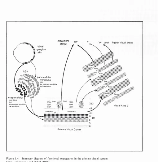
In monkeys, X cells are more common than Y and this is thought to reflect the higher spatial and chromatic acuity in the primate (Shapley and Perry, 1986). In monkey, the X ganglion cells synapse onto neurones in the parvocellular layers in the LGN and are thus called P-cells, and Y cells synapse onto the magnocellular layer and are called M-cells (see LGN section below).
1.3.2 Lateral Geniculate Nucleus:
Six stacked layers can be recognised in the LGN (three layers for each eye). The synaptic terminals from the two eyes distribute themselves in the 6 layers so as to produce six topographic maps of the contralateral half-field of vision. There are 4 parvocellular layers (2 for central 20 deg. of vision and two for peripheral vision) and two magnocellular layers.
stria te cortex
4a 4b 4 c a 4cfl
LGN
P a r v o c e llu la r ( sm a ll cell)
laminae RIGHT EYE
l e f te y e
RIGHT
M a g n o c e llu la r ( la r g e cell) l a m in a e
Figure 1.1: Diagram o f a coronal section through the left LGN o f the macaque showing main projections o f P and M cells in LGN to striate cortex .P-cells project primarily to 4CP and 4A.
The magnocellular layer only represents 8-10% of the total volume of the LGN (Kaas et al, 1978). The LGN layers also have different projection sites; the parvocellular cells projecting to cortical layer 4a and the deeper p sublayer of 4C (4Cp), while the magnocellular cells project to the superficial sublayer of 4C (4C a) (Valverde, 1985). A diagram showing these projections can be found in Figure 1.1.
Parvocellular LGN cells are mostly served by colour-opponent receptive fields and respond in a sustained fashion to visual stimuli, although a small number (15-20%) lack colour sensitivity. They have small receptive fields, linear summation properties and have axons which conduct at medium velocities to the striate cortex. Magnocellular LGN cells lack colour-opponent receptive fields, and are not very colour sensitive. They respond in a transient fashion, have relatively large receptive fields and tend to have non-linear spatial summation properties. They conduct at faster velocities to the cortex (by about 4ms.-Lennie et al., 1990) and have greater contrast sensitivity than parvocellular cells (Schiller and Colby, 1983). The receptive fields of LGN cells in general are nearly circular with the greatest width being less than that of cortical cells. It has been suggested that the separation of parvocellular layers from magnocellular layers corresponds to the distinct processing for contrast (magnocellular) and colour (parvocellular) at the LGN level (Shapley, 1986). Grating contrast sensitivity is much higher for magnocellular cells than parvocellular cells (by 10 to 20 times) and it is thought that magnocellular cells respond and mediate the perception of pattems at low contrasts and at low to intermediate spatial frequencies, whereas parvocellular cells are involved with perception at high contrasts and high spatial frequencies.
1.3.3 Cortex
areas in macaque, and 10 in man have been described and the majority bave been found to be homologous. Topographically, however, human areas are about twice as wide as the macaque counterparts, with the exception of human V3 and VP, which are disproportionately enlarged in man, furthermore distance from foveal areas V2, V3 and VP to V5 (MT) is also disproportionately expanded in humans and it has been suggested that this is related in form processing (Tootell et al., 1996). Cortical areas in man have been studied using PET (positron emission tomography), fMRI (functional magnetic resonance imaging) and magneto- encephalography (MEG). In primates, areas V I to V5 appear to have an important role in determining VEP characteristics with respect to activation by features such as spatial and temporal frequencies, luminance change, the visual field and areas of stimulation, and on/off stimuli versus reversing stimuli which has a motion contribution.
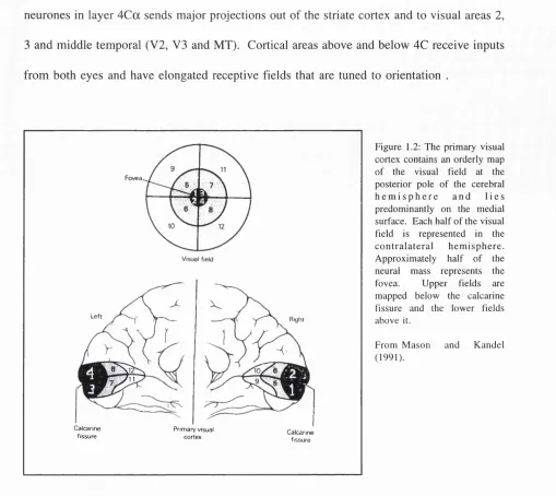
The main projection from the LGN is to the striate cortex (area 17 or V I) and in monkey it is retinotopic. Cortical cytoarchitecture tends to be distinct. Physiological recordings have shown that the area contains a single representation of the visual field which is foveally weighted. The representation of the central 1 degree of the fovea extends over about 15mm of cortex, whereas, at 10 degree eccentricity, 1 degree is only represented by about 1mm (Van Essen et al., 1984) - see figure 1.2.
neurones in layer 4C a sends major projections out of the striate cortex and to visual areas 2, 3 and middle temporal (V2, V3 and MT). Cortical areas above and below 4C receive inputs from both eyes and have elongated receptive fields that are tuned to orientation .
Fovea.
C alcarine P rim ary visual C alcarine
fissu re cortex fissu re
F ig u r e 1.2: T h e p ri m ary v i s u a l c o r t e x c o n t a i n s an o r d e r ly m a p o f the v is u a l f i e l d at the po s t e r io r p o l e o f the cer e br al h e m i s p h e r e a n d l i e s p r e d o m i n a n t l y o n the m e d i a l s u r fa c e . E a c h h a l f o f th e v i s u a l f ie ld is re p r e s e n te d in the c o n t r a l a t e r a l h e m i s p h e r e . A p p r o x i m a t e l y h a l f o f the neural m a s s r e p r e s e n ts the f o v e a . U p p e r f i e l d s are m a p p e d b e l o w the c a lc a r in e fis s u r e an d the l o w e r fi e l d s a b o v e it.
F r o m M a s o n a n d K a n d e l ( 1 9 9 1 ) .
In the striate cortex a common type of orientation tuned neurons are known as 'complex cells' (Hubei and Wiesel, 1968). They respond weakly to stationary targets or give ON and OFF responses when the stimulus goes on and off. Complex cells tend to be both orientation and direction specific. LGN cells do not show direction selectivity, thus this property of cortical cells, like orientation, is created by cortical connections. 'Simple cells' in the striate cortex resemble LGN cells in that they have excitatory and inhibitory regions in their receptive fields. However instead of being concentrically arranged, they are organised in parallel strips and respond well to a moving bar oriented parallel to the parallel strips.
Simple and complex cells differ in the linearity of their spatial summation. Hubei and Wiesel (1962, 1968) originally identified a third subset of striate cells, the hypercomplex cells, however others have classified them as subsets of simple and complex cells (Dreher, 1972). The defining hypercomplex property is the presence of inhibitory zones at one or both ends of the excitatory region of the receptive field, with the consequence that these cells respond to say stimulating bars of preferred orientation only if they are of a particular length.
# #
4
K
VI □
V2 B
V3 □
VP □
V3a [ g
V4v ■
MT/V5
VIP/
SP O
pMSTd/ B
MSTd
L O D
F igu re 1.3: D i a g r a m s o f h u m a n ( A and C ) and m a c a q u e (B and D ) brai ns s h o w i n g to p o g r a p h y o f p r e s u m p t iv e cor ti cal v is u a l areas. A and B s h o w o n e h e m i s p h e r e o f the h u m a n and m a c a q u e brains in f o l d e d n or m al state, and C and D s h o w the s a m e a n a to m i c a l (right h e m i s p h e r e ) and fu n c tio n a l data in 'flattened' cortical format. H u m a n v is u a l areas are base d on data from fun c tio n a l m a g n e t i c r e s o n a n c e im a g in g ( f M R I ) , and m a c a q u e v isu a l areas are b a se d on p u b li s h e d data. N a m e s for h u m a n v is u a l areas h a v e bee n adapted from c o r r e s p o n d i n g areas in m a c a q u e b a se d on t o p o g r a p h i c a l and fu n c tio n a l e v i d e n c e for h o m o l o g y , for e x a m p le ; for V I , V 2 , V 3 , ventral p ost er io r ( V P ) , V 3 A and m i d d l e te m p o r a l ( M T , or V 5 ) . O the r are as are V 4 v (ventral V 4 ) , V IP (ventral intraparietal), S P O ( s u p e r io r parietal o c c i p i t a l) , p M S T d (p o s t e r io r d i v i s i o n o f d ors al med ia l su per io r t em pora l area), M S T d (d or sa l d i v i s i o n o f M S T ) , LIP (l ateral in traparietal), L S P O (lateral su p e r io r parietal o c c ip it a l) , LO (lateral occ ip ita l) .
A d a p te d fr o m T o o t e l l et al., ( 1 9 9 6 ) .
Lower visual field is represented in V3, whereas a corresponding representation of the upper field is found in the ventral posterior area, which is no longer thought of as part of V3 due to different cytoarchitecture. V3 receives inputs from layer 4B of the striate cortex and from the thick stripes of V2 (Felleman et al 1988). M ost of the cells in V3 are strongly orientational and directional specific as well as being sensitive to binocular disparity, but not many are chromatically sensitive. There are distinctive cells that are narrowly tuned for several orientations or directions of motion (Felleman and Van Essen, 1987).
V4 is an area that extends from the anterior bank of the lunate sulcus on to the prelunate gyrus and contains a large number of neurones that are chromatically sensitive (Zeki, 1973). It receives direct but small input from VI with the majority coming from V2 and V3. The projection from V2 arise in one set of cytochrome oxidase stripes (thin ones in squirrel monkey and in the interstripe (DeYoe and Van Essen, 1985; Shipp and Zeki, 1985), the former by implication being the source of the chromatically opponent signals. However not all neurones in V4 are involved in colour analysis and receptive fields, despite being large, have spatial and orientational selectivities just like those in V I. Hey wood and Cowey (1987) showed that bilateral damage to V4 in macaque impaired chromatic discrimination and also resulted in gross disturbance of pattem and orientation discriminations. Cells in V4 (as well as those in V3 and V5) are selective for binocular disparity. These binocular disparity V4 cells receive projections from the parvocellular stream via the V I (inter blob) - V2 (inter stripe) stream (see figure 8.2 -De Yoe and Van Essen, 1988).