review article peer reviewed | OpeN acceSS
Localization and function of VIP and PACAP in the heart
Magdalena Chottova Dvorakova, Jana Slavikova
AbstrAct
Vasoactive intestinal polypeptide (VIP) and pituitary adenylate cyclase-activating peptide (PAcAP) are structurally closely related neuropeptides, which exert prominent pharmacological effects on heart function through three types of receptors: VPAc1, VPAc2, and PAc1. All of them are present in the heart. Localization and function of these endogenous peptide molecules and their receptors in the heart will be presented
Keywords: Heart, Peptide molecules, Pituitary adenylate cyclase-activating peptide (PAcAP), Vasoactive intestinal polypeptide (VIP)
How to cite this article
Dvorakova MC, Slavikova J. Localization and function of VIP and PACAP in the heart. Edorium J Physiol 2015;1:1–6.
Article ID: 100001P07MD2015
Magdalena Chottova Dvorakova1, Jana Slavikova1
Affiliations: 1Associate Professor, Department of Physiology, Charles University in Prague, Faculty of Medicine in Pils-en, Czech Republic.Junior Researcher, Biomedical Centre, Charles University in Prague, Faculty of Medicine in Pilsen, Czech Republic.
Corresponding Author: Magdalena Chottova Dvorakova, Department of Physiology, Medical Faculty, Charles Univer-sity, Alej Svobody 1655/76, 323 00 Pilsen, Czech Repub-lic; Tel : +420 377 593 343, Fax: +420 377 593 349; Email: [email protected]
Received: 05 January 2015 Accepted: 06 February 2015 Published: 18 March 2015
*********
doi:10.5348/P07-2015-1-RA-1
IntroductIon
Vasoactive intestinal peptide (VIP) is a linear 28-amino-acid peptide with molecular weight of 3,326 Da that is generated by enzymatic cleavage from its precursor molecule, preproVIP. The 170 amino-acid precursor is metabolized by a signal peptidase in the endoplasmic reticulum to yield the 148-amino-acid proVIP. ProVIP is cleaved by prohormone convertases to prepro VIP125–155 and prepro VIP81–110 (precursor of peptide histidine methionine, PHM). Both of them are then cleaved by carboxypeptidase-B like enzymes to VIP-G and PHM-G. The VIP-G and PHM-G can then be metabolized by PAM enzymes to VIP and PHM [1]. VIP was first isolated as a vasorelaxant from porcine gut by Said and Mutt [2]. The amino acid sequence of VIP in human, cow, sheep, goat, dog, rabbit and rat is identical to that of the porcine peptide [1]. VIP is structurally related to another neuropeptide pituitary adenylate cyclase-activating peptide (PACAP). They belong to the secretin peptide family including also secretin, peptide histidine isoleucin, peptide histidine methionine (human counterpart to peptide histidine isoleucin), glucagon, glucagon-like peptide, glucose-dependent insulinotropic polypeptide and growth hormone-releasing factor [3]. VIP and PACAP are best discussed together because they share receptors as well as functions (Table 1).
The human VIP peptide is 70% identical to N-terminus of both PACAP variants (Table 2). VIP sequence has been very well conserved during the evolution from protochordates to mammals, suggesting an important biological function [6]. Both peptides have a broad spectrum of biological functions including neurotransmitter, secretagogue, neuroprotective, neurotrophic and differentiation roles as well as effect on growth and survival of cells in the developing nervous system [7].
Localization
Vasoactive intestinal polypeptide (VIP) is produced (1) by neurons in different areas of the central and peripheral nervous system, (2) by endocrine cells such as cells of the endocrine pancreas and pituitary lactotrophes, and (3) by inflammatory and immune cells. In the nervous systems, it acts as a multifunctional neurotransmitter and neuromodulator. In the peripheral nervous system, VIP-containing neurons are either intrinsic neurons involved in local reflexes, or postganglionic neurons under preganglionic cholinergic control. VIP is synthesized in neuronal cell bodies and is then transported along axons or dendrites to dense core vesicles located in presynaptic nerve terminals [1].
PACAP is produced by neurons of central and peripheral nervous system and several nonneural tissues such as the adrenal gland, gonads, immune cells, and pancreas [6].
In the heart, VIP is of extrinsic (from vagal or sympathetic efferents) or intrinsic origin, however, the predominant part of cardiac VIP-immunoreactive (IR) innervation is intrinsic, which consists of postganglionic parasympathetic nerve cell bodies localized within various atrial subepicardial regions and their fibers innervating the heart. In the heart atria, VIP-IR fibers are observed in the area of sinoatrial (SA) node where a dense network of VIP-IR fibers is seen closely apposed to the nodal cells. Other parts of the conductive system (atrioventricular node and Purkinje fibers) are also innervated by VIP-IR nerve fibers, however, the innervation is much less pronounced. Additionally, VIP-IR fibers have been detected along the terminal arterioles in the vicinity of atrial cardiomyocytes with endocrine activity. In the ventricles, VIP-IR fibers are present very rarely, more in the right than in the left ventricle, mainly surrounding coronary vasculature. In the atrial part of coronary circulation, VIP-IR fibers occur within adjacent small atrial arteries and arterioles in a density exceeding that of VIP-IR nerves in the remaining segments of the coronary vasculature [8]. In the nerve fibers from postganglionic parasympathetic neurons, VIP is co-localized with the “classical” neurotransmitter acetylcholine (ACh). VIP is stored in large dense core vesicles, and its release is enhanced by high frequency vagal stimulation in contrast to the release of ACh from small vesicles, which is favored by low frequency stimulation [7].
PACAP is much more widely distributed than VIP in the central nervous system, while they often appear to be co-localized in the same nerve cell bodies, and nerve fibers in peripheral organs. In tissues, PACAP38 is the predominant form, and PACAP27 makes up less than 10% of the total PACAP content. In the heart, PACAP27 and PACAP38 immunoreactive nerve cell bodies and nerve fibers were identified within cardiac ganglia and interganglionic fibre tracts. Additionally, PACAP38 has been detected in the coronary arteries. Additionally,
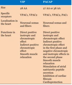
Table 1: Size, specific receptors, localization and function of VIP and PACAP in the heart
VIP PAcAP
Size 28 AA 27 AA or 38 AA
Specific
receptors VPAC1, VPAC2 VPAC1, VPAC2, PAC1 Localization in
the heart Neuronal somas
and fibers Neuronal somas and fibers
Functions in
the heart Direct positive inotropic and chronotropic effect
Indirect positive chronotropic effect
Smooth muscle relaxation
Direct positive inotropic and chronotropic effect Indirect positive chronotropic effect in the first phase and negative chronotropic and inotropic effects in the second phase Smooth muscle relaxation
Stimulation of atrial natriuretic peptide secretion
Inhibition of cardiac fibrosis
Cardioprotection
Table 2: Chromosomal location of VIP and PACAP gene and their amino acid sequence. Bold letters in the amino acid sequences represent differences between VIP and PACAP-27
VIP PAcAP 27
Chromosomal location
Human 6q25 18p11
Rat 1p11 9q37
Amino acid sequence
Human,
Rat HSDAVFTDnYtRLR
immunohistochemical observations suggest that PACAP is localized mainly in the nerve fibres within the intracardiac ganglia, which are predominantly of extrinsic origin [9].
Thus, it is likely that endogenous PACAP and VIP might affect the firing of intracardiac cholinergic neurons in a species-dependent fashion but only VIP would directly influence the sinus node and most other regions of the heart under normal circumstances [10]. Additionally, VIP and PACAP derived from an extrinsic source and coming to heart with blood could play important role in regulation of cardiac functions [11].
receptors
VIP and PACAP exert their effects through three different specific receptors, which belong to the family of guanine nucleotide binding protein (G protein)-coupled receptors with seven conserved transmembrane domains and a molecular weight of 43-80 kDa. They trigger mainly adenylyl cyclase activation through Gs protein, but also can activate phospholipase C. The balance between couplings to adenylyl cyclase versus phospholipase C may be related to the presence of accessory proteins such as the receptor activity modifying proteins RAMPs [12]. Two subtypes of these receptors possess comparable affinity for VIP and PACAP, and, therefore, have been named VPAC1 and VPAC2. The VPAC1 receptor (but not VPAC2 or PAC1) has been shown to be able to interact with RAMPs; in this case ligand specificity is not altered but the VPAC1 receptor-RAMP2 heteromer displays altered signal transduction specificity, with significant enhancement of agonist-mediated phosphoinositide hydrolysis with no change in cyclic AMP stimulation [13]. The third receptor binds VIP with 1,000-fold lower affinity than PACAP, and has been designated PAC1 [9]. Numerous isoforms of the PAC1 receptor, corresponding to 17 known splice variants of the same gene, were identified. The human PAC1 gene is composed of 18 exons, ten being constitutively expressed (exons 2, 3, 7-13 and 18) while seven (exon 4-6 and 14-17) are regulated. A complex process of differential splicing generates the numerous receptor isoforms that display distinct pharmacological profiles. Recent reports suggest that at least one of these isoforms also acts as a high affinity receptor for both VIP and PACAP [14]. All these receptors (VPAC1, VPAC2 and PAC1) belong to the class II family of G protein-coupled receptors. Northern blot analysis confirms presence of all three types of VIP/ PACAP receptors in the heart [5]. Immunohistochemistry showed that in the rat heart, VPAC1 receptor is present in macrophages, while VPAC2 receptor was detected on the smooth muscle cells of coronary arteries. Additionally, small amount of cardiomyocytes express VPAC2 immunoreactivity [15]. PAC1 have been detected in cardiomyocytes and intracardiac neurons [9, 16]. According to our knowledge, expression of the specific
PAC1 receptor variants has not been studied in the heart yet.
Actions In the Heart
cardiac ganglia
Intracardiac ganglia play a major role in neuronal control of the heart and are believed to be capable of independently monitoring and influencing cardiac function. They receive not only cholinergic preganglionic inputs, but also peptidergic afferents and sympathetic postganglionic signals. VIP and PACAP are co-localized with acetylcholine in preganglionic parasympathetic fibers, which densely innervate majority of intracardiac ganglionic cells, additionally, both of this neuropeptide are expressed by cholinergic cardiac neurons. On the surface of these neurons are localized several types of specific receptors for VIP and VPAC meaning, that cell membrane of one neuron may contain VPAC1 and/ or VPAC2 and /or some isoform of PAC1 receptor [16]. Simultaneous activation of VPAC2 and PAC1 receptors by PACAP elicits a synergistic enhancement of neuronal excitability and produces changes in the active membrane properties that are not seen with stimulation of either receptor alone. This process is dependent on VPAC2 receptor-induced Ca2+ release from caffeine- and
ryanodine-sensitive intracellular stores. VIP, by acting exclusively on VPAC receptors, evokes a depolarization of lesser magnitude than PACAP and fails to increase action potential firing [17]. These would offer an explanation for distinct effects of VIP and PACAP on the heart. Whereas both VIP and PACAP have positive chronotropic effect and cause smooth muscle relaxation, PACAP, but not VIP, evokes pronounced negative chronotropic and inotropic effects that are mediated by activation of intrinsic cardiac neurons [9].
cardiomyocytes
by activating intracardiac parasympathetic nerves in the atrium.
coronary circulation
The presence of VIP and PACAP nerve fibers and their receptors in the coronary circulation strongly suggests that both peptides are important in the regulation of coronary blood flow. They are able to produce a vasodilatation but while PACAP displays identical vascular responses in all vessel segments, the vasodilatory effect of VIP differs from one vascular bed to another. The regional differences in vascular responses to the two peptides may be explained (1) by the involvement of a new unknown receptor, or (2) by the interaction of various substances produced and present in the tissue, or (3) by the presence of vessel-specific different populations of the receptor subtypes [28]. In the heart, the vasodilatory effects of VIP on arteries are much greater than on veins because of the greater VIP receptor density in arterial vessels [29]. The potency of VIP depends on the existing level of coronary vessel tone, which may differ from species to species [30]. Moreover, the vasodilatory effect of the peptides is not limited to the coronary arteries. VIP as well as PACAP produces significant arterial dilatation in other body organs such as brain, endocrine glands, and the respiratory system [28, 31, 32]. The requirement of intact endothelium for the vasodilator effects of VIP depends upon the vascular site studied. VIP induced relaxation of rat aortic aorta and the human uterine artery is largely dependent upon the presence of an intact endothelium [33, 34] but the vasodilator effect of VIP at most other sites is endothelium-independent [35-38].
concLusIon
The presence of vasoactive intestinal polypeptide (VIP) and pituitary adenylate cyclase-activating peptide (PACAP) peptides in the heart and results of several functional studies suggest that these peptides plays an important role in the regulation of heart functions including e.g. frequency of beating, contractile ability of myocardium or regulation of coronary blood flow.
*********
Acknowledgements
This study was elaborated within the project ED2.1.00/03.0076 from European Regional Development Fund and by the Charles University Research Fund (project number P36).
Author contributions
Magdalena Chottova Dvorakova – Substantial contributions to conception and design, Acquisition of data, Analysis and interpretation of data, Drafting the cardiac myocytes tension development and the rate and
extent of contraction. VIP has a more potent positive inotropic effect on the right atrial than left ventricular muscle in human, which would play an important role in maintaining cardiac output under critical conditions, where atrial contraction is fundamental [9, 22]. However, the inotropic response to VIP may diminish with increasing VIP dose and/or with time, which is probably due to VIP receptor desensitization in the myocardium [1]. PACAP exerts actions on cardiomyocytes very similar to VIP in the first phase, but in the second phase it causes a decrease of heart rate and contractile force by interaction with cardiac parasympathetic nerves via the PAC1 receptor [6]. Different cardiac responses to VIP and PACAP in the atria and ventricles might be due to differences in the expression and density of specific receptors.
Additionally, PACAP stimulates secretion of atrial natriuretic peptide through the PAC1 receptor, which indicates that PACAP could play the role of a neurohumoral factor to inhibit the remodeling at cardiac hypertrophy [21].
cardiac nonmyocytes
VPAC2 receptor mRNAs have been detected also in cardiac fibroblasts. PACAP inhibits DNA and collagen syntheses in cultured nonmyocytes, which is mediated through a cAMP-dependent process. PACAP is able to inhibit cardiac fibrosis through VPAC2 receptor [21]. Additionally, PACAP immunoreactivity has been detected in macrophages infiltrating heart [23], which could play a role in cardioprotective effect of PACAP demonstrated by several studies [21, 24–26].
conductive tissue
article, Revising it critically for important intellectual content, Final approval of the version to be published Jana Slavikova – Substantial contributions to conception and design, Acquisition of data, Analysis and interpretation of data, Drafting the article, Revising it critically for important intellectual content, Final approval of the version to be published
Guarantor
The corresponding author is the guarantor of submission.
conflict of Interest
Authors declare no conflict of interest.
copyright
© 2015 Magdalena Chottova Dvorakova et al. This article is distributed under the terms of Creative Commons Attribution License which permits unrestricted use, distribution and reproduction in any medium provided the original author(s) and original publisher are properly credited. Please see the copyright policy on the journal website for more information.
rEFErEncEs
1. Henning RJ, Sawmiller DR. Vasoactive intestinal peptide: Cardiovascular effects. Cardiovasc Res 2001 Jan; 49(1):27–37.
2. Said SI, Mutt V. Polypeptide with broad biological activity: Isolation from small intestine. Science 1970 Sep 18;169(3951):1217–8.
3. Arimura A. Pituitary adenylate cyclase activating polypeptide (PACAP): Discovery and current status of research. Regul Pept 1992 Feb 18;37(3):287–303. 4. Miyata A, Arimura A, Dahl RR, et al. Isolation of a
novel 38 residue-hypothalamic polypeptide which stimulates adenylate cyclase in pituitary cells. Biochem Biophys Res Commun 1989 Oct 16; 164(1):567–4. 5. Vaudry D, Gonzalez BJ, Basille M, Yon L, Fournier
A, Vaudry H. Pituitary adenylate cyclase-activating polypeptide and its receptors: From structure to functions. Pharmacol Rev 2000 Jun;52(2):269–324. 6. Sherwood NM, Krueckl SL, McRory JE. The origin and
function of the pituitary adenylate cyclase-activating polypeptide (PACAP)/glucagon superfamily. Endocr Rev 2000 Dec;21(6):619–70.
7. Fahrenkrug J, Hannibal J. Neurotransmitters co-existing with VIP or PACAP. Peptides 2004 Mar;25(3):393–401.
8. Weihe E, Reinecke M, Forssmann WG. Distribution of vasoactive intestinal polypeptide-like immunoreactivity in the mammalian heart. Interrelation with neurotensin- and substance P-like immunoreactive nerves. Cell Tissue Res 1984;236(3):527–40.
9. Dvoráková MC. Cardioprotective role of the VIP signaling system. Drug News Perspect 2005 Jul-Aug;18(6):387–91.
10. Chang Y, Lawson LJ, Hancock JC, Hoover DB. Pituitary adenylate cyclase-activating polypeptide: Localization and differential influence on isolated hearts from rats and guinea pigs. Regul Pept 2005 Jul 15;129(1-3):139–46.
11. Hoover DB, Girard BM, Hoover JL, Parsons RL. PAC(1) receptors mediate positive chronotropic responses to PACAP-27 and VIP in isolated mouse atria. Eur J Pharmacol 2013 Aug 5;713(1-3):25–30. 12. Laburthe M, Couvineau A, Tan V. Class II G
protein-coupled receptors for VIP and PACAP: Structure, models of activation and pharmacology. Peptides 2007 Sep;28(9):1631–9.
13. Christopoulos A, Christopoulos G, Morfis M, et al. Novel receptor partners and function of receptor activity-modifying proteins. J Biol Chem 2003 Jan 31;278(5):3293–7.
14. Muller JM, Debaigt C, Goursaud S, et al. Unconventional binding sites and receptors for VIP and related peptides PACAP and PHI/PHM: An update. Peptides 2007 Sep;28(9):1655–66.
15. Dvoráková MC, Pfeil U, Kuncová J, et al. Down-regulation of vasoactive intestinal peptide and altered expression of its receptors in rat diabetic cardiomyopathy. Cell Tissue Res 2006 Mar;323(3):383–93.
16. DeHaven WI, Cuevas J. Heterogeneity of pituitary adenylate cyclase-activating polypeptide and vasoactive intestinal polypeptide receptors in rat intrinsic cardiac neurons. Neurosci Lett 2002 Aug 2;328(1):45–9.
17. DeHaven WI, Cuevas J. VPAC receptor modulation of neuroexcitability in intracardiac neurons: dependence on intracellular calcium mobilization and synergistic enhancement by PAC1 receptor activation. J Biol Chem 2004 Sep 24;279(39):40609–21.
18. Saetrum Opgaard O, Knutsson M, de Vries R, Tom B, Saxena PR, Edvinsson L. Vasoactive intestinal peptide has a direct positive inotropic effect on isolated human myocardial trabeculae. Clin Sci (Lond) 2001 Dec;101(6):637–43.
19. Tiaho F, Nerbonne JM. VIP and secretin augment cardiac L-type calcium channel currents in isolated adult rat ventricular myocytes. Pflugers Arch 1996 Sep;432(5):821–30.
20. Baron A, Monnier D, Roatti A, Baertschi AJ. Pituitary adenylate cyclase-activating polypeptide activates K(ATP) current in rat atrial myocytes. Am J Physiol Heart Circ Physiol 2001 Mar;280(3):H1058–65. 21. Sano H, Miyata A, Horio T, Nishikimi T, Matsuo H,
Kangawa K. The effect of pituitary adenylate cyclase activating polypeptide on cultured rat cardiocytes as a cardioprotective factor. Regul Pept 2002 Nov 15;109(1-3):107–3.
22. Chottová Dvoráková M, Kuncová J, Pfeil U, et al. Cardiomyopathy in streptozotocin-induced diabetes involves intra-axonal accumulation of calcitionin gene-related peptide and altered expression of its receptor in rats. Neuroscience 2005;134(1):51–8. 23. Alston EN, Parrish DC, Hasan W, Tharp K, Pahlmeyer
through gp130-dependent and independent mechanisms. Neuropeptides 2011 Feb;45(1):33–42. 24. Rácz B, Gasz B, Gallyas F Jr, et al. PKA-Bad-14-3-3
and Akt-Bad-14-3-3 signaling pathways are involved in the protective effects of PACAP against ischemia/ reperfusion-induced cardiomyocyte apoptosis. Regul Pept 2008 Jan 10;145(1-3):105–15.
25. Roth E, Wéber G, Kiss P, et al. Effects of PACAP and preconditioning against ischemia/reperfusion-induced cardiomyocyte apoptosis in vitro. Ann N Y Acad Sci 2009 Apr;1163:512–6.
26. Mori H, Nakamachi T, Ohtaki H, et al. Cardioprotective effect of endogenous pituitary adenylate cyclase-activating polypeptide on Doxorubicin-induced cardiomyopathy in mice. Circ J 2010 Jun;74(6):1183– 90.
27. Accili EA, Redaelli G, DiFrancesco D. Activation of the hyperpolarization-activated current (if) in sino-atrial node myocytes of the rabbit by vasoactive intestinal peptide. Pflugers Arch 1996 Mar;431(5):803–5. 28. Dalsgaard T, Hannibal J, Fahrenkrug J, Larsen
CR, Ottesen B. VIP and PACAP display different vasodilatory effects in rabbit coronary and cerebral arteries. Regul Pept 2003 Feb 28;110(3):179–88. 29. Luu TN, Dashwood MR, Chester AH, Tadjkarimi S,
Yacoub MH. Action of vasoactive intestinal peptide and distribution of its binding sites in vessels used for coronary artery bypass grafts. Am J Cardiol 1993 Jun 1;71(15):1278–2.
30. Hoover DB. Effects of guinea pig vasoactive intestinal peptide on the isolated perfused guinea pig heart. Peptides 1989 Mar-Apr;10(2):343–7.
31. Matran R. Neural control of lower airway vasculature. Involvement of classical transmitters and
neuropeptides. Acta Physiol Scand Suppl 1991;601:1– 54.
32. Ito H, Bell D, Tamamori M, et al. Calcitonin gene-related peptide (CGRP) and hypertrophy of cardiomyocytes. Heart Vessels 1997;Suppl 12:15–7. 33. Davies JM, Williams KI. Endothelial-dependent
relaxant effects of vaso-active intestinal polypeptide and arachidonic acid in rat aortic strips. Prostaglandins 1984 Feb;27(2):195–202.
34. Jovanovic A, Jovanovic S, Tulic I, Grbovic L. Predominant role for nitric oxide in the relaxation induced by vasoactive intestinal polypeptide in human uterine artery. Mol Hum Reprod 1998 Jan;4(1):71–6. 35. Duckles SP, Said SI. Vasoactive intestinal peptide as
a neurotransmitter in the cerebral circulation. Eur J Pharmacol 1982 Mar 12;78(3):371–4.
36. D’Orleans-Juste P, Dion S, Mizrahi J, Regoli D. Effects of peptides and non-peptides on isolated arterial smooth muscles: role of endothelium. Eur J Pharmacol 1985 Aug 7;114(1):9–21.
37. Beny JL, Brunet PC, Huggel H. Effect of mechanical stimulation, substance P and vasoactive intestinal polypeptide on the electrical and mechanical activities of circular smooth muscles from pig coronary arteries contracted with acetylcholine: Role of endothelium. Pharmacology 1986;33(2):61–8.
38. Barnes PJ, Cadieux A, Carstairs JR, Greenberg B, Polak JM, Rhoden K. Vasoactive intestinal peptide in bovine pulmonary artery: localisation, function and receptor autoradiography. Br J Pharmacol 1986 Sep;89(1):157–62.
Access full text article on