Publicly Accessible Penn Dissertations
2019
Characterization And Perturbation Of Functional
Networks That Support Human Memory
Ethan Andrew Solomon
University of Pennsylvania, [email protected]
Follow this and additional works at:
https://repository.upenn.edu/edissertations
Part of the
Neuroscience and Neurobiology Commons
This paper is posted at ScholarlyCommons.https://repository.upenn.edu/edissertations/3382
For more information, please [email protected].
Recommended Citation
Solomon, Ethan Andrew, "Characterization And Perturbation Of Functional Networks That Support Human Memory" (2019). Publicly Accessible Penn Dissertations. 3382.
Human Memory
Abstract
Episodic memory is essential to our daily lives, as it attaches meaning to the constant stream of sensory inputs to the brain. However, episodic memory often fails in a number of common neurocognitive disorders. Effective therapies remain elusive, owing to the complexity of brain networks and neural processes that support episodic encoding and retrieval. In particular, it is not understood how inter-regional communication within the brain supports memory function, though such communication may be critical to the highly integrative nature of episodic memory. To uncover the patterns of memory-related functional connectivity, we asked a large cohort of neurosurgical patients with indwelling electrodes to perform a verbal free-recall task, in which patients viewed lists of simple nouns and recalled them a short time later. As patients performed this task, we collected intracranial EEG (iEEG) from electrodes placed on the cortical surface and within the medial temporal lobe (MTL). First, we examined whole-brain functional networks that emerged during the encoding and retrieval phases of this task, using spectral methods to correlate frequency-specific signals between brain regions. We identified a dynamic network of regions that exhibited enhanced theta (3-8 Hz) connectivity during successful memory processing, whereas regions tended to desynchronize at high frequencies (30-100 Hz). Next, using only electrodes placed within the MTL, we asked whether functional coupling was also observed among this mesoscale subnetwork of highly specialized regions that play an outsize role in memory. Recapitulating our earlier findings, we noted broadly enhanced theta connectivity within the MTL, centering on the left entorhinal cortex during successful encoding operations. Finally, to determine whether such low-frequency functional connections reflect correlative or causal relations in the brain, we applied direct electrical stimulation via electrodes placed within the MTL. We found that low-frequency connections (5-13 Hz) predicted the emergence of theta activity at distant regions in the brain – particularly when stimulation occurred near white matter – indicating the potential causal relevance of iEEG-based functional connections. Taken together, these studies underscore the importance of low-frequency functional coupling to memory across spatial scales, and suggest this form of coupling indicates a causal relation between brain regions.
Degree Type Dissertation
Degree Name
Doctor of Philosophy (PhD)
Graduate Group Bioengineering
First Advisor Michael J. Kahana
Keywords
hippocampus, iEEG, memory, networks, oscillations
THAT SUPPORT HUMAN MEMORY
Ethan A. Solomon
A DISSERTATION in
Bioengineering
Presented to the Faculties of the University of Pennsylvania in
Partial Fulfillment of the Requirements for the
Degree of Doctor of Philosophy
2019
Supervisor of Dissertation Graduate Group Chairperson
_________________________________ _________________________________
Michael J. Kahana Ravi Radhakrishnan Professor, Psychology Professor, Bioengineering
Dissertation Committee
Danielle S. Bassett, Eduardo D. Glandt Faculty Fellow and Associate Professor of Bioengineering Lyle H. Ungar, Professor of Computer and Information Science
ii
iii
Acknowledgements
There is a very long list of people I have to thank for making my PhD possible. The work in these pages would not have happened without – literally – dozens of people from my personal and professional lives, and they each deserve my deep gratitude (and credit!).
First, I want to thank my mentor, Michael Kahana, for building a lab that I was privileged to be a part of. Mike has spent years creating a fantastic research enterprise, with a laser-like focus on statistical rigor and prolific – yet careful – data collection. His efforts have paid off. There is no place else in the world where I could have been equally as immersed in the electrophysiology of human memory, a subject of which I knew little when I first joined the lab in 2015. Under Mike’s mentorship, I published my first manuscript. I attended and presented at conferences around the world. I learned to write grants. And, of course, I learned a lot about how little we understand episodic memory. I think I still don’t quite
grasp how tirelessly Mike has worked to create an environment where all this is possible. For those efforts, and for his mentorship over the past few years (and hopefully many more to come), he has my sincere thanks.
“It takes a village” might not be a mantra that wins elections, but it certainly makes for successful DARPA grants. I have so many DARPA RAM colleagues and friends to thank for their incredibly hard work over the past four years: Paul Wanda, the best all-in-one
iv
Zeinab Helili and Alison Xu, the amazing researchers/clinical site testers; and Dan Rizzuto, who is now trying to turn our research into something people can actually use.
I’ve been told that Prof. Christoph Weidemann was part of the DARPA RAM project, but I
never had the pleasure of seeing him at the biweekly calls. Strange – must have been the
days I wasn’t there. Regardless, he deserves my deep gratitude for providing wonderful mentorship and friendship since I first joined the lab. Though he’s now back in the UK, I’ll
always remember him, because he left one of his suitcases in my basement.
As the lab moves into a post-DAPRA world, I have even more fantastic scientists to thank for keeping me on my toes, challenging me at lab meetings, and being great friends. I’ve become
a better scientist just by sitting in close spatial proximity to Senior Member of the Main Lab
Nora Herweg, and I’m excited to collaborate with her on a number of surely
-groundbreaking ideas. My co-graduate students and good friends Rivka Cohen, Logan Fickling, Dan Schonhaut, and Ada Aka have been incredibly supportive and kind, and make for stellar lab meeting questioners. The same goes for Jesse Pazdera and Effie Li, who are also my co-graduate students, as far as I’m concerned. Nicole Kratz and Deb Gaspari are the
lab’s unsung heroes, making sure the trains run on time, day-in, day-out. All of these people are the reason I never really want to work from home, even if I can.
I also want to acknowledge the wonderful support from the MD/PhD program, particularly Maggie Krall and Skip Brass. They truly run the Best MSTP in the Galaxy®.
Finally, I want to thank my family, for encouraging my interest in science and medicine from a young age. I am confident that I would not be writing these words without their constant love and support. And to Hannah, who even between night shifts in the PICU somehow summoned the will to listen thoughtfully to my practice presentations on theta
v
ABSTRACT
CHARACTERIZATION AND PERTURBATION OF FUNCTIONAL NETWORKS
THAT SUPPORT HUMAN MEMORY
Ethan A. Solomon Michael J. Kahana
Episodic memory is essential to our daily lives, as it attaches meaning to the constant stream of sensory inputs to the brain. However, episodic memory often fails in a number of common neurocognitive disorders. Effective therapies remain elusive, owing to the
complexity of brain networks and neural processes that support episodic encoding and retrieval. In particular, it is not understood how inter-regional communication within the brain supports memory function, though such communication may be critical to the highly integrative nature of episodic memory. To uncover the patterns of memory-related
vi
processing, whereas regions tended to desynchronize at high frequencies (30-100 Hz). Next, using only electrodes placed within the MTL, we asked whether functional coupling was also observed among this mesoscale subnetwork of highly specialized regions that play an outsize role in memory. Recapitulating our earlier findings, we noted broadly enhanced theta connectivity within the MTL, centering on the left entorhinal cortex during successful encoding operations. Finally, to determine whether such low-frequency functional
connections reflect correlative or causal relations in the brain, we applied direct electrical stimulation via electrodes placed within the MTL. We found that low-frequency connections (5-13 Hz) predicted the emergence of theta activity at distant regions in the brain –
vii
Table of Contents
ACKNOWLEDGEMENTS ... III
ABSTRACT... V
LIST OF TABLES ... X
LIST OF ILLUSTRATIONS ...XI
CHAPTER 1: INTRODUCTION ... 1
Major Contributions ... 3
CHAPTER 2: BACKGROUND ... 5
Principles of Human Episodic Memory ... 5
Intracranial EEG ... 9
Network Neuroscience ... 11
Brain Stimulation ... 14
CHAPTER 3: WHOLE-BRAIN ELECTRICAL NETWORKS ...17
Abstract ... 17
Introduction ... 18
Results... 20
Quantification of brain-wide connectivity phenomena ... 20
Identification of network hubs ... 24
Temporal modulation of connectivity effects ... 27
Relationship between connectivity and spectral power ... 29
Generalization of network phenomena to memory retrieval ... 33
Filtering for oscillatory activity ... 34
viii
Methods ... 40
Participants ... 40
Free-recall task ... 41
Electrocorticographic recordings ... 42
Anatomical localization ... 42
Data analyses and spectral decomposition... 43
Network construction and analyses ... 45
Hub analysis ... 46
Power-synchrony analysis ... 47
Retrieval analysis ... 48
Oscillations analysis ... 49
Supplemental Figures ... 51
CHAPTER 4: MESOSCALE NETWORKS IN THE MTL ...56
Abstract ... 56
Introduction ... 57
Results... 59
Theta networks of memory encoding and retrieval ... 62
Temporal dynamics of memory-related connectivity ... 64
Relationship between connectivity and spectral power ... 70
Memory effects by frequency band ... 73
Discussion... 74
Methods ... 78
Participants ... 78
Free-recall task ... 78
Electrocorticographic recordings ... 79
Data analyses and spectral methods ... 81
Network analyses ... 83
Hub analysis ... 84
Analysis of spectral power ... 85
Retrieval analysis ... 86
Supplemental Figures ... 87
CHAPTER 5: PERTURBATION OF BRAIN NETWORKS...90
Abstract ... 90
ix
Results... 92
Calculating network-mediated activation ... 92
NMAθ is correlated with proximity to white matter... 95
Network properties of MTL stimulation ... 98
Alternative measures of connectivity ... 101
Discussion... 108
Methods ... 111
Participants ... 111
Electrocorticographic recordings ... 112
Anatomical localization ... 112
Functional connectivity estimation ... 113
Stimulation paradigm ... 113
Spectral power analysis ... 114
Estimating network-mediated activation ... 115
Network properties of stimulation ... 116
Alternative connectivity metrics... 117
Supplemental Figures ... 119
CHAPTER 6: CONCLUSIONS ... 124
x
List of Tables
3.T1. List of ROIs included in the core memory network.
5.T1. Stimulation sites with significant network-mediated activation.
xi
List of Illustrations
2.1. Processes of episodic memory encoding and retrieval.
2.2. Verbal free recall task.
2.3. Intracranial EEG.
2.4. Intracranial measures of functional connectivity.
2.5. General network structure and the node strength statistic.
2.6. Example trace of an intracranial stimulation event.
3.1. Network construction and basic analysis.
3.2. Synchrony effects from 3-120 Hz.
3.3. Network hubs.
3.4. Timecourse of ROI participation in memory networks.
3.5. Power-synchrony correlations across the whole brain.
3.6. Correlations in the core memory network.
3.7. Generalization to memory retrieval processes.
3.8. Synchronization of low gamma (30-60 Hz) oscillatory activity.
3.S1. Free-recall behavioral results.
3.S2. Left hemispheric theta (3-8 Hz) hub timecourses.
3.S3. Power-synchrony correlations in the theta band.
3.S4. Memory-associated brainwide spectral power and phase synchrony in the encoding interval.
4.1. Task structure and analysis methods.
4.2. Structure of theta networks supporting episodic memory.
4.3. Time-varying dynamics of left EC to left DG coupling.
4.4. Timing analysis of key encoding connections.
4.5. Timing analysis of key retrieval connections.
4.6. Dynamics of spectral power associated with memory encoding and retrieval.
4.7. Network-wide synchrony by frequency band.
4.S1. Number of subjects contributing to each region-pair.
4.S2. Memory-related spectral power in all MTL subregions.
4.S3. Comparison of permutation Z-scores against 1-sample T statistics.
5.1. Comparison of pre- vs. post-stimulation theta (5-8 Hz) power in an example subject.
5.2. Method for determining network-mediated activation (NMA).
5.3. Proximity to white matter predicts NMA.
xii
5.5. Alternative measures of connectivity.
5.6. Stimulation-induced power across frequency bands.
5.7. Power response at higher frequencies.
5.S1. MRI and electrode placements in white matter.
5.S2. Analysis of stimulation parameters on evoked power and theta network-mediated activation (NMA).
5.S3. Correlation of NMA and distance to nearest white matter.
1
Chapter 1:
CHA PTER 1:
Introduction
Memory is an ever-present facet of our daily experience. It allows us to effortlessly tune our behaviors to the situation at hand –seeing a labmate’s face, for example, releases a flood of associated information: what this person’s name is, your prior interactions with them, and whether you should congratulate them on their recently-completed thesis. Memory attaches rich meaning to the stream of sensory information we encounter in every moment of our lives. It is hard to imagine what life would be like without memory entirely, though we know that even the first hints of normal, age-related decline in memory function can be deeply unsettling.
As central as it is to our lives, we do not know how the brain gives rise to human episodic memory. Behavioral assays of memory – starting in earnest more than 125 years ago – have described key features of how we learn and forget, but do not offer a mechanism for how memories are formed and retrieved in the brain. Clinical case studies, neuroimaging, and invasive neurosurgical recordings have begun to unravel that mystery by isolating
particular brain structures that exhibit enhanced activity during memory operations. To be sure, we can now confidently declare that regions of the medial temporal lobe (MTL) play a key role in episodic memory1, but the precise manner in which these structures encode
2
complex neural circuitry that gives rise to these processes pose a serious challenge to effective therapeutic interventions. Nearly 6 million Americans live with memory loss
associated with Alzheimer’s dementia, including 10% of those over the age of 65. The
burden of dementia worldwide stands at 50 million people2. Worse yet, there are no
effective therapies to restore memory function.
Increasingly, clinicians and scientists have turned to brain stimulation as a tool to better understand – and hopefully improve – human memory3. Stimulation techniques come in
several forms, but all rely on the general idea that electrical perturbations of brain activity can alter cognition and behavior4. Is it possible that stimulation in the right part of the
brain, at the right time, can restore the normal operation of dysfunctional memory circuits? Developing such an approach depends on answering two fundamental questions: (1) What is the normal pattern of neural activity that supports episodic memory, and (2) What changes in neural activity are induced by electrical stimulation?
To fully answer these questions, it is insufficient to only characterize where in the brain we observe memory-related activity. Memory involves the integration of information across widespread cortical areas, necessitating rapid, complex patterns of inter-regional
communication between brain structures5. Functional magnetic resonance imaging (fMRI)
has been used to study memory networks in the human brain6–10, but this method lacks the
high temporal resolution necessary to track rapid fluctuations in neural activity that accompany memory formation and retrieval. Intracranial electroencephalography (iEEG), recorded from neurosurgical patients with drug-resistant epilepsy, affords high spatial and temporal resolution and is ideally suited to examine the detailed structure of electrical networks that engage during memory encoding and retrieval11.
3
Chapter 2 covers relevant background material, including foundational principles of human memory, EEG recording and analysis, fundamentals of network neuroscience, and a review of recent work in intracranial brain stimulation.
Chapter 3 cover our work to extract memory-related iEEG networks across the whole brain. Here, we ask whether functional connectivity in particular frequency bands, namely theta and gamma, best correlates with memory performance, and whether such networks exhibit structure that varies over time and space. We identified a widespread dynamic network of enhanced theta activity underlying successful episodic encoding and retrieval operations.
Chapter 4 extends the work in Chapter 3 by asking whether low-frequency networks are also observable in the mesoscale. Specifically, we assayed memory-related functional connectivity between subregions of the hippocampus and medial temporal lobe. We found that, during encoding, time-varying theta connectivity centers on the left entorhinal cortex, but this network substantially reorganizes during retrieval.
Chapter 5 describes our efforts to understand how electrical stimulation events may propagate through functional networks, and whether stimulation events have differential effects depending on the network topology of a stimulated node. We confirmed that low-frequency functional connections predict the propagation of stimulation events in the MTL. Finally, in Chapter 6 we provide a summary of the thesis work and discuss key open questions in network neuroscience and human memory.
Major Contributions
The major contributions of this work are:
4
2. Describing the functional connectivity networks within the medial temporal lobe; asking whether mesoscale networks between nearby brain regions recapitulate network principles from the whole-brain.
3. Understanding the perturbation of functional networks with intracranial electrical stimulation; asking whether patterns of functional connectivity explain the
5
Chapter 2:
CHAPTER 2:
Background
Principles of Human Episodic Memory
“Episodic” memory refers specifically to the ability to recall information about past
experiences, and it will be the focus of most of the investigations presented here. Episodic memory has been systematically studied for over 125 years. In the late 19th century,
Hermann Ebbinghaus famously conducted memorization studies on himself, characterizing the rate at which he could learn and forget lists of random syllables12. The powerful notion
at the root of his work – and virtually all subsequent studies until the modern day – was that episodic memory relies on associations between stored items. The associative nature of memory is salient in our everyday lives; being in a particular place, seeing an old friend, or smelling a favorite food tends to bring forth information of related events or people. More broadly, we now conceptualize memory as a process that links new information –content–
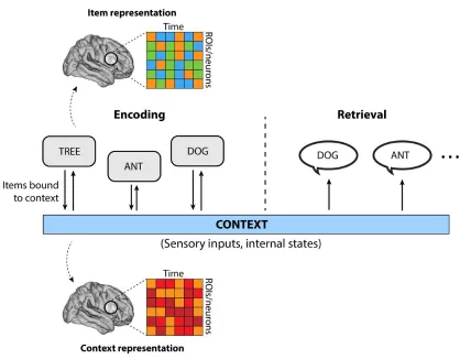
with a prevailing context, or the combined experience of ambient sensory inputs and internal mental states13.
6
asked to remember pairs of words, and are cued with only one word from each pair. In free recall paradigms, subjects are simply asked to recall as many items as possible, in the absence of any explicit cues. Implicitly, however, subjects use two cues. First, the prevailing context, such as sitting in the same testing room where the items were originally learned, serves to spur the retrieval of those memories. Second, memories act as the cues themselves
– recalling one prior word will tend to cue the recall of words experienced in close temporal proximity14, much as reminiscing with old about high school prompts the spontaneous
retrieval of childhood family memories (Figure 2.1).
The free recall paradigm is a particularly powerful tool to study human episodic memory. The method has high ecological validity; much of our everyday lives involve the
spontaneous retrieval of past experiences or knowledge, cued by nothing more than the current collection of sensory inputs and internal mental states. Relatedly, the retrieval period in free recall constitutes an endogenous cognitive search process, guided by the way in which knowledge is inherently organized in the brain. It is conceivable that cued recall paradigms force subjects to adopt particular strategies that hinge on the presentation of an arbitrary cue stimulus. Conversely, free-recall gains real-world validity at the expense of experimental control – subjects may adopt any number of strategies to support successful encoding or retrieval, potentially reducing the ability to detect statistically consistent effects across subjects. Furthermore, the absence of an explicit cue stimulus during retrieval
7
Figure 2.1. Processes of episodic memory encoding and retrieval. Episodic memory can be conceptualized as a linking of new items and the prevailing context. Context reflects the ongoing collection of sensory inputs and internal mental states that occur as new information (items) are encoded into memory. Drifting context forms the basis for later retrieval of past experiences. The cognitive neuroscience of memory seeks to understand the way in which items and context are neurally represented, and the underlying mechanisms that link the two.
8
How can free recall be used to understand the fundamental mechanisms that give rise to human episodic memory? How exactly are associations formed between items and their contexts, and how do we engage those associations to accurately recall prior experiences?
Figure 2.2. Verbal free recall task. In the verbal free recall task, subjects are instructed to remember 12-item lists of simple nouns, each presented successively on a computer screen. After a brief arithmetic distractor task, subjects are asked to freely recall as many words from the prior list as possible.
Recent theoretical and empirical work has combined notions of context-based episodic memory with insights from spatial navigation to conceptualize memory as an embedding of
information in a “cognitive space.”15 This space, like physical space, places items with strong
associations near each other, such as if two events occurred in a similar context. Items might also be closely associated – and therefore embedded closely in a cognitive space – via pre-existing knowledge like the semantic content of word items. Retrieval therefore
constitutes an “exploration” of cognitive spaces, akin to the free exploration of a physical
9
Intracranial EEG
Noninvasive neuroimaging, in particular functional magnetic resonance imaging (fMRI), has been extensively used to understand the brain activity that correlates with behavior and cognition8,16–18. However, fMRI’s poor temporal resolution limits its utility to tease apart
neural mechanisms that support rapid computations occurring on millisecond timescales. Indwelling electrodes placed directly in brain tissue can capture activity at this timescale, reflecting electric field changes induced by the aggregated activity of thousands of neurons near the electrode11,19. This local field potential (LFP) can be safely recorded in human
neurosurgical patients undergoing clinical monitoring for medication-resistant epilepsy. Patients are typically implanted with dozens or hundreds of such electrodes to help clinicians identify epileptogenic tissue for resection and eloquent areas to surgically avoid (Figure 2.3). During their hospital stay, patients are asked to participate in research studies and neural activity is recorded as subjects perform cognitive tasks (such as verbal free recall).
iEEG’s superior temporal resolution revealed that – as was known from animal studies –
human cognition is correlated with rhythmic fluctuations in the LFP, called oscillations20,21.
Neural oscillations have been observed in every part of the brain and occur at timescales ranging from 1 Hz to 100 Hz (and potentially beyond). The exact purpose and generation of neural oscillations is the subject of intense research, though current thinking suggests that the functional role of oscillations is differentiated by their rate. Low-frequency oscillations, including the theta (4-8 Hz), alpha (9-13 Hz), and beta (15-25 Hz) ranges, have been observed in diverse cortical areas as humans behave and perform cognitive tasks22–24.
Gamma frequencies, between 30 and 100 Hz, also correlate with cognition and behavior, but it is not clear to what extent iEEG recordings in this range reflect rhythmic oscillatory activity versus overall changes in the firing rate of neurons proximal to the recording electrode25–29.
10
decompose iEEG signals – a voltage measure – into component parts within particular frequency bands of interest. The exact decomposition method differs in accordance with the hypothesis at hand and experimental constraints, but common methods include the Hilbert transform, Morlet wavelet convolution, and multitapers. The subsequent frequency-filtered signals are usually next analyzed to extract spectral power, reflecting the overall energy contained in a given frequency. Filtered signals can also be analyzed for spectral phase, or the position (i.e. angle) of a sinusoidal wave at a given point in time. Together, the collection of power and phase measurements at a set of frequencies are the fundamental unit of analysis in any iEEG study.
Figure 2.3. Intracranial EEG. A. Surgical placement of a grid electrode in a patient with medication-resistant epilepsy. Grid electrodes are placed subdurally, allowing direct recording of electrical potentials from the cortical surface (electrocorticography; ECoG). B. Post-operative CT scan coregistered to pre-operative T1 MRI, highlighting the placement of a linear depth electrode in the right temporal lobe (red arrow). Depth electrodes enable the recording of LFPs from the medial temporal lobe. Image in (a) reproduced from https://mnepilepsy.org/services/ .
Despite excellent temporal resolution, iEEG is not without its pitfalls. Though the method also affords precise spatial resolution – electrodes are typically only a few millimeters in diameter – electrodes are only placed in the brain according to clinical considerations, not research agendas. Accordingly, placement differs drastically from patient-to-patient, and structures believed critical to a particular cognitive process may not be sampled at all. Moreover, it is only ethical to perform invasive brain surgery in people with a dire clinical need – usually medication-resistant epilepsy. Though it is likely that the general neural mechanisms supporting cognition and behavior are the same in epileptic brains as
11
exhibit highly pathologic activity even outside the seizure focus30–33. It remains an open
question as to how much these pathologies affect our measurements and judgments of human neural function.
Network Neuroscience
Since the early 20th century, neuroscience has sought to correlate brain structure and
function with the generation of behavior. The most straightforward way to do this is make a measurement of activity within the brain (e.g. scalp/intracranial EEG, microwire recordings, fMRI, calcium imaging, etc.), measure an interesting behavioral output, and ask whether the two are correlated. This approach has yielded key insights into brain function and
consequent Nobel prizes. Indeed, this approach largely defines the common understanding of neuroscience among the general public; a part of the brain lights up when a mouse or a person does something interesting.
But even since the earliest days of modern neuroscience, it has been clear that the brain does not function through the isolated activity of particular regions34. Rather, the brain is a
highly interconnected organ, with each neuron or chunk of cortex receiving thousands of axonal inputs from other places, and sending out thousands more. The brain’s inherent
structure suggests connectivity is a critical piece of the puzzle to understanding how collections of neurons generate movement, language, memory, thought, and emotion. Until recently, connectivity was largely the domain of anatomists studying white matter tracts in cadaveric brains. Starting in the 1980s and accelerating in the 1990s,
neuroscientist began to use new tools to uncover functional correlations in brain activity.
Instead of asking, “are these two brain regions physically connected?”, fMRI and intracranial recordings enabled us to ask, “do these two brain regions behave in concert?”16 The idea of
functional connectivity has evolved into one of neuroscience’s most prolific and exciting
12
Figure 2.4. Intracranial measures of functional connectivity. Intracranial function connectivity can be computed by several methods, including multitaper coherence. Coherence reflects the normalized cross-spectral density between iEEG recordings from disparate brain regions, at a given frequency. The coherence between all possible pairs of electrodes is given by an adjacency matrix (right), which represents a whole-brain network and can be subject to further graph-theoretic analysis.
Functional connectivity is essentially a measure of timeseries correlations. Whether these timeseries are fluctuations in the BOLD signal recorded through fMRI, or the theta-filtered signal from iEEG electrodes, we can ask whether up-and-down fluctuations of these signals co-occur (with or without lags) between distant brain regions (Figure 2.4). We now know that functional connectivity, like measures of local neural activity, is correlated with cognitive and behavioral events. The activity of the prefrontal cortex and hippocampus, for example, becomes correlated as human subjects engage in episodic memory tasks10,35.
Critically, it bears emphasizing that functional connectivity is not a measure of causal relations; inter-regional correlations do not imply that that one region directly influences another, or that information directly flows from one to the next36. Experimental
perturbations (see “Brain Stimulation”) remain the only gold-standard way to assess the causal role of brain activity in generating behavior.
Moving beyond pairwise correlations, more recent work in neural connectivity
conceptualizes the brain as a network of interconnected regions. In this framework, it is the coordinated activity among all regions of the brain that ultimately supports complex behaviors37,38. Neuroscientists have borrowed tools from the mathematical field of graph
13
connected to other nodes by edges. In real brains, edges reflect structural or functional
connections, depending on a researcher’s particular hypothesis or exploratory question.
Edges are often given weights in accordance with the magnitude of functional correlations or structural connections, though it is also possible to threshold weights and simply analyze a network of binary, connected-or-not network.
Figure 2.5. General network structure and the node strength statistic. Left: Schematic network with circles representing nodes and lines representing edges. The node strength is given by the sum of connection weights to a given node, indicated on the blue colored node.
It is now routine to use graph-theoretic measures to assess the higher-order structure of these functional/structural brain networks. Higher-order structure reflects the complex topology of brain networks – certain recurring patterns (or motifs) of nodes and edges have been observed in naturally-occurring networks, including the brain39. The node strength
statistic, for example, measures the sum total of all connection weights to a given node.
Nodes with high node strengths are called “hubs,” which in the brain may indicate regions
that strongly influence or orchestrate the activity of many others (Figure 2.5). Other statistics, such as the clustering and betweenness coefficients, capture more complex patterns of interconnectedness and serve as useful summaries of the role a particular node plays in a broader network. Taken together, graph-theoretic analysis of brain networks is a new and powerful tool for linking features of network topology with interesting cognitive or behavioral variables.
14
analyses presented here are fundamentally correlational; functional connectivity is measured as correlated changes in iEEG signals across space, and connectivity itself is further correlated with behavioral variables relating to episodic memory. However, the last study presented here leverages another innovation: the use of intracranial brain stimulation to assess the causal role of brain activity.
Brain Stimulation
Electrical brain stimulation is not a fundamentally new technique. In fact, neurologists and psychiatrists have been delivering electrical pulses to human brain tissue for almost 100 years, both as clinical and research endeavors40. These efforts have been instrumental to
understanding the causal role of certain brain structures in behavior; as early as 1937, it became apparent that particular areas of the cortex were responsible for speech or
sensation, which became altered upon electrical stimulation of the area41. Electroconvulsive
therapy, in which a current is passed through the brain noninvasively, has for over 50 years been used as a last-resort therapy for refractive psychiatric illness, particularly
depression42. More recently, deep brain stimulation (DBS) through indwelling electrodes
has been used to effectively treat Parkinson’s and related disorders43.
15
Figure 2.6. Example trace of an intracranial stimulation event. iEEG recording from a depth electrode before, during, and after a 500 ms square pulse stimulation event. Stimulation itself elicits a recording artifact (visible as high-amplitude spikes during the stimulation interval) and ensuing change in neural activity in the post-stimulation period.
Despite its relatively long history, the way in which electrical stimulation affects neural activity is largely unknown. Depending on parameters and methods, stimulation can enhance or decrease neural excitability and firing rate local to the targeted area, but it is unclear whether such modulations are facilitating ongoing neural processes or merely injecting noise40. Even more mysterious is the way in which stimulation events are
propagated through the brain via endogenous mechanisms. In monkeys, it has been shown that direct electrical stimulation propagates through known anatomical connections in the visual system44. In humans, stimulation events were also noted to move through structural
and functional connections, as measured via fMRI45,46. Beyond these basic studies, it is not
understood (1) precisely how ensembles of neurons react to applied currents, (2) how neurons transmit the perturbation to distant regions, and (3) how exogenous stimulation manifests as alterations in behavior or cognition.
16
stimulation can be used to enhance memory, while others suggest it decreases memory performance49–53. Notably, these studies span a wide range of stimulation amplitudes,
frequencies, anatomical targets, and task-related timing. Indeed, there is now a consensus that stimulation for therapeutic benefit must optimize (1) the time at which stimulation is delivered, relative to ongoing neural activity, and (2) the location of stimulation given a desired change in behavior. Unfortunately, solving this optimization problem requires a far more advanced understanding of (1) how the brain generates behavior, and (2) how stimulation alters neural activity.
17
CHAPTER 3:
CHAPTER 3:
Whole-Brain Electrical Networks
Solomon, E. A., et al. "Widespread theta synchrony and high-frequency desynchronization
underlies enhanced cognition." Nature communications 8.1 (2017): 1704.
Abstract
The idea that synchronous neural activity underlies cognition has driven an extensive body of research in human and animal neuroscience. Yet, insufficient data on intracranial
electrical connectivity has precluded a direct test of this hypothesis in a whole-brain setting. Through the lens of memory encoding and retrieval processes, we construct whole-brain connectivity maps of fast gamma (30-100 Hz) and slow theta (3-8 Hz) spectral neural activity, in a dataset of 294 neurosurgical patients fitted with indwelling electrodes. Here we report that gamma networks desynchronize and theta networks synchronize during encoding and retrieval. Further, for nearly all brain regions we studied, gamma power rises as that region desynchronizes with gamma activity elsewhere in the brain, establishing gamma as a largely asynchronous phenomenon. The abundant phenomenon of theta synchrony is positively correlated with a brain region’s gamma power, suggesting a
18
Introduction
The brain gives rise to behavior and thought through the coordinated activity and transfer of information between disparate regions5. Despite over a century of investigation into the
brain's interconnectedness34 however, the nature of these inter-regional interactions
remains unknown. Our understanding of connectivity in the brain originates from studies that use indirect measures of neural activity, like blood-oxygen-level dependent (BOLD) functional MRI, extracranial electroencephalography (EEG), and magnetoencephalography (MEG)54. While these techniques provide a useful picture of how distant brain regions act in
concert during cognition, they lack the spatial or temporal precision of direct electrical recordings in the brain37. Until recently, the limited availability of such intracranial data
made it difficult to assess the connectivity dynamics of the whole brain as it performs cognitive tasks.
Recent studies using direct brain recordings in neurosurgical patients have made it possible to robustly investigate neural synchronization, the coordinated activity of ensembles of neurons in different parts of the brain. Synchronization is an appealing mechanism for explaining how the brain stores memories, processes sensory inputs, or performs any operation that involves interlinking representations of the outside world54, and it generally
occurs on different timescales - or frequencies - of neural activity. In particular, gamma-band (30-100 Hz) synchronization is frequently invoked as a means for the brain to
communicate between regions, since the fast nature of an oscillatory gamma signal is timed appropriately for rapid perceptual operations or induction of synaptic
strengthening21,25,55,56. Support for this idea comes mostly from animal studies25,56–58, though
some human EEG studies also report cognitively induced low-gamma and short-range synchronicity59–61. However, others have argued that this body of work is conceptually and
empirically deficient to defend the broad notion that high-frequency activity supports a meaningful neural connection29,62–65. Notably, conduction delays between cortical areas
19
synchrony in humans report significant periods of desynchronization59,60,66 and steep
drop-offs in synchrony at higher frequencies61. These critiques raise the possibility that gamma
does not serve to support communication between cortical regions, though this hypothesis has not been directly tested.
If activity in the gamma range is not synchronous, it may instead reflect the aggregation of rapid, stochastic firing in a population of neurons near an electrode, not an oscillatory modulation of activity that indicates coordinated activity across space27,67. Were this true,
the general neural activation of a brain region – captured by the spectral power recorded at a cortical electrode – would rise as the synchronicity of that region with others would tend to fall. However, this form of broadband asynchronous activity may coexist with
narrowband synchronous oscillations,68,69 and both may contribute to spectral changes at
frequencies in the gamma band. In this case, it remains untested whether the oscillatory component of a gamma-band signal underlies long-range synchronization, and to what extent high-frequency activity during cognition reflects synchronous oscillations versus asynchronous broadband activity.
If high-frequency activity is not the principal mediator of inter-regional synchronization, low-frequency interactions may be a promising alternative. Synchrony in the slower theta-band (3-8 Hz) has been reliably found to correlate with cognition in humans and animals70–
73, and theta oscillations are also linked to modulations of gamma activity74,75. However,
low-frequency networks have not been characterized on a brain-wide scale, making it difficult to differentiate general principles of brain function from dynamics that may be particular to specific structures. It is possible that canonical regions such as the medial temporal lobe and prefrontal cortex participate in low-frequency networks while less well-studied regions break from this trend. Moreover, low-frequency interactions have not yet been directly related to modulations of spectral power on a brain-wide scale, though
probing these interactions may reveal the relationship between a region’s functional
connectivity and local processing.
In this study, our goal is to determine what principles underlie how neural activity is
20
and synchrony are related: To what extent is inter-regional communication mediated by low- versus high-frequency interactions? As the local high-frequency activity of a region increases, does its synchrony concomitantly decrease? How often do we observe high-frequency oscillations during cognition, and are they associated with long-range
connectivity? While 294 subjects perform memory encoding and retrieval tasks – processes which rely on the integration and binding of information – we record iEEG and construct whole-brain networks of high- and low-frequency phase interactions. To determine how synchrony changes over time and space, we parse these networks with graph-theoretic tools that identify hubs of the network, and then correlate the spatio-temporal pattern of synchrony at these hubs with simultaneously-measured spectral power. Though our focus is on gamma- and theta-band synchrony, we consider whether connectivity dynamics in these bands are better captured by broader frequency ranges, such as broadband low (< 30 Hz) and broadband high (> 30 Hz) . We observe widespread desynchronization of high-frequency activity and synchronized low-high-frequency activity during memory processes, which correlate with regions of enhanced high-frequency power. Our findings support the notion that macroelectrode-scale recordings largely reflect asynchronous neural firing at high frequencies, but also suggest a low-frequency mechanism for interregional
communication.
Results
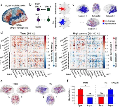
Quantification of brain-wide connectivity phenomena
To assess connectivity between brain regions, we collected intracranial
regularly-21
spaced frequencies in the 45-100 Hz range, referred to collectively as “high gamma,” though
we make no prior assumption as to whether these frequencies capture predominantly broadband asynchronous or oscillatory synchronous effects. Some analyses are extended to the 30-60 Hz range, referred to as “low gamma.”
We first sought to quantify the grand-average modulation in high gamma (HG) and theta connectivity during item encoding that correlates with subsequent successful recall of that item – in other words, the relative level of synchronization comparing successful to
unsuccessful encoding events. To measure this, we averaged the modulation in HG or theta connectivity across all possible electrode pairs that spanned every pair of anatomically-defined regions of interest (ROIs) in all subjects (ROIs are based on automated Talairach atlas labeling77, e.g. superior frontal gyrus, middle temporal gyrus, etc. See Methods for
22
Figure 3.1. Network construction and basic analysis.(a) 3D visualization of all surface electrodes
included in our dataset, colored by the Talairach atlas labels used in this article’s analysis. (b)
23
significantly greater than chance (P<0.01), and there was a significant frequency-synchrony
interaction (P<0.01, chi-square test). Dotted lines indicate mean chance level, shaded area +/- 1 STD. Encoding networks showed markedly different properties between HG- and theta-band frequencies. As measured by the summed connection weights across the entire network, HG asynchrony and theta-band synchrony significantly correlated with successful encoding (Figure 3.1f; P < 0.01 via permutation test of summed connection weights; see Methods). And though the network-wide level of synchronous activity in HG was not significant (permutation P = 0.892), this does not preclude the possibility that specific connections among ROIs are associated with successful memory encoding. Similarly, the overall level of theta-asynchronous interactions was not greater than chance (permutation P >0.99). Extending this analysis to higher and lower frequencies revealed significant asynchrony (Figure 3.2a; permutation P < 0.05) in frequencies between 30 Hz and 120 Hz, including the typical 30-60 Hz low gamma band. Significant synchrony in frequencies between 3 Hz and 28 Hz – theta, alpha, and beta bands – was also observed (permutation P < 0.01). The brainwide connectivity z-score is given as a heatmap for each assessed frequency and timepoint in Figure 2b.
24
connection weights in the network, compared to the sum expected by chance. This analysis is performed in successive 200ms windows spanning the encoding interval. Red reflects increased synchrony associated with successful memory encoding, blue reflects decreased synchrony (see Methods for details). Vertical black lines indicate word onset and offset.
Our findings of brain-wide HG asynchrony and theta synchrony during successful memory encoding suggest that it is low-frequency connections which support information
integration or coordinated brain activity during memory formation. However, we must first answer two deeper questions to determine whether there is a relationship between the neural activity of a region and the state of its connections to the rest of the brain: First, what is the brain-wide spatiotemporal pattern of synchrony/asynchrony during memory
processing, and how does it relate to the pattern of local spectral power? Second, are there differing fundamental sources of neural activity that may have different power-synchrony relationships?
Identification of network hubs
Given that we observed significant levels of synchrony or asynchrony in low and high frequency ranges, we next asked whether there is anatomic specificity to these phenomena. Are positive and negative connections homogenously distributed throughout the brain, or are there specific regions that exhibit greater modulation of connectivity during successful memory encoding?
To determine the most highly-connected (or highly-disconnected) ROIs, we turned to basic principles of graph theory. We used the node strength statistic (the sum of the
unthresholded weights of every connection to a given node, here defined as an ROI) to identify which brain regions act as highly-connected "hubs" in the memory network during the word presentation interval (0-1600 ms), the epoch with the greatest task-related modulation78. We defined hubs as ROIs with significantly greater node strength than
25
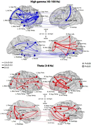
significantly increase or decrease their overall connectivity when a word is successfully encoded (0.006 < P < 0.033, FDR corrected). The theta-network exhibits 32 synchronous hubs widely dispersed across the cortex (0.005 < P < 0.049, FDR corrected), but no hubs of asynchronous activity. Theta and HG hubs are depicted in Figure 3.3, along with their strongest connections (Z > 2.5).
Taken together, these findings demonstrate that frontal, temporal, and medial temporal lobe (MTL) cortical regions became desynchronized from each other in HG during memory encoding. A smaller subset of right mesial frontal regions expressed synchronous activity with each other and functionally connect to temporal and parietal cortex. In the slower theta rhythm, the brain exhibited generally correlated activity, with numerous fronto-temporal, temporal-parietal, and interhemispheric functional connections.
Our finding that there is widespread theta-synchronization during memory encoding follows from prior scalp and intracranial studies, which have shown that low-frequency entrainment is associated with cognition70–73,79. These findings also mirror findings in the
fMRI literature of low-frequency networks that converge on the MTL in memory tasks10.
26
27
Dashed lines indicate cross-hemispheric connections. Some labels are excluded from certain views to maintain readability. To aid visualization, hemispheres are reflected from their true position in the skull.
Temporal modulation of connectivity effects
To better characterize the role of these hubs in memory encoding, we asked whether a hub's participation in the HG or theta network changes over time. We assessed this by computing the node strength statistic at each 200 ms non-overlapping time window spanning 200 ms prior to 200 ms after the word presentation interval (see Methods). ROIs exhibited their strongest modulation of network participation between 400 ms and 1200 ms after onset of a word, with a particularly robust decrease in HG connectivity of left MTL structures between 800-1000 ms (significant hippocampus, parahippocampus, and uncus ROIs, P < 0.05 via permutation test of node strengths; see Methods for details).
Correspondingly, the right MTL exhibited an increase in theta synchrony between 800-1200 ms (permutation P < 0.05). Theta synchrony in the right frontal lobe (significant middle, medial, inferior, and superior frontal cortices, permutation P < 0.05) peaked earlier, between 600-800 ms, while right temporal (significant middle, transverse, superior, and inferior temporal cortices, permutation P < 0.05) synchrony peaked between 1000-1200 ms (left cortical areas follow a similar pattern, see Figure 3.S2). In Figure 3.4, we show
timecourses of node strength for ROIs in a subset of broader brain regions that contained hubs as identified previously (see Figure 3.S2 for additional timecourses).
It is not surprising that we observed strong modulation of connectivity in both frequency bands during the item presentation interval, since this time period is also known to feature the greatest change in spectral power78. What is not known, however, is how the
28
Figure 3.4. Timecourse of ROI participation in memory networks. Node strength as a function of time for 6 key regions that contain hubs in the theta or high gamma networks: right and left MTL, frontal lobe, and temporal lobe. Blue-shaded lines indicate asynchronous hub strength over time, while red indicates synchronous hub strength. Vertical lines
29
Relationship between connectivity and spectral power
Having established the spatio-temporal dynamics of synchrony during performance of a memory task – noting the presence of MTL hubs that peak in their activity during the item presentation interval, for instance – we are now equipped to ask how these connectivity dynamics relate to spectral power, or the general neural activation of a region. Answering this question fills an important gap in knowledge about the nature of connectivity in the brain, by showing how connectivity and power relate across a diverse array of cortical regions during memory processing.
We used the node strength of each ROI as a basis for a spectral power-synchrony correlation, asking whether a region's overall participation in the whole-brain network correlates with that region's modulation of spectral power. For each ROI, we computed the power-synchrony (node strength) correlation across time and frequency in HG. We further asked how power and synchrony correlate across all ROIs and time after averaging effects within frequency band, enabling cross-band correlations.
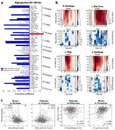
We found that only one ROI exhibited a significant positive correlation between HG power and synchrony – the left transverse temporal gyrus - after Benjamini-Hochberg correction for multiple comparisons (Figure 3.5a; Pearson correlation, r = 0.27, corrected P = 0.017). Twenty-four regions exhibited a significant negative correlation (Figure 3.5a; Pearson correlation, -0.48 < r < -0.23, 4.6 × 10⁻⁶ < P < 0.037). Example power-synchrony heatmaps
are given for four regions in Figure 3.5b, depicting significant (corrected P < 0.05) negative correlations in the right parahippocampus, left medial temporal lobe, and left frontal cortex. Across all ROIs (74) and timepoints (10) together, the HG power-synchrony Pearson
correlation was -0.339 , P = 0.002 via a permutation test of synchrony and power
correlation (Figure 3.5c; see Methods for details). In theta, within-ROI correlations showed 3 ROIs each of positive and negative power-synchrony relationships (corrected P < 0.05; Figure 3.S3), but the general effect across all time and ROIs together was negative though not significant (Pearson correlation, r = -0.12, permutation P = 0.23; Figure 3.5c).
30
(Pearson correlation, r = 0.11, permutation P = 0.2; Figure 3.5c). The brainwide spectral power and synchrony at all frequencies from 3 Hz to 120 Hz are shown in Figure 3.S4. Measuring correlations across all ROIs together may obscure meaningful relationships within the subset of ROIs that actively participate in memory processing. We therefore sought to assess whether regions of the "core" memory network – those ROIs that significantly modulate their neural activity during successful memory encoding – exhibit power-synchrony dynamics that are different from the rest of the brain. These regions are said to exhibit a subsequent memory effect (SME)80. We found a total of 37 ROIs with no
significant difference between HG power during successful versus unsuccessful encoding, and classified these as outside the core memory network. Next, we matched these ROIs against the 37 ROIs with the largest SMEs, representing the core memory network (see Table 3.T1 for ROI classifications and z-scores). Among these two ROI subsets, we again computed power-synchrony correlations across all regions and all timepoints during the word encoding interval. In both groups, HG power and synchrony were inversely correlated (Figure 3.6; Pearson correlation, r = -0.38 in-network and r = -0.158 out-of-network, P < 0.001 and P < 0.05 via permutation test; see Methods). However, only in the core memory network was theta synchrony significantly predictive of HG power (Pearson correlation, r = 0.25, permutation P = 0.003; see Methods). The difference in correlation between
31
32
within frequency band. HG power and HG synchrony are significantly inversely related (P < 0.001, permutation test), HG synchrony is positively correlated with theta power (P = 0.005), while other tested relationships do not meet significance.
Figure 3.6. Correlations in core memory network. (a) Power-synchrony correlations in the core memory network: the 37 ROIs with significant HG-power subsequent memory effect (SME). (b) Power-synchrony correlations across the 37 ROIs with the no significant HG-power effects. Among the core memory network – consisting mostly of left frontal, temporal, and MTL cortex – z-scored gamma power and z-scored synchrony were significantly anticorrelated, while theta synchrony and gamma power were significantly positively correlated (top row; P < 0.001 and P < 0.01 via
permutation test, respectively). Among regions that did not exhibit strongly modulated HG activity in successful memory encoding, HG power and synchrony were still inversely correlated (Pearson correlation, r = -0.158, P < 0.05), but theta synchrony was not significantly predictive of HG power (r
33
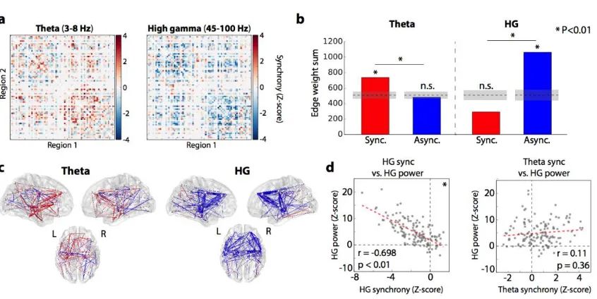
Generalization of network phenomena to memory retrieval
To establish whether memory retrieval is also characterized by desynchronized HG activity and synchronized theta-band activity, we identified all of the 500 ms time windows in each
subject’s recall period that precede onset of a response vocalization, and compared
connectivity dynamics against 500 ms time windows that are not followed by any vocalization for at least 2 seconds ("unsuccessful memory search"). Procedures are
otherwise identical to those described in Figure 3.1 and Methods – phase locking values in successful retrieval are compared to unsuccessful memory search, and these differences are pooled across subjects and ROIs. The result is a whole-brain connectivity map that reflects how phase synchrony is correlated with successful memory retrieval versus unsuccessful memory search (Figure 3.7a, 3.7c).
We found that the same network-level patterns of connectivity held true in the retrieval contrast compared to the encoding contrast. The HG retrieval network is characterized by a significant degree of asynchronous activity (Figure 3.7b; P < 0.01 via permutation test of edge weight sum; see Methods) and an insignificant overall level of synchronous activity (permutation P > 0.99). In theta-band, there is a greater degree of synchronous activity compared to asynchronous (permutation P < 0.01; Figure 3.7b). The relationship between power and synchrony also holds true in the analysis of recall. Even without sub-selecting for a core memory network as in Figure 6, we find an inverse HG power-synchrony correlation (Pearson correlation, r = -0.67, permutation P < 0.01; Figure 3.7d), although theta
34
Figure 3.7. Generalization to memory retrieval processes. (a) Adjacency matrices, reflecting relative recall vs. baseline synchronization, organized as in Figure 3.1d. (b) Summed positive and negative connection weights in each network, showing a strong desynchronization effect in gamma-band and a synchronization in theta (P < 0.01 for both). There was a significant frequency-synchrony interaction (P < 0.01, chi-square test). (c) 3D representation of gamma (top) and theta (bottom) retrieval networks, organized as in Figure 3.1e. (d) Correlation of spectral power and phase synchronization across all regions (74) and timepoints spanning a retrieval trial (2). HG power and synchrony were significantly inversely correlated (Pearson correlation, r = -0.698, P < 0.01 via permutation test), while an ROI's theta synchrony was positively but not significantly predictive of HG power (r = 0.11, P = 0.36)
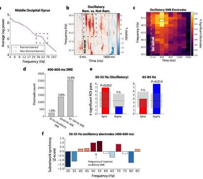
Filtering for oscillatory activity
Findings of HG desynchronization associated with successful memory encoding and retrieval suggest stochastic, non-oscillatory neural activity. However, it is possible that a mixture of two fundamental signals that occupy the same frequency band: some
components may be oscillatory, facilitating inter-regional communication, while others reflect asynchronous neural spiking activity. If the asynchronous component is much stronger or more commonplace than the oscillatory component, our results may be unable to capture true high-frequency synchronization that correlates with successful memory operations.
To answer whether high-frequency synchronization is driven by oscillatory dynamics, we
35
utilizing a validated oscillation-detection routine (“Better Oscillation Detection” method, see
Methods for details; see Figure 8a for an example)81. We identified the specific frequency
and time at which a given electrode showed reliably increased oscillatory activity associated with trials that were later remembered, as compared to those forgotten
(“oscillatory SME”; Fig. 3.8b for an example). Among the subset of electrodes with oscillatory SMEs, we reconstructed our phase-synchronization networks to determine whether enhanced oscillatory activity was associated with increased inter-regional synchronization.
261 electrodes in our dataset exhibited increased oscillatory activity associated with successful memory, maximally occurring at 52 Hz, between 400 and 600 ms after word onset (Fig. 3.8c). This is 1% of the total electrodes assessed; 5.6% of electrodes exhibited a 30-60 Hz SME and 10.4% of electrodes that exhibited a 65-100 Hz power SME in the same time window (Figure 3.8d). In the phase synchronization subnetwork that can be
36
Figure 3.8. Synchronization of low gamma (30-60 Hz) oscillatory activity. (a) Example of an electrode exhibiting a gamma oscillation in the middle occipital gyrus, as detected by BOSC (see Methods for details). Red line reflects average log power across all remembered events, blue line reflects average log power across not-remembered events. An isolated peak in the power spectrum is indicated, between approximately 36 and 74 Hz. (b) For the electrode in (a), heatmap of the t-statistic reflecting the relative frequency of oscillations detected in remembered versus
37
the chance mean and standard deviation at this significance level indicated in the gray shaded area. Left: Counts observed at 50-55 Hz, near the frequency of maximal oscillatory SMEs (52 Hz). Right: Counts observed in the 65-85 Hz range among the same electrode subset. The frequency/synchrony interaction is not significant (P = 0.11). (f) Average network synchrony (z-score) for the subnetwork of regions sampled in the 52 Hz oscillatory electrode subset, measured by summing the subnetwork connection weights at each frequency in the 400-600 ms window, and comparing to the sum expected by chance.
Discussion
We set out to uncover fundamental principles that govern the electrophysiological networks of activity in the human brain. As 294 subjects performed a verbal free-recall memory task, we analyzed three frequency bands that have been strongly implicated in neural synchronization24: theta (3-8 Hz), low gamma (30-60 Hz), and high gamma (45-100
Hz). Gamma networks exhibited strong desynchronizations between brain regions,
especially those that saw an increase in gamma power. Theta networks were characterized by enhanced synchrony, especially among regions with strong increases in HG power. Moreover, hubs of theta network activity tend to localize in frontal, temporal, and medial temporal cortices – regions that are known to play a strong role in memory encoding and retrieval82.
Here we report findings that address whether theta or gamma band neural activity drives synchronization during memory processing. Gamma activity as a general biological mechanism of information transmission25,54,55,70 is not backed by many compelling
observations in the human brain. We found a profound decrease in HG synchronization that is associated with successful memory encoding and retrieval, especially among regions that see heightened overall HG activation. This relation is consistent with the hypothesis that broadband high-frequency activity in the human brain – as detected by macroelectrodes on the cortical surface – largely reflects the aggregation of fast, stochastic spiking activity of a population of neurons29. It refutes the notion that this kind of broadband signal
38
Far from suggesting that brain regions are cut off from their neighbors, the observation that highly-active memory regions significantly increase their theta synchronization offers a low-frequency mechanism by which the brain coordinates its many parts – a brain-wide finding that was suggested by prior studies which could only examine specific
interactions73,84. Furthermore, our results demonstrate how theta networks exhibit
time-varying structure, highlighting frontotemporal hubs that strengthen their connections starting 500 ms after onset of an item to be remembered. The fMRI connectivity literature parallels this, demonstrating broad, low-frequency networks that act to support human memory by convergence on the MTL7,10,85,86. The extent to which whole-brain iEEG-based
networks overlap with fMRI networks is unexplored territory.
A small subset of electrodes exhibited increased narrowband gamma-oscillatory power associated with successful memory encoding. We observed increased long-range phase synchronization among this subset at the frequency of maximal oscillatory activity. This indicates that, in some instances, gamma-band activity is organized into coherent, oscillatory waves that may serve to coordinate activity between different regions. The rareness of this phenomenon should not be understated; statistically reliable oscillatory SMEs were detected in only 1% of electrodes in our 294-subject dataset.
A prior study by Burke, et al. in 2013 also suggested a general decrease in gamma
synchronization and increase in theta during memory encoding in humans, but only at the level of lobe-wise interactions84. The findings presented here extend that work in several
important ways. First, we establish that decreases in synchrony accompany increases in high-frequency power, and that this fundamental relationship between power and synchrony manifests itself throughout the human brain. Second, we examined synchrony dynamics at a much finer spatial scale, allowing for the possibility that aggregation by lobe obscured synchronous gamma activity between nearby regions. Third, we teased out oscillatory effects in gamma, demonstrating that while synchrony is observed in rare