Fabry disease: Identification of 50
novel
a
-galactosidase A mutations
causing the classic phenotype and
three-dimensional structural analysis
of 29 missense mutations
Junaid Shabbeer,
1Makiko Yasuda,
1Stacy D. Benson
2and Robert J. Desnick
1*
1Department of Human Genetics, Mount Sinai School of Medicine of New York University, New York, NY 10029, USA 2Department of Chemistry, Oklahoma State University, Stillwater, OK 74078, USA
*Correspondence to: Tel:þ1 212 659 6700; Fax:þ1 212 360 1809; E-mail: [email protected]
Date received (in revised form): 16th December 2005
Abstract
Fabry disease, an X-linked recessive inborn error of glycosphingolipid catabolism, results from the deficient activity of the lysosomal exoglycohydrolase,a-galactosidase A (EC 3.2.1.22;a-Gal A). The molecular lesions in thea-Gal Agene causing the classic phenotype of Fabry disease in 66 unrelated families were determined. In 49 families, 50 new mutations were identified, including: 29 missense mutations (N34K, T41I, D93V, R112S, L166G, G171D, M187T, S201Y, S201F, D234E, W236R, D264Y, M267R, V269M, G271S, G271V, S276G, Q283P, A285P, A285D, M290I, P293T, Q312H, Q321R, G328V, E338K, A348P, E358A, Q386P); nine nonsense mutations (C56X, E79X, K127X, Y151X, Y173X, L177X, W262X, Q306X, E338X); five splicing defects (IVS4-1G.A, IVS5-2A.G, IVS5þ3A.G, IVS5þ4A.G, IVS6-1G.C); four small deletions (18delA, 457delGAC, 567delG, 1096delACCAT); one small insertion (996insC); one 3.1 kilobase Alu-Alu deletion (which included exon 2); and one complex mutation (K374R, 1124delGAG). In 18 families, 17 previously reported mutations were identified, with R112C occurring in two families. In two classically affected families, affected males were identified with two mutations: one with two novel mutations, D264Y and V269M and the other with one novel (Q312H) and one previously reported (A143T) mutation. Transient expression of the individual mutations revealed that D264Y and Q312H were localised in the endoplasmic reticulum and had no detectable or markedly reduced activity, whereas V269M and A143T were localised in lysosomes and had approximately 10 per cent and approximately 35 per cent of expressed wild-type activity, respectively. Structural analyses based on the enzyme’s three-dimensional structure predicted the effect of the 29 novel missense mutations on the mutant glycoprotein’s structure. Of note, three novel mutations (approximately 10 per cent) were predicted not to significantly alter the glycoprotein’s structure; however, they were disease causing. These studies further define the molecular heterogeneity of thea-Gal Amutations in classical Fabry disease, permit precise heterozygote detection and prenatal diagnosis, and provide insights into the structural alterations of the mutant enzymes that cause the classic phenotype.
Keywords:Fabry disease,a-galactosidase A,a-Gal A, mutations, molecular modelling, transient expression
Introduction
Fabry disease is an X-linked recessive inborn error of glyco-sphingolipid catabolism resulting from the deficient activity of the lysosomal exoglycohydrolase,a-galactosidase A (EC 3.2.1.22;a-Gal A).1 The enzymatic defect causes the pro-gressive accumulation of globotriaosylceramide (GL-3) and related glycosphingolipids with terminala-linked galactosyl moieties in the plasma and in tissue lysosomes throughout the body. In classically affected males who have little, if any,
with cardiac and/or renal disease and lack the major classical manifestations, including angiokeratoma, acroparesthesias, hypohidrosis and ocular abnormalities.2 – 5
To date, a variety of mutations have been identified which cause the classic phenotype, including missense, nonsense and splice-site mutations, as well as partial gene rearrangements, including small and large intragenic deletions and insertions1,6,7 (the Human Gene Mutation Database). Most mutations have been private (ie unique to one or a few families), with the exception of certain mutations found in unrelated individuals that occurred at CpG dinucleotides, known hot spots for mutation.8,9Of note, non-coding sequence variants have been identified ina-Gal Aalleles from normal individuals and patients with Fabry disease10,11(the SNP database), with the exception ofD313Y, an exonic sequence variant that encodes an enzyme with 60–70 per cent of wild-type activity, but does not cause disease in males or females with this variant.12,13
Mutation detection in Fabry disease is important for several reasons. First, heterozygote detection by enzyme assay of carriers for this X-linked recessive disease is unreliable because obligate carriers can have normal activity due to random X-chromosomal inactivation.14 – 16Secondly, safe and effective treatment for Fabry disease bya-Gal A replacement therapy has recently become available worldwide.17 – 20Because affected males should be treated early to prevent the serious complications of the disease, and because carriers may be asymptomatic or have very mild manifestations,1,21,22 it is important to identify affected males and carriers for medical monitoring and early treatment.23Thirdly, recentin vitro studies24have demonstrated that certaina-Gal A missense mutations result in mutant proteins that are misfolded and degraded in the endoplasmic reticulum (ER). Some of these misfolded mutant proteins, particularly those that have residual enzymatic activity (.1 per cent), have been rescued by pharmaceutical chaperones, such as galactose and deoxyga-lactonojirimycin, which are reversible competitive inhibitors ofa-Gal A.25 – 28In fact, intravenous administration of galactose (1 g/kg) to a 56-year-old male with the cardiac variant phenotype resulted in marked improvement of his cardiac manifestations.29Thus, it is of interest to predict which of the missense mutations that cause the classic Fabry pheno-type might be rescuable based on the enzyme’s 3D struc-ture.30,31These mutations can then be studiedin vitroto determine their rescuability.
In this paper, 50 new and 17 previously reporteda-Gal A mutations were identified in 66 unrelated classically affected families. Two unrelated patients with the classic phenotype had mutant alleles with two missense mutations, each of which was expressedin vitro. In addition, the structural alterations resulting from the 29 novel missense mutations were predicted based on the 3D structure of the wild-type enzyme homodi-meric glycoprotein. Several of these mutations resulted from misfolding and are candidates to determine their rescuability by pharmacological chaperones.
Materials and methods
Patient specimens
Peripheral blood was collected from the probands of 66 unrelated families with the classic phenotype of Fabry disease. Thea-Gal A activity was determined in the plasma and/or lymphocytes as previously described.32Genomic DNA was extracted using the Puregene isolation kit according to manufacturer’s instructions (Gentra Systems, Minneapolis, MN, USA). All specimens were obtained with informed consent and the approval of the Institutional Review Board of the Mount Sinai School of Medicine of New York University.
Mutation analysis
Mutation analysis was performed as previously described.33 Briefly, each of thea-Gal Aexons and adjacent flanking and/ or intronic sequences was amplified by means of polymerase chain reaction (PCR) from genomic DNA. Each amplicon was then analysed by denaturing high-performance liquid chromatography, and abnormally running fragments were sequenced using an ABI Prism 3700 Capillary Array
Sequencer with the ABI PrismeBigDyeeTerminator Ready Reaction Mix (Perkin-Elmer-Cetus, Norwalk, CT, USA). Each mutation was confirmed by repeat PCR amplification and sequencing of the opposite strand, and/or by co-segre-gation of the lesion and disease phenotype in other members of each family. In addition, 100 normal chromosomes were examined to rule out a polymorphism for each missense mutation.
Southern blot analysis
Genomic DNA (10mg), extracted from normal controls and the proband, was digested with 100 units (U) of HindIII and PvuII for 16 hours. After electrophoresis on a 1 per cent agarose gel, DNA was transferred onto a Hybond-Nþ membrane (Amersham GE Healthcare, Piscataway, NJ, USA) in 0.4M NaOH solution using standard procedures. The filter was ultraviolet-irradiated and then hybridised with a randomly primed32P-labelled PCR-generated probe spanning exons 1 and 2 of thea-Gal Agene. After overnight hybridisation using PerfectHyb Plus hybridisation buffer (Sigma Aldrich), membranes were washed in diluted standard saline citrate buffers with 0.1 per cent sodium dodecyl sulphate. Bands were visualised after exposure and analysed on a Molecular Dynamics STORM 860 phosphoimager (GE Healthcare).
Microsatellite studies
DXS8100, DXS8063 and DXS8096. Forward primers were fluorescent dye-labelled (Invitrogen Life Technologies, Carlsbad, CA, USA). Genomic DNA was amplified in 10ml volumes with 50 ng of genomic DNA, 2 mM MgCl2, 10 mM Tris-HCl, pH 8.3, 50 mM KCl, 200 nM of each primer, 0.2 mM deoxyribonucleoside triphosphates and 2 UTaqDNA polymerase (AmpliTaq Gold, Applied Biosystems, Foster City, CA, USA). The reaction mixtures were initially incubated at 958C for 10 minutes, and then underwent 27 cycles of amplifications with denaturation at 948C for 30 seconds, annealing at 588C for 30 seconds, extension at 728C for 30 seconds and a final extension step at 728C for 7 minutes. Microsatellites were analysed with an ABI Prism 3100 Genetic Analyzer using GeneScan Analysis Software (Version 3.1.2) and Genotyper Software (Version 2.5) (Perkin-Elmer-Cetus, Norwalk, CT, USA).
Conservation of missense mutations
Each of the missense lesions was analysed to determine the relative conservation of the substituted amino acid by com-parison with four mammalian and 22 non-mammalian eukaryotica-Gal A orthologues and eighta -N-acetylgalac-tosaminidase (a-Gal B) orthologues in the GenBank database. These searches were performed using the MacVector program (Oxford Molecular Group). Highly conserved residues were defined as those that were present in three of the four (75 per cent) mammalian orthologues in at least 17 (77 per cent) of the eukaryotic orthologues and in six (75 per cent) of the eukaryotic orthologues of the relateda-Gal Bgene.34
In vitro
expression studies
The full-length wild-typea-Gal AcDNA was cloned into the pAsc8 vector.35Mutant constructs, carrying the individual D264Y, V269M, Q312H or A143T mutations or the double mutations (D264Y/V269M and A143T/Q312H), were gen-erated by site-directed mutagenesis PCR (Stratagene, La Jolla, CA, USA) using the primers: GGACCAGGGGGTTG-GAATT ACCCAGATATGTTAGTG (D264Y sense), CAC-TAACATATCTGGGTAATTCCAACCC CCTGGTCC (D264Y antisense), GACCCAGATATGTTAATGATTGGC-AACTTTGG (V269M sense), CCAAAGTTGCCA-ATCATTAACATATCTGGGTC (V269M antisense), GCTGGAAATAAAACCTGCACAGGCTTCCCTGGGAG (A143T sense), CTCCCAGGGA AGCCTGTGCAGGTTT-TATTTCCAGC (A143T antisense), CAAGCCAAAGCTC-TCCTTC ATGATAAGGACGTAACTGCCA (Q312H sense) and TGGCAGTTACGTCCTTATCATG-AAGGAGAGCTTTGGCTTG (Q312H antisense).
All constructs were confirmed by re-sequencing and plasmid preparations were made using the Qiagen plasmid midi kit (Valencia, CA, USA). The wild-type and each mutant construct were individually transfected into COS-7 cells and analysed for intracellulara-Gal A activity and subcellular localisation, as previously described.13
Structural analysis of
a
-Gal A missense
mutations
The 3.25 A˚ X-ray structure of humana-Gal A30was the basis of the structural analysis. Side-chain positions were compared with an independently derived human a-Gal A model based on the chickena-Gal B X-ray structure.36Mutations were modelled and visualised in the program O37and energy minimised with the CNS program.38
Results
Mutation detection
Table 1 summarises the 50 novel and 17 previously reported mutations detected in 66 unrelated patients with classic Fabry disease. PCR amplification of thea-Gal Aexons and adjacent intronic or flanking sequences from genomic DNA, and electrophoresis of the amplicons, did not reveal any gene rearrangements (.50 base pairs [bp]), except one in which exon 2 did not amplify. Southern blot analysis indicated a large deletion of,3 kilobases (kb), which included exon 2. To determine the precise breakpoints of the deletion, several random primer pairs in introns 1 and 2 were used to amplify the proband’s genomic DNA. Using a sense primer (GCTA-ATGGCAAGACCCTG) located at g2765 in intron 1 and an antisense primer (AAATCCCCCAGTTCTGCTGAGCTA) at g7218 in intron 2, an approximately 4.5 kb PCR fragment was expected; however, these primers amplified a 1.3 kb fragment. Sequencing of this PCR fragment identified the breakpoints in Alu repetitive sequences at g3260 and at g6410, resulting in an Alu–Alu rearrangement that deleted 3,152 bp, including the entire exon 2 (Figure 1).
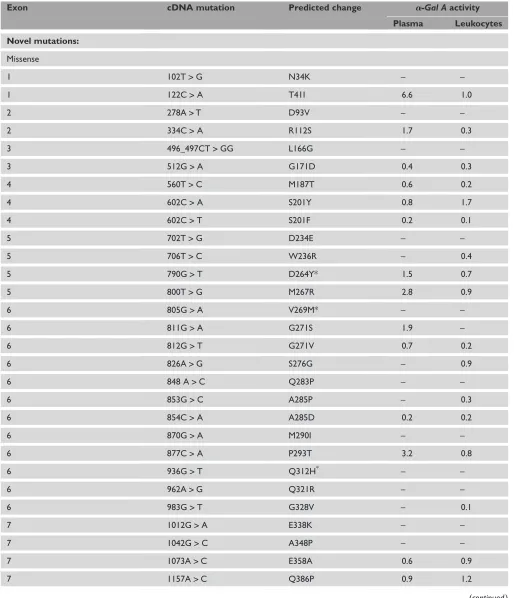
Table 1. a-Gal A mutations in 66 unrelated probands with classic Fabry disease.
Exon cDNA mutation Predicted change a-Gal Aactivity
Plasma Leukocytes Novel mutations:
Missense
1 102T>G N34K – –
1 122C>A T41I 6.6 1.0
2 278A>T D93V – –
2 334C>A R112S 1.7 0.3
3 496_497CT>GG L166G – –
3 512G>A G171D 0.4 0.3
4 560T>C M187T 0.6 0.2
4 602C>A S201Y 0.8 1.7
4 602C>T S201F 0.2 0.1
5 702T>G D234E – –
5 706T>C W236R – 0.4
5 790G>T D264Y* 1.5 0.7
5 800T>G M267R 2.8 0.9
6 805G>A V269M* – –
6 811G>A G271S 1.9 –
6 812G>T G271V 0.7 0.2
6 826A>G S276G – 0.9
6 848 A>C Q283P – –
6 853G>C A285P – 0.3
6 854C>A A285D 0.2 0.2
6 870G>A M290I – –
6 877C>A P293T 3.2 0.8
6 936G>T Q312H* – –
6 962A>G Q321R – –
6 983G>T G328V – 0.1
7 1012G>A E338K – –
7 1042G>C A348P – –
7 1073A>C E358A 0.6 0.9
7 1157A>C Q386P 0.9 1.2
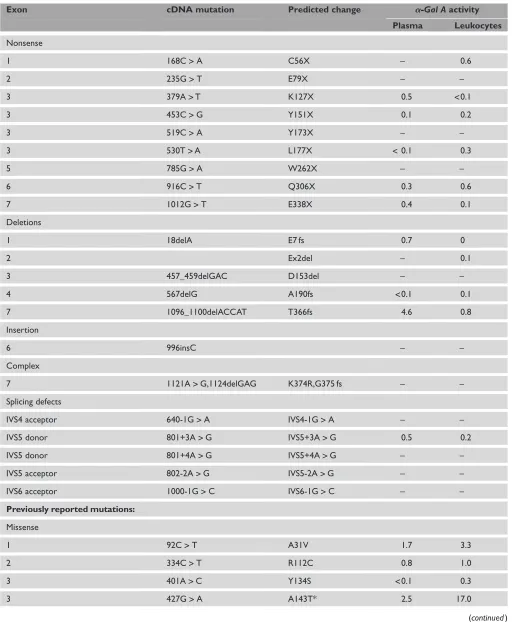
Table 1.Continued.
Exon cDNA mutation Predicted change a-Gal Aactivity
Plasma Leukocytes
Nonsense
1 168C>A C56X – 0.6
2 235G>T E79X – –
3 379A>T K127X 0.5 <0.1
3 453C>G Y151X 0.1 0.2
3 519C>A Y173X – –
3 530T>A L177X <0.1 0.3
5 785G>A W262X – –
6 916C>T Q306X 0.3 0.6
7 1012G>T E338X 0.4 0.1
Deletions
1 18delA E7 fs 0.7 0
2 Ex2del – 0.1
3 457_459delGAC D153del – –
4 567delG A190fs <0.1 0.1
7 1096_1100delACCAT T366fs 4.6 0.8
Insertion
6 996insC – –
Complex
7 1121A>G,1124delGAG K374R,G375 fs – –
Splicing defects
IVS4 acceptor 640-1G>A IVS4-1G>A – –
IVS5 donor 801+3A>G IVS5+3A>G 0.5 0.2
IVS5 donor 801+4A>G IVS5+4A>G – –
IVS5 acceptor 802-2A>G IVS5-2A>G – –
IVS6 acceptor 1000-1G>C IVS6-1G>C – –
Previously reported mutations:
Missense
1 92C>T A31V 1.7 3.3
2 334C>T R112C 0.8 1.0
3 401A>C Y134S <0.1 0.3
3 427G>A A143T* 2.5 17.0
The previously reported lesions included seven missense mutations (A31V, R112C, Y134S, A143T, P259R, G328A and L414S), six nonsense mutations (Y152X, Q157X, Q221X, W226X, R227X and R342X) and four small del-etions (26delA, 1031delTC, 1209delAAG and 1235delCT). The R112C substitution was identified in two probands that were found to be unrelated from microsatellite studies. Of note, the R112S, R112C, Y152X, R227X and R342X mutations occurred at CpG dinucleotides, known mutational hot spots.9 Although previously reported sequence variants were detected ina-Gal Aalleles from normal individuals and patients with Fabry disease10 – 13(the SNP Database), no new non-pathological sequence variants were detected in this study.
Expression and subcellular location of the
double missense mutations
To determine the functional effects of each substitution in the two affected males whosea-Gal Aalleles had two missense lesions, expression studies in COS-7 cells were carried out (Table 2). TheV269Mallele had approximately 10 per cent of the mean expressed wild-type activity and the mutant enzyme protein was localised immunohistologically to the lysosomes, whereas theD264Yallele had no detectable activity and the mutant protein remained in the ER. TheA143Tallele had approximately 35 per cent of expressed wild-type activity and localised to the lysosome, whereas theQ312Hallele had approximately 5 per cent of expressed wild-type activity and was detected predominantly in the ER. Constructs with the double
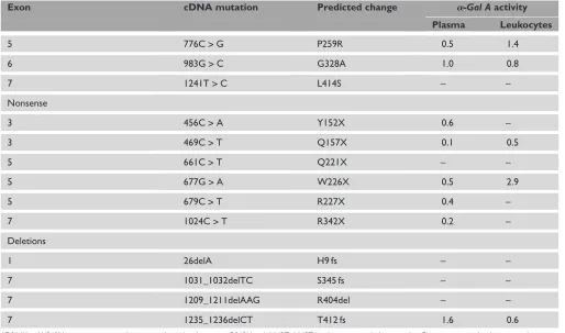
Table 1.Continued.
Exon cDNA mutation Predicted change a-Gal Aactivity
Plasma Leukocytes
5 776C>G P259R 0.5 1.4
6 983G>C G328A 1.0 0.8
7 1241T>C L414S – –
Nonsense
3 456C>A Y152X 0.6 –
3 469C>T Q157X 0.1 0.5
5 661C>T Q221X – –
5 677G>A W226X 0.5 2.9
5 679C>T R227X 0.4 –
7 1024C>T R342X 0.2 –
Deletions
1 26delA H9 fs – –
7 1031_1032delTC S345 fs – –
7 1209_1211delAAG R404del – –
7 1235_1236delCT T412 fs 1.6 0.6
* D264Y and V269M were present together in a single proband, as were Q312H and A143T. A143T has been reported alone in other Fabry patients with a late-onset phenotype.
Intron 1: ·ACTCCACCTCCCGGGTTTAAGCAGTTCTCCTG···TCGTAGTCTCCTGAGTAGCTGGGATTACAGGCACACC···g3291
Intron 2: ·CCTCCACCTCTTGGGTTCAAGTGATTCTCCTG···CCTCAGCCTCCCAAGTAGCTGGGACTACAGGCGCACA···g6447 Patient: ·ACTCCACCTCCCGGGTTTAAGCAGTTCTCCTG··CCTCAGCCTCCCAAGTAGCTGGGACTACAGGCGCACA···
*
* *
*
mutations,D264Y/V269M or A143T/Q312H,expressed no detectable enzymatic activity and were retained in the ER, consistent with being detected in patients with the classic phenotype.
Molecular modelling of the
a
-Gal A missense
mutations
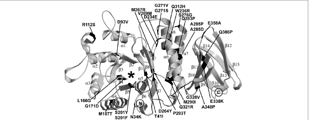
Based on a refined model of thea-Gal A crystal structure (Benson, S.D.et al., unpublished results), the locations and predicted structural alterations of each of the 29 novel missense mutations were determined (Figure 2 and Table 3). The degree of conservation of the residues involved in the novel missense mutations is shown in Table 4.N34K:Asparagine-34 is located in the N-terminal loop, where its side chain forms hydrogen bonds with the side-chain of asparagine-224. The substituted lysine is too large for this position and its positive
charge could interfere with the proper glycosylation at aspar-agine-192.T41I:Threonine-41 is part of the firstb-strand of theb/a-barrel, and the b-strands align the active site. The threonine side-chain points into a small pocket away from the active site. Although there is room for the side-chain of the substituted isoleucine, hydrogen bonding between threonine-41 and histidine-225 would be lost, most likely resulting in protein misfolding.D93V:Aspartate-93 is on the rim of the active site and provides the negative charge environment sur-rounding the galactosyl residue that will be cleaved. Although it is not one of the active aspartate residues, it interacts with the hydroxyl group at the C6 of the terminal galactose and orientates the substrate for cleavage. The substitution of a valine would destroy the interaction with the C6 hydroxyl group and, presumably, would prevent proper binding and orientation of the substrate for cleavage.R112S:Arginine-112 is located on thea2-helix of the N-terminalb/a-barrel, some distance from the active-site pocket. Its side-chain points toward the disulphide bond between 52 and cysteine-94, possibly stabilising the bond. A substitution to serine is predicted to destabilise both the disulphide bond and the pocket that the arginine occupies, leading to protein mis-folding.L166G:Leucine-166 is on theb4-strand of the N-terminalb/a-barrel that lines the edge of the active-site pocket. A substitution of a glycine is predicted to enlarge the active-site pocket, probably causing lower specificity and efficiency because it would be more difficult to orientate the substrate. Glycine is also more flexible, adding to the inability of the active pocket to stabilise the substrate. G171D: Glycine-171 is located at the end of theb4-strand along the edge of the active site and next to the enzymatically active aspartate-170. Glycine is a small and flexible amino acid
suit-Table 2. In vitroexpression of the double missense mutations in COS-7 cells.
a-Gal A
mutation
Enzymatic activity*
Immunohistochemical localisation
V269M 10 Lysosomes
D264Y <1 ER
V269M/D264Y <1 ER
A143T 35 Lysosomes
Q312H 5 ER
A143T/Q312H <1 ER
* Expressed as % of mean wildtype activity. Abbreviation: ER5endoplasmic reticulum.
able for this position. The substitution of an aspartate would cause a charged side-chain to be buried into a small hydro-phobic pocket, which would disrupt the active site and mis-place the active aspartate-70. This would prevent substrate binding.M187T:Methionine-187 is located on the outer
edge in the middle of thea4-helix of theb/a-barrel and its side-chain is buried. Although the substitution of a threonine should be tolerated, it will not occupy the same amount of space and would probably disrupt proper folding of the enzyme.S201Y/F:Serine-201 is located on theb5-strand of
Table 3. Predicted structural alterations caused by the novel missense mutation.
Residue Fabry mutation
Importance ina-Gal A Residue/side-chain
accessible surface (A˚2) Solventexposure of residue
N34 K Disrupts hydrogen bonding 42.83/18.23 Buried T41 I Buried, lines active site 8.87/2.18 Buried D93 V Active site; prevents substrate binding 2.38/2.38 Buried
R112 S Misfolding 19.69/15.48 Buried
L166 G Active site; prevents substrate binding 4.19/4.19 Buried G171 D Active site; prevents substrate binding 3.04/0.00 Buried
M187 T Misfolding 0.05/0.05 Buried
S201 Y Active site, prevents substrate binding 7.71/7.71 Buried F Active site; prevents substrate binding
D234 E Dimer interface; disrupts dimer association 1.44/0.84 Buried W236 R Dimer interface; buries a charge 20.31/18.66 Buried D264 Y Active site; prevents substrate binding 12.79/11.00 Buried M267 R Misfolding; buries charge 4.87/3.78 Buried
V269 M Misfolding 1.76/0.00 Buried
G271 V Misfolding; phi/psi constraints 15.13/0.00 Intermediate S Misfolding; phi/psi constraints
S276 G Misfolding; loss of hydrogen bonding 5.32/3.27 Buried
Q283 P Misfolding 0.00/0.00 Buried
A285 P Misfolding 0.01/0.01 Buried
D Misfolding, buries charge
M290 I Misfolding 3.29/3.10 Buried
P293 T Misfolding 1.71/0.01 Buried
Q312 H Unknown 40.14/28.62 Intermediate
Q321 R Misfolding, disrupts hydrogen bonding 39.66/33.15 Intermediate
G328 V Misfolding 0.83/0.00 Buried
E338 K Unknown 5.92/5.42 Buried
A348 P Misfolding 7.98/7.45 Buried
E358 A Misfolding; loss of charge 33.13/28.70 Intermediate
the N-terminalb/a-barrel and has its side-chain directed into the lower part of the active site cavity. The substitution of either a tyrosine or phenylalanine in this position would decrease the size of the active site. Additionally, a tyrosine or phenylalanine would displace lysine-168, which appears to be critical in properly orientating the substrate into the active site for cleavage, thus abolishing enzymatic activity.D234E: Aspartate-234 is located on the outer edge of theb/a-barrel
in the loop connecting the b6-strand anda6-helix, and is at the dimer interface. It interacts only with the backbone nitrogens of phenylalanine-273 and glycine-274 of its poly-peptide and not with the other subunit. Although a change to the same charged, but slightly larger, glutamate is not a drastic change, it presumably interferes with dimerisation and renders the enzyme non-functional.W236R:Tryptophan-236 is in the loop between theb6-strand anda6-helix on the outer
Table 4. Conservation of novel missense mutations ina-Gal Aanda-Gal Borthologues.
Eukaryotic (22) Mammalian (4) a-Gal B
orthologues (8)
Conservation score*
N34K 13 3 7 ++
T41I 1 3 0 2
D93V 18 3 8 +++
R112S 5 3 7 +
L166G 4 3 0 2
G171D 7 3 8 +
M187T 16 4 5 ++
S201F/Y 15 4 8 ++
D234E 13 3 6 ++
W236R 13 3 7 ++
D264Y 20 3 8 +++
M267R 14 3 7 ++
V269M 3 3 1 2
G271V/S 21 3 7 +++
S276G 11 3 7 ++
Q283P 7 3 7 +
A285P/D 9 3 7 +
M290I 10 3 1 +
P293T 17 3 8 +++
Q312H 5 3 6 +
Q321R 20 3 8 +++
G328V 9 3 7 +
E338K 14 3 6 ++
A348P 1 3 6 +
E358A 3 3 1 2
Q386P 3 3 6 +
edge of theb/a-barrel and fills a large cavity close to the dimer interface where there is a pocket. This cavity has hydrophobic residues along one side towards the core of the protein and hydrophilic residues on the side closest to the solvent. An arginine substitution with a positive charge on its side-chain could position its charge near the hydrophilic residues, whereas the hydrophobic middle of the side-chain could interact with the hydrophobic side of the cavity. Argi-nine would not occupy the large pocket of the tryptophan, however, leading to destabilisation of the enzyme. Tryptophan residues are probably instrumental in early establishment of a protein’s hydrophobic core during protein folding, so that this substitution would also interfere with folding.D264Y: Aspartate-264 is on theb7-strand of theb/a-barrel and has its side-chain in the active site. A change to tyrosine would constrict the active site and remove the negative charge that assists in orientating the substrate. This substitution would cause either misfolding or markedly impair substrate binding. M267R:Methionine-267 is on a loop between theb7-strand anda7-helix that aligns the entrance to the active site and has its side-chain pointing into the active site. A change to arginine would constrict the active site and add an additional positive charge. It would also interfere with lysine-168, which appears to help to align the substrate in the active pocket. Thus, this substitution would markedly interfere with substrate binding.V269M: Valine-269 is on a loop between the
b7-strand anda7-helix that surrounds the entrance to the active site, but is slightly more removed from the active site than methionine-267. It is in a small hydrophobic pocket and a substitution with methionine would cause some con-striction of this area leading to misfolding and impairment of substrate binding.G271V/S:Glycine-271 is located in a turn between theb7-strand anda7-helix in the N-terminal b/a
barrel. The phi/psi angles are disallowed for the other 19 amino acids. Glycine-271 is in a buried hydrophobic area surrounded by polar residues. A side-chain of either valine or serine could be accommodated at this position, but the rigidity of this turn requires a residue with the flexibility of glycine. Therefore, these changes would lead to misfolding.S276G: Serine-276 is on the outer edge of theb/a-barrel at the start of thea7-helix near the dimer interface. Its side-chain is involved in extensive hydrogen bonding with the backbone and side-chain nitrogens of glutamine-279 and with the backbone carbonyl oxygens of phenylalanine-273 and leucine-275. Because this hydrogen bonding network stabilises this part of the enzyme, a change to glycine would disrupt proper folding.Q283P:Glutamine-283 is part of thea7-helix in the N-terminalb/a-barrel. A proline at this position would dis-rupt proper folding of the enzyme.A285P/D:Alanine-285 is part of thea7-helix in the b/a-barrel and lies buried in the interface between the N- and C-terminal domains. The proline substitution would disrupt the helix, interfering with the proper folding of the enzyme. The helix also is beside the C-terminal domain, and this interaction would be disturbed.
Substitution of an aspartate residue, which is negatively charged, would be extremely unstable and would disrupt folding because there are no surrounding residues to counter this charge.M290I:Methionine-290 is at the end of the
a7-helix, where it occupies a large hydrophobic pocket. An isoleucine at this position would not occupy the same volume and would destabilise this section of the enzyme, thus dis-rupting proper folding of the enzyme. P293T:Proline-293 occurs just before theb8-strand of the N-terminalb/abarrel and is buried in a central portion of the enzyme some distance from the active site. Prolines often provide rigidity to the protein that promotes proper folding. A threonine substitution at this position should be tolerated, but there will probably be a cost to the folding dynamics of the protein. Any protein that does form should have enzymatic activity, but there may be little, if any, stable enzyme.Q312H:The lasta-helix (a8) of the N-terminalb/abarrel is actually formed from two helices separated by two residues (glutamine-312 and aspartate-313) that are not in the more extended conformation. The glutamine-312 side-chain is exposed to solvent on one side and the side-chain of tryptophan-81 on the other. The structure should be able to accommodate the substituted histidine, whose side-chain could even make a more favourable inter-action with the side-chain of tryptophan-81. This mutation is also distant from the active site. Structurally, it is difficult to determine why this mutation would be deleterious; however, the pKa of histidine suggests that it would be protonated in the lysosome and thus have a positive charge, which could interfere with the organisation in this area of the protein. Q321R:Glutamine-321 occurs in the lasta-helix (a8) of the N-terminalb/a-barrel. Its side-chain is mostly exposed, but it hydrogen bonds with the side-chain of threonine-39. An arginine substitution would add a positive charge to this area, and its longer side-chain would prevent the interaction with threonine-39, possibly destabilising this area and preventing proper folding.G328V:Glycine-328 is located in the loop between thea8-helix of the N-terminal domain and the
occurs right after a loop, however, one would predict that a small amount of protein would fold properly and be func-tional.E358A: Glutamate-358 is in a loop after theb11-strand of the C-terminal domain; it is completely solvent-exposed and an alanine substitution could easily fit into this position. Glutamate-358 does hydrogen bond to tryptophan-236 and lysine-240, however, stabilising this loop region, which is near the dimer interface. Alanine would not support these interactions and lead to misfolding.Q386P: Glutamine-386 is located in theb13-strand of the C-terminal domain. A proline replacement does not contain an amide hydrogen, so it will not maintain the hydrogen bonding of theb-sheets. This mutation would interfere with the proper folding of the C-terminal domain.
Discussion
Mutation analysis of thea-Gal A gene in 66 unrelated pro-bands with Fabry disease identified 50 new mutations, demonstrating the extensive molecular genetic heterogeneity underlying this lysosomal storage disease. Of the new mutations, several were notable, including a 3.1 kb deletion (only the fifth large deletion detected in this ‘Alu-rich’ gene [Human Gene Mutation Database]), an allele with two adja-cent base substitutions (L166G) and two alleles, each with two missense mutations (D264Y/V269M, and Q312H/A143T).
Although all types of mutations have been found to cause Fabry disease,1there have been relatively few large gene deletions, considering the fact that thea-Gal Agene is an Alu-rich gene with about one Alu per kb. Previously, only four deletions over 1 kb were reported among the over 400 mutations causing this disease, and only one of these four was due to Alu– Alu recombination.1The large deletion reported here most probably resulted from unequal, but homologous, recombina-tion between the highly homologous and similarly orientated Alu sequences in introns 1 and 2, thereby resulting in the approximately 3.1 kb loss, including all of exon 2 (Fig. 1).
The L166G mutation is unusual, in that it involves two base changes, a double transversion of CT to GG at cDNA posi-tions 496 and 497. A single event most likely resulted in the other complex mutation that gave the missense mutation K374R, located 3 bp upstream of a GAG deletion.
The D264Yand V269M mutations were found in a classically affected male. Both mutations were located in exon 6. Structural analysis predicted that V269M would constrict the active site, but that the protein would fold properly and retain some, albeit reduced, activity. By contrast, the aspartate at position 264 lines the active-site pocket, and the change to tyrosine predicts a marked constriction of the active site. Also, the negative charge that probably assists in orientating the substrate would be lost, markedly altering folding and enzyme activity. These structural predictions were confirmed byin vitroexpression assays which demonstrated that theV269Mallele had about 10 per cent of the mean wild-type expressed activity, which was detectable in the
lysosomes (Figure 2), whereas theD264Yallele expressed no detectable enzyme activity, and the enzyme protein was detected immunologically in the ER.
Most mutations causing Fabry disease are private, occurring in a single or few families;1,7however, several mutations at CpG dinucleotides, known mutational hot spots, occur more often in unrelated families. These include R112S, R112C, A143T, Y152X, R227X and R342X in unrelated classically affected families. Here, the R112S mutation is first reported in a classically affected male. In addition, other residues are encoded at CpG dinucleotides, including T39, R49, R118, C142, D153, R220, R301, D315, V316, R356, R363, I367 and A368. Mutations have been reported at all of these CpG sites, except at codons 39, 118, 315, 316, 367 and 368. Four of the mutations reported here (8 per cent) were detected only in the affected proband and were not present in either of the probands’ parents. Three of thesede novomutations occurred at CpG sites (R112C, Y152X and Q157X). One otherde novo mutation occurred at S201F.
In this paper, we have mapped the novel missense mutations onto the native, properly folded enzyme to better understand their 3D locations and proximity to the active site. It is easy to understand why mutations at residues aligning the active-site pocket, such as aspartate-93, aspartate-264 or methionine-267, perturb enzyme function because they are critical in binding and orientating the substrate for
hydrolysis.30Other missense mutations interfere with the proper folding of the enzyme, leading to retention in the ER. The enzyme exists as a homodimer, but there is no indication of functional cooperativity between the two subunits. Two of the mutations occur near the dimer interface (aspartate-234 and tryptophan-236), although they do not interact with the other subunit. The structural analyses predict that three of the substitutions (M187T, Q312H and A348P) would be tolerated or have residual activity. These mutations all resulted in the classic phenotype, however, and only four mutations had significant residual activity in the plasma or leukocytes. The structural studies are based on the properly folded enzyme and cannot provide all of the information needed to determine the folding mechanism of this complex glycoprotein. When the structural analyses are coupled with other in vitroexpression studies, however, they can give a clearer insight into genotype–phenotype correlations. The mutations that are predicted to be accommodated by structural analysis might be functional if allowed to fold and are candidates for studies with pharmacological chaperones.
native structure and cause protein misfolding. Those located around the active site interfere with orientation and/or bind-ing with the substrate, thereby alterbind-ing enzyme function. A few located on or near the dimer interface alter proper dimerisa-tion of the glycoprotein. None of the mutadimerisa-tions described here replaced any of the three asparagines at N-glycosylation sites, but N34K presumably could affect glycosylation at N192, where asparagines-34 interacts with the side-chains of asparagines-192. Several of the normally substituted amino acids were predicted to be structurally accommodated and therefore tolerated (ie T41I, M187T, G271V, G271S, P293T, Q312H and A348P); however, these replacements all caused severe loss of enzyme function or stability because they resulted in the classic phenotype. Thus, predicting the phenotype based on the structural alterations of the mutations may underestimate the severity of the substitution on the protein’s ability to fold into a functional configuration. Clearly, most of the novel missense mutations described here altered folding and presumably led to the glycopolypeptide’s
aggregation/retention in the ER and subsequent proteosomal degradation in the cytosol, consistent with the classic phenotype of Fabry disease.
Acknowledgments
This work was supported, in part, by grants from the National Institutes of Health, including a research grant (R37 DK34045, Merit Award), a grant (5 MO1 RR00071) for the Mount Sinai General Clinical Research Center Program from the National Center of Research Resources and a basic research grant from the Genzyme Corporation. M.Y. is the recipient of an NIH postdoctoral training fellowship in Mental Retardation and Developmental Disabilities (5T32 HD07105).
Electronic database information
Human Gene Mutation Database: http://www.hgmd.org. GenBank: http://www.ncbi.nlm.nih.gov.
Single Nucleotide Polymorphism (SNP) Database:
http://www.ncbi.nlm.nih.gov/entrez/query.fcgi?CMD¼search&DB¼snp.
References
1. Desnick, R.J., Ioannou, Y.A., Eng, C.M.et al.(2001), ‘a-Galactosidase A deficiency: Fabry disease’, in Scriver, C.R., Beaudet, A.L., Sly, W.S.et al.
(eds), ‘The Metabolic and Molecular Bases of Inherited Disease’, 8th edn, McGraw-Hill, New York, NY, pp. 3733–3774.
2. Elleder, M., Bradova, V., Smid, F.et al.(1990), ‘Cardiocyte storage and hypertrophy as a sole manifestation of Fabry’s disease. Report on a case simulating hypertrophic non-obstructive cardiomyopathy’,Virchows Arch. A Pathol. Anat. Histopathol.Vol. 417, pp. 449–455.
3. Nakao, S., Kodama, C., Takenaka, T.et al.(2003), ‘Fabry disease: Detection of undiagnosed hemodialysis patients and identification of a “renal variant” phenotype’,Kidney Int.Vol. 64, pp. 801–807.
4. Nakao, S., Takenaka, T., Maeda, M.et al.(1995), ‘An atypical variant of Fabry’s disease in men with left ventricular hypertrophy’,N. Engl. J. Med.
Vol. 333, pp. 288–293.
5. von Scheidt, W., Eng, C.M., Fitzmaurice, T.F.et al.(1991), ‘An atypical variant of Fabry’s disease with manifestations confined to the myocar-dium’,N. Engl. J. Med.Vol. 324, pp. 395–399.
6. Ashley, G.A., Shabbeer, J., Yasuda, M.et al.(2001), ‘Fabry disease: Twenty novela-galactosidase A mutations causing the classical phenotype’,J. Hum. Genet.Vol. 46, pp. 192–196.
7. Shabbeer, J., Yasuda, M., Luca, E. and Desnick, R.J. (2002), ‘Fabry dis-ease: 45 novel mutations in the a-galactosidase. A gene causing the classical phenotype’,Mol. Genet. Metab.Vol. 76, pp. 23–30.
8. Barker, D.F., Schafer, M. and White, R. (1984), ‘Restriction sites containing CpG show a higher frequency of polymorphism in human DNA’,CellVol. 36, pp. 131–138.
9. Cooper, C. and Youssoufian, H. (1988), ‘The CpG dinucleotide and human genetic disease’,Hum. Genet.Vol. 78, pp. 151–155. 10. Davies, J.P., Winchester, B.G. and Malcolm, S. (1993), ‘Sequence
variations in the first exon of alpha-galactosidase A’,J. Med. Genet.Vol. 30, pp. 658–663.
11. Fitzmaurice, T.F., Desnick, R.J. and Bishop, D.F. (1997), ‘Human
a-galactosidase A: High plasma activity expressed by the -30G!A allele’,
J. Inherit. Metab. Dis.Vol. 20, pp. 643–657.
12. Froissart, R., Guffon, N., Vanier, M.T.et al.(2003), ‘Fabry disease: D313Y is an alpha-galactosidase A sequence variant that causes pseudo-deficient activity in plasma’,Mol. Genet. Metab.Vol. 80, pp. 307–314. 13. Yasuda, M., Shabbeer, J., Benson, S.D.et al.(2003), ‘Fabry disease:
Characterization of alpha-galactosidase A double mutations and the D313Y plasma enzyme pseudodeficiency allele’,Hum. Mutat.Vol. 22, pp. 486–492.
14. Brown, R.M. and Brown, G.K. (1993), ‘X-chromosome inactivation and the diagnosis of X-linked disease in females’,J. Med. Genet.Vol. 30, pp. 177–184.
15. Lyon, M. (1961), ‘Gene action in the X-chromosome of the mouse (Mus musculus L.)’,NatureVol. 190, pp. 372–373.
16. Willard, H.F. (2001), ‘The sex chromosome and X chromosome inacti-vation’, in Scriver, C.R., Beaudet, A.L., Sly, W.S.et al.(eds), ‘The Meta-bolic and Molecular Bases of Inherited Disease’, McGraw-Hill, New York, NY, pp. 1191–1212.
17. Eng, C.M., Banikazemi, M., Gordon, R.et al.(2001), ‘A phase 1/2 clinical trial of enzyme replacement in Fabry disease: Pharmacokinetic, substrate clearance, and safety studies’,Am. J. Hum. Genet.Vol. 68, pp. 711–722.
18. Eng, C.M., Guffon, N., Wilcox, W.R.et al.(2001), ‘Safety and efficacy of recombinant humana-galactosidase A replacement therapy in Fabry’s disease’,N. Eng. J. Med.Vol. 345, pp. 9–16.
19. Schiffmann, R., Kopp, J.B., Austin, H.A.et al.(2001), ‘Enzyme replace-ment therapy in Fabry disease: A randomized controlled trial’,JAMA
Vol. 285, pp. 2743–2749.
20. Wilcox, W.R., Banikazemi, M., Guffon, N.et al.(2004), ‘Long-term safety and efficacy of enzyme replacement therapy for Fabry disease’,Am. J. Hum. Genet.Vol. 75, pp. 65–74.
21. Galanos, J., Nicholls, K., Grigg, L.et al.(2002), ‘Clinical features of Fabry’s disease in Australian patients’,Intern. Med. J.Vol. 32, pp. 575–584. 22. MacDermot, K.D., Holmes, A. and Miners, A.H. (2001),
‘Anderson-Fabry disease: Clinical manifestations and impact of disease in a cohort of 60 obligate carrier females’,J. Med. Genet.Vol. 38, pp. 769–775. 23. Desnick, R.J., Brady, R., Barranger, J.et al.(2003), ‘Fabry disease, an
under-recognized multisystemic disorder: Expert recommendations for diagnosis, management, and enzyme replacement therapy’,Ann. Intern. Med.Vol. 138, pp. 338–346.
24. Fan, J.Q. (2003), ‘A contradictory treatment for lysosomal storage dis-orders: Inhibitors enhance mutant enzyme activity’,Trends Pharmacol. Sci.
Vol. 24, pp. 355–360.
25. Asano, N., Ishii, S., Kizu, H.et al.(2000), ‘In vitroinhibition and intra-cellular enhancement of lysosomala-galactosidase A activity in Fabry lymphoblasts by 1-deoxygalacto-nojirimycin and its derivatives’,Eur. J. Biochem.Vol. 267, pp. 4179–4186.
26. Desnick, R.J. and Schuchman, E.H. (2002), ‘Enzyme replacement and enhancement therapies: Lessons from lysosomal disorders’,Nat. Rev. Genet.Vol. 3, pp. 954–966.
28. Ishii, S., Yoshioka, H., Mannen, K.et al.(2004), ‘Transgenic mouse expressing human mutant a-galactosidase A in an endogenous enzyme deficient background: A biochemical animal model for studying active-site specific chaperone therapy for Fabry disease’,Biochim. Biophys. Acta
Vol. 1690, pp. 250–257.
29. Frustaci, A., Chimenti, C., Ricci, R.et al.(2001), ‘Improvement in cardiac function in the cardiac variant of Fabry’s disease with galactose-infusion therapy’,N. Eng. J. Med.Vol. 345,
pp. 25–32.
30. Garman, S.C. and Garboczi, D.N. (2004), ‘The molecular defect leading to Fabry disease: Structure of human alpha-galactosidase’,J. Mol. Biol.
Vol. 337, pp. 319–335.
31. Matsuzawa, F., Aikawa, S.I., Doi, H.et al., (2005), ‘Fabry disease: Correlation between structural changes ina-galactosidase, and clinical and biochemical phenotypes’,Hum. Genet. Vol. 117, pp. 317–328.
32. Desnick, R.J., Allen, K.Y., Desnick, S.J.et al.(1973), ‘Fabry’s disease: Enzymatic diagnosis of hemizygotes and heterozygotes.a-Galactosidase activities in plasma, serum, urine, and leukocytes’,J. Lab. Clin. Med.
Vol. 81, pp. 157–171.
33. Shabbeer, J., Robinson, M. and Desnick, R.J. (2005), ‘Detection of
a-galactosidase A mutations causing Fabry disease by denaturing high
performance liquid chromatography’,Hum. Mutat.Vol. 25, pp. 299–305.
34. Wang, A., Bishop, D. and Desnick, R. (1990), ‘Humana-N -acetylgalactosaminidase: Molecular cloning, nucleotide sequence, and expression of a full-length cDNA’,J. Biol. Chem.Vol. 265, pp. 21859–21866.
35. Higgins, M.E., Davies, J.P., Chen, F.W. and Ioannou, Y.A. (1999), ‘Niemann-Pick C1 is a late endosome-resident protein that transiently associates with lysosomes and the trans-Golgi network’,Mol. Genet. Metab.
Vol. 68, pp. 1–13.
36. Garman, S.C., Hannick, L., Zhu, A. and Garboczi, D.N. (2002), ‘The 1.9 A structure ofa-N-acetylgalactosaminidase: Molecular basis of glycosidase deficiency diseases’,Structure (Camb.)Vol. 10, pp. 425–434.
37. Jones, T.A., Zou, J.Y., Cowan, S.W. and Kjeldgaard, M. (1991), ‘Improved methods for building protein models in electron density maps and the location of errors in these models’,Acta Crystallogr.Vol. A47, pp. 110–119.